Chapter 44
Sarcoidosis
ROBERT S. WEINBERG
Main Menu   Table Of Contents

Search

INCIDENCE
ETIOLOGY
SYSTEMIC SARCOIDOSIS
OCULAR SARCOIDOSIS
DIAGNOSIS
COURSE AND MANAGEMENT
REFERENCES

Sarcoidosis is a systemic granulomatous disease of undetermined etiology and pathogenesis. The diagnosis is established by a compatible clinical picture, together with laboratory or biopsy confirmation of the noncaseating granuloma, the pathologic hallmark of the disease. To the ophthalmologist, sarcoidosis must be considered in the differential diagnosis of almost any type of ocular inflammation, because sarcoidosis may affect any part of the eye or ocular adnexa. Because this systemic disease may present with ocular abnormalities, and because the ophthalmologist may be the first physician to suspect the presence of sarcoidosis, the ophthalmologist must be familiar with the general aspects of the disease.
Back to Top
INCIDENCE
Sarcoidosis has a worldwide distribution, affecting all ages, races, and both genders. Estimates of prevalence of the disease, all patients with the disease at a given time period, range from 1 to 40 per 100,000 population.1 Because some patients may be asymptomatic, prevalence data may be difficult to confirm. In the United States, the incidence of sarcoidosis, the number of new cases in a given time period, is approximately three times higher in blacks than whites.2 The disease appears to be more common in Scandinavia and in Ireland than in other parts of the world.3 Women appear to have a slightly higher incidence of sarcoidosis.4 The disease affects all age groups but is rare in children, more common in adults under 40 and may occur in families.4
Back to Top
ETIOLOGY
The etiology of sarcoidosis remains unknown. Many pathogenetic theories have been proposed including exposure to environmental stimuli, such as clay, metal dusts, or pine pollen; infection with a variety of organisms, such as atypical mycobacteria, fungi, and viruses; autoimmunity; and a genetic predisposition to the development of the disease. Identification of the molecular mechanisms involved in the development of the noncaseating granuloma may help to clarify the pathogenesis of sarcoidosis.5 Finding the antigen or antigens that trigger the response is the first step in clarifying the etiology. The demonstration of mycobacterial DNA in sarcoid granulomas in a few patients suggests a possible antigen.6 The response to antigen exposure with acquired cellular immunity against the antigen, and a nonspecific inflammatory response mediated by immune effector cells leads to the development of the sarcoid granuloma.1
Back to Top
SYSTEMIC SARCOIDOSIS
Multisystem granuloma formation is the characteristic disease process in sarcoidosis. Assessment of disease activity in sarcoidosis is difficult because the disease may be asymptomatic, persist without progression, and may not require treatment. Sarcoidosis may be a transient illness in many patients, but it can be a chronic disabling or fatal problem in others. The clinician ought to remember that sarcoidosis is multisystemic and that intensity of inflammatory reaction in one organ does not necessarily imply general or systemic involvement. The lungs are most frequently affected, with pulmonary involvement in approximately 90% of patients.7 Pulmonary manifestations range from asymptomatic hilar adenopathy detected on routine chest radiograph to pulmonary fibrosis (Table 1). Up to 80% of patients with Type 0 or I disease may improve spontaneously.7 Symptomatic extrapulmonary sarcoidosis occurs in the skin and in the lymphatic system in approximately 25% of patients and in the heart, liver, spleen, joints, bone, and nervous system in less than 10% of patients.1,7 Ocular disease occurs in about 25% of patients with sarcoidosis.1,7,8


TABLE 1. Clinical Staging of Pulmonary Sarcoidosis Based on Chest Radiography at Time of Presentation


TypeCharacteristics
IBilateral hilar adenopathy
IIBilateral hilar adenopathy with diffuse pulmonary infiltrates
IIIDiffuse pulmonary infiltrates without adenopathy
IVPulmonary fibrosis


Back to Top
OCULAR SARCOIDOSIS
Uveitis is the most common and most serious form of intraocular involvement in sarcoidosis. However, any part of the globe or ocular adnexa may be involved. The skin of the lids may have sarcoid nodules, which are accessible to biopsy. Conjunctival sarcoidosis appears a discrete yellowish nodules, usually smaller than 2 mm in diameter. (Fig. 1A). Although these nodules usually occur in the fornices or on the tarsal conjunctiva, they may also be present on the bulbar conjunctiva or on the conjunctiva overlying the lacrimal gland. If conjunctival nodules are present, they are easily biopsied (see Fig. 1B). Conjunctival biopsy can be done at the slit lamp biomicroscope using only topical anesthesia. Blind conjunctival biopsy, that is, biopsy of an area of conjunctiva where no nodules are visible, is less likely to yield helpful information than excisional biopsy of a visible nodule.

Fig. 1. A. Conjunctival nodules in sarcoidosis. this 36?year?old black woman has yellow gelatinous conjunctival nodules characteristic of sarcoidosis. B. Biopsy of a conjunctival nodules demonstrating giant cell and noncaseating granuloma compatible with a diagnosis of sarcoidosis. C. Enlarged prolapsable lacrimal gland in a 23?year?old black woman with sarcoidosis. With the patient looking down and to the right, the upper lid is retracted manually, thus allowing the lacrimal gland to prolapse. the glandular tissue itself has a nodular consistency. D. A 24?year?old black woman with sarcoid uveitis. There are medium and small keratic precipitates, anterior chamber reaction, and posterior synechiae. E. Peripheral so?called candlewax dripping in sarcoid uveitis. Yellow?white lesions are seen in the midperiphery of the fundus. These lesions are characteristic of sarcoidosis. F. Solitary choroidal granuloma in a black man with biopsy?proven systemic sarcoidosis.

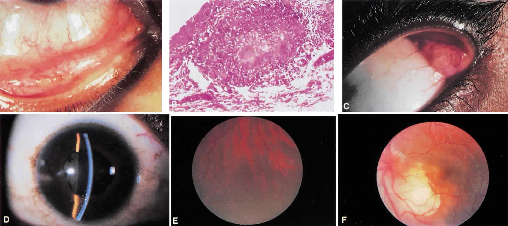
Lacrimal gland enlargement (see Fig. 1C) or dacryoadenitis may be an early finding in sarcoidosis, but it is the initial manifestation of the disease in approximately 1% of patients.9 The differential diagnosis of lacrimal gland enlargement is listed in Table 2. A biopsy demonstrating noncaseating granulomas can differentiate lacrimal gland enlargement caused by sarcoidosis.


TABLE 2. Differential Diagnosis of Lacrimal Gland Enlargement

  Sjögren's syndrome
  Sarcoidosis
  Lymphoma
  Leukemia
  Systemic lupus erythematosus
  Wegener's granulmatosis
  Amyloidosis
  Other connective tissue disease


Corneal sarcoidosis may be either interstitial keratitis or localized stromal opacification secondary to endothelial decompensation in areas where keratic precipitates once were present.10 Neither type of corneal involvement is specific for sarcoidosis. The sclera may rarely be a site of activity in sarcoidosis. Granulomatous scleritis is a localized form of nodular scleritis, which may be seen in sarcoidosis, and posterior scleritis has been reported in sarcoidosis.11

Uveitis in sarcoidosis may be granulomatous or nongranulomatous, acute or chronic, localized or diffuse, and monocular or bilateral. However, anterior uveitis is the most frequent type of uveitis seen in patients with sarcoidosis. Either acute or chronic iridocyclitis may occur. Acute iritis presents most often in women, between ages 20 and 30, with pain, redness, and photophobia. There is generally moderate anterior chamber flare and cells, and keratic precipitates are usually small or fine. The presence of large keratic precipitates is suggestive of inflammation of long duration, but may be seen acutely. (see Fig. 1D). Large or mutton-fat keratic precipitates are not specific for sarcoidosis. Iris nodules on the pupillary border (Koeppe nodules) or in the midstroma (Busacca nodules) may be present. Vitreous cells are frequently present, and when accompanied by anterior chamber reaction, without posterior involvement, the condition is called iridocyclitis. Acute sarcoid iridocyclitis does occur. Visual acuity may be normal, but with associated cystoid macular edema, it may decrease. Acute iridocyclitis may be unilateral or bilateral.

Chronic iridocyclitis in sarcoidosis is frequently associated with cystoid macular edema. Patients with this condition may not have pain or redness, but they present with blurred vision and floating spots. These patients tend to be older (35 to 50 years of age) than those presenting with acute iritis.12 Keratic precipitates, either small or large, moderate anterior chamber cell and flare, posterior and anterior synechiae, and vitreous cells may all be present. Fluorescein angiographic evidence of cystoid macular edema is to be expected if visual acuity is decreased. Activity in chronic iridocyclitis in sarcoidosis tends to be bilateral.

Posterior segment intraocular inflammation frequently is present in sarcoidosis. Vitreous cellular reaction may range from minimal with few vitreous cells or clumps of cells, vitreous snowballs, to the more unusual presentation of dense vitreous debris. Histologically vitreous snowballs contain epithelioid cells.13 Retinal periphlebitis occurring in the midperiphery of the retina is highly suggestive of sarcoidosis. Small exudates near retinal veins, termed candle wax drippings may be present (see Fig. 1E). Neovascularization may occur. Choroidal granulomas may be small and multiple, resembling Dalen-Fuchs nodules, or single and very large, resembling a choroidal tumor, as seen in Figure 1F. Chronic cystoid macular edema may cause decreased vision in patients with long-standing sarcoid uveitis. The optic nerve may be involved with disc neovascularization, disc edema in chronic inflammation, or with granuloma formation within the optic nerve.

Back to Top
DIAGNOSIS
Establishing a diagnosis of sarcoidosis requires a compatible history, suggestive physical findings, and a biopsy demonstrating epithelioid cell granulomas. Classically, associated granulomas are noncaseating, but small areas of fibrinoid necrosis may be found in sarcoid granulomas.7 Abnormal laboratory tests may provide suggestive evidence of the disease, but only with a biopsy can the diagnosis be certain. Sarcoidosis must be in the differential diagnosis of almost any form of uveitis. Conversely, however, the differential diagnosis of sarcoid uveitis depends on the anatomic area of the eye that is primarily involved with inflammation (Table 3).


TABLE 3. Differential Diagnosis of Sarcoid Uveitis

  Acute iritis
  Human leukocyte antigen B-27 uveitis
  Idiopathic iritis
  Syphilis
  Chronic iridocyclitis
  Chronic cyclitis
  Syphilis
  Vitritis
  Reticulum cell sarcoma
  Whipple's disease
  Panuveitis
  Behçet's syndrome
  Infectious uveitis (fungal)
  Syphilis
  Toxoplasmosis
  Tuberculosis
  Vogt-Koyanagi-Harada syndrome


Because of lack of specificity, no single laboratory or radiologic test is pathognomonic for sarcoidosis.1 Multiple test results, if abnormal, suggest sarcoidosis. These include serum calcium, calcitriol, angiotensin-converting enzyme (ACE), lysozyme, and soluble interleukin-2 receptors (sIL-2R).14 Serum calcium is elevated in approximately 10% of patients, secondary to 1,25-dihydroxyvitamin D3, calcitriol, produced at sites of active disease. ACE, produced by epithelioid cells of granulomas is elevated in between 40% and 90% of patients but it may also be elevated in diabetes, berylliosis, primary biliary cirrhosis, histoplasmosis, tuberculosis, and Hodgkin's disease.7,14 Serum lysozyme, produced by macrophages and epithelioid cells, may be more sensitive, but less specific than ACE, in suggesting a diagnosis of sarcoidosis.15 Pulmonary T lymphocytes in patients with active sarcoidosis produce IL-2 and elevated sIL-2R levels have been found in the serum of patients with active, but not inactive sarcoidosis. Again, the test result is not specific for sarcoidosis, and elevated sIL-2R can be found in lymphoproliferative disease, bronchial carcinoma, connective tissue disease, and tuberculosis, among other conditions.14 Gallium scanning, once thought to be helpful in the diagnosis of sarcoidosis, is nonspecific, and can be abnormal in other inflammatory disease.7 The combination of two tests, ACE and gallium scan, may increase specificity for the diagnosis of ocular sarcoidosis.16 Other tests thought to be useful diagnostically in the past, cutaneous anergy and the Kveim test, are no longer used.

Although many sites are available for biopsy to confirm a clinical impression of sarcoidosis, biopsy of a visible conjunctival nodule provides the highest yield with the least morbidity. When the lacrimal gland is enlarged or nodular in a patient suspected of having sarcoidosis, transconjunctival biopsy of the lacrimal gland may be diagnostic.17

Back to Top
COURSE AND MANAGEMENT
Sarcoidosis is a chronic disease that may be manifest by exacerbations and remissions. Uveitis in this disease may exhibit either a monophasic course, generally with retention of good vision, or a chronic relapsing course with a greater likelihood of significant visual loss.18 Topical corticosteroids and cycloplegia are indicated in the management of the anterior segment manifestations of sarcoidosis.

Corticosteroids, administered either topically, periocularly, or systemically, remain the therapy of choice in sarcoid uveitis. Generally, unless bilateral disease is present, or the patient refuses periocular corticosteroid therapy, ocular manifestations of sarcoidosis may be managed without the use of systemic corticosteroids. Although no controlled clinical studies have investigated other therapeutic approaches to sarcoid uveitis, there appear to be some effective alternatives to systemic corticosteroids for treating systemic sarcoidosis. Steroid-sparing therapy for systemic sarcoidosis includes low dose methotrexate, cyclosporine, and chlorambucil.19–22 Hydroxychloroquine has been suggested for management of cutaneous sarcoidosis.23 These drugs may be helpful for patients unresponsive to corticosteroids or in whom corticosteroids are contraindicated.

Back to Top
REFERENCES

1. Newman, LS, Rose CS, Maier LA: Sarcoidosis. N Engl J Med 336: 1224, 1997

2. Rybicki BA, Major M, Popovich J Jr et al: Racial differences in sarcoidosis incidence: A 5-year study in a health maintenance organization. Am J Epidemiol 145:234, 1997

3. James DG, Hosoda Y. Epidemiology. In James DG (ed): Sarcoidosis and Other Granulomatous Disorders, pp 729–743. Los Angeles: Marcel Dekker, 1994

4. Hosoda Y, Yamaguchi M, Hiraga Y: Global epidemiology of sarcoidosis. Clin Chest Med 18:681, 1997

5. Moller DR: Cells and cytokines involved in the pathogenesis of sarcoidosis. Sarcoidosis Vasculitis Diff Lung Dis 16:24, 1999

6. Mangiapan G, Hance AJ: Mycobacterioa and sarcoidosis: An overview and summary of recent molecular biological data. Sarcoidosis 12:20, 1995

7. Johns CJ, Michele TM: The clinical management of sarcoidosis: A 50 year experience at the Johns Hopkins Hospital. Medicine 78:65, 1999

8. Jabs DA, Johns CJ: Ocular involvement in chronic sarcoidosis. Am J Ophthalmol 102:297, 1986

9. Nowinski T, Flanigan J, Ruchman M: Lacrimal gland enlargement in familial sarcoidosis. Ophthalmology 90:919, 1983

10. Lucchese N, Tessler H: Keratitis associated with chronic iridocyclitis. Am J Ophthalmol 92:717, 1981

11. Dodds EM, Lowder CY, Barnhorst DA et al: Posterior scleritis with annular ciliochoroidal detachment. Am J Ophthalmol 120:677, 1995

12. Smith JA, Foster CS: Sarcoidosis and its ocular manifestations. Int Ophthalmol Clin 36:109, 1996

13. Gass JDM, Olson CL: Sarcoidosis with optic nerve and retinal involvement: A clinicopathologic case report. Trans am Acad Ophthalmol Otolaryngol 77:739, 1973

14. Costabel U, Teschler H: Bochemical changes in sarcoidosis. clin Chest med 18:827, 1997

15. Tomita H, Sato S, Matsuda R et al: Serum lysozyme levels and clinical features of sarcoidosis. Lung 177:161, 1999

16. Power WJ, Neves RA, Rodriguez A et al: The value of combined serum angiotensin-converting enzyme and gallium scan in diagnosing ocular sarcoidosis. Ophthalmology 102: 2007, 1995

17. Weinreb: diagnosing sarcoidosis by transconjunctival biopsy of the lacrimal gland. Am J Ophthalmol 97:573, 1984

18. Dana MR, Merayo-Lloves J, Schaumberg et al: Prognosticators for visual outcome in sarcoid uveitis. Ophthalmology 103:1846, 1996

19. Baughman RP: Methotrexate for sarcoidosis. Sarcoidosis Vasulitis Diff Lung Dis 15:147, 1998

20. Dev S, McCallum RM, Jaffe GJ: Methotrexate treatment for sarcoid-associated panuveitis. Ophthalmology 106:111, 1999

21. York EL, Kovitavhongs T, Man SF et al: Cyclosporine and chronic sarcoidosis. Chest 98:1026, 1990

22. Israel HL, McComb BL: Chlorambucil treatment of sarcoidosis. Sarcoidosis 8:35, 1991

23. Jones E, Callen JP: Hydroxychloroquine is effective therapy for control of cutaneous sarcoidal granulomas. J Am Acad Dermatol 23:487, 1990

Back to Top