Chapter 41
Lens-Induced Uveitis and Related Intraocular Inflammations
NARSING A. RAO and ROBERT F. SEE
Main Menu   Table Of Contents

Search

PHACOANAPHYLACTIC ENDOPHTHALMITIS
PHACOGENIC NONGRANULOMATOUS UVEITIS
PHACOLYTIC GLAUCOMA
INTRAOCULAR LENS-RELATED INFLAMMATIONS AND LOCALIZED ENDOPHTHALMITIS
ACKNOWLEDGMENTS
REFERENCES

Lens-induced uveitis is a diagnostic category that encompasses three main clinical and pathologic entities characterized by intraocular inflammation that is induced by lens protein. The first entity, phacoanaphylactic endophthalmitis, is believed to be an autoimmune disorder. The second entity, phacogenic nongranulomatous uveitis, previously referred to as phacotoxic uveitis, is a nonspecific, chronic intraocular inflammatory reaction to lens protein. The third entity, considered to be a lens-induced uveitis by some authors but not by others, is phacolytic glaucoma. Patients with phacolytic glaucoma exhibit some of the features of intraocular inflammation, but histologically the process consists of a macrophage phagocytic response to cataractous lens protein.1–4 These three entities overlap to a certain extent in their clinical presentation, and patients characteristically have cells in the anterior chamber and vitreous cavity and at least some degree of uveitis or intraocular inflammation. These three entities can be differentiated histopathologically, however, largely by the type of cellular infiltration and by the pattern of inflammation around the lens.

When the lens capsule is disrupted by either surgical trauma or accidental injury and lens protein is released into the ocular cavities, many people do not develop clinically significant intraocular inflammation. However, in a few people, a macrophage foreign-body type of reaction may be observed. If a persistent, nongranulomatous inflammation develops with signs of uveitis, such intraocular inflammation is usually classified as a phacogenic nongranulomatous uveitis (phacotoxic uveitis). In rare instances, a granulomatous inflammation may develop around the disrupted lens, in which case the intraocular inflammation is called phacoanaphylactic endophthalmitis.5 In contrast to the aforementioned inflammatory responses to a disrupted lens, patients classified as having phacolytic glaucoma show features of a hypermature cataract and an intact lens capsule, through which altered lens protein escapes. These patients develop a macrophage phagocytic response to the altered lens protein.

Back to Top
PHACOANAPHYLACTIC ENDOPHTHALMITIS
Phacoanaphylactic endophthalmitis, also known as phacoanaphylactic uveitis, is a granulomatous inflammatory process associated with a disrupted lens. This entity was first reported to be granulomatous inflammation by Straub6 in 1919. In 1922, Verhoeff and Lemoine7, who referred to this entity as “endophthalmitis phacoanaphylactica,” proposed an autoimmune response to lens protein as the cause of this granulomatous inflammation. Clinically, this inflammation follows traumatic or surgical disruption of the lens. The inflammation usually begins within 14 days of lens injury and characteristically presents as unilateral, severe uveitis in the traumatized eye. Occasionally, this ocular inflammation develops following spontaneous rupture of the lens capsule or in combination with sympathetic ophthalmia. In most cases the ocular inflammation is severe, with large numbers of cells in the anterior chamber and vitreous cavity. Large keratic precipitates are generally present, and extensive posterior synechia formation often occurs. Fragments of lens cortex may be observed in the anterior chamber or in the vitreous cavity, and in most cases the lens material exhibits cataractous changes. In some cases the inflammation is intense enough to cause hypopyon (Fig. 1). If the inflammation continues unchecked, damage to the eye may be severe, because the inflammation can lead to formation of a cyclitic membrane, tractional retinal detachment, and, eventually, phthisis bulbi.4

Fig. 1. Hypopyon and marked conjunctival hyperemia (A) and large keratic precipitates (B) in phacoanaphylactic endophthalmitis. This ocular inflammation first appeared 2 weeks following extracapsular cataract surgery.

A milder form of phacoanaphylactic uveitis is known to occur following extracapsular cataract extraction, with or without posterior chamber intraocular lens implantation. In these cases the granulomatous inflammation develops at between 1 week and 6 months after the surgical procedure. This form of phacoanaphylaxis is believed to reflect limited immune response to a small amount of lens protein remaining in the ocular cavity.8,9 In some patients a sympathetic response may occur in the opposite eye.

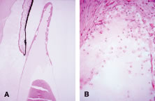
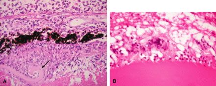
Histopathologically, enucleated eyes with severe phacoanaphylactic endophthalmitis show a zonal type of granulomatous inflammation concentrated around the disrupted lens capsule and exposed lens cortex or nucleus5 (Fig. 2). Typically, a zone of polymorphonuclear leukocytes is present around the lens cortex; the cortical fibers show degenerative changes in the form of fragmentation and disintegration. Surrounding the polymorphonuclear leukocytes is a zone of mononuclear cells, which consists primarily of macrophages and epithelioid cells but is admixed with a few scattered multinucleated giant cells. Some of these giant cells show features of foreign body type cells, whereas others exhibit features typical of Langhans' cells (see Fig. 2B). A third zone, made up of lymphocytes, surrounds these epithelioid cells. This lymphocytic zone may also contain fibroblasts or granulation tissue. The surrounding iris, ciliary body, and anterior choroid are usually infiltrated by lymphocytes and plasma cells. This lymphoplasmacytic infiltration is marked in the anterior uvea and tends to be of mild to moderate degree in the posterior uvea. The retina may show some perivascular lymphocytic infiltration. In many cases, extensive iridolenticular adhesions are present (see Fig. 2A), often accompanied by chronic inflammatory cells in the anterior chamber and in angle structures. The endothelial surface of the cornea may contain collections of mononuclear cells.

Fig. 2. A. Enucleated eye of phacoanaphylaxis shows disrupted lens capsule (arrow), iridolenticular adhesion and granulomatous inflammation around the lens. B. Note prominent multinucleated giant cells around disrupted lens cortex in phacoanaphylaxis.

Eyes enucleated during more chronic stages of phacoanaphylactic endophthalmitis may show only a few polymorphonuclear leukocytes around the disrupted lens. However, a zone of granulomatous inflammation can be seen, made up of epithelioid cells admixed with large number of multinucleated giant cells, as can a surrounding zone of lymphocytes and fibroblasts. These eyes may also exhibit fibrosis that encloses the granulomatous inflammatory mass. Such eyes usually show a cyclitic membrane, tractional retinal detachment, osseous metaplasia of the retinal pigment epithelium, and phthisis.

ETIOLOGY

Even though the precise cause of the initial events that evoke the inflammation in phacoanaphylactic endophthalmitis is unknown, the inflammation is believed to be initiated by an autoimmune process directed at the lens protein. Reports in the past have suggested that lens protein is organ specific and, under normal conditions, is sequestered from the immune system by the lens capsule.10,11 However, in recent years it has been documented that most healthy people, with no demonstrable ocular disease, have anti-lens antibodies,12 and when they do undergo extracapsular cataract surgery, they rarely develop phacoanaphylaxis. Based on these observations it is less likely that phacoanaphylaxis is an immune reaction to the so-called sequestered lens antigen that is released by traumatic or surgical disruption of the lens capsule. Moreover, experimental animal studies have shown that lens proteins are not sequestered solely in the lens capsule and, in addition, immunologically these proteins are not organ specific. Based on experimental animal studies, Marak13 proposed a hypothesis for the pathogenesis of phacoanaphylaxis. According to this theory, development of phacoanaphylactic endophthalmitis is dependent on abrogation of normal tolerance to lens protein.

Experimentally, granulomatous inflammation can be induced around surgically disrupted lens in various strains of rats that have been sensitized to lens protein.13 Histopathologically, the enucleated eyes of these animals show features very similar to those observed in human phacoanaphylaxis. Immunologic investigations of such animals demonstrated that the lens-induced uveitis is an antibody-mediated autoimmune disease that develops when normal immune tolerance (T-cell tolerance) is terminated by stimulation of B cells with gram-negative bacterial lipopolysaccharide.13 Based on such experimental studies, it appears that phacoanaphylactic endophthalmitis in humans could be an anti-lens protein antibody-mediated localized immune complex disease that is triggered by traumatic release of lens protein and stimulation of B cells.

DIFFERENTIAL DIAGNOSIS

The intraocular inflammatory disorders that should be differentiated from phacoanaphylactic endophthalmitis include infectious (bacterial or fungal) endophthalmitis, phacogenic nongranulomatous uveitis, phacolytic glaucoma, localized endophthalmitis, and sympathetic ophthalmia. Exogenous bacterial endophthalmitis following a penetrating wound to the globe or following cataract extraction may present with signs of severe intraocular inflammation and hypopyon. However, unlike phacoanaphylactic endophthalmitis, it is unusual to observe large keratic precipitates in bacterial endophthalmitis. Some cases of bacterial or fungal endophthalmitis may be difficult to differentiate from phacoanaphylaxis. In such cases a definitive diagnosis can be established by histologic examination and by culture studies of the intraocular inflammatory exudate.

Exogenous fungal endophthalmitis may present with several clinical features that are typical of low-grade phacoanaphylactic endophthalmitis. These include delayed onset of ocular inflammation, hypopyon and inflammatory exudate in the anterior chamber and vitreous cavity, and puffball vitreous opacities. Furthermore, some of these patients may exhibit keratic precipitates similar to those noted in phacoanaphylactic endophthalmitis, but cultures and histologic studies of the intraocular exudate should point to the proper diagnosis. Phacoanaphylactic endophthalmitis may prove difficult to differentiate from phacogenic nongranulomatous uveitis, phacolytic glaucoma, and localized endophthalmitis, entities that are discussed in detail subsequently in this text.

Phacoanaphylactic endophthalmitis occurs typically as a uniocular inflammatory process involving the traumatized eye but can present occasionally as bilateral intraocular inflammation.11 Such bilateral lens-induced intraocular inflammation must be differentiated from sympathetic ophthalmia, which is always a bilateral disease. Patients with sympathetic ophthalmia rarely develop hypopyon. Moreover, sympathetic ophthalmia typically results in angiographic findings of hyperfluorescence and focal leakage at the level of the retinal pigment epithelium during the early arteriovenous phase, with pooling of dye in the subretinal space during late stages of angiography. Echographic evaluation should also be useful because phacoanaphylactic endophthalmitis causes inflammatory swelling of the anterior uvea, whereas sympathetic ophthalmia induces marked swelling of the posterior uvea. It should be noted that in some cases of penetrating injury to the globe, both phacoanaphylactic endophthalmitis and sympathetic ophthalmia may coexist in one eye, and the opposite eye may show just sympathetic ophthalmia.

LABORATORY INVESTIGATIONS

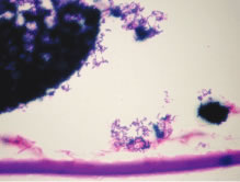
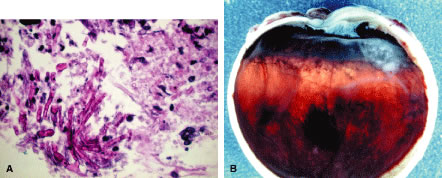
Even though phacoanaphylactic uveitis could be mediated by anti-lens antibody, serologic detection of such antibody is not diagnostic. As noted earlier, many people without any ocular disease show circulating anti-lens antibody in their serum. Similarly, patients who have undergone uncomplicated extracapsular cataract extraction, with no subsequent intraocular inflammation, have demonstrated serologic evidence of anti-lens antibody. Echography in typical cases of phacoanaphylaxis reveals a swollen iris, ciliary body, and anterior choroid and evidence of marked inflammation around the lens. Histopathologic examination of an anterior chamber tap or vitrectomy specimens can be helpful also in establishing the correct diagnosis, because these specimens will typically contain epithelioid histiocytes mixed with polymorphonuclear leukocytes and giant cells (Fig. 3). Cultures of these specimens are negative for bacteria and fungi.

Fig. 3. Vitrectomy specimen exhibiting granulomatous inflammation around disrupted lens cortex. Note multinucleated giant cell (arrow).

TREATMENT

The management of phacoanaphylactic endophthalmitis includes surgical removal of the offending antigen, namely the lens material. Such a surgical procedure should be carried out early in the course of intraocular inflammation, and, in severe cases, the patient should also be treated with topical and systemic corticosteroids. Even though the corticosteroids can reduce the severity of ocular inflammation, these agents do not eliminate the source of the ocular inflammation. Meticulous surgical removal of the lens material, supplemented with cycloplegic and corticosteroid administration, may result in preservation of useful vision with minimal ocular morbidity.

In patients with a history of phacoanaphylactic endophthalmitis following trauma or extracapsular cataract extraction, it is essential to recognize the risk for similar inflammation to develop in the fellow eye following extracapsular cataract extraction. Accordingly, removal of a cataractous lens by intracapsular cataract extraction is recommended in such cases.

Back to Top
PHACOGENIC NONGRANULOMATOUS UVEITIS
Phacogenic nongranulomatous uveitis is a nongranulomatous intraocular inflammation induced by lens protein. This entity was previously referred to as phacotoxic uveitis.1–5 In the early 1900s this was believed to be caused by toxic material liberated by lens disruption,11 but subsequent studies failed to show any toxic properties of the lens substance.5 Based on these findings, some investigators deny the existence of a phacotoxic uveitis.4,11 However, even though the lens may not contain any discernible toxin capable of inciting nongranulomatous inflammation, there is some experimental evidence suggesting such uveitis can be induced following surgical disruption of the lens capsule.14 Based on such experimental and clinical studies, it is now believed that phacogenic nongranulomatous uveitis may be a variant of phacoanaphylaxis and, thus, warrants inclusion in the category of lens-induced uveitis.

Phacogenic nongranulomatous uveitis shares several clinical features with phacoanaphylactic uveitis, including development of intraocular inflammation following traumatic or surgical disruption of the lens capsule, the appearance of inflammation generally within 2 to 3 weeks after the injury, and formation of iridolenticular adhesions. However, phacogenic nongranulomatous uveitis differs from phacoanaphylaxis in some important respects; for example, keratic precipitates are lacking in most patients with phacogenic nongranulomatous uveitis, and those that do form are small and nongranulomatous.

Phacogenic nongranulomatous uveitis can be easily mistaken for phacolytic glaucoma, because both of these entities can result in increased intraocular pressure and inflammatory cells in the anterior chamber. However, phacolytic glaucoma occurs only in patients with a mature or hypermature cataract who have no evidence of traumatic or surgical disruption of the lens capsule.

Histologically, phacogenic nongranulomatous uveitis can be differentiated from other forms of lens-induced uveitis in that it typically exhibits a nongranulomatous inflammation around the disrupted lens.5 This cellular infiltrate consists of lymphocytes, histiocytes, and some polymorphonuclear leukocytes; epithelioid cells, giant cells, and the zonal pattern are not seen in this entity. The macrophages rarely contain eosinophilic granular material, which is commonly noted in cases of phacolytic glaucoma.

The etiology of phacogenic nongranulomatous uveitis is unknown, but because it appears that this inflammation may be a variant of phacoanaphylactic endophthalmitis, it is most likely mediated by an autoimmune process directed to the lens protein.

The management of phacogenic nongranulomatous uveitis is similar to that of phacoanaphylaxis. Surgical removal of the lens is essential, as is administration of topical and systemic corticosteroids to suppress the inflammation.

Back to Top
PHACOLYTIC GLAUCOMA
Phacolytic glaucoma occurs in patients who have a hypermature cataract and presents as an acute glaucoma with marked flare and large cells in the anterior chamber.4,11 The cornea is usually edematous and generally without any evidence of keratic precipitates. Some of these patients show evidence of anterior chamber angle recession or rupture of the posterior lens capsule. In general, the lens capsule surrounding the hypermature cataractous lens is intact, although it is markedly thinned (Fig. 4A). Some patients with phacolytic glaucoma exhibit conjunctival injection.

Fig. 4. Phacolytic glaucoma. A. Enucleated eye shows Morgagnian cataract. B. Macrophages containing lens material are seen in the anterior chamber angle and trabecular meshwork. (Courtesy of Alan Snyder, MD.)

The large cells present in the anterior chamber are mostly mononuclear phagocytes containing lenticular material. These macrophages accumulate on the lens capsule and clog the trabecular meshwork, thereby causing an elevation in intraocular pressure15 (see Fig. 4B). The diagnosis can be established by paracentesis of anterior chamber fluid and examination of the material for macrophages laden with lens material.

The treatment of phacolytic glaucoma consists of reduction of intraocular pressure by acetazolamide and/or hyperosmotic agents, followed by surgical removal of the entire lens. Most patients respond well to this approach, but the patient with angle recession combined with phacolytic glaucoma may require long-term topical or systemic antiglaucoma therapy following extraction of the lens.

Back to Top
INTRAOCULAR LENS-RELATED INFLAMMATIONS AND LOCALIZED ENDOPHTHALMITIS
The development of acute or chronic infectious endophthalmitis is a well-known complication of intraocular surgery. Various types of intraocular infections and inflammations develop following intraocular lens implantation surgery. These include inflammation attributed to mechanical irritation of intraocular tissue by the intraocular lens, introduction of toxic substances at the time of surgery, a foreign-body type of inflammatory response to suture material, activation of the complement system by the intraocular lens, development of delayed autoimmunity to remnants of lens protein, and localized endophthalmitis from various infectious agents of low virulence. The latter, known as infectious endophthalmitis, is seen in patients who have undergone mainly extracapsular cataract extraction with implantation of a posterior chamber intraocular lens. The infectious agents that are most commonly cultured from such localized intraocular infections are Propionibacterium acnes, P. granulosum, Staphylococcus epidermidis, Candida parapsilosis, Torulopsis candida, Bacillus cirulans, Acinetobacter calcoaceticus, Alcaligenes xylosoxidans, and others.16–24 Among these infectious causes of localized endophthalmitis, the intraocular inflammation produced by P. acnes is perhaps the most commonly reported clinical entity.18

P. acnes is an anaerobic gram-positive rod that is a common inhabitant of eyelids, conjunctiva, skin, and other organs. In conjunctiva, this anaerobe is present in 10% to 40% of persons who have no overt ocular disease. However, this organism is known to cause blepharitis, keratitis, canaliculitis, and orbital cellulitis. Furthermore, P. acnes has an affinity for attachment to prostheses, including intraocular lenses, and to indwelling catheters and is known to induce chronic infection in tissues at the site of implantation of these foreign materials.

The localized endophthalmitis caused by P. acnes following extracapsular cataract extraction and posterior chamber intraocular lens implantation typically presents with delayed onset, occurring 2 to 6 months or longer after the surgical procedure. This inflammation is usually associated with several of the following clinical features: chronic course, transient positive response to localized corticosteroid therapy, presence of large keratic precipitates, development of hypopyon, enlarging white plaque on the peripheral lens capsule (Fig. 5), and worsening of inflammation following neodymium: yttrium-aluminum-garnet (Nd: YAG) capsulotomy. Optic disc edema with a visual field defect has also been reported.25

Fig. 5. Localized endophthalmitis with a white plaque (arrow) (noted clinically to have been enlarging) on the posterior lens capsule.

Histopathologically, the lens capsular sacs and vitrectomy material obtained from patients with P. acnes endophthalmitis characteristically show large numbers of gram-positive rods sequestered within the lens capsular sac (Fig. 6). These organisms are usually seen in close association with degenerating lens material.19 Only rarely are mononuclear cells or other inflammatory cells present within the lens capsular sac. However, mononuclear phagocytes, some of which fuse to form giant cells, are commonly present on the external surface of the capsule and within the vitreous gel. These mononuclear cells may show phagocytosed organisms, but only rarely are free bacteria found within the vitreous gel or aqueous.

Fig. 6. Propionibacterium acnes endophthalmitis. Large numbers of gram-positive organisms are present within the lens capsular sac.

The delayed onset of endophthalmitis from P. acnes infection is believed to be due to sequestration of bacteria within the lens capsular sac.19,26–28 This sac provides a barrier to the spread of organisms into the vitreous or aqueous.17 By the same token, the capsular sac protects the organism from recognition and phagocytosis by circulating mononuclear phagocytes and polymorphonuclear leukocytes. However, when the bacterial growth reaches a certain critical mass and particularly when these organisms gain access to vitreous gel following Nd:YAG capsulotomy, the organisms are freed of the protective barrier of the capsule and are exposed to circulating leukocytes, which leads to the development of intraocular inflammation.19 Furthermore, the mild and chronic nature of the clinical course of most P. acnes infections may reflect the relatively low virulence of these organisms.

Patients with delayed-onset endophthalmitis should undergo testing to confirm the etiologic diagnosis by culture methods and by histologic examination.29 Because most of these organisms are sequestered in the lens capsular sac, aqueous cultures may not yield a positive diagnosis. Similarly, vitreous tap specimens from some of these patients may show lack of growth. However, it is still recommended that a vitreous tap be performed in cases of localized endophthalmitis and that the material be used for culture on anaerobic media. Moreover, such cultures should be maintained for a minimum of 2 weeks. Ideally, any surgically removed capsular material should be submitted for culture studies and for histopathologic examination. In addition to anaerobic culture, other media should be employed because organisms such as S. epidermidis and fungi (Figs. 7 and 8) can cause similar localized endophthalmitis.17 Although it is not yet widely available, polymerase chain reaction has been successful in detecting a causative organism, including P. acnes, in culture-negative aqueous and vitreous samples of patients with chronic endophthalmitis.30,31

Fig. 7. Granulomatous keratic precipitates (A) and a white capsular plaque (B) in a patient with suspected Proprionobacterium acnes endophthalmitis. C. The capsulectomy specimen revealed the presence of Candida (arrow). Note the anterior capsule at the top and the posterior capsule at the bottom (arrowhead).

Fig. 8. The patient developed localized endophthalmitis following extracapsular cataract extraction with posterior chamber intraocular lens implantation. Diagnostic vitrectomy showed septate branching hyphae and cultures grew Fusarium species (A). The eye later came to enucleation and a white organized exudate is seen in the region of the pars plana (B). The exudate revealed septate branching hyphae.

The treatment of P. acnes-induced endophthalmitis varies depending on severity and extent of intraocular inflammation.29 Mild cases, in which the fundus can be well visualized, are generally managed with intensive topical and systemic antibiotics in addition to intravitreal injection of vancomycin (1 mg) at the time that the intraocular material is obtained for culture. In advanced cases, with poor visual acuity or poor visualization of the fundus, vitrectomy along with injection of vancomycin (1 mg) is advised. In both groups, inflammation will resolve in as many as half of the cases without further surgical intervention.27,32,33 However, the definitive therapy for those patients who do not respond to conservative management includes removal of all capsular remnants, intraocular lens removal or exchange, and injection of vancomycin (1 mg).29 This procedure has been found to be curative in almost all cases.32,33 Because of the localized nature of endophthalmitis, the previously suggested medical or surgical management regimens usually result in marked improvement in visual acuity with up to half of patients retaining 20/40 or better vision.32,33

Back to Top
ACKNOWLEDGMENTS
Supported in part by National Institutes of Health core grant EY03040 and by an unrestricted grant from Research to Prevent Blindness, New York, New York.
Back to Top
REFERENCES

1. Irvine SR, Irvine AR Jr. Lens-induced uveitis and glaucoma. Part I. Endophthalmitis phaco-anaphylactica. Am J Ophthalmol 1952;35:177

2. Irvine SR, Irvine AR Jr. Lens-induced uveitis and glaucoma. Part II. The “phacotoxic” reaction. Am J Ophthalmol, 1952;35:370

3. Irvine SR, Irvine AR Jr. Lens-induced uveitis and glaucoma. Part III. “Phacogenic glaucoma”; lens induced glaucoma; mature or hypermature cataract; open iridocorneal angle. Am J Ophthalmol 1952;35:489

4. Smith RE, Nozik RA. Uveitis: A Clinical Approach to Diagnosis and Management. Baltimore: Williams & Wilkins, 1983

5. Spencer WH. Ophthalmic Pathology: An Atlas and Textbook. 3rd ed. Philadelphia: WB Saunders, 1985

6. Straub M. Inflammation of the eye caused by lenticular material dissolved in eye lymph (monograph in Dutch). Amsterdam, de Bussy, 1919. Cited by Woods AC. An adventure in ophthalmic literature: Manuel Straub and the tradition of toxicity in lens protein. Am J Ophthalmol 1959;48:463

7. Verhoeff FH, Lemoine AN. Endophthalmitis phaco-anaphylactica. Am J Ophthalmol 1922;5:737

8. Apple DJ, Mamalis N, Steinmetz RL et al. Phacoanaphylactic endophthalmitis associated with extracapsular cataract extraction and posterior chamber intraocular lens. Arch Ophthalmol 1984;102:1528

9. Abrahams IW. Phakoanaphylaxis as a cause of granulomatous uveitis following extracapsular cataract surgery. Ann Ophthalmol 1987;19:211

10. Manski W, Wirostko E, Halbert SP et al. Autoimmune phenomenon in the eye. In Miescher PA, Muller Eberhard Muller-Eberhard (eds). Textbook of Immunopathology. New York: Grune & Stratton, 1976

11. Duke-Elder S. System of Ophthalmology. 2nd ed. London: H. Kimpton, 1958

12. Hackett ETA. Anti-lens antibody in human sera. Lancet 1964;2:663

13. Marak GE. Abrogation of tolerance to lens protein. In Sears ML (ed). New Directions in Ophthalmic Research. New York: Yale University Press, 1981:47–58

14. Marak GE, Font RL, Rao NA. Strain differences in autoimmunity to lens protein. Ophthalmic Res 1981;13:320

15. Flocks M, Littwin CS, Zimmermann LE. Phacolytic glaucoma: A clinicopathologic study of one hundred thirty-eight cases of glaucoma associated with hypermature cataract. Arch Ophthalmol 1955;102:37

16. Meisler DM, Palestine AG, Vastine DW et al. Chronic Propionibacterium endophthalmitis after extracapsular cataract extraction and intraocular lens implantation. Am J Ophthalmol 1986;102:733

17. Roussel TJ, Culbertson WW, Jaffe NS. Chronic postoperative endophthalmitis associated with Propionibacterium acnes. Arch Ophthalmol 1987;105:1199

18. Meisler DM, Mandelbaum S. Propionibacterium-associated endophthalmitis after extracapsular cataract extraction. Review of reported cases. Ophthalmology 1989;96:54

19. Piest KL, Kincaid MC, Tetz MR et al. Localized endophthalmitis: A newly described cause of the so-called toxic lens syndrome. J Cataract Refract Surg 1987;13:498

20. Rao NA, Nerenberg AV, Forster DJ. Torulopsis candida (Candida famata) endophthalmitis simulating Propionibacterium acnes syndrome. Arch Ophthalmol 1991;109:1718

21. Mandelbaum S, Forster RK. Postoperative endophthalmitis. Int Ophthalmol Clin 1987;27:95

22. Gopal L, Ramaswamy AA, Madhavan HN et al. Postoperative endophthalmitis caused by sequestered Acinetobacter calcoaceticus. Am J Ophthalmol 2000;129:388

23. Tandon A, Tay-Kearney ML, Metcalf C et al. Bacillus circulans endophthalmitis. Clin Exp Ophthalmol 2001;29:92

24. Aaberg TM Jr, Rubsamen PE, Joondeph BC et al. Chronic postoperative gram-negative endophthalmitis. Retina 1997;17:260

25. Kouyoumdjian GA, Larkin TP, Blackburn PJ et al. Optic disk edema as a presentation of Propionibacterium acnes endophthalmitis. Am J Ophthalmol 2001;132:259

26. Sawusch MR, Michels RG, Stark WJ et al. Endophthalmitis due to Propionibacterium acnes sequestered between IOL optic and posterior capsule. Ophthalmic Surg 1989;20:90

27. Winward KE, Pflugfelder SC, Flynn HW Jr et al. Postoperative Propionibacterium endophthalmitis. Treatment strategies and long-term results. Ophthalmology 1993;100:447

28. Meisler DM, Zakov ZN, Bruner WE et al. Endophthalmitis associated with sequestered intraocular Propionibacterium acnes. Am J Ophthalmol 1987;104:428

29. Zambrano W, Flynn HW Jr, Pflugfelder SC et al. Management options for Propionibacterium acnes endophthalmitis. Ophthalmology 1989;96:1100

30. Lohmann CP, Linde HJ, Reischl U. Improved detection of microorganisms by polymerase chain reaction in delayed endophthalmitis after cataract surgery. Ophthalmology 2000;107:1047

31. Therese KL, Anand AR, Madhavan HN. Polymerase chain reaction in the diagnosis of bacterial endophthalmitis. Br J Ophthalmol 1998;82:1078

32. Clark WL, Kaiser PK, Flynn HW Jr et al. Treatment strategies and visual acuity outcomes in chronic postoperative Propionibacterium acnes endophthalmitis. Ophthalmology 1999;106:1665

33. Aldave AJ, Stein JD, Deramo VA et al. Treatment strategies for postoperative Propionibacterium acnes endophthalmitis. Ophthalmology 1999;106:2395

Back to Top