Chapter 40
Acute Anterior Uveitis
WILLIAM A. GODFREY and KEITH A. WARREN
Main Menu   Table Of Contents

Search

SPONDYLOARTHROPATHIES
ANKYLOSING SPONDYLITIS
INFLAMMATORY BOWEL DISEASE
PSORIATIC ARTHRITIS
WHIPPLE'S DISEASE
ACUTE SARCOIDOSIS
GLAUCOMATOCYCLITIC CRISIS (POSNER-SCHLOSSMAN SYNDROME)
BEHÇET'S DISEASE
HERPES SIMPLEX, HERPES ZOSTER, AND OTHER VIRAL IRIDOCYCLITIS DISORDERS
VASCULAR OCCLUSIVE DISEASE (ANTERIOR SEGMENT ISCHEMIA)
SCLERITIS
KAWASAKI'S DISEASE
SYPHILIS
VOGT-KOYANAGI-HARADA SYNDROME
SYMPATHETIC OPHTHALMIA
ENDOPHTHALMITIS
MASQUERADE SYNDROMES
REFERENCES

Acute anterior segment inflammation may involve the cornea, sclera, iris, ciliary body, pars plana, and anterior peripheral choroid or retina. These areas of primary involvement with inflammation share overlapping signs and symptoms. Acute iridocyclitis is a more specific anatomic diagnosis, with inflammatory activity primarily involving the anterior uvea (i.e., the iris and ciliary body). In scleritis, the sclera may be the primary area of involvement, “spilling” inflammatory cells and reaction into the anterior segment, and may have some of the characteristics of an acute iridocyclitis, as in scleritis due to rheumatoid arthritis. The retina and choroid may be primarily involved in an inflammatory process and “spill” inflammatory products into the anterior segment, as in Toxoplasma retinochoroiditis. The cornea may be involved primarily, as in herpes simplex or herpes zoster keratitis, with cells also visible in the anterior chamber. These conditions may also involve the iris and ciliary body and may be a keratouveitis. The lens also may be the primary site of initiating inflammation, as is seen in lens-induced uveitis (Table 1). These areas of inflammation (scleritis, keratitis) are covered in other chapters; however, they may present with findings that suggest acute anterior iridocyclitis. The iris is rarely involved by itself, and “iritis” should usually be considered as a mild, limited iridocyclitis, rather than as a separate entity.


TABLE 1. Anterior Intraocular Inflammation

  Anterior “spillover”
  Scleritis (e.g.,rheumatoid arthritis)
  Keratitis (e.g.,herpes simplex keratitis)
  Sclerokeratitis (e.g.,herpes simplex keratitis with limbitis)
  Anterior segment intraocular primary involvement
  Iridocyclitis
  Lens-induced iridocyclitis
  Intermediate uveitis (chronic cyclitis or pars planitis)
  Posterior “spillover”
  Chorioretinitis (e.g.,Toxoplasma retinochoroiditis)
  Vitritis (e.g.,Candida endophthalmitis)
  Peripheral acute retinal necrosis


Acute anterior segment inflammation is characterized by rapid onset of symptoms of pain, redness, photophobia, and blurring of vision of variable degree. Signs of lid swelling; narrowing of palpebral fissure; conjunctival, episcleral, and scleral edema; vascular enlargement; and tenderness to touch are discernible on gross examination. Findings of white blood cells on the corneal endothelium, fibrin, keratic precipitates, cells and protein in the anterior chamber, miosis, posterior synechiae, and anterior vitreal cells and debris may require more sophisticated instruments, such as the slit lamp, for adequate evaluation. These findings may be quantitated to give important aids in management and characterized to give clues as to the etiology of the disease.1 The initial and recurrent episodes of acute iridocyclitis are usually similar in their clinical course and are among the more common clinical intraocular inflammatory conditions encountered in practice.

The inflammation associated with acute iridocyclitis is frequently a self-limiting process and may resolve after 2 to 6 weeks, even without treatment. However, damage done by the inflammation may be devastating to the eye if proper management is not instituted. The management of the acute inflammation is directed at minimizing this damage so that the eye will be able to function once the inflammation is resolved. An initial acute iridocyclitis may be the only episode, even in conditions that are frequently characterized by repeated recurrences.

The property of transparency of certain ocular tissues and fluids must be maintained to enable the transmission of a clear image to the retina. Minimal inflammation present in the eye may be a serious threat and may cause permanent damage to vision by disrupting transparency. This same level of inflammation elsewhere would remain unrecognized and of no systemic concern, and would cause little response that would be recognized by usual systemic testing methods in which tissue transparency is not the gauge of pathology. Thus systemic, low-grade inflammatory processes being dealt with sometimes as the result of the body's frequent fight for survival against agents such as viruses, bacteria, and parasites, and that are successfully overcome by the immune system, may not be recognized as disease by usual testing methods. These processes may be recognized as disease in the eye, where the effect on this different physical property of transparency can be seen. The ability of the physician to observe this pathologic change and its potential damage to this unique physiologic property of transparency that results in vision, makes it possible to detect low, but in this location significant, levels of inflammation. It also makes it possible to observe current immunologic processes and activity as an indication of response to treatment. This may be useful from a systemic standpoint: that is, the ocular tissues can be used as a sensitive and current way of monitoring systemic processes that are damaging to other tissues in clinically less recognized ways elsewhere.

There are multiple causes of acute inflammation of the anterior segment. Most of these involve immunologic mechanisms that are still unclear, even if the underlying disease process is known. Management can be carried out effectively on an empiric basis and is usually oriented toward nonspecific suppression of the inflammatory response with topical, periocular, or oral corticosteroids and dilation of the pupil to prevent formation of posterior synechiae. A major exception to this is when the anterior segment inflammation is due to an infectious agent and when a specific antibiotic should be used for that agent. An example of this is the acute anterior chamber reaction seen with metastatic bacterial ocular involvement, or sometimes seen secondary to an intraocular involvement from a bacterial conjunctivitis in patients with corneal or scleral defects, such as filtering bleb for glaucoma. Another example is secondary syphilis, in which an otherwise sometimes unremarkable acute iridocyclitis requires specific antibiotic therapy. These disorders are covered in other sections but must be part of the differential diagnosis when working through the problems of etiologies and management in acute anterior segment inflammation. In many cases, regardless of extensive evaluation, the etiology remains uncertain and the diagnosis frequently becomes apparent as time passes and additional manifestations evolve.

Back to Top
SPONDYLOARTHROPATHIES
The spondyloarthropathies are now recognized as a group of diseases that have a high incidence of associated ocular inflammatory disease. They include ankylosing spondylitis, Reiter's syndrome (also called reactive arthritis), inflammatory bowel disease, psoriatic arthritis, and less well defined, incomplete forms (Table 2). They are characterized by spondylitis or sacroiliitis, peripheral joint disease, and commonly increased incidence of the histocompatibility locus class I antigen HLA-B27. The presence of HLA-B27 is highly correlated with the occurrence of eye disease in these and other clinical conditions (Table 3).2 The spondyloarthropathies are “seronegative” (i.e., there is no rheumatoid factor). Onset of clinical disease occurs most frequently in the young adult years between 16 and 40 years of age. Studies have shown radiographic evidence of spondylitis in up to 25% of HLA-B27-positive “normal” or asymptomatic first-degree relatives of HLA-B27-positive ankylosing spondylitis patients. Approximately 2% of the HLA-B27 positive white population will develop clinically significant ankylosing spondylitis. Incidence is equal in both sexes.3


TABLE 2. Spondyloarthropathies

  Ankylosing spondylitis
  Reactive arthritis or postinfectious arthritis (also called Reiter's syndrome)
  Inflammatory bowel disease

  Ulcerative colitis
  Crohn's disease or regional enteritis


  Psoriatic arthritis
  Undifferentiated


TABLE 3. Clinical Presentations of HLA-B27-Associated Acute Iridocyclitis

  Inflammatory chest wall pain and back pain without radiographic spondylitis
  Mild arthritis—especially in women, persons with back injuries, or those with occupations that predispose them to back injury or back stress
  Eye inflammation is presenting tissue involvement then subsequent full-blown systemic involvement with spondyloarthropathy, which occurs at a later date
  Juvenile spondyloarthropathies
  Incomplete spondyloarthropathies (e.g., Reiter's syndrome with balanitis, keratoderma blennorrhagicum or heel pain alone, ankylosing tarsitis, neck stiffness, but not urethritis)
  Iridocyclitis where no spondyloarthropathy develops (i.e., localizing factors for joint involvement do not occur)
  Associated recognized spondyloarthropathy (see Table 2).


HLA-B27 is a serologically identified allele of the HLA B locus, which is one of the three recognized loci encoding the class I major histocompatibility complex (MHC) molecules on chromosome 6 in the human genome. The gene product that becomes the cell surface marker for HLA-B27-positive persons appears on certain cells and serves as one of the markers for the immune system to recognize these cells as self. They also act as antigen presentation molecules in cooperation with other molecules to facilitate antigen processing. The structure of the HLA-B27 surface molecule has been identified and characterized by serology testing into molecular subtypes of HLA-B27 (noted as B*2701 through B*2707). B*2705 is the more common subtype and is identified as that found in 90% of Whites, Eskimos, and Native Americans. The different subtypes have not been shown to correlate with different types of spondyloarthropathies and vary by small differences in amino acid sequences.4

Identification and characterization of the crystalline structure and of the peptides that can bind in its binding grove have helped to clarify possible mechanisms involving molecules from bacterial cell wall, but these relationships seem far from being clearly established.5–7

Clinical data seem to indicate that the HLA-B27 molecule or similar molecules are involved in the production of the disease process of the spondyloarthropathies. From the results of one study, transgenic rats appear to be a suitable animal model for human spondyloarthropathy.7 These rats were engineered to express the HLA-B27 gene and γ-macroglobulin gene; disease developed only in those rats with 50 to 150 copies of the HLA-B27 gene, and many of the clinical spondyloarthropathy findings developed even in a germ-free environment.7

Back to Top
ANKYLOSING SPONDYLITIS
Ankylosing spondylitis is characterized by inflammatory stiffening of the spine and rib cage by calcification of the nonsynovial (cartilaginous) synchondrosis of the intervertebral spaces; the apophyseal, costovertebral, neurocentral articulations; and the sacroiliac joints. The joints such as the cartilaginous manubriosternal and the symphysis pubis articulations and sternoclavicular joints are also sometimes involved. Involvement of peripheral synovial joints, such as hips, knees, and shoulders, although less frequent, does occur. Involvement of smaller joints is unusual. Nonarticular disease occurs in the aortic wall, lung, and anterior segment of the eye.8 Diagnostic criteria are shown in Table 4.


TABLE 4. Diagnostic Criteria of Ankylosing Spondylitis*

  Low back pain of over 3 months' duration, unrelieved by rest
  Pain and stiffness in the thoracic cage
  Limited chest expansion
  Limited motion in the lumbar spine
  Past or present evidence of iridocyclitis
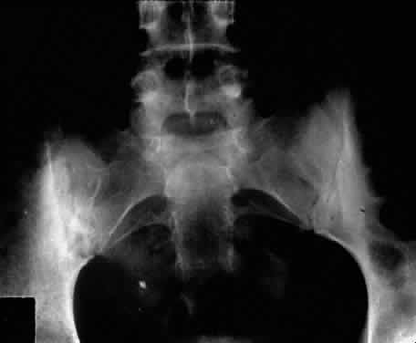
  Bilateral radiographic sacroiliitis
  Radiographic syndesmophytosis


*Diagnosis requires four of the five clinical criteria or bilateral radiographic sacroiliitis and one other criterion.
(Bluestone R: Ankylosing spondylitis. In McCarty DJ (ed): Arthritis and Allied Conditions, p 819. Philadelphia, Lea & Febiger, 1985)


Ankylosing spondylitis is distinctly different from rheumatoid arthritis, although earlier literature did not make this distinction. The sacroiliac joint is usually the most easily visible area of involvement and is best seen in oblique views on radiography of the sacroiliac joint as periarticular sclerosis and irregular loss of the joint space. These patients are seronegative for rheumatoid factor and may have an elevated erythrocyte sedimentation rate.3,6

The presence of HLA-B27 antigen in 90% to 95% of white patients and 50% of black patients with ankylosing spondylitis is well documented, and the genetic predisposition to this disease has been recognized for many years. The relationship, however, is still incompletely understood at this point. HLA-B27 antigen is present in 6% to 14% of the white American population and 3% to 4% of the black American population. It is clear that people with this HLA type are at greater risk of developing one of the spondyloarthropathies, particularly ankylosing spondylitis, but as yet poorly defined environmental factors must be involved in triggering the development of the disease process.3,8 The development of acute iridocyclitis is even more strongly correlated with HLA-B27 antigen than is its association with joint changes. Of patients with HLA-B27 antigen, 20% to 25% will develop symptoms or radiographic evidence of spondylitis at some time in their life, and milder forms of the disease are commonly not diagnosed but believed to be due to back “strain” or injury. The incidence of clinical spondylitis is about equal between men and women, but the disease tends to be milder and more difficult to diagnose in women; its prevalence appears to approach 2% in those in the North American population who are HLA-B27 positive and who have been studied.

The mechanism of how HLA-B27 antigen is involved in this disease process is unclear. HLA determinants are located on chromosome 6. These determine cell surface markers that enable the immune system to recognize each cell as self. One theory that has been advanced suggests that the HLA-B27 antigen may be similar to antigens of the cell wall of certain exogenous agents or may be changed by the agents in a manner whereby the immune system has difficulty recognizing or responding appropriately to the agent or the altered HLA-B27 antigen. In one study, patients with ankylosing spondylitis had lower in vitro responsiveness of lymphocytes to Klebsiella antigens than HLA-B27-positive and HLA-B27-negative controls.9 Antisera to certain isolates of Klebsiella lysed the lymphocytes of HLA-B27-positive patients with ankylosing spondylitis, but not the lymphocytes of HLA-B27-positive or HLA-B27-negative controls. This suggests that perhaps some Klebsiella antigens cross-react with a gene product closely associated with HLA-B27 or a Klebsiella-modified B27 antigen in patients with ankylosing spondylitis. Cross-reacting antigens have been identified in HLA-B27 and Klebsiella, Shigella, and Yersinia.10

The role of Klebsiella has not been clarified, and no other agent has been substantiated with respect to ankylosing spondylitis. Chlamydia has been suggested in some cases of Reiter's syndrome, especially in instances of nonspecific urethritis.11–13 Shigella, Salmonella, and Yersinia have been involved in clinical epidemics of postinfectious arthropathies.14 Additional theories involve HLA-B27 linkage with a specific immune response gene that predisposes patients to the disease, perhaps making them more susceptible to infection. The role of these factors remains to be clarified.

The acute iridocyclitis associated with ankylosing spondylitis is characterized by rapid onset of pain, photophobia, and blurred vision. Conjunctival, episcleral, and scleral injection and edema are seen. Poorly defined keratic precipitates are seen in the lower half of the corneal endothelium, and the anterior chamber has heavy flare that may be uneven. If the process is severe, there may be clot formation in the pupil space.15,16 Cells in the anterior chamber may be so numerous that hypopyon will occur. Glaucoma can result from anterior chamber reaction blockage of the angle in the acute phase of inflammation and from pupil block from synechiae. Synechiae form early and, if not broken, will form lasting adhesions. Mydriatic and cycloplegic therapy is needed early in treatment. Spillover of inflammatory cells and inflammatory debris into the vitreous may occur, and the presence of disc blurring and macular edema is sometimes observed. This is sometimes also associated with hypotony. Posterior subcapsular cataracts and diffuse lens clouding are seen with severe prolonged episodes and repeated acute recurrences.15,16

The typical episode lasts from 2 to 6 weeks. Aggressive suppression of the inflammatory reaction with topical corticosteroids is usually sufficient and reduces tissue damage, if an early intensive schedule is used, rather than increasing the drop schedule as the reaction increases. These patients may need to be seen daily when they are acutely active and may need to be seen every 2 to 3 days until the process is stable or clearly resolving. Treatment must be continued for several weeks as the process is resolving or reactivation will occur. Oral corticosteroids can be given for short periods of time. Some patients experience elevation of intraocular pressure with corticosteroid therapy, particularly as the eye improves and the ciliary body is again more able to produce aqueous humor. The long-acting effects of periocluar injection of corticosteroids may become a more serious problem with persistent corticosteroid-induced glaucoma than the episode of acute iridocyclitis that was being treated.

Frequent episodes of recurrent iridocyclitis may cause significant disability that results in loss of work, discomfort, and structural damage to the eye. These patients may benefit from longer term treatment with oral nonsteroidal anti-inflammatory agents, such as indomethacin or naproxen. These medications may help to reduce the severity and frequency of recurrences, but the pain-decreasing effects of these medications may make it more difficult for the patient to recognize an acute recurrence of the iridocyclitis. Patients should be examined for exacerbations of the inflammation if they note any change in vision, even minor symptoms.

REITER'S SYNDROME OR POSTINFECTIOUS REACTIVE ARTHRITIS

Reiter's syndrome is a clinical syndrome usually described as arthritis, conjunctivitis, or iridocyclitis and nonbacterial urethritis or cervicitis. A better definition may be needed, because not only may these not all be present, but also dysentery and mucocutaneous disease with balanitis, oral ulceration, or keratoderma blennorrhagicum may be part of the clinical picture. Enthesopathy of the plantar fascia or Achilles tendon is also suggestive of Reiter's syndrome. It is more commonly identified in males, but may be more frequent in females than previously thought.14 The incidence reported by Noer17 in US Navy personnel over a 10-year period was 4 in 100,000 men per year. HLA-B27-positive persons have approximately a 25% risk for Reiter's syndrome development after Shigella infection.18 Although the cause is unknown, the high correlation with the presence of HLA-B27 (75%)19 is clearly recognized. The previous discussion in the section on spondyloarthropathies concerning this HLA-B27 association demonstrates a genetic predisposition in a high percentage of these patients. In Reiter's syndrome, infectious agents are suggested by reports of clinically indistinguishable acute disease after epidemic dysentery and sexually transmitted nongonococcal urethritis thought to be due to Chlamydia20 or possibly Mycoplasma (Ureaplasma urealyticum).21,22 Large epidemics of dysentery have been linked to multiple occurrences of arthritis, urethritis, and iridocyclitis.17,23 Shigella, Campylobacter, Salmonella, and Yersinia have all been implicated.24 Microbial antigens have been identified in the synovium after infectious with Chlamydia, Yersinia, and Salmonella.25–27 Salmonella typhimurium is a frequently associated Salmonella pathogen causing reactive arthritis.

This seronegative arthritis usually involves larger joints and the weight-bearing joints of the lower extremities. The knees and ankles are most frequently involved, with redness and diffuse swelling. Multiple joint involvement is usual. Periostitis and tendinitis may occur, especially involving the Achilles tendon, producing heel pain. Sacroiliac radiographic changes are present in up to 32% of patients.14 Children may be affected rarely.28 Reiter's syndrome tends to affect young adults in the range of 16 to 40 years of age. The diagnosis is sometimes hard to establish because the urethritis or cervicitis may be forgotten or suppressed and the enteritis and other symptoms may have been mild or not identified as abnormal. If a urethritis or cervicitis is present, cultures should be considered to make sure no treatable organism is present, such as gonococcus. Serologic testing for syphilis will help to rule out this sometimes-associated venereal problem.

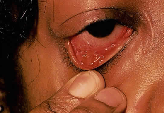
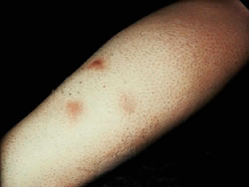
Low back pain due to insertional tendinitis and sacroiliitis is common. The dermal lesions typified by keratoderma blennorrhagicum, described as a hyperkeratotic erythematous dermatitis resembling pustular psoriasis, may not be present until later in the disease. It usually involves the hands and feet but may involve other areas. Superficial ulcers of the mucous membranes are frequent.29 Enteritis is usually a prolonged diarrheal episode with frequent passage of bloody, loose stools, but it may be a 24-hour episode of increased bowel activity.29

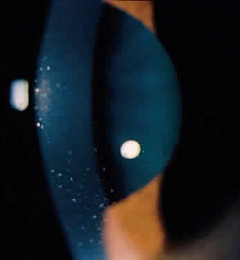
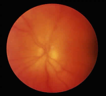
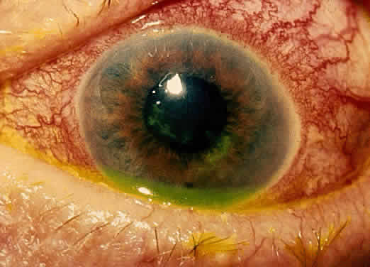
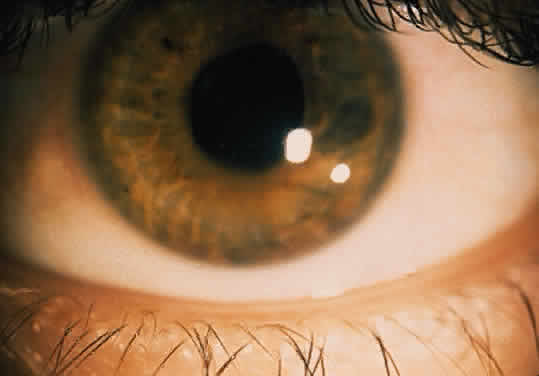
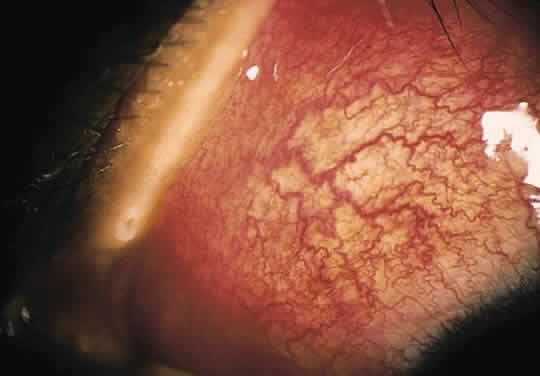
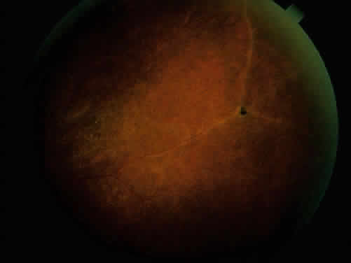
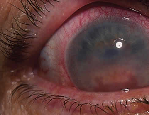
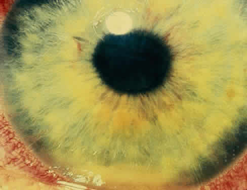
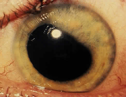
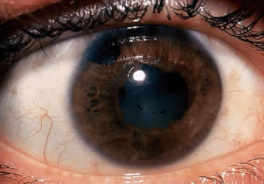
The conjunctivitis is usually described as mucopurulent and may be sterile. It may be accompanied by an iridocyclitis, episcleritis, or scleritis. Punctate and subepithelial corneal involvement has been reported. The iridocyclitis may be the presenting ocular manifestation. Recurrences are usually associated with an acute iridocyclitis, which is of rapid onset with conjunctival and episcleral edema and injection. The corneal endothelium has cellular debris and poorly defined, small-sized to medium-sized keratic precipitates.30 Heavy flare and cells and a very early tendency toward formation of posterior synechiae is characteristic, more so than in most other forms of acute iridocyclitis (Figs. 1, 2, and 3). Even the most aggressive pupil dilation management is sometimes inadequate for preventing synechiae. A peripheral iridectomy may be necessary to prevent iris bombé and angle closure if the synechiae cannot be broken enough to establish an opening for aqueous through the pupil (Fig. 4). The heavy flare is sometimes so plasmoid that cells are immobile and a fibrinlike clot may be seen in the pupil opening as the inflammation resolves. Cells and inflammatory debris may be seen in the vitreous, and blurring of the disc margins and macular edema may occur with severe or prolonged episodes. Lens clouding and posterior subcapsular cataracts occur with prolonged or repeated episodes. Hypotony can occur after a severe or prolonged course and may persist after resolution. Occasionally, secondary glaucoma may occur, owing to the anterior chamber reaction, in which case it will resolve as the inflammation resolves. With repeated recurrences, damage to the trabecular meshwork may occur, and prolonged recalcitrant glaucoma may result that may be poorly responsive to any medical or surgical management. This can be a serious factor in permanent visual loss with this type of iridocyclitis.15,16,30,31

Fig. 1. Early onset complete posterior synechiae in iridocyclitis with Reiter's disease.

Fig. 2. Partial dilation of pupil in Figure 1.

Fig. 3. Completed dilation of pupil in Figure 1 with pigment on anterior surface of the lens.

Fig. 4. Complete pupillary synechiae with pupil block requiring peripheral iridectomy in severe iridocyclitis with Reiter's disease.

The usual course is 2 to 6 weeks for acute iridocyclitis. Topical corticosteroids and mydriatics should be used early and aggressively to reduce tissue damage. Frequent follow-up visits are necessary until the process is clearly resolved. The use of systemic corticosteroids may be necessary and should be used with the same indications and precautions as in other inflammatory disease and for a short period of time. Prolonged topical treatment is necessary for several weeks after the inflammation has cleared, because early withdrawal of topical corticosteroids will frequently result in return of inflammatory changes.

Occasionally, this process will become chronically active or recur at frequent enough intervals to require long-term management. Oral nonsteroidal anti-inflammatory agents, such as indomethacin or naproxen, may be helpful and may be needed for many months for two reasons: (1) to help reduce the level of activity and the frequency of recurrences in the ocular inflammation; and (2) to allow decreasing the corticosteroid dosage to decrease the rate of cataract formation and other associated side effects of corticosteroids. The decreased awareness of pain sometimes seen with these medications may alter the patient's recognition of recurrences. If they have any change in symptoms, they should be examined to evaluate for recurrence of an acute episode of inflammation.

Back to Top
INFLAMMATORY BOWEL DISEASE
Ulcerative colitis and regional enteritis, such as Crohn's disease or granulomatous colitis, have been reported to be associated with acute iridocyclitis. Ulcerative colitis is characterized by mucosal ulcerations of the rectal area, colon, and occasionally terminal ileum. The mucosa is inflamed and friable and may contain crypt abscesses. Toxic megacolon, hemorrhage, and spontaneous bowel perforation may occur with exacerbations. Carcinoma may be a late complication. Regional enteritis may variably involve large and small bowel with transmural inflammation, including granulomas, fissuring, discrete ulcerations, serositis, and mesenteric lymphadenitis. Strictures, fistulas, and inflammatory masses are seen as complications. The causes of these conditions are unknown.32–35

The associated arthritis and iridocyclitis, however, are similar in both conditions. Peripheral arthritis is seen in approximately 12% of patients with ulcerative colitis and approximately 20% of patients with regional enteritis. Radiographic evidence of sacroiliitis has been reported to show an incidence of approximately 18% and to be equal in both sexes in both conditions. Clinical spondylitis is seen in association with bowel inflammation in 4% to 7% of these conditions. The overall incidence of HLA-B27 in these two conditions may be slightly elevated, but is similar to that in the normal population. HLA-B27 is present in 53% to 75% of patients who have both spondylitis and inflammatory bowel disease. The sacroiliac involvement is frequently asymmetric (Fig. 5). It is not clear whether the spondylitis is secondary to the inflammatory bowel disease or to an overlap of the conditions. Spondylitis occurs before the bowel disease in 25% of patients, and there seems to be little correlation in the activity of symptoms between the sacroiliitis and the bowel disease. Colectomy does not affect the sacroiliitis, even when the bowel disease is improved. This is in contrast to the activity of the peripheral arthritis, which does seem to parallel the activity of the inflammatory bowel disease in 60% to 74% of patients. The peripheral arthritis also precedes the bowel disease in 7% to 11% of patients.33,36,37 It is not clear how these relate to the associated iridocyclitis.

Fig. 5. Asymmetric sacroiliitis with sclerosis and narrowing of right sacroiliac joint in a 22-year-old woman with inflammatory bowel disease and iridocyclitis.

Ocular inflammatory changes include conjunctivitis, episcleritis, keratitis, acute and chronic iridocyclitis, and posterior changes as well.38–43 The acute iridocyclitis is milder and frequently not accompanied by as much pain, photophobia, and redness as in the acute iridocyclitis associated with other spondyloarthropathies. Mild injection of the conjunctival and episcleral vessels and blurred vision may be the only symptoms of an acute flare of activity. The endothelium of the cornea usually has cellular debris and poorly formed keratic precipitates in the lower cornea. The anterior chamber contains cells and moderate flare, and a protein clot is not infrequent in the pupil, with early formation of posterior synechiae (Fig. 6); however, the synechiae are usually more easily broken than in ankylosing spondylitis and Reiter's syndrome.44 Treatment with topical corticosteroids and mydriatics is usually effective. The activity of the iridocyclitis has been reported to follow the activity course of the inflammatory bowel disease. Systemic treatment of inflammatory bowel disease has been noted to help the ocular inflammation. Decreasing the systemic treatment occasionally allows ocular inflammation to become visible and requires close observation and appropriate increase in local therapy.39,44–46 Surgical resection of inflamed bowel for other medical reasons has sometimes been reported to coincide with lessening of ocular inflammation,44,46 but these observations need better evaluation.42 Posterior segment manifestations, including retinal and choroidal vascular disease, are also sometimes noted.47,48

Fig. 6. Partial posterior synechiae and fibrin clot in pupil with iridocyclitis and Crohn's disease.

Back to Top
PSORIATIC ARTHRITIS
Acute and chronic iridocyclitis occurs with psoriatic arthritis. Psoriatic arthritis is sometimes confused with Reiter's syndrome because of the similarity of skin lesions (i.e., pustular psoriasis and keratoderma blennorrhagicum). In patients with keratoderma blennorrhagicum, other psoriatic lesions develop as well.49,50 In psoriatic arthritis, distal interphalangeal joints may be involved and nail lesions are common. The correlation of widespread ankylosis with spondylitis (40%) and sacroiliitis (21%) is frequently observed,51 but the presence of HLA-B27 has a higher correlation with these associated complications.52 Psoriatic arthritis is found in approximately 20% to 34% of patients with psoriasis, and estimates of 0.10% in the general population are considered reasonable.51

Conjunctivitis is seen in approximately 20%, scleritis in approximately 2%, and an association between HLA-B27 and iridocyclitis in approximately 10% of patients.53 The iridocyclitis seen in connection with this condition tends to be similar to that seen with ankylosing spondylitis and is managed in a similar manner. There may be multiple recurrences, and the iridocyclitis may become chronic, unrelenting, and, at times, difficult to control.

Back to Top
WHIPPLE'S DISEASE
Iridocyclitis associated with Whipple's disease or intestinal lipodystrophy may have an acute onset, but more characteristically the iridocyclitis is chronic and associated with vitritis and retinal involvement.54 In this multisystem disease, the tissue is infiltrated with granulomas containing macrophages with periodic-acid-Schiff (PAS)-positive material. Macrophages containing PAS-positive material and what resembles degenerating bacteria have been observed in the eye.55 Whipple's disease is caused by an uncultured microbe that is a gram-positive actinomycete unrelated to other microbes.56 The PAS-positive material is derived from the bacteria. The disease is not easily spread and must be related to an as yet unidentified host immune deficiency for this organism, as these patients do not have a propensity for infection with other organisms. The arthritis and associated systemic involvement, including malabsorption, diarrhea, and weight loss with inflammation in multiple organs (e.g., pneumonia, pleuritis, pericarditis, thyroiditis, papilledema), may go undiagnosed for years. There seems to be an association between Whipple's disease and sacroiliitis and spondylitis. Back pain was present in 19%, and there also appears to be an increased incidence of HLA-B27 antigen.32 This diagnosis should be considered in acute, recurrent, or chronic ocular inflammatory disease of uncertain etiology that is associated with these types of systemic disease. Tetracycline was reported to decrease the ocular inflammatory activity in one case,54 and it reduced other types of systemic involvement.32 Other antibiotics have been used effectively as well,32 and the disease is now considered completely reversible by treatment with other antibiotics.57 Long-term treatment is usually necessary. These patients, once identified, need to be followed indefinitely because of relapses of active disease.
Back to Top
ACUTE SARCOIDOSIS
Sarcoidosis remains a systemic granulomatous condition of unknown etiology. It is recognized by histologic appearance and clinical features and sometimes by exclusion of other etiologies. The granuloma formation is probably a final common tissue response pathway to many different antigens that are not readily cleared from the tissue. The histologic pictures of diseases associated with a number of environmental agents, such as beryllium, pine dust, or peanut dust are similar to those of sarcoidosis.58,59 This seems to be a reasonable explanation for the inability to find one specific causative agent despite the relative frequency of the disease, which is estimated to be between 30 and 64 per 100,000 based on clinical examination. Autopsy studies, however, show that the true incidence may be 10 times this rate, suggesting that most cases go undiagnosed.60,61 Fatigue is sometimes the only symptom of systemic involvement. Skin lesions, conjunctival granulomas (Fig. 7), lymph node biopsy specimens, and other involved tissues show high CD4+ T-cell concentrations in epitheloid cell noncaseating granulomas. Certain body compartments may be more involved, and systemic circulating T-cell populations or other systemic indicators of inflammatory activity may show no abnormality. The diagnosis may be easier when the chest x-ray shows the typical mediastinal lymph node enlargement, or when systemic levels of angiotensin-converting enzyme or serum lysozyme are elevated.62–64 Genetic factors apparently play a role, and immune complexes have been noted; however, these influences remain to be clarified.

Fig. 7. Conjunctival nodules in patient with sarcoidosis.

Patients with acute anterior inflammation due to sarcoid usually present with a red, painful scleritis or more frequently an iridocyclitis. This is more frequently bilateral in the acute presentation, and the keratic precipitates are not well defined (Fig. 8). Posterior synechiae may form with broad-base adhesions of the iris to the lens, and elevated intraocular pressure is sometimes seen. The synechiae can usually be broken with intensive pupil dilation, and topical corticosteroids usually produce a rapid suppression of the inflammation, but recurrence of the inflammation is likely if treatment is decreased too early.

Fig. 8. Small indistinct keratic precipitates with fibrinlike debris in patient with acute sarcoid iridocyclitis and pulmonary disease.

The acute sarcoid course frequently becomes recurrent and chronic and may recur after months or years of inactivity. The recurrent and chronic course is likely (1) to be less symptomatic, with little vascular injection; and (2) to have the sharply defined peripheral corneal precipitates and Koeppe nodules at the pupil margin more commonly associated with chronic sarcoid iridocyclitis. These are also likely to be the sites of peripheral anterior synechiae and posterior synechiae if not treated appropriately.

Treatment is complicated by the tendency for sarcoid to clear and undergo spontaneous remission, and then to recur at a later date. Topical treatment is usually sufficient. Consideration can sometimes be given to periocular corticosteroid injection. This is particularly useful when topical corticosteroids are ineffective or compliance is a factor, or when cystoid macular edema is a persistent problem. If there are no contraindications, the injection can be given subconjunctivally in the inferior fornix for a more anterior effect. For more posterior inflammation, including cystoid macular edema, a periocular injection can be given more posteriorly from a superotemporal approach after the application of adequate topical anesthesia. It should be noted that there are multiple potential side effects of periocular corticosteroid injections, including corticosteroid-induced glaucoma, cataract formation, subconjunctival hemorrhage, ptosis, and inadvertent intracameral injection. Oral corticosteroids, if indicated, can frequently be used in moderate to low alternate-day dosages if chronic use is needed to control the inflammatory process. This is usually better tolerated and produces less severe and fewer systemic side effects.65,66

Back to Top
GLAUCOMATOCYCLITIC CRISIS (POSNER-SCHLOSSMAN SYNDROME)
Glaucomatocyclitic crisis is recurrent open-angle glaucoma associated with mild iridocyclitis that follows a predictable episodic course. It is almost always unilateral. The pupil of the affected eye may be larger. There does not seem to be any associated systemic disease, and few symptoms are encountered. There is little evidence of injection of the conjunctiva. A few keratic precipitates may be present. The anterior chamber contains a few cells. The anterior chamber angle is open throughout the episode, and the intraocular pressure may stay elevated for several weeks and then return to normal. Inflammatory changes may be present for a month or longer. Between recurrences, no abnormalities may be noted. Results of the usual provocative tests for glaucoma have been reported to be normal.67–69 More recent studies have indicated that there may be an increased incidence of elevated intraocular pressure between episodes, as well as decreased facility of outflow, increased pressure response to topical corticosteroids, disc cupping, and field loss in the involved and noninvolved eye. There is a suggestion that glaucomatocyclitic crisis may be associated with primary open-angle glaucoma.70,71

The course of glaucomatocyclitic crisis has been described as self-limiting, usually leaving no residual damage. Topical corticosteroids and medical management of the intraocular pressure with topical timolol or oral or topical carbonic anhydrase inhibitors during the active episode are usually adequate. The response may be variable, and sometimes the inflammation and pressure will return as treatment is discontinued. One must be alert for patients with other causes of inflammation presenting with similar findings.72 Mild herpes simplex uveitis can mimic this syndrome and may be the cause in many patients.

Back to Top
BEHÇET'S DISEASE
Behçet's disease is a clinical syndrome characterized by acute-onset iridocyclitis with hypopyon, posterior intraocular inflammation, recurrent aphthous stomatitis, genital aphthous ulceration, and multiple systemic manifestations, including arthropathy, gastrointestinal disease, subcutaneous nodules (Fig. 9), erythema nodosum, cutaneous vasculitis, neurologic involvement, thrombophlebitis, and other areas of vasculitis, both arterial and venous. Most of the disease process can be related to effects of multifocal vasculitis. There is no definite diagnostic test for Behçet's disease, and the clinical course may resemble other conditions.

Fig. 9. Subcutaneous nodules in Behçet's disease.

A simplified set of diagnostic criteria has been proposed based on the following six common clinical manifestations73: (1) recurrent aphthous stomatitis, (2) genital aphthous ulcers, (3) uveitis, (4) synovitis, (5) cutaneous vasculitis, and (6) meningoencephalitis. Diagnosis usually requires at least three of the six criteria, with aphthous ulceration being a necessary finding. Aphthous ulceration and one other criterion may be sufficient for diagnosis of incomplete forms.73 More rigid criteria have been proposed in Japan, where this disease is more common, and involves combinations of “major” and “minor” criteria for diagnosis of complete and incomplete forms of Behçet's disease.74,75 Briefly, the major signs are (1) recurrent aphthous ulcerations of the mouth, (2) skin lesions, (3) eye lesions, and (4) genital ulcerations. The minor signs are (1) arthritis, (2) gastrointestinal lesions, (3) epididymitis, (4) vascular lesions, and (5) central nervous system disease. For complete Behçet's disease, all of the four major lesions must be present. In the incomplete type, three of the four major criteria are present, or the patient has eye disease plus one other major criterion. International criteria require recurrent oral ulceration and two of the following: (1) recurrent genital lesions, (2) eye lesions, (3) skin lesions, or (4) positive pathergy.76,77

This disease is more common in Japan and in the eastern Mediterranean countries. It is reported to have a prevalence of 1 in 10,000 in Japan78 and to cause 15% of all cases of uveitis.79 It has been shown to occur in Minnesota at an incidence of 1 per 300,000 per year.78 In San Francisco it causes 0.4% of all cases of uveitis.80 It does not seem to occur frequently in Japanese-Americans.78 Behçet's disease has been shown in Japan to have a strong correlation with HLA-B5 and particularly the B5 split antigen B51.80 The ocular disease seems even more closely related to HLA-B5.81 Additional environmental factors seem to be involved. Men are more commonly affected than women in most series, and the disease is more common in young adults.

Aphthous stomatitis and vulvitis alone in young girls or women are usually self-limiting and can be due to many causes besides Behçet's disease, even though they are necessary for the diagnosis of Behçet's disease. The ulcers in Behçet's disease are usually the first manifestation (70%) and resemble uncomplicated canker sores. They are frequently multiple, are painful, and heal in 3 to 30 days, usually without scar formation. Genital ulcers of the vulva and vagina in females and of the scrotum and penis in males are similar to the oral ulcers, but less painful.73

Arthritis occurs in 50% to 60% of patients and involves large and small joints. The knees and ankles are most commonly involved. Though recurrent, the arthritis is usually not destructive. Cutaneous lesions, including pustules, papules, and nodules, are thought to be related to vasculitis. Nodules resemble erythema nodosum, but unlike erythema nodosum, these may become ulcerated. A nonspecific condition resulting in formation of a pustule following a sterile needle stick has been called “pathergy” and has been reported to be as high as 40% in Behçet's disease.44 Others have not found this condition to be present. Fever, headache, neck stiffness, and cerebrospinal fluid pleocytosis is found along with the meningoencephalitis. Bacterial and other causes must be ruled out.73,82,83

Studies, including inoculations of cerebrospinal fluid into rabbits and posterior uveal tissue into chorioallantoic membrane then passed into mice, causing encephalitis, have suggested a viral cause.84–86 Abnormalities in the immune system have been reported, but as yet have failed to give a comprehensive explanation for the disease process. Immune complex disease would be compatible with a drop in complement just before a flare of the intraocular inflammation and has been shown to occur, suggesting activation of complement pathways.87 Abnormalities in the secretory component of IgA in saliva and gastrointestinal secretions have been reported and should suggest an abnormality that might allow a foreign antigen to gain access to the circulation.88 There are reports that the disease can be activated by certain foods, such as English walnuts.89 The presence of damaged membrane fragments, presumably from complement activity on the cell wall,90 helps to implicate this cellular damage in the rapid onset of symptoms and probably plays a role in hypopyon and vascular occlusive events observed in the eye.81 Heat shock proteins from Streptococcus sanguis or other bacterial agents have also been implicated,82 and antibodies against ocular and oral antigens have been reported.83

The acute iridocyclitis is suggestive of vascular occlusive changes, or acute tissue damage, with sudden onset and massive outpouring of cells into the anterior chamber or vitreous (Fig. 10). Vascular injection is not as pronounced as the number of cells in the anterior chamber or vitreous would suggest, and protein in the anterior chamber may be minimal. The cells settle to the dependent portion of the anterior chamber, forming a hypopyon. There may be little tendency toward formation of keratic precipitates. The process may be unilateral or bilateral. Episodes usually are recurrent, and recurrences produce increased tissue damage, progressing to more chronic involvement. All vascular tissues of the eye may become involved, including episcleritis, scleritis, iridocyclitis, retinal and choroidal vasculitis, and optic neuritis. Retinal focal ischemic areas of edema and optic nerve edema (Fig. 11) are seen in the acute phases, and retinal and optic nerve atrophy along with retinal pigment epithelial changes resembling secondary retinitis pigmentosa, attenuation, and sheathing of retinal vessels are seen in the later stages of ocular involvement.76,91–93

Fig. 10. Hypopyon, iris sector ischemia, posterior synechiae and cataract formation in severe iridocyclitis with Behçet's disease.

Fig. 11. Optic nerve and retinal edema with cells in the vitreous in posterior involvement with acute Behçet's disease.

Ocular inflammation responds inconsistently to local use of corticosteroids, and high doses of oral corticosteroids may be palliative. Mydriasis is usually needed. Depending on the duration of inflammation, frequent topical corticosteroids may be sufficient. The course must be watched closely and management based on response to treatment. If the inflammation is prolonged, treatment must be directed at the systemic disease. Reports show variable response to many agents, including colchicine, corticosteroids, transfer factor, levamisole, and immunosuppressants. Treatment for ocular involvement should be carried out together by an ophthalmologist and internist because of the associated complications. Chlorambucil has been reported to be beneficial, especially in posterior ocular involvement, but is still less than satisfactory in many patients and may have serious systemic side effects. The potentially serious side effects of chlorambucil and other immunosuppressive agents, including oncogenic and cytotoxic potential effects that may lead to death, must be carefully weighed against the benefits to be gained, and informed consent should be obtained before the use of these agents may be considered.73,94–98 Cyclosporin A and tacrolimus (FK506) have been used to treat uveitis, but have had mixed results.99,100 Currently, no available treatment is effective in all situations.

Back to Top
HERPES SIMPLEX, HERPES ZOSTER, AND OTHER VIRAL IRIDOCYCLITIS DISORDERS
Acute iridocyclitis may be caused by herpes simplex (Fig. 12), herpes zoster, and other suspected viral disorders. The accompanying skin manifestations and associated corneal lesions are helpful in establishing the diagnosis. These should be considered when the acute symptoms of redness, pain, photophobia, and tearing are found with poorly defined keratic precipitates, cells in the anterior chamber, sector iris atrophy (Fig. 13), and other acute anterior segment inflammatory changes, especially if unilateral.

Fig. 12. Acute herpes simplex iridocyclitis and subsequent keratitis.

Fig. 13. Corneal endothelial cells, indistinct keratic precipitates, and sector iris ischemia and atrophy (2 to 3 o'clock) with herpes simplex iridocyclitis.

Back to Top
VASCULAR OCCLUSIVE DISEASE (ANTERIOR SEGMENT ISCHEMIA)
Acute iridocyclitis that occurs after vascular occlusion of anterior segment vessels is seen with conditions such as (1) acute anterior necrosis following surgical procedures that compromise anterior segment blood supply (e.g., removal of multiple muscles in retinal surgery,101 or multiple muscle strabismus procedures102); (2) giant cell arteritis or aortic arch syndromes; (3) other forms of vasculitis; and (4) vascular occlusive disease where anterior segment arterial supply is affected.102,103 Disseminated intravascular coagulation syndromes,104 clotting abnormalities, hyperviscosity syndromes, and hemoglobinopathy—including sickle cell disease and other such diseases—can sometimes manifest as vascular occlusive episodes. Posterior involvement is usually the predominant manifestation in these systemic diseases.

The anterior symptoms and findings may be of acute onset. The conjunctiva and episclera are mildly injected. The cornea has scattered endothelial cellular debris with little tendency to form keratic precipitates. Severe ischemia may be accompanied by diffuse episcleral hyperemia, hypotony, corneal edema, and wrinkles in Descemet's membrane. A few cells and flare are present in the anterior chamber, unless the vascular supply has been dramatically obstructed, in which case the anterior chamber reaction will be more severe. There is a tendency for posterior synechiae to occur. The iris may be poorly reactive and may not dilate well with mydriatics. Keratopathy, hypotony or glaucoma, pupil dilation, iris atrophy, and neovascularization (rubeosis irides) are sometimes seen. Symptoms of amaurosis fugax and pain without photophobia may be present. Vision may be decreased more than expected from the clinical findings. The anterior vitreous usually also contains cells and debris with little tendency toward clumping.105

Diagnosis is usually suspected from the history of extensive anterior segment vascular supply injury, the clinical appearance of anterior segment reaction, or in the case of other causes of vascular occlusion, from the accompanying posterior segment disease, systemic symptoms, and history. An erythrocyte sedimentation rate should be obtained on an emergency basis for anyone having suspected giant cell arteritis (cranial arteritis or temporal arteritis) or polymyalgia rheumatica. The diagnosis should be clarified immediately, and if the diagnosis can be substantiated, treatment should probably be started with systemic corticosteroids without delay. This diagnosis probably should be made in cooperation with an internist and as quickly and firmly as possible on the basis of a tissue diagnosis (if indicated), elevated sedimentation rate, compatible clinical course, and associated findings.106,107

Additional consideration should be given for other causes of vasculitis or vascular occlusive disease, including evaluation for diabetes, antinuclear antibody, blood pressure evaluation, carotid flow studies for carotid occlusive disease, auscultation of the neck vessels for a bruit, and a good general medical evaluation for any additional systemic disease.108

Back to Top
SCLERITIS
Iridocyclitis may be present in association with many acute conditions that primarily involve the sclera. Sometimes tissue damage extending to involve intraocular structures will produce acute iridocyclitis. This is a complication in many of the conditions in which scleritis is the primary ocular inflammatory involvement, including rheumatoid arthritis, polyarteritis nodosa, polychondritis, Wegener's granulomatosis (Fig. 14), dermatomyositis, systemic lupus erythematosus, and nonspecific vasculitis. A posterior scleritis or an orbital vasculitis or inflammation may secondarily involve the anterior segment and may present a particularly diagnostically difficult set of signs and symptoms with a secondary low-grade iridocyclitis.

Fig. 14. Acute scleritis and iridocyclitis with Wegener's granulomatosis.

Back to Top
KAWASAKI'S DISEASE
Kawasaki's disease or “neurocutaneous lymph node syndrome in children” is an acute multisystem disease with pathologic features resembling a vasculitis. It is usually seen in children, more commonly in those of Japanese descent. It is of unknown etiology, although an as yet unidentified infectious etiology has not been ruled out. It has been confused with infantile or childhood polyarteritis nodosa.

Acute iridocyclitis has been reported as part of Kawasaki's disease. Ocular involvement with vasculitis that is part of this disease may manifest as usually bilateral photophobia, nonexudative conjunctival injection, and a mild anterior segment inflammation, with cells and poorly defined keratic precipitates on the corneal endothelium and cells in the anterior chamber and anterior vitreous. The course is usually several weeks in duration and not characteristically recurrent.109,110

The eye disease is part of the systemic vasculitis. A fever of unknown etiology lasts 1 to 3 weeks. Other clinical features include lymphadenopathy, a sore throat, a maculopapular rash over the trunk, “strawberry” tongue, fissured lips, and desquamation of the skin at the junction of the nails and skin on the fingertips.111–113 Involvement of the aorta and coronary arteries is a serious complication and may be suggested by the clinical course, electrocardiographic abnormalities, and echocardiography.114,115

Back to Top
SYPHILIS
Acute iridocyclitis is associated with secondary syphilis and interstitial keratitis. This association should be considered in any patient with an acute iridocyclitis with any suggestion of syphilis-associated disease, retinal vasculitis (Fig. 15), interstitial keratitis (Fig. 16),116 or syphilis per se.116–120

Fig. 15. Acute retinal vasculitis with iridocyclitis and vitritis due to syphilis in previously unknown HIV-positive patient.

Fig. 16. Acute iridocyclitis with interstitial keratitis.

Back to Top
VOGT-KOYANAGI-HARADA SYNDROME
Patients may present with both acute iridocyclitis and meningeal symptoms of headache and neck stiffness.120–123 Posterior inflammatory changes include exudative retinal detachment and macular edema, vitreous inflammation, and optic nerve edema.124 Poliosis, alopecia, vitiligo, and auditory symptoms are frequently encountered, but are usually part of the longer term course of the disease.121–125
Back to Top
SYMPATHETIC OPHTHALMIA
Patients with sympathetic ophthalmia may present with acute symptoms and findings of an acute iridocyclitis. The history of trauma or injury to one eye and later onset of inflammation in the other eye should alert one to the possibility of this diagnosis.
Back to Top
ENDOPHTHALMITIS
Endophthalmitis should be considered when acute inflammation of the anterior segment is encountered. Pain is usually a significant finding. Filtering blebs for glaucoma, suture tracts, injury, recent intraocular surgical history, site of infection or abscess, and so on should also raise the suspicion of this diagnosis. Cells and heavy flare with persistent synechia formation and vitreous inflammation is seen. Hypopyon may be present. There is usually little tendency to form keratic precipitates. Among other possible bacterial causes, Listeria monocytogenes, Neisseria gonorrhea, Neisseria meningitides, Haemophilus influenzae, Mycobacterium leprae, and Mycobacterium tuberculosis are worthy of special note and may cause an endogenous endophthalmitis. Nonbacterial infections with Candida albicans and Coccidioidomycosis should also be given special consideration.126–128 Many other less frequently noted organisms are potential causes of endogenous endophthalmitis that can cause an acute anterior inflammation, and all need immediate attention. Appropriate identification of the causative agent and antibiotic therapy is needed as soon as possible. After cataract surgery, especially extracapsular surgery, an endophthalmitis caused by Propionibacterium acnes has been noted with some frequency.129 This infection may consist of a low-grade smoldering inflammation with white plaquelike lens capsular opacities and cellular precipitates on the corneal endothelium that are poorly formed over the lower cornea and may have pigment present as well. Occasionally, there are more acute symptoms of redness, hypopyon, and decreasing visual acuity. Identification by culture and anaerobic cultures may be needed. Treatment with an appropriate antibiotic is usually sufficient. Occasionally vitrectomy or removal of residual cortex is necessary.
Back to Top
MASQUERADE SYNDROMES
Many conditions that are not primarily inflammatory may resemble an acute iridocyclitis. Most masquerade syndromes are more chronic in their course, but some neoplastic processes, especially lymphoma, intraocular foreign bodies, unsuspected trauma, retinal detachment, and other conditions can present as acute inflammatory changes.
Back to Top
REFERENCES

1. Hogan MJ, Kimura SJ, Thygeson P: Signs and symptoms of uveitis (part 2): 1. Anterior uveitis. Am J Ophthalmol 47:155, 1959

2. Brewerton DA, Caffrey M, Nicholls A et al: Acute anterior uveitis and HLA-27. Lancet 2:994, 1973

3. Van der Linden S: Ankylosing spondylitis. In Kelley WN, Arris ED, Ruddy S et al (eds): A Textbook of Rheumatology, p 969. Baltimore, Williams & Wilkins, 1997

4. Lopez de Castro JA: HLA-B27 and HLA 2 subtypes structure, evolution and function. Immunol Today 10:239, 1989

5. Lipsky PE: The spondyloarthropathies. In Frank MM, Austen KI, Claman HN, Unanui ER (eds): Samter's Immunologic Diseases, 5th ed, p 769. Boston, Little, Brown & Co, 1995

6. Arnett FC: Spondyloarthropathies. In Rich RR (ed): Clinical Immunology: Principles and Practice, p 1166. St. Louis, Mosby, 1996

7. Hammer RE, Maika SD, Richardson JA et al: Spontaneous inflammatory disease in transgenic rats expressing HLA-B27-associated human disorders. Cell 63:1099, 1990

8. Arnett FC: Ankylosing spondylitis. In Koopman J (ed): Arthritis and Allied Conditions, p 1197. Baltimore, Williams & Wilkins, 1997

9. Geczy AF, Seager K, Bashir HV et al: The role of Klebsiella in the pathogenesis of ankylosing spondylitis: II. Evidence for a specific B27-associated marker on the lymphocytes of patients with ankylosing spondylitis. J Clin Lab Immunol 3:23, 1980

10. Van Bonheimer CG, Grumet FC, Zanen HC: Identification of HLA-B27-M1 and -M2 cross reacting antigens in Klebsiella, Shigella and Yersinia. Immunology 52:607, 1984

11. Kousa M, Saikku P, Richmond S et al: Frequent association of chlamydial infection with Reiter's syndrome. Sex Transm Dis 5:7, 1978

12. Dawson CR, Schachter J: TRIC agent infections of the eye and genital tract. Am J Ophthalmol 63:1288, 1967

13. Dawson CR, Schachter J, Ostler HB et al: Inclusion conjunctivitis and Reiter's syndrome in a married couple. Arch Ophthalmol 83:300, 1970

14. Cush JJ, Lipsky LE: Reiter's syndrome and reactive arthritis. In Koopman WJ (ed): Arthritis and Allied Conditions, p 1209. Baltimore, Williams & Wilkins, 1997

15. Kimura SJ, Hogan MJ, O'Connor GR et al: Uveitis and joint disease: clinical findings in 191 cases. Arch Ophthalmol 77:309, 1967

16. Duke-Elder S, Perkins ES: Diseases of the uveal tract. In Duke-Elder S (ed): Systems of Ophthalmology, Vol 9, p 545. St. Louis, CV Mosby, 1966

17. Noer HR: An “experimental” epidemic of Reiter's syndrome. JAMA 197:117, 1966

18. Calin A, Fries JF: An “experimental” epidemic of Reiter's syndrome revisited: follow-up evidence of genetic and environmental factors. Ann Intern Med 84:564, 1976

19. Morris R, Metzger AL, Bluestone R et al: JL-A27: A clue to the diagnosis and pathogenesis of Reiter's syndrome. N Engl J Med 290:554, 1974

20. Keat AC, Thomas BJ, Taylor-Robinson D et al: Evidence of Chlamydia trachomatis infection in sexually acquired reactive arthritis. Ann Rheum Dis 39:431, 1980

21. Taylor-Robinson D, McCormack WM: The genital mycoplasmas. N Engl J Med 302:1003, 1980

22. Ford DK: Relationship between Mycoplasma and the etiology of nongonococcal urethritis and Reiter's syndrome. Ann NY Acad Sci 143:501, 1967

23. Paronen I: Reiter's disease: a study of 344 cases observed in Finland. Acta Med Scand 212(suppl):1, 1948

24. Keat A: Reiter's syndrome and reactive arthritis in perspective. N Engl J Med 309:1606, 1983

25. Schumacher HR, Magge S, Cherian V, Sleckman J et al: Light and electron microscopic studies on the synovial membrane in Reiter's syndrome: immunochemical identification of chlamydial antigens in patients with early disease. Arthritis Rheum 31:917, 1988

26. Merilahti-Palo R et al: Bacterial antigens in synovial biopsy specimens in Yersinia-triggered reactive arthritis. Ann Rheum Dis 50:87, 1991

27. Granfors K et al: Salmonella lipopolysaccharide in synovial cells from patients with reactive arthritis. Clin Exp Immunol 76:361, 1989

28. Vergnani RJ, Smith RS: Reiter's syndrome in a child. Arch Ophthalmol 91:165, 1974

29. Fan PT, Yan Yee DT: Reiter's syndrome. In Kelley WN, Harris ED, Ruddy S et al (eds): A Textbook of Rheumatology, p 983. Philadelphia, WB Saunders, 1997

30. Ostler HB, Dawson CR, Schachter J et al: Reiter's syndrome. Am J Ophthalmol 71:986, 1971

31. Duke-Elder S: Reiter's disease. In Duke-Elder S (ed): Diseases of the Outer Eye: Cornea and Sclera, Vol 8, System of Ophthalmology, p 527. London, Henry Kimpton, 1965

32. Wollheim FA: Enteropathic arthritis. In Kelley WN, Harris ED, Ruddy S et al (eds): Textbook of Rheumatology, p 1006. Philadelphia, WB Saunders, 1997

33. Ferguson RH: Enteropathic arthritis. In McCarty DJ (ed): Arthritis and Allied Conditions, p 867. Philadelphia, Lea & Febiger, 1985

34. Singleton JW: Clinical features of laboratory findings and course of ulcerative colitis. In Kusner JB, Shorter RG (eds): Inflammatory Bowel Disease, p 335. Baltimore, Williams & Wilkins, 1995

35. Lashner BA: Clinical features, laboratory findings and course of Crohn's disease. In Kusner JB, Shorter RG (eds): Inflammatory Bowel Disease, p 344. Baltimore, Williams & Wilkins, 1995

36. Rubin RH, Present DH: Differential diagnosis of chronic ulcerative colitis and Crohn's disease of the colon: One, two or many diseases. In Kusner JB, Shorter RG (eds): Inflammatory Bowel Disease, p 355. Baltimore, Williams & Wilkins, 1995

37. Wright R, Lumsden K, Luntz MH et al: Abnormalities of the sacroiliac joints and uveitis in ulcerative colitis. Q J Med 34:229, 1965

38. Knox DK, Schachat AP, Mustonen E: Primary, secondary and coincidental ocular complications of Crohn's disease. Ophthalmology 91:163, 1984

39. Billson FA, DeDombal FT, Watkinson G et al: Ocular complications of ulcerative colitis. Gut 8:102, 1967

40. Ellis P, Gentry JH: Ocular complications of ulcerative colitis. Am J Ophthalmol 58:779, 1964

41. Ranbin GB: Extraintestinal and systemic manifestations of inflammatory bowel disease. Med Clin North Am 74:39, 1190

42. Petrelli EA, McKinley M, Troneali FJ: Ocular manifestations of inflammatory bowel disease. Ann Ophthalmol 14:356, 1982

43. Retsky JE, Kraft SC: The intraintestinal manifestation of inflammatory bowel disease. In Kusner JB, Shorter RG (eds): Inflammatory Bowel Disease, p 474. Baltimore, Williams & Wilkins, 1995

44. Korelitz BI, Coles RS: Uveitis (iritis) associated with ulcerative colitis and granulomatous colitis. Gastroenterology 52:78, 1967

45. Hopkins DJ, Horan E, Burton H et al: Ocular disorders in a series of 332 patients with Crohn's disease. Br J Ophthalmol 58:732, 1974

46. Macoul KL: Ocular changes in granulomatous ileocolitis. Arch Ophthalmol 84:95, 1970

47. Ernst BG, Lowder SY, Meisler DM, Gutman FA: Posterior segment manifestations of inflammatory bowel disease. Ophthalmology 98:1278, 1991

48. Ruby AJ, Janipol LM: Crohn's disease and retinal vascular disease. Am J Ophthalmol 110:349, 1991

49. Wright V, Reed WB: The link between Reiter's syndrome and psoriasis arthritis. Ann Rheum Dis 23:12, 1964

50. Gladman DD: Psoriatic arthritis: recent advances in pathogenesis and treatment. Rheum Dis Clin North Am 18: 247, 1992

51. Gladman DD: Psoriatic arthritis. In Kelley WN, Harris ED, Ruddy W et al (eds): A Textbook of Rheumatology, p 999. Philadelphia, WB Saunders, 1997

52. Bennett RM: Psoriatic arthritis. In McCarty DJ (ed): Arthritis and Allied Conditions, p 850. Philadelphia, Lea & Febiger, 1985

53. Lambert JR, Wright V: Eye inflammation in psoriatic arthritis. Ann Rheum Dis 35:354, 1976

54. Knox DL, Bayless TM, Yardley JH et al: Whipple's disease presenting with ocular inflammation and minimal intestinal symptoms. Johns Hopkins Med J 123:175, 1968

55. Font RL, Rao NA, Issarescu S et al: Ocular involvement in Whipple's disease: light and electron microscopic observations. Arch Ophthalmol 96:1431, 1978

56. Relman DA et al: Identification of the uncultured bacillus of Whipple's disease. N Engl J Med 327:293, 1992

57. Klinath RD et al: Antibiotic treatment and relapse in Whipple's disease: long term follow up of 88 patients. Gastroenterology 88:1867, 1987

58. Parada NA, Center DM, Berman JS: Sarcoidosis. In Rech RR (ed): Clinical Immunology Principles and Practice, p 1250. St. Louis, Mosby, 1996

59. Thomas P, Hunninglake G: Current concepts of the pathogenesis of sarcoidosis. Am Rev Respir Dis 135:747, 1987

60. Chaves AD: Prevalence and demographic characteristics of sarcoidosis in New York City. In Turiaf J, Chabot J (eds): La Sarcoidose. Paris, Masson, 1967

61. Hagerstrad I, Linell F: Prevalence of sarcoidosis in the autopsy material from a Swedish town. Acta Med Scand 176(suppl 425):171, 1964

62. Weinberg RS, Tessler HH: Serum lysozyme in sarcoid uveitis. Am J Ophthalmol 82:105, 1976

63. Weinreb RN, Barth R, Kimura SJ: Limited gallium scans and angiotensin converting enzyme in granulomatous uveitis. Ophthalmology 87:202, 1980

64. Weinreb RN, Tessler HH: Laboratory diagnosis of ophthalmic sarcoidosis. Surv Ophthalmol 28:653, 1984

65. James DG, Jones W: Ocular and neurosarcoidosis. In: Sarcoidosis and Other Granulomatous Disorders. Philadelphia, WB Saunders, 1985

66. Jabs DA, Johns CJ: Ocular involvement in chronic sarcoidosis. Am J Ophthalmol 102:297, 1986

67. Posner A, Schlossman A: Syndrome of unilateral recurrent attacks of glaucoma with cyclitis symptoms. Arch Ophthalmol 39:517, 1948

68. Theodore FH: Observations of glaucomatocyclitic crisis (Posner-Schlossman syndrome). Br J Ophthalmol 36:207, 1952

69. Posner A, Schlossman A: Further observations on the syndrome of glaucomatocyclitic crisis. Trans Am Acad Ophthalmol Otolaryngol 57:531, 1953

70. Kass MA, Becker B, Kolker AE: Glaucomatocyclitic crisis and primary open angle glaucoma. Am J Ophthalmol 75: 668, 1973

71. Raitta C, Vannas A: Glaucomatocyclitic crisis. Arch Ophthalmol 95:608, 1977

72. Epstein DL: Chandler and Grant's Glaucoma, p 363. Philadelphia, Lea & Febiger, 1986

73. Valente RMS, Hall S, O'Duffy JD, Conn DL: Vasculitis and related disorders (Behçet's disease). In Kelley WN, Harris ED Jr, Ruddy S et al (eds): Textbook of Rheumatology, p 1114. Philadelphia, WB Saunders, 1997

74. Behçet's Disease Research Committee of Japan: Guide to diagnosis of Behçet's disease. Jpn J Ophthalmol 18:291, 1974

75. Michelson JB, Chisari FV: Behçet's disease. Surv Ophthalmol 26:190, 1982

76. Davatchi F, Shahram F, Akbarian M et al: Accuracy of existing diagnostic criteria for Behçet's disease. In Wechsler B, Godeau P (eds): Behçet's Disease. Amsterdam, Excerpta Medica, 1993

77. O'Neil TW, Rigby AS, Silman AJ et al: Validation of international study group criteria for Behçet's disease. Rev Med Intern 14(suppl 1):31, 1993

78. Ball EV: Behçet's disease. In Bennett JC, Plum F (eds): Cecil Textbook of Medicine, p 1506. Philadelphia, WB Saunders, 1996

79. Bloch-Michel E, Urganciouglu M: Behçet's syndrome or disease. In Campinchi R, Faure JP, Bloch-Michel E et al (eds): Uveitis, Immunologic and Allergic Phenomena, p 488 [Golden B, Givoiset MM transl]. Springfield, IL, Charles C Thomas, 1973

80. Ohno S: HLA and Behçet's. Acta Ophthalmol 62(suppl 165):40, 1984

81. O'Connor GR: Epidemiology and pathogenesis of the ocular and cerebral forms of Behçet's disease. In Inaba G (ed): Behçet's Disease: Pathogenetics, Mechanisms and Clinical Future, p 115. Tokyo, Tokyo University Press, 1982

82. Lehner T, Lavery E, Smith R et al: Association between 65-kilodalton heat shock protein Streptococcus sanguis and the corresponding antibodies in Behçet's syndrome. Infect Immunol 59:1434, 1991

83. Klok AM, deVries J, Rothove A et al: Antibodies against ocular and oral antigens in Behçet's disease associated with uveitis. Curr Eye Res 8:957, 1989

84. Selzer FN: The isolation of a virus as the cause of Behçet's disease. Am J Ophthalmol 36:301, 1953

85. Kiui H, Tahara Y, Nakamizo K et al: Further studies on eyes with Behçet's syndrome. J Clin Ophthalmol 14:529, 1960

86. Mortada A, Imam ZE: Viral etiology of Behçet's syndrome. Br J Ophthalmol 48:250, 1964

87. Shimada K, Kogure M, Kawashima T et al: Reduction of complement in Behçet's disease and drug allergy. Med Biol 52:234, 1974

88. Abdou N, Schumacher HR, Colman RW et al: Behçet's disease: possible role of secretory component deficiency, synovial inclusions and fibrinolytic abnormality in various manifestations of the disease. J Lab Clin Med 91:409, 1978

89. Marquardt JL, Snyderman R, Oppenheim JJ: Depression of lymphocyte transformation and exacerbation of Behçet's syndrome by ingestion of English walnuts. Cell Immunol 9:263, 1973

90. Lehner T, Almeida JD, Levinsky RJ: Damaged membrane fragments and immune complexes in the blood of patients with Behçet's syndrome. Clin Exp Immunol 34:206, 1978

91. Colvard DM, Robertson DM, O'Duffy JD: The ocular manifestations of Behçet's disease. Arch Ophthalmol 95:1813, 1977

92. James DG, Spiteri MA: Behçet's disease. Ophthalmology 89:1279, 1982

93. Pamir MN, Kansu T, Erbengi A et al: Papilledema in Behçet's syndrome. Arch Neurol 38:643, 1981

94. Mamo JG, Azzam SA: Treatment of Behçet's disease with chlorambucil. Arch Ophthalmol 84:446, 1970

95. Godfrey WA, Epstein WV, O'Connor GR et al: The use of chlorambucil in intractable idiopathic uveitis. Am J Ophthalmol 78:415, 1974

96. Mamo JG: Treatment of Behçet's disease with chlorambucil. Arch Ophthalmol 94:580, 1976

97. Tabbara KF: Chlorambucil in Behçet's disease: a reappraisal. Ophthalmology 90:906, 1983

98. Ben Ezra O, Cohen E: Treatment and visual prognosis in Behçet's disease. Br J Ophthalmol 70:589, 1986

99. Caspes-Velu LE, Decaux G, Libert J: Cyclosporine in Behçet's disease resistant to conventional therapy. Ann Ophthalmol 21:111, 1989

100. Mochizuki M and the Japanese FK 506 Study Group on Refractory Uveitis in Behçet's disease: A clinical trial of FK 506 in refractory uveitis of Behçet's disease. Rev Med Interne 14(suppl 1):50, 1993

101. Boniuk M, Zimmerman LE: Necrosis of the uvea, sclera and retina following retinal detachment surgery. Arch Ophthalmol 66:318, 1961

102. Girard L, Beltranena F: Early and late complications of extensive muscle surgery. Arch Ophthalmol 64:576, 1960

103. Hedges TR: The aortic arch syndromes. Arch Ophthalmol 71:28, 1964

104. Font RL, Naumann G: Ocular histopathology in pulseless disease. Arch Ophthalmol 82:784, 1969

105. Cogan DG: Ocular involvement in disseminated intravascular coagulopathy. Arch Ophthalmol 93:1, 1975

106. Knox DL: Ischemic ocular inflammation. Am J Ophthalmol 60:995, 1965

107. Hunder GG: Giant cell arteritis and polymyalgia rheumatica. In Kelley WN, Harris ED, Ruddy S et al (eds): A Textbook of Rheumatology, p 1123. Philadelphia, WB Saunders, 1997

108. Healey LA: Polymyalgia rheumatica and giant cell arteritis. In Wyngaarden JB, Smith LH (eds): Cecil Textbook of Medicine, p 1946. Philadelphia, WB Saunders, 1985

109. Soter NA, Franks AG Jr: Necrotizing vasculitides. In Kelley WN, Harris ED, Ruddy S et al (eds): A Textbook of Rheumatology, p 502. Philadelphia, WB Saunders, 1997

110. Rennebohm RM, Burke MJ, Crowe W et al: Anterior uveitis in Kawasaki's disease. Am J Ophthalmol 91:535, 1981

111. Googe JM, Brady SE, Argyle JC et al: Choroiditis in infantile periarteritis nodosa. Arch Ophthalmol 103:81, 1985

112. Melish ME: Kawasaki syndrome (the mucocutaneous lymph node syndrome). Pediatr Ann 11:255, 1982

113. Ambrus JL Jr: Small and medial vessel primary vasculitides. In Rich RE (ed): Clinical Immunology, Principles and Practice, p 1198. St. Louis, Mosby, 1996

114. Cassidy JT, Petty RE: Textbook of Pediatric Rheumatology, p 372. Philadelphia, WB Saunders, 1995

115. Koren G, Lavi S, Rose V et al: Kawasaki disease: review of risk factors for coronary aneurysm. J Pediatr 108:388, 1986

116. Bloch-Michel E: Uveitis caused by syphilis. In Campinchi R, Faure JP, Bloch-Michel E et al (eds): Uveitis: Immunologic and Allergic Phenomena, p 390 [Golden B, Givoiset MM transl]. Springfield, IL, Charles C Thomas, 1973

117. Grant WM: Late glaucoma after interstitial keratitis. Am J Ophthalmol 79:87, 1975

118. Schwartz LK, O'Connor GR: Secondary syphilis with iris papules. Am J Ophthalmol 90:380, 1980

119. Belin MW, Baltch AL, Hay PB: Secondary syphilitic uveitis. Am J Ophthalmol 92:210, 1981

120. Ross WH, Sutton JFS: Acquired syphilitic uveitis. Arch Ophthalmol 98:496, 1980

121. Rousselie F, Haut J: Uveomeningitis. In Campinchi R, Faure JP, Bloch-Michel E et al (eds): Uveitis: Immunologic and Allergic Phenomena, p 479 [Golden B, Givoiset MM transl]. Springfield, IL, Charles C Thomas, 1973

122. Ohno S, Char DH, Kimura SJ et al: Vogt-Koyanagi-Harada syndrome. Am J Ophthalmol 83:735, 1977

123. Snyder DA, Tessler HH: Vogt-Koyanagi syndrome. Am J Ophthalmol 90:69, 1980

124. Kanter PJ, Goldberg MF: Bilateral uveitis with exudative retinal detachment: angiographic appearance. Arch Ophthalmol 91:13, 1974

125. Rubsamen PE, Gass DM: Vogt-Koyanagi-Harada syndrome: clinical course, therapy and long-term visual outcome. Arch Ophthalmol 109:682, 1991

126. Heidemann DG, Trese M, Murphy SF et al: Endogenous Listeria monocytogenes and endophthalmitis presenting as keratouveitis. Cornea 9:179, 1990

127. Rodenbiker HT, Ganley JP: Ocular coccidioidomycosis. Surv Ophthalmol 24:263, 1980

128. Forster RK, Zachary IG, Cottingham AJ et al: Bacterial endophthalmitis: further observation on the diagnosis, course and treatment of endophthalmitis. Am J Ophthalmol 81:52, 1976

129. Miesler DM, Mandelbaum S: Propionibacterium-associated endophthalmitis after extracapsular cataract extraction: review of reported cases. Ophthalmology 96:54, 1989

Back to Top