Chapter 39
Keratouveitis
STEPHEN D. McLEOD and JOEL SUGAR
Main Menu   Table Of Contents

Search

HERPES SIMPLEX KERATOUVEITIS
HERPES ZOSTER KERATOUVEITIS
SCLERITIS AND KERATOUVEITIS
REFERENCES

Keratouveitis refers to a clinical picture of active corneal disease associated with anterior chamber inflammation. In certain cases an inflammatory process, such as active herpes simplex virus (HSV) or herpes zoster virus (HZV), involves multiple anterior chamber tissues including the cornea, but in other situations the anterior chamber inflammation appears to be secondary to corneal disease. This “reflex” anterior chamber inflammation is seen commonly in association with dendritic HSV disease, nonviral infectious keratitis, and epithelial disease in the setting of diabetes or aphakic bullous keratopathy. A rare idiopathic, dominantly-inherited familial form of recurrent keratouveitis also has been described in two Swedish families.1 Although HSV and HZV ocular disease are the most common causes of infectious keratouveitis, bacterial, acanthamoebal, and rarely, vaccinia infection2 should be considered among the differential diagnoses. Endogenous bacterial endophthalmitis that can occur in nonimmunocompromised patients is a rare cause of keratouveitis,3 but scleritis is an important noninfectious cause of this disease.
Back to Top
HERPES SIMPLEX KERATOUVEITIS

CLINICAL FEATURES

Ocular herpes simplex infection is a leading cause of corneal blindness in the developed world. The estimated prevalence is roughly 150 per 100,000 population.4 Experimental infection can be induced in other animals, but humans are the only natural hosts of HSV, and infection is ubiquitous among them. It is estimated that by 15 to 25 years of age, 70% of the population has been infected, and this proportion rises to 97% by 60 years of age.5 Transmission of the virus is by direct contact, and it has an incubation period of 3 to 6 days. The initial infection is subclinical in 85% to 99% of cases, and all infected individuals continue to carry the virus. The oral mucosa is most commonly infected by contaminated saliva droplets, and the virus can be transmitted to the eye by way of the trigeminal nerve. Ocular HSV-2 infection is likely spread directly by oculogenital contact or by contaminated fingers, but hematogenous spread also has been demonstrated.

The clinical manifestations of HSV disease can be categorized as congenital or neonatal infection, primary infection, or recurrent disease. Currently, neonatal ocular HSV can include conjunctivitis, epithelial or stromal keratitis, cataract, iris atrophy and synechiae, chorioretinitis, optic neuritis, and iridocyclitis.6 The visual prognosis is best for disease that is restricted to the anterior segment, because immune-mediated destruction of these structures is limited in neonates.

In primary disease, the oral mucosa is much more commonly involved than the eye, and the disease is often subclinical or mild. Periocular disease is characterized by a vesicular or ulcerative blepharitis, and ocular disease by a unilateral, acute follicular conjunctivitis that can become pseudomembranous or demonstrate dendrites. Keratitis, which is seen in one third to one half of cases, tends to lag conjunctivitis and lid disease by 1 to 2 weeks. Corneal signs are usually confined to the epithelium and are diffuse and variable in morphology. Infrequently, stromal keratitis or iridocyclitis can occur in primary disease.

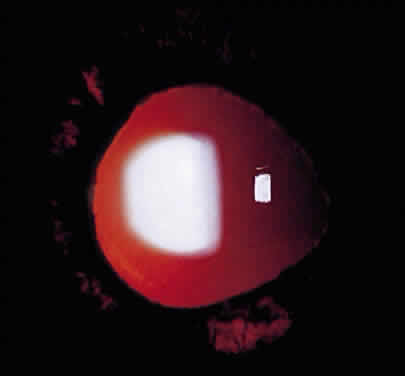
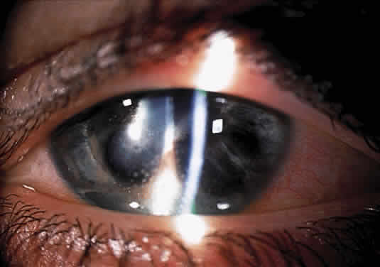
Recurrent disease most often affects the cornea, and can affect any or all layers. The characteristic epithelial lesions are dendrites: thin, meandering, arborizing epithelial ulcerations, sometimes with terminal bulbs at the ends of fine branches. Stromal disease may be immune or infectious, or invoke combined mechanisms. The commonly described patterns of stromal herpetic disease include disciform edema, necrotizing stromal keratitis, and immune ring formation. In disciform edema, the dominant feature is disk-shaped stromal edema, without neovascularization or necrosis, that can be focal in milder disease but extensive and diffuse in more severe disease. Folds in Descemet's membrane may accompany corneal thickening, and keratic precipitates accumulate under the area of edema (Fig. 1). Necrotizing stromal keratitis is characterized by inflammatory necrosis and infiltration of the cornea with polymorphonuclear leukocytes, macrophages, lymphocytes, and plasma cells.7 This cellular infiltration produces stromal edema and necrosis. The necrotizing focus can be located at any level of the corneal stroma, with or without epithelial ulcer ation. Ring infiltrates often develop. In some cases, diffuse patches of infiltration appear beneath an intact epithelium, followed by local neovascularization; this is the so-called interstitial keratitis pattern. Lipid deposition, stromal melting, descemetocele formation, and perforation are all potential complications of advanced necrotizing stromal keratitis. If the peripheral cornea is involved, inflammation and necrosis can spread to the sclera, producing a sclerokeratitis.

Fig. 1. Herpes simplex virus keratouveitis. Anterior chamber inflammation with multiple endothelial precipitates and overlying corneal stromal edema.

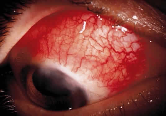
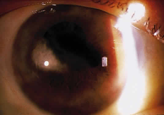
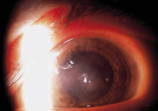
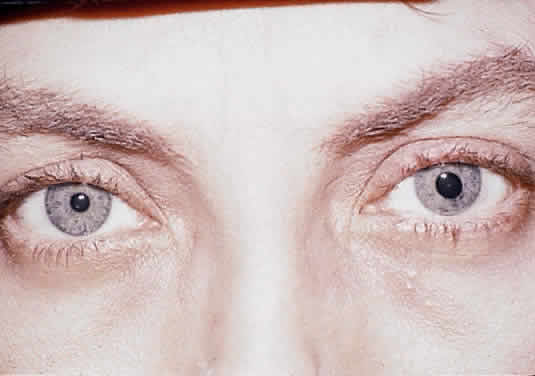
Herpetic keratouveitis can occur in association with any form of herpetic keratitis. Anterior chamber inflammation that accompanies epithelial disease is thought to be caused by reflex irritation and is characteristically both mild and transient. However, the iridocyclitis that invariably accompanies necrotizing disease tends to be much more severe, and is not necessarily correlated with the apparent severity of the keratitis. Indeed, although uveitis dominates the clinical picture in some cases of ocular HSV, subtle corneal findings are limited to faint cellular infiltration of the stroma. Uveitis that accompanies necrotizing disease is typically granulomatous and often recurrent. In more severe cases, perilimbal injection is marked, and the cornea can become thickened and edematous. Dense, fibrinous flare with heavy, anterior chamber cell and medium-sized white keratic precipitates may be distributed widely over the endothelium (Fig. 2). A hypopyon and synechiae can form, and elevated intraocular pressure that exacerbates this often painful uveitis may develop. Marked dilation of iris blood vessels and spontaneous hyphemas sometimes occur. Episodes of inflammation are frequently marked by progressive iris atrophy and sphincter damage, leading to corectopia and anisocoria (Figs. 3 and 4).

Fig. 2. Herpes simplex virus disciform keratitis. Central corneal edema and underlying keratic endothelial precipitates.

Fig. 3. Herpes simplex virus keratouveitis. Multiple features of herpetic keratouveitis, including corneal stromal scarring, keratic endothelial precipitates, anterior synechiae, and diffuse iris atrophy.

Fig. 4. Herpes simplex virus iridocyclitis of the left eye, leading to iris sphincter atrophy and anisocoria.

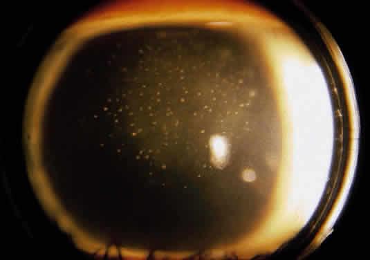
Disciform keratitis is usually accompanied by a mild to moderate uveitis. The typical clinical picture is one of regional stromal edema, which directly overlies an area of endothelial inflammatory precipitates, and mild to moderate anterior chamber reaction. A type of HSV-related corneal edema has been observed in which a line of endothelial dysfunction that is reminiscent of corneal allograft rejection develops and progresses from the periphery centrally, accompanied by anterior chamber inflammation and overlying corneal edema.8–10 This endotheliitis may be accompanied by trabeculitis, which leads to elevated intraocular pressure.

PATHOPHYSIOLOGY

Uveitis associated with endotheliitis, disciform keratitis, and necrotizing stromal keratitis can be attributed to both immunologic reaction and live virus invasion. HSV viral particles have been recovered from the aqueous of patients with HSV iridocyclitis.11,12 Nevertheless, in a rabbit model of HSV uveitis, Oh demonstrated that live but not inactivated HSV introduced to HSV antigen-negative animals stimulated a gradual, delayed iridocyclitis, whereas either live or inactivated virus introduced to eyes that had recovered from primary disease rapidly stimulated an inflammatory response.13 These observations suggest that live virus is implicated in the iridocyclitis of primary disease, but that recurrent disease may be mediated by immunologic mechanisms not necessarily dependent upon the presence of live virus. Sundmacher and Neumann-Haefelin have suggested that clinical features such as elevated intraocular pressure, acute focal iritis, and endotheliitis are particularly suggestive of the presence of live HSV in the anterior chamber.12

Live virus also has been isolated from the aqueous of patients with herpetic endotheliitis and stromal edema.8,9 It thus appears that in some cases of disciform keratitis, live virus might infect endothelial cells that demonstrate swelling and pleomorphism. It has been postulated that endothelial damage might result from direct viral cytolysis, as well as from subsequent antigen-antibody-complement (AAC) mediated immunologic attack.12 Most experimental evidence, however, seems to support the theory that endothelial dysfunction and subsequent corneal swelling of disciform keratouveitis is caused by a delayed-type hypersensitivity and immunologic attack directed against endothelial cells that express herpes antigens.12,14,15

TREATMENT

Because the keratouveitis of epithelial HSV is mild and considered reactive, therapy is restricted to treatment of the epithelial disease with a topical antiviral agent, and treatment of the iridocyclitis with a topical cycloplegic agent. Treatment with topical corticosteroids is discouraged because the suppression of the inflammatory response induced by these agents may lead to reduced clearance of antigen, which allows deeper spread of infection and extends the period of exposure to this stimulus, leading to destructive inflammation.16 No evidence suggests that systemic antiviral agents provide added benefit.17

Disciform and necrotizing stromal disease share the potential for irreversible corneal opacification, in the former because of irrevocable endothelial decompensation, and in the latter because of stromal scarring. Associated uveitis also introduces the risk of synechiae formation and glaucoma. All of these conditions must be accounted for in the treatment regimen.

Mild to moderately severe disciform keratitis that is unaccompanied by signs of live virus, such as elevated intraocular pressure or focal iris necrosis, can be treated without the use of topical antiviral agents; however, because it is difficult to prove the absence of live virus, some clinicians prefer to routinely apply a topical antiviral. If concurrent iridocyclitis of any significance is present, the results of a prospective, randomized clinical trial have suggested that oral acyclovir administered for several weeks, along with a topical antiviral agent and topical corticosteroid, may enhance resolution of inflammation.18 These cases tend to be exquisitely responsive to topical corticosteroid administration. Initially, corticosteroids may be applied as frequently as every 2 hours, and topical antiviral agents 4 times a day. Both are usually tapered for several weeks, as signs of inflammation resolve. Cycloplegic agents also should be used to reduce discomfort caused by ciliary body spasm, and to prevent the formation of synechiae. Elevated intraocular pressure should be treated with appropriate agents.

Linear endotheliitis with iridocyclitis is treated in a similar fashion, with topical corticosteroids, topical antiviral agents, and cycloplegic agents. In some cases, oral antiviral medication may prove beneficial in controlling inflammation and preventing a recurrence of disease.10 Associated elevated intraocular pressure should be treated with appropriate medication.

Severe disciform disease and necrotizing stromal disease with significant iridocyclitis can be resistant to therapy. Because there may be live virus present, topical antiviral agents should be used, and agents with enhanced stromal penetration are favored in this situation. Topical acyclovir has excellent stromal penetration and antiviral action but unfortunately is not universally available; alternatives such as trifluridine are commonly used four times a day and tapered over a number of weeks. Inflammatory destruction can be significant, and a prospective, randomized clinical trial has indicated that frequent application (initially eight times a day) of topical corticosteroid effectively shortens the duration of stromal keratitis and reduces persistence or progression of HSV stromal keratitis.19 Because, as noted above, oral acyclovir administered for several weeks may enhance the resolution of herpetic iridocyclitis, systemic antiviral treatment should be administered along with topical medication. In this situation, elevated intraocular pressure is common and should be treated with topical agents. Cycloplegic agents also should be used. Miotics that may contribute to inflammation and synechiae formation should be avoided. Because attacks of severe herpetic keratouveitis are often prolonged, all medicines must be tapered slowly and cautiously, with careful patient follow up.

Back to Top
HERPES ZOSTER KERATOUVEITIS
Primary infection with the herpes zoster virus causes varicella disease, and reactivation of the virus leads to zoster. Ninety percent of the population demonstrates serologic evidence of exposure to VZV by age 60,20 but only 20% will have experienced an episode of zoster reactivation.21 Immunosuppression associated with disease or advancing age increases the risk of zoster reactivation. Second episodes of zoster are rare, and again are related to immunocompromised states.

The herpes zoster virus establishes its latent phase in the satellite cells of sensory ganglia; the thoracic and lumbar dermatomes are most frequently involved clinically, but the trigeminal ganglion is often also infected. Herpes zoster ophthalmicus (HZO) indicates reactivation of virus in the first division of the trigeminal nerve. Virtually all ocular and adnexal tissues can be involved, with vesicular eruption of the forehead and upper eyelid, follicular or necrotizing conjunctivitis, episcleritis or scleritis, keratitis, uveitis, glaucoma, necrotizing retinitis, retinal vascular occlusion, extraocular muscle palsy, and optic neuritis.22 Tissue damage is mediated through inflammation and vasculitic ischemia caused by viral invasion. Currently it is not entirely clear whether this vasculitis is a direct result of viral invasion, or whether it is mediated by an immune response to viral antigen within the vasculature.

Zoster-associated corneal disease occurs in approximately two thirds of individuals with HZO, and uveitis in about 40%.23,24 Corneal disease can take many forms, including coarse punctate keratopathy, pseudodendrites, serpiginous ulceration, anterior stromal infiltrates, delayed mucus plaques, disciform keratitis, and keratouveitis with endotheliitis. Coarse punctate keratopathy is characterized by elevated patches of epithelium that contain live virus and stain with rose bengal. Although these patches can resolve spontaneously, sometimes they extend deeply to form anterior stromal infiltrates, or coalesce to form elevated dendritiform lesions. Unlike the dendrites of HSV, HZV-related lesions are thicker and more ropy and have an elevated appearance; they also lack terminal bulbs and stain poorly with fluorescein. However, like the epithelial lesions of HSV, these lesions contain live virus.

HZV dendritiform lesions appear most often during the acute event, but can occur many weeks later and typically resolve spontaneously in a few weeks. These lesions should be distinguished from mucus plaques, which usually appear 3 to 4 months after the acute event and vary in appearance from day to day. These plaques are gray, elevated, linear or branching lesions that stain with rose bengal and are loosely adherent to underlying degenerating epithelium and inflamed stroma. With the polymerase chain reaction method, viral DNA has been isolated from these lesions, which were formerly thought to be purely inflammatory.25 After the early epithelial disease, anterior stromal infiltrates sometimes appear.23 It is not clear whether these infiltrates result from an immune reaction to viral antigen or represent direct viral cytotoxicity.

Inflammation of the endothelium, which can occur relatively early in the course of disease, leads to stromal and epithelial edema. Endotheliitis is typically accompanied by signs of anterior chamber inflammation. Keratic precipitates often appear under affected regions of the cornea, but swelling can be diffuse. The corneal endothelium is a favored site of attack, and acute endothelial cell loss has been noted during HZO keratouveitis.26 Endothelial damage may be wrought by viral invasion,27 or by immunologic attack, and can lead to permanent corneal decompensation. It has been suggested that the elevated intraocular pressure that frequently accompanies HZO keratouveitis probably contributes to endothelial cell attenuation, but cell loss has been observed in the absence of elevated pressure.26 Infrequently, an immune ring composed of antigen-antibody complex, complement, and polymorphonuclear leukocytes develops within the edematous cornea.

Although iridocyclitis is often noted in association with endothelial disease, signs of anterior chamber inflammation may be obscured by the corneal opacification. Iridocyclitis tends to occur within 1 to 2 weeks of disease onset28 but can appear many months later. Whereas early inflammation is presumably an immune response to viral invasion, later inflammation is likely caused by an immune reaction against persistent viral antigen. Although corneal disease is almost invariably accompanied by some degree of anterior chamber inflammation, iridocyclitis may occur independently of keratitis. Iridocyclitis is often associated with elevated intraocular pressure and is generally mild to moderate in intensity. More severe iridocyclitis may be associated with eruptive iris lesions29 and is characterized by extensive keratic precipitates, anterior and posterior synechiae, hypopyon, and hemorrhage. Hemorrhage is caused by ischemic vasculitis and necrosis of the iris30 that may be isolated31 or part of an HZO-related, anterior segment ischemia syndrome which, in its most severe form, can lead to phthisis.32 Sectoral occlusion of the iris vasculature leads to atrophic sectoral patches in approximately 20% of cases24,30 and can produce hypopigmentation or, infrequently, hyperpigmentation33 and iris sphincter damage (Fig. 5). The keratouveitis that ensues with the acute inflammatory event may take a protracted course and persist for many months. Histopathologic examination of corneal specimens that had experienced smoldering inflammation for many months demonstrated a granulomatous inflammatory reaction with giant cells in the vicinity of Descemet's membrane.34 Many months after acute HZO, deep, central disciform stromal edema that may be associated with an underlying immune ring sometimes develops. Keratic precipitates usually are not evident. This clinical presentation most likely represents a delayed-type hypersensitivity reaction. The edema may evolve into a diffuse pattern of infiltration and edema, accompanied by vascularization, scarring, lipid deposition, and thinning.23

Fig. 5. Herpes zoster virus keratouveitis. Iris transillumination occurs through regions of iris stromal atrophy.

Another form of keratitis, which typically occurs 2 to 20 weeks after the acute attack, is peripheral crescentic ulceration accompanied by corneal edema, cellular infiltration, and a mild iridocyclitis. These lesions often produce stromal thinning and vascularization and may lead to corneal perforation.23,35

Sclerokeratitis sometimes develops early in the disease course, although it has been noted to occur acutely 2 to 3 months after the acute episode of HZO.36 The scleritis component may be nodular or brawny and is usually associated with uveitis.36 A locus of episcleritis or scleritis in the limbal region can induce adjacent corneal infiltration, which may extend across the cornea, accompanied by vascularization, lipid deposition, thinning and scarring, and anterior chamber inflammation. It has been suggested that sclerokeratitis results from local vasculitis and leads to ischemia and inflammation of the limbal region that extends to the cornea and anterior chamber.

TREATMENT

The immediate goals of treatment in the acute phase of HZO are control of viral replication and inflammation. Systemic antiviral agents play an important role in the management of herpes zoster ophthalmicus, especially if they are administered in the first 72 hours of onset of skin lesions.37 Oral acyclovir is usually given in doses of 800 mg five times a day for 7 to 10 days; it has been demonstrated to accelerate the resolution of skin rash and the healing of skin lesions, reduce the period of lesion formation and viral shedding, and reduce the incidence of episcleritis, keratitis, and iritis.37

Alternative antiviral medications that require less frequent dosing and may be superior to acyclovir in reducing the duration of zoster-associated pain include the nucleoside analogue famciclovir, which is given in doses of 500 mg a day for 7 days, and valacyclovir, a prodrug of acyclovir, which is given in doses of 1000 mg three times a day for 7 to 10 days.38 Both are more bioavailable with oral administration than oral acyclovir. Plasma acyclovir concentrations reached with valacyclovir are comparable to those achievable only with intravenous acyclovir, but famciclovir's active metabolite, penciclovir triphosphate, has an intracellular half-life nearly ten times that of acyclovir.

Along with systemic antiviral therapy, systemic corticosteroids should be considered in the treatment of such severe inflammatory complications of HZO as severe uveitis, scleritis, and orbital inflammation. Moreover, systemic corticosteroids combined with systemic antivirals appear to reduce acute zoster-associated pain during the acute event. However, immunosuppressive systemic corticosteroids increase the risk of disseminated viral infection during reactivation. This is a particular concern in HIV-positive individuals, some of whom may go unrecognized. It is therefore recommended that systemic corticosteroids are restricted to patients 50 years of age or older who appear to have age-normal immune function, and for whom corticosteroids are not otherwise contraindicated.

In general, topical antiviral agents are not commonly used for local therapy of HZO keratitis and uveitis because despite in vitro susceptibility, in most cases the disease is clinically unresponsive to commercially available topical antiviral drugs other than acyclovir. However, Pavan-Langston and associates have reported that some delayed zoster dendritiform lesions respond to topical antiviral therapy.25 Therefore, the role of these agents remains to be defined.

Unlike the epithelial disease of herpes simplex, the disease course of epithelial HZO is not necessarily aggravated by topical corticosteroids. Topical corticosteroids also are extremely useful and effective in controlling iridocyclitis and the inflammatory manifestations of corneal disease, including disciform keratitis, endotheliitis, and keratouveitis.23 If herpes simplex is the suspected causative agent rather than zoster, topical antiviral agents should be administered concurrently. More severe disease may require doses as frequent as every hour, but milder disease often can be controlled with administration three or four times daily. As inflammation is brought under control, drops should be tapered slowly and cautiously for many weeks to months, as dictated by clinical response. In the acute stages, cycloplegic agents also should be used to relieve the discomfort of photophobia and prevent the formation of synechiae.

Elevated intraocular pressure that is caused by inflammation usually responds to corticosteroid treatment, but ocular hypotensive agents may be necessary. Miotics should be avoided, as should topical carbonic anhydrase inhibitors if there is associated endotheliitis and corneal decompensation.

Back to Top
SCLERITIS AND KERATOUVEITIS
Scleritis describes a spectrum of disease that ranges from self-limited moderate inflammation of the sclera to severe, necrotizing, blinding disease that is associated with a significant mortality rate from systemic disease.39 Scleritis is often complicated by corneal involvement and by uveitis; the severity of both tends to parallel that of the scleritis. Based on Moorfield's experience, it appears that anterior uveitis is present in about 40% of eyes with scleritis of all classifications.40 Whereas approximately two thirds of the cases of iridocyclitis are mild, one third are more severe, with a small minority being grave. Although not uncommon in any form of scleritis (based on the classification of Watson and Hayreh41) iridocyclitis appears most commonly in necrotizing scleritis and posterior scleritis. Signs of anterior uveitis appear in more than 50% of these patients, and signs of posterior uveitis are seen in virtually all patients with posterior scleritis.

Iridocyclitis may appear somewhat belatedly in an episode of scleritis and is most likely to appear if non-necrotizing scleritis assumes a necrotizing form. The anterior uveitis is typically characterized by a mild to moderate degree of anterior chamber cell and flare, with small, scattered keratic precipitates. Some eyes develop elevated intraocular pressure and peripheral anterior or posterior synechiae. Dilated iris vessels can appear in the sector of scleral inflammation. Iridocyclitis can be prolonged, and tends to resolve only as the scleral inflammation is brought under control.

Wilhelmus and associates40 found anterior uveitis to be statistically associated with corneal infiltration, but not with corneal thinning. As noted earlier, corneal infiltration is a relatively common finding in the various forms of anterior scleritis. Tuft and Watson42 found that corneal infiltration was associated with approximately 50% of cases of nodular scleritis, 40% of cases of necrotizing scleritis, and 20% of cases of diffuse scleritis. It is uncommon for corneal infiltration to accompany posterior scleritis. In the same report, peripheral ulceration occurred in 40% of cases of necrotizing scleritis and 13% of cases of nodular scleritis, but in only 3% of cases of diffuse and posterior scleritis.

Although the corneal changes associated with all forms of anterior scleritis can be diffuse, there is a tendency for the corneal infiltrates of nodular scleritis to appear adjacent to the area of nodular inflammation (Fig. 6). Corneal infiltration associated with scleritis can take the form of stromal keratitis or sclerosing keratitis. Sclerosing keratitis appears with all forms of anterior scleritis, and is the most common form of corneal infiltration. This is the classic “cotton candy” infiltrate that first appears as diffuse, discrete, crystalline infiltrates within local or diffuse corneal edema. The crystalline infiltrates persist as the edema resolves, and can coalesce to form extensive deep, dense stromal opacities Fig. 7). Associated corneal neovascularization is common. Stromal keratitis can be seen with both nodular and diffuse scleritis. Patches of edema and infiltration appear, usually acutely and often in the limbal region, and progress to form discrete stromal opacities. These lesions can be diffuse or focal, and diffuse patches may coalesce. Corneal neovascularization also can develop in association with the stromal keratitis of scleritis.

Fig. 6. Sclerokeratitis. Inflammatory infiltration of the corneal stroma adjacent to a focus of nodular scleritis.

Fig. 7. The characteristic “cotton candy” infiltrate of sclerosing keratitis after scleritis and keratouveitis.

Necrotizing scleritis also may be complicated by extensive corneal melting, referred to as keratolysis. Typically, the cornea adjacent to an area of intense scleral inflammation becomes edematous, thickened, and opacified. Keratic precipitates may form beneath the involved stroma. As inflammation progresses, there is extensive erosion of corneal stromal tissue and loss of limbal architecture. The local vessels become tortuous and abnormal, and invade the melting cornea at all levels.43 More insidious peripheral corneal erosion that resembles the peripheral gutters of rheumatoid arthritis can also complicate less severe, diffuse scleritis. Peripheral corneal edema and opacification progresses to erosion of the epithelium and superficial stroma, and the formation of a well-demarcated gutter that is frequently invaded by blood vessels. Both forms of peripheral corneal erosion are accompanied by some degree of vascular occlusion and ischemia.43

Immunohistopathologic evidence suggests that scleritis is primarily an immune complex-mediated vasculitis, whereby granulomatous inflammation of the sclera involves episcleral and scleral vessels.44 Conjunctival and scleral biopsies of eyes with scleritis reveal that the thrombosis, fibrinoid necrosis, and inflammatory vascular infiltration in inflamed regions is frequently associated with immune complex deposition. Circulating immune complexes, which may be deposited in scleral vessels and thus contribute to local inflammation, also can be identified in most cases.44 Evidence also suggests that scleral antigens may be the targets of immunologic attack.45 Anterior segment fluorescein angiography studies of eyes with various forms of scleritis have demonstrated that in more benign varieties of diffuse and nodular anterior scleritis there is rapid filling of the vessels and a short transit time, but necrotizing scleritis is characterized by venular occlusion, hypoperfusion, and ischemia.46 The destructive stromal keratitis that accompanies this intense inflammation of the adjacent limbus and sclera is associated with local vascular inflammation and occlusion that reflect a systemic vasculitis.43

The treatment of scleritis, with its attendant complications of keratitis, uveitis, and glaucoma requires systemic therapy. In particular, necrotizing scleritis has a poor outcome if not treated promptly and aggressively, and is associated with a high rate of mortality from systemic disease.39 Depending on the severity of the scleral inflammation and associated systemic disease, appropriate therapy may range from administration of systemic nonsteroidal antiinflammatory agents to multiple potent immunosuppressive agents. Recommendations for aggressive immunosuppressive therapy are beyond the scope of this chapter, and are addressed elsewhere. However, although keratitis, uveitis, and glaucoma are expected improve with systemic treatment of the scleritis, local treatment can be a helpful supplement. Topical cycloplegic agents can help prevent the formation of synechiae and reduce discomfort, but topical ocular antihypertension agents can help to reduce intraocular pressure during stages of acute elevation. Topical corticosteroids can be helpful in reducing discomfort, but are by no means a substitute for systemic therapy. Subconjunctival injections of corticosteroids are contraindicated in scleritis because they have been observed to lead to perforation at the site of injection.

NONSPECIFIC “REFLEX” KERATOUVEITIS

Keratouveitis also may be seen with nonspecific irritation of the corneal epithelium. This occurs most strikingly in patients with bullous corneal edema who, after bulla rupture, may develop sufficient inflammation to produce a hypopyon. Other ocular epithelial trauma also may produce significant anterior chamber inflammation, despite minimal keratitis. Such “reflex” iridocyclitis is treated with cycloplegic agents and appropriate management of the corneal epithelial disease.

The reflex anterior chamber inflammation that follows active corneal disease probably represents a form of neurogenic inflammation in which corneal irritation stimulates the release of neuropeptides from anterior segment, unmyelinated sensory nerves.47,48 Substance P was the first neuropeptide identified in the aqueous humor during neurogenic inflammation. This tachykinin has multiple inflammatory properties, including stimulation of polymorphonuclear lymphocyte/monocyte chemotaxis and phagocytosis, vasodilatation, and smooth muscle contraction. It also induces mast cell degranulation, macrophage eicosanoid production and release, and T-lymphocyte proliferation.49,50 Retrobulbar injection of capsaicin leads to release of, among other neuropeptides, substance P, which is considered to be partly responsible for the subsequent keratouveitis that is confined to the anterior segment.51

In the rabbit model, the neuropeptide calcitonin gene-related peptide (CGRP) is also thought to make a significant contribution to neurogenic inflammation.52 CGRP that is released by sensory afferent neurons produces rapid anterior uveal vasodilatation, breakdown of the blood-aqueous barrier, miosis, prostaglandin release, and elevated aqueous levels of cyclic AMP.48,53 The last appears to be particularly implicated in the breakdown of the blood-aqueous barrier.54 Evidence suggests that the tachykinin neurokinin A may also play a significant role in ocular neurogenic inflammation.55

Although most studies of neurogenic inflammation and related immunoreactive components have been based on rabbit models, CGRP immunoreactive nerves have been identified in thin, varicose nerve fibers mainly associated with blood vessels in the human eye.56 Most of these fibers appear to be located in the ciliary body; most are associated with blood vessels but a moderate number have been identified within the ciliary muscle. A relatively sparse concentration are also found in the iris and cornea. Iris CGRP immunoreactive nerves are associated with blood vessels, but corneal CGRP immunoreactive nerves are found immediately beneath the epithelium or as free nerve endings in the epithelium. CGRP immunoreactive ganglion cells also can be identified in the trigeminal ganglion.56 These observations lend support to the argument that animal models of neurogenic inflammation may be extended to humans.

Back to Top
REFERENCES

1. Eriksson A, Fagerholm P, Olsson K: Keratouveitis: Two families with a dominantly inherited disorder. Acta Ophthalmol Scand 74:473, 1996

2. Lee, SF, Buller R, Chansue E et al: Vaccinia keratouveitis manifesting as a masquerade syndrome. Am J Ophthalmol 117:480, 1994

3. Heidemann DG, Trese M, Murphy SF et al: Endogenous Listeria monocytogenes endophthalmitis presenting as keratouveitis. Cornea 9:179, 1990

4. Liesegang TJ, Melton LJ III, Daly PJ, Ilstrup DM: Epidemiology of ocular herpes simplex: Incidence in Rochester, Minn, 1950 through 1982. Arch Ophthalmol 107:1155, 1989

5. Smith IW, Peutherer JF, MacCallum FO: The incidence of Herpesvirus hominis antibody in the population. J Hyg 65:394, 1967

6. Nahmias AJ, Visintine AM, Caldwell DR, Wilson LA: Eye infections with herpes simplex viruses in neonates. Surv Ophthalmol 21:100, 1976

7. Pepose JS: Herpes simplex keratitis: Role of viral infection versus immune response. Surv Ophthalmol 35:345, 1991

8. Vannas A, Ahonen R: Herpetic endothelial keratitis: A case report. Acta Ophthalmol 59:296, 1981

9. Robin JB, Steigner JB, Kaufman HE: Progressive herpetic corneal endotheliitis. Am J Ophthalmol 100:336, 1985

10. Olsen TW, Hardten DR, Meiusi RS, Holland EJ: Linear endotheliitis. Am J Ophthalmol 117:468, 1994

11. Kaufman HE, Kanai A, Ellison ED: Herpetic iritis: Demonstration of virus in the anterior chamber by fluorescent antibody techniques and electron microscopy. Am J Ophthalmol 71:465, 1971

12. Sundmacher R, Neumann-Haefelin D: Herpes simplex virus-positive and -negative keratouveitis. In Silverstein AM, O'Connor R (eds): Immunology and Immunopathology of the Eye p 225. New York: Masson, 1979

13. Oh JO: Role of immunity in the pathogenesis of Herpes simplex uveitis. In Silverstein AM, O'Connor R (eds): Immunology and Immunopathology of the Eye p 248. New York: Masson, 1979

14. Nagy RM, McFall RC, Sery TW et al: Scanning electron microscope study of herpes simplex virus in experimental disciform keratitis. Br J Ophthalmol 62:838, 1978

15. Vannas A, Ahonen R, Mäkitie J: Corneal endothelium in Herpetic keratouveitis. Arch Ophthalmol 101:913, 1983

16. Kibrick S, Takahashi GH, Leibowitz HM: Local corticosteroid therapy and reactivation of herpetic keratitis. Arch Ophthalmol 86:694, 1971

17. Herpetic Eye Disease Study Group: A controlled trial of oral acyclovir for the prevention of stromal keratitis or iritis in patients with herpes simplex virus epithelial keratitis: The epithelial keratitis trial. Arch Ophthalmol 115:703, 1997

18. Herpetic Eye Disease Study Group: A controlled trial of oral acylovir for iridocyclitis caused by herpes simplex virus. Arch Ophthalmol 114:1065, 1996

19. Wilhelmus KR, Gee L, Hauck WW et al: Herpetic eye disease study: A controlled trial of topical corticosteroids for herpes simplex stromal keratitis. Ophthalmology 101:1883, 1994

20. Gershon AA, Steinberg SP: Antibody responses to varicella-zoster virus and the role of antibody in host defense. Am J Med Sci 282:12, 1981

21. Liesegang TJ: Diagnosis and therapy of herpes zoster ophthalmicus. Ophthalmology 98:1216, 1991

22. Karbassi M, Raizman MB, Schuman JS: Herpes zoster ophthalmicus. Surv Ophthalmol 36:395, 1992

23. Liesegang TJ: Corneal complications from herpes zoster ophthalmicus. Ophthalmology 92:316, 1985

24. Womack LW, Liesegang TJ: Complications of herpes zoster ophthalmicus. Arch Ophthalmol 101:42, 1983

25. Pavan-Langston D, Yamamoto S, Dunkel EC: Delayed herpes zoster pseudodendrites. Polymerase chain reaction detection of viral DNA and a role for antiviral therapy.Arch Ophthalmol 113:1381, 1995

26. Reijo A, Antti V, Jukka M: Endothelial cell loss in herpes zoster keratouveitis. Br J Ophthalmol 67:751, 1983

27. Maudgal PC, Missotten L, De Clercq E, et al: Varicella-zoster virus in the human corneal endothelium: A case report. Bull Soc Belge Ophthalmol 190:71, 1980

28. Cobo M, Foulks GN, Liesegang T et al: Observations on the natural history of herpes zoster ophthalmicus. Curr Eye Res 6:195, 1987

29. Marsh RJ, Easty DL, Jones BR: Iritis and iris atrophy in herpes zoster ophthalmicus. Am J Ophthalmol 78:255, 1974

30. Naumann G, Gass JDM, Font RL: Histopathology of herpes zoster ophthalmicus. Am J Ophthalmol 533:65, 1968

31. Edgerton AE: Herpes zoster ophthalmicus: Report of cases and review of literature. Arch Ophthalmol 34:40, 114, 1945

32. Crock G: Clinical syndromes of anterior segment ischaemia. Trans Ophthalmol Soc UK 87:513, 1967

33. Klein BA, Farka TG: Pseudomelanoma of the iris after herpes zoster ophthalmicus. Am J Ophthalmol 57:392, 1964

34. Hedges TR III, Albert DM: The progression of the ocular abnormalities of herpes zoster: Histopathological observations of nine cases. Ophthalmology 89:165, 1982

35. Mondino BJ, Brown SI, Mondzelewski JP: Peripheral corneal ulcers with herpes zoster ophthalmicus. Am J Ophthalmol 86:611, 1978

36. Marsh RJ: Herpes zoster keratitis. Trans Ophthalmol Soc UK 93:181, 1973

37. Cobo LM, Foulks GN, Liesegang T et al: Oral acyclovir in the treatment of acute herpes zoster ophthalmicus. Ophthalmology 93:763, 1986

38. Kost RG, Straus SE: Postherpetic neuralgia: Pathogenesis, treatment, and prevention. N Engl J Med 335:32, 1996

39. Foster CS, Forstot SL, Wilson LA: Mortality rate in rheumatoid arthritis patients developing necrotizing scleritis or peripheral ulcerative keratitis: Effects of systemic immunosuppression. Ophthalmology 91:1253, 1984

40. Wilhelmus KR, Watson PG, Vasavanda AR: Uveitis associated with scleritis. Trans Ophthal Soc UK 101:351, 1981

41. Watson PG, Hayreh SS: Scleritis and episcleritis. Br J Ophthalmol 60:163, 1976

42. Tuft SJ, Watson PG: Progression of scleral disease. Ophthalmology 98:467, 1991

43. Watson PG: Vascular changes in peripheral corneal destructive disease. Eye 4:65, 1990

44. Fong LP, de la Maza MS, Rice BA et al: Immunopathology of scleritis. Ophthalmology 98:472, 1991

45. York LJ, McCluskey PJ, Wakefield D: Scleral autoantibodies in external eye disease. Proc Aust Soc Exp Pathol 17:17, 1985

46. Watson PG, Bovey E: Anterior segment fluorescein angiography in the diagnosis of scleral inflammation. Ophthalmology 92:1, 1985

47. Pernow B: Role of tachykinins in neurogenic inflammation. J Immunol 135:812s, 1985

48. Bill A, Stjernschantz J, Mandahl A et al: Substance P: Release on trigeminal nerve stimulation, effects in the eye. Acta Physiol Scand 106:371, 1979

49. Payan DG, Goetzl EJ: Modulation of lymphocyte function by sensory neuropeptides. J Immunol 135:783s, 1985

50. O'Doriso MS, Wood CL, O'Doriso TM: Vasoactive intestinal peptide and neuropeptide modulation of the immune response. J Immunol 135:792s, 1985

51. Wldrep JC, Crosson CE: Induction of keratouveitis by capsaicin. Curr Eye Res 7:1173, 1988

52. Krootila K, Uusitalo H, Palkama A: Effect of topical and intracameral methysergide on calcitonin gene-related peptide-induced irritative changes in the rabbit eye. J Ocul Pharmacol 8:121, 1992

53. Wahlested C, Beding B, Ekman R et al: Calcitonin gene-related peptide in the eye: Release by nerve stimulation and effects associated with neurogenic inflammation. Regul Pept 16:107, 1986

54. Krootila K: CGRP in relation to neurogenic inflammation and cAMP in the rabbit eye. Exp Eye Res 47:307, 1988

55. Beding-Barnekow B, Brodin E: Neurokinin A, neurokinin B and neuropeptide K in the rabbit iris: A study comparing different extraction methods. Regul Pept 25:199, 1989

56. Uusitalo H, Krootila K, Palkama A: Calcitonin gene-related peptide (CGRP) immunoreactive sensory nerves in the human and guinea pig uvea and cornea. Exp Eye Res 48: 467, 1989

Back to Top