Chapter 38
An Overview of Albinism and Its Visual System Manifestations
ELIAS I. TRABOULSI and W. RICHARD GREEN
Main Menu   Table Of Contents

Search

INTRODUCTION
FAMOUS ALBINOS
EPIDEMIOLOGY
BIOCHEMISTRY OF MELANIN FORMATION
GENETICS OF DISORDERS OF MELANOGENESIS
CLINICAL ASPECTS OF OA AND OCA
PATHOPHYSIOLOGY OF VISUAL IMPAIRMENT IN ALBINISM
ANCILLARY TESTS
ALBINISM AND SYSTEMIC DISORDERS
MANAGEMENT OF PATIENTS WITH ALBINISM
REFERENCES

INTRODUCTION
The term “albinism” (from albus, white) is applied to a group of inherited disorders that are characterized by decreased or absent melanin pigment in tissues, together with developmental abnormalities of the eye and visual pathways. The skin and hair color of patients with albinism depends at least in part on the amount of melanin pigment present and the underlying metabolic defect. Pigmentation can be completely absent throughout the lifetime of the individual, or may increase to nearly normal levels in the first few years of life. In addition to decreased melanin in ocular tissues, patients with albinism have characteristic anatomical defects in the visual system, such as foveal hypoplasia and abnormal decussation of optic nerve fibers. Other common ocular abnormalities include errors of refraction, reduced visual acuity, iris transillumination defects, nystagmus, and strabismus.1,2 The variability in the extent of skin, hair, and eye hypopigmentation, reduction of visual acuity, and nystagmus occasionally makes the diagnosis of albinism challenging. However, a history of marked skin hypopigmentation in infancy, and careful examination for iris transillumination defects, underdevelopment of the fovea, and other subtle clinical signs make it possible to detect mild forms of albinism. Electrophysiologic and molecular genetic testing can be used to confirm a clinical diagnosis in the rare case in which the clinical diagnosis remains in doubt.

A number of inherited systemic disorders feature skin, hair, and ocular hypopigmentation in addition to other abnormalities. Some of these disorders, such as Hermansky-Pudlak's syndrome (HPS) and Chédiak-Higashi's syndrome (CHS), are associated with the characteristic ocular and visual system abnormalities of albinism, but others, such as Waardenburg's syndrome, are not. The latter should not be classified under the general rubric of albinism, but rather as disorders associated with hypopigmentation.3–5

Back to Top
FAMOUS ALBINOS
Evidence that Noah (of the Old Testament) was an albino was presented by Sorsby.6 Another famous albino was W.A. Spooner, a British classicist and warden of New College at Oxford University, whose amusing errors of speech came to be known as “spoonerisms.” These aberrations of speech were said to be related to his nystagmus, which caused a jumbling of information from the printed page. Other famous albinos include Edward the Confessor, the Saxon king from 1042 to 1066, and Tamerlane, the central Asian conqueror in the Middle Ages.
Back to Top
EPIDEMIOLOGY
The prevalence of all types of albinism in Northern Ireland has been estimated at 1:10,000.7 In the United States, albinism affects 1:18,000 and 1:10,000 of the white and black populations, respectively. Tyrosinase-positive oculocutaneous albinism (OCA2) is the most common form, with an incidence of 1:37,000 in U.S. whites and 1:15,000 in U.S. blacks. Tyrosinase-negative OCA (OCA1) is the second most common type, and occurs in 1:39,000 U.S. whites and 1:28,000 U.S. blacks. OCA1B (yellow type) is most prevalent among the Amish,8 but has also been reported in other ethnic groups. OCA4 (brown albinism) is more frequent in Nigerians, with an incidence of 1:1,100.9 X-linked Nettleship-Falls ocular albinism (OA) has an estimated incidence of 1:150,000 persons.10 HPS is commonly encountered in Puerto Ricans (1:1,800).11 CHS is very rare, and has been reported in Venezuelan, Caucasian, Japanese, and black patients.12
Back to Top
BIOCHEMISTRY OF MELANIN FORMATION
Melanin synthesis (Fig. 1) starts with the amino acid tyrosine, which is oxidized to dihydroxyphenylalanine (DOPA) by the enzyme tyrosinase. DOPA is then oxidized to dopaquinone by the same enzyme. Dopaquinone is spontaneously transformed to leucodopachrome, and then to dopachrome. The latter is decarboxylated to 5,6-dihydroxyindole, and then oxidized to indole 5,6-quinone. Indole-quinone is converted to melachrome, and then polymerizes to eumelanin (pigment with brown to black color). Dopaquinone can react with cysteine under high concentrations of the latter to form cysteinyl-dopa, which is oxidized to pheomelanin (a pigment with a yellow to red color). DOPA can also react with glutathione to form pheomelanin.

Fig. 1. Melanin synthesis pathway.

Melanogenesis is identical in melanocytes of the hair, skin, and eye. Melanosome formation begins with the fusion of vesicles from the endoplasmic reticulum and Golgi apparatus. Stage I vesicles contain the enzyme tyrosinase, but no melanin is present. These vesicles fill up with a collection of parallel filamentary structures prior to melanin synthesis (stage II). Melanin granules then deposit on the filaments (immature melanosome, stage III) and progressively saturate them, forming a dense pigmented structure (mature melanosome, stage IV). Skin and hair melanocytes are capable of exporting stage IV melanosomes into adjacent cells, while ocular melanosomes remain within the cell membranes of melanocytes. Melanogenesis in retinal pigment epithelial (RPE) cells is completed shortly after birth, but it may continue for several years in the uveal tract. Patients with OCA have a normal number of melanosomes that are incompletely melanized.

Back to Top
GENETICS OF DISORDERS OF MELANOGENESIS
In 1904 Durham13 discovered that albino animals lacked tyrosinase activity. Kugelman and Van Scott14 later demonstrated, with the use of the hair bulb tyrosine assay, that some albinos showed evidence of tyrosinase activity. This led to the classification of OCA into tyrosinase-negative and tyrosinase-positive categories.

Traditionally, clinicians have divided albino patients into tyrosinase-negative and -positive categories on the basis of clinical findings and the occasional use of the hair-bulb incubation test. They also distinguish between patients with associated systemic disorders (such as HPS or CHS) and those without. These are practical classification schemes, but it would be better to integrate them into one that incorporates recent information on the complex molecular genetics of these disorders. Patients with no tyrosinase activity at all have white hair and no ocular or skin pigment. They have been designated as tyrosinase-negative, while those with any amount of pigmentation have been classified as tyrosinase-positive. The limitation of such a classification is that it relies on clinical features rather than the underlying genetic basis. For example, although the molecular defect in OCA1A and OCA1B is in the enzyme tyrosinase, patients with the former are clinically classified as tyrosinase-negative, and patients with the latter as tyrosinase-positive. So-called autosomal-recessive OA is merely a very mild form of OCA1 in which hypopigmentation is most prominent in the eye, with almost normal skin pigmentation. If strict clinical definition criteria were used, all albinism would be oculocutaneous, except for X-linked Nettleship-Falls OA.

Several genetic loci for human albinism and a spectrum of mutations in each of the responsible genes have been identified in the last two decades (Table 1).5 We now recognize that classic tyrosinase-negative albinism results from mutations that render tyrosinase totally inactive. However, there is a broad spectrum of cutaneous and ocular pigmentation associated with tyrosinase mutations. So-called tyrosinase-positive OCA, in which hair bulb cells form melanin when they are incubated in DOPA or tyrosine, can be caused by mutations in several genes, including tyrosinase, the P gene (vide infra), and others.

TABLE 1. Molecular Genetic Classification of Nonsyndromic Albinism


Clinical Type of Albinism Abreviation OMIM # Defective gene Gene map
Tyrosinase-negative OCAOCA1A203100Tyrosinase11q14-q21
Yellow type OCAOCA1B606952Tyrosinase11q14-q21
Temperature-sensitive OCAOCA1TS203100Tyrosinase11q14-q21
Tyrosinase-positive OCAOCA2203200P gene or Tyrosinase15q11.2-q12 11q14-q21
Rufous albinism (xanthism)OCA3278400TYRP1 Tyrosinase-related protein9p23
Brown albinismBOCA203290P gene15q11.2-q12
OCA type IVOCA4606574MATP5p
Nettleship-Falls OA, X-linked OAOA1300500Melanosomal G protein-coupled receptorXp22.3


TYROSINASE

The human tyrosinase gene is located on chromosome 11q14-21 and contains 5 exons encoding a protein of 529 amino acids. The tyrosinase polypeptide contains two copper-binding sites, a signal peptide, a region of conserved cysteine residues that create an epidermal growth factor (EGF)-like domain, and a transmembrane region. It is a monophenol mono-oxygenase. A large number of mutations, deletions, and polymorphisms have been discovered in the tyrosinase gene (www.cbc.umn.edu/tad/), and recent investigations have tried to elucidate the mechanism by which changes in the tyrosinase protein lead to the clinical manifestations of the disease.15 The majority of tyrosinase gene mutations identified to date have been found in individuals who have the classic tyrosinase-negative phenotype. One type of OCA1—yellow albinism or OCA1B—has been shown to result from a number of tyrosinase mutations that encode an enzyme with less than 10% of residual activity.16 Another OCA1 phenotype results from an R402Q mutation that produces a temperature-sensitive tyrosinase that is active at temperatures below 35°C and is relatively inactive at 37°C.17,18 This is the result of the inefficient exiting of folded tyrosinase from the endoplasmic reticulum at the higher temperature. Individuals with this form of the enzyme have white axillary hair, nearly white scalp hair, light brown arm hair, and dark brown leg hair, and their skin is hypopigmented. Hair color in this variant depends on regional skin temperature. Equivalent animal models include the Siamese cat and the Himalayan mouse.

P GENE

Mutations in the P gene, the human homologue of the mouse pink-eyed dilution gene (p) are responsible for OCA2.19 The human gene is on chromosome 15q and encodes an integral, melanosomal membrane protein of 110 kDa. This protein has 12 putative membrane spanning regions and appears to modulate the processing and transport of tyrosinase.20 The hypopigmentation pattern found in Africans and African-Americans (in whom this is the most common cause of albinism) is characterized by yellow hair and white skin with localized pigmented regions. The irides are partially or completely pigmented with a tan-colored melanin. A 2.7 kb deletion mutation of the P gene is found in 25% to 50% of all African-Americans, and about 80% of South African, Tanzanian, and other sub-Saharan African blacks with OCA2. Mutations in the P gene also cause Brown OCA.21

TYRP1

Rufous OCA occurs in blacks and is characterized by bright copper-red coloration of the skin and hair, and dilution of the color of the iris. It is caused by mutations in the TYRP1 gene.22 TYRP1 has multiple functions, including stabilizing tyrosinase and regulating the production of black eumelanin (see Fig. 1).23

MATP

Mutations in the MATP gene underlie OCA4, a newly identified form of human albinism. OCA1–3 result from the aberrant processing and/or sorting of tyrosinase. The disruption of tyrosinase trafficking occurs at the level of the endoplasmic reticulum (ER) in OCA1 and OCA3, and at the post-Golgi level in OCA2. In OCA4 melanocytes tyrosinase is abnormally secreted from the cells. This process is similar to that seen in OCA2 melanocytes, which results from a mutation of the pink-eyed dilution (p) gene. The P protein and MATP have 12 transmembrane regions and are thought to function as transporters. The abnormal transportation of tyrosinase in OCA4 disrupts the normal maturation process of the melanosomes.24,25 Rundshagen et al26 found mutations in MATP in five of 176 German albinos.

OA1

The gene for X-linked ocular albinism, OA1, was mapped to Xp22.3-p22.2 and was later identified as a membrane protein that is necessary for the maturation of melanosomes.27 In the presence of abnormalities in OA1, melanosomes may fail to bud off the endoplasmic reticulum and enlarge as melanin is deposited, leading to the formation of giant melanosomes (Fig. 2). About 48% of the reported mutations in the OA1 gene are intragenic deletions, and about 43% are point mutations.28,29

Fig. 2. Electron micrograph of an epidermal melanocyte in Nettleship-Falls X-linked OA. Four macromelanosomes and some normal-sized melanosomes (arrow) are shown (original magnification, 8,000×). (Reprinted from Ref. 54.)

Back to Top
CLINICAL ASPECTS OF OA AND OCA
The severity of the pigmentary dilution in OCA depends on the genetic subtype, constitutive (racial) pigmentation, and age of the patient. Theoretically, a black adult with some tyrosinase acitivity is expected to be darker than a white infant with the same degree of tyrosinase activity. Thus, hair color in OCA ranges from white to dark yellow or light reddish brown (Fig. 3), and iris color ranges from very light blue, with a pink reflex through an undilated iris, to light hazel. The skin may be without pigment or it may have some pigment, especially in nevi and freckles. van Dorp30 observed that some patients with OCA may have normal pigmentation, patients with X-linked OA may be underpigmented, patients with HPS may have a dark complexion, and pigment dilution varies in all types other than OCA1A.

Fig. 3. Teenager with tyrosinase-positive OCA. Note the iris transillumination, esotropia, and reddish-brown hair color.

The ocular manifestations of patients with albinism vary in severity among the different types of this heterogeneous group of conditions but share characteristic common features.31 Sensory nystagmus is present very early in life but improves in amplitude and frequency with increasing age. This is frequently the presenting sign of the disorder. In most cases the nystagmus is pendular and horizontal. Occasionally a null region and compensatory face turn may be present. A detailed discussion of eye-movement abnormalities in patients with albinism is beyond the scope of this review. Reduced visual acuity is present from birth, and ranges from 20/40 to 20/400 in most cases. Vision usually remains stable, but may improve slightly with age in some patients. Poor binocular vision and reduced stereopsis may be present; however, many patients can pass parts of the clinical stereo tests.32 Iris color ranges from light blue with a pink reflex in OCA1A to hazel in other types, and may change as more pigment is deposited with age. Iris transillumination is best detected by means of the slit-lamp and retroillumination in undilated patients. In some cases the outline of the lens can be seen through the iris (Fig. 4). Hypopigmentation of the iris pigment epithelium results in light scatter and contributes to the subjective complaint of photophobia in some (but certainly not all) patients. Fundus hypopigmentation is also variable. A dilated-fundus examination may reveal a prominent choroidal vascular pattern resulting from decreased or absent RPE melanin (Fig. 5). The optic nerve head may be grayish in patients with OCA1A, or it may be hypoplastic (Fig. 6). Absent macular xanthophyll pigment and a diminished foveal reflex indicate foveal hypoplasia (Fig. 7).33,34 The latter is considered one of the main factors underlying reduced visual acuity in albinism. Abnormal macular vascular patterns may be present in which retinal vessels course through the foveal avascular zone instead of arching around it, which is a hallmark of foveal hypoplasia.35 Whether foveal hypoplasia in albinos is directly related to reduced macular RPE melanin is unknown. Optic nerve hypoplasia is a well documented but not very common finding in albinos.36 Megalocornea, aniridia, posterior embryotoxon, Axenfeld's anomaly, glaucoma,37,38 retinal coloboma, and abortive cryptophthalmos have also been reported in rare individual patients, and except for mild anterior segment dysgenesis are probably coincidental.

Fig. 4. Retroillumination of the iris in an older patient with OA who had undergone cataract extraction and intraocular lens implantation. The outline of the lens is clearly visible.

Fig. 5. Prominent choroidal vascular pattern in a patient with OCA. Note the absence of RPE and choroidal pigmentation.

Fig. 6. Optic nerve hypoplasia in a patient with OCA.

Fig. 7. Foveal hypoplasia in a patient with OCA2. There is no foveal reflex, and some fine blood vessels are crossing the horizontal raphe.

Back to Top
PATHOPHYSIOLOGY OF VISUAL IMPAIRMENT IN ALBINISM
All albino mammals have abnormalities of visual system development. Three main cellular disorders that lead to reduced vision have been identified: a reduction in the number of rod photoreceptors, the underdevelopment of the central retinal specialization or foveal hypoplasia, and a misrouting of some temporal retinal ganglion cell axons.39 Ganglion cells are produced early in retinal development, and the organization of the retinogeniculate pathway depends on some retinal ganglion cell axons crossing at the optic chiasm to project to the contralateral dorsal lateral geniculate nucleus in the thalamus. In albinos, fewer retinal ganglion cells project to the ipsilateral side, leading to a disruption of lateral geniculate organization and to disorganization of visual information in the cortex (Fig. 8). Tyrosinase activity is required during some phases of the ipsilateral retinogeniculate pathway development. In the RPE, tyrosinase is first expressed on embryonic day 10,40 and pigment formation starts on embryonic day 11.41 The onset of melanin formation starts in the periphery and is graded across the retina. Because melanization occurs at the same time as neuroblast divisions and patterning, tyrosinase expression and the graded onset of pigment formation may be a developmental signal that sets up positional information in the retina. Ganglion cells that project ipsilaterally are produced between embryonic days 11 and 16.41 Tyrosinase expression may also be necessary to ensure that the neuroblasts divide properly. Finally, tyrosinase may be necessary for the pathfinding of the ganglion cell axonal to the lateral geniculate body, which takes place between embryonic days 12.5 and 18.5. After the ganglion cell axons reach the lateral geniculate body, there is a period of refinement of connections and ganglion cell death. Tyrosinase expression in the eye may be necessary to ensure that cells die or are maintained appropriately during this period.42

Fig. 8. Pattern of optic nerve fiber decussation in normal (left) and albino (right) individuals.

ABNORMAL DECUSSATION OF OPTIC NERVE FIBERS

Carroll and colleagues43 presented evidence that human albinos have the same misrouting of the retinogeniculate projections that has been found in albinos of other species. When pigmentation is incomplete, the developing optic tracts revert to an almost completely crossed pattern at the chiasm, as is the case in more primitive panoramic systems. In normal humans, 45% of the axons that originate in the temporal half of the retina remain uncrossed as they pass through the chiasm and project to the ipsilateral lateral geniculate nucleus. The greatest number of these fibers subserves the central 20° of the temporal retina. In albinos, most of these fibers decussate at the chiasm and synapse in the contralateral lateral geniculate nucleus, leaving only 10% to 20% of fibers uncrossed (Fig. 8). These abnormalities of decussation lead to a predominantly monocular representation of the central visual field in each occipital cortex. Monocular visual-evoked responses hence lead to markedly asymmetric responses from the two hemispheres.44,45 All OCA or OA conditions in humans and animals with nystagmus tested to date have shown either electrophysiologic or anatomic evidence of a decussation defect in the optic tracts. Evidence that anomalous decussation also exists in the auditory system was presented by Creel and colleagues.46 It is thought that 1% to 2% of the human population may be heterozygous for albinism, and that this may have an adverse effect on binocular depth perception. The locus ceruleus and substantia nigra are normally pigmented in albinos. They owe their pigmentation to neuromelanin, which is synthesized by tyrosine hydroxylase rather than tyrosinase.

LIGHT SCATTERING

Light scattering within the eye may play some role in the pathophysiology of visual impairment in albinism, but its contribution must be minimal. For example, patients with autosomal-dominant OCA have translucent irides and an albinotic fundus, but normal visual acuity. Some of these patients complain of photophobia, which may be a consequence of light scattering. Tinted lenses can help. Corneal contact lenses with an opaque iris portion have not significantly improved visual acuity in albinos.47

LIGHT-INDUCED RETINAL DAMAGE

Light-induced retinal damage does not appear to play any appreciable role in the visual impairment of albinos. There is histopathologic and ultrastructural evidence that extraordinarily intense light can produce nonthermal retinal damage even in normally pigmented animals.48 Albino animals have lower thresholds for such damage. This has been attributed to an ineffectual iris diaphragm, because normally pigmented animals show somewhat compatible thresholds with pupillary dilatation. There are no data to suggest that visual function in human albinos deteriorates with age. Nevertheless, it seems reasonable to limit exposure to unnatural, moderately intense light sources. It has been hypothesized that light-generated free radicals are responsible for nonthermal light damage. Feeney and Berman49 suggested that melanin normally plays a protective role by reducing such free radicals.49

CONGENITAL NYSTAGMUS

The nystagmus that is invariably present from birth in albinism may contribute to visual impairment, and is occasionally misdiagnosed as congenital motor nystagmus or nystagmus secondary to retinal dystrophy. Black patients with OA are often misdiagnosed as having motor-defect congenital nystagmus. Contrary to the clinical impression, waveform analysis does not confirm a consistent difference in the type of nystagmus between sensorial causes of congenital nystagmus (such as albinism) and the primary motor defect type of congenital nystagmus.50 Both pendular and jerk-type patterns can be found in each group, depending on the test conditions. Also, in both groups the nystagmus consists of horizontal and rotary excursions. In motor-defect congenital nystagmus, some patients assume a compensatory head position to find a zone of least nystagmus and best visual acuity. Eye muscle surgery on such patients to shift the null point to the primary position of gaze can be effective in eliminating abnormal head positions and improving function. Although albinos typically do not assume such head positions, the author has observed a number of such patients. An occasional patient with albinism may require eye muscle surgery to correct the torticollis. An improvement in visual acuity is not to be expected, because of the sensorial defect. Four-rectus-muscle maximal recession, and disinsertion and reattachment of the rectus muscles to their original insertion site have been advocated as effective methods to reduce the amplitude of nystagmus and improve the vision of patients with albinism. The experience with such procedures to date has been very limited, and they have not been widely adopted.

MACULAR HYPOPLASIA

Macular hypoplasia is probably the most significant vision-limiting factor in albinos. The clinical presence of presumed “hypoplasia” has been appreciated for a long time, but it was difficult to interpret early histopathologic studies of albino eyes. There seems to be a consensus that the macular luteal pigment is absent. Postmortem serial sections through the presumed macular retina of the eyes of oculocutaneous albinos51–53 (Fig. 9) and an ocular albino54 have been studied. In each case, no foveal pit or umbo was found, but the central ganglion and nuclear cell layers were present and of normal thickness. Mietz and co-workers53 examined the eyes of a 99-year-old tyrosinase-negative albino woman. They found a posterior embryotoxon, absence of melanin in all ocular structures, and absence of a foveal pit. In the normal anatomic position of the foveola, the retina was of normal thickness, with five to seven ganglion cell layers. In another study, the morphology of the outer segments in the presumed foveal area suggested an absence of the rod-free area, and the presumed foveal cones resembled extrafoveal cones in shape.52 Optical coherence tomography has also shown the absence of a foveal pit in albinism.55 The etiology of foveal hypoplasia in albinism is thought to be related to the decreased amount of melanin in the retinal pigment epithelium.

Fig. 9. Sections through the center of the anatomic location of the fovea reveal no foveal pit. (Reprinted from Ref. 51.)

In the following section the salient clinical features of the various types of albinism are discussed.

TYROSINASE-POSITIVE OCA (OCA2)

OCA2 is the most common form of albinism. It is clinically differentiated from tyrosinase-negative albinism or OCA1A by the presence of some pigment in the hair, skin, and eyes of all patients beyond infancy (Fig. 3). Pigmented nevi and freckles develop around the age of 5 to 6 years. There is susceptibility to basal cell and squamous cell carcinoma of the skin, and cutaneous malignant melanoma has been reported in a few cases.56,57 Electron microscopy reveals lightly melanized melanosomes in the skin and hair of older patients. Infants with OCA2 may be clinically indistinguishable from those with OCA1A, but they acquire pigmentation with age. The hair bulb incubation test or mutation analysis of the tyrosinase and P genes can help differentiate the two disorders in infancy. Mutations in the P gene in patients with tyrosinase-positive OCA suggest that at least in some patients, albinism is due to a decreased availability of tyrosine in the melanocytes because of a problem in its transportation into the cell.58,59 The P protein is thought to be important in the transport of tyrosine. In the mouse, the p (pink-eyed dilution) mutation results in albinism. OCA2 maps to 15q11.2-q12, a region associated with Prader-Willi's and Angelman's syndromes, both of which are characterized by hypopigmentation in addition to dysmorphic and developmental abnormalities.60,61

TYROSINASE-NEGATIVE OCA (OCA1)

OCA1 is the second most common type of OCA. Patients with OCA1 do not have clinically or histopathologically discernible pigmentation. Pigmented nevi and freckles do not occur. Electron microscopy of the skin and hair reveals no melanization of melanosomes. In vitro hair bulb incubation in L-tyrosine does not produce melanin. There is a total absence of tyrosinase enzyme activity. In a study of tyrosinase gene mutations, Tripathi and co-workers62 concluded that in Caucasians OCA1 results from a great variety of uncommon alleles. About 90% of the patients in their study had a total of 29 mutations, and 80% of missense substitutions were clustered in two relatively small regions of the tyrosinase polypeptide. Not surprisingly, in Tripathi et al's study, six of 17 patients who were initially classified as having tyrosinase-positive OCA had mutations in the tyrosinase gene. Several other review studies of mutations in the tyrosinase gene have been published.63–66

YELLOW OCA

Clinically, this form of OCA is characterized by yellow hair with a reddish cast in older patients.67,68 Of the different subtypes of albinism known today, the yellow-mutant form (or yellow albinism, as suggested by King, who stated that such albinos are offended by being called mutants) is probably the most difficult to diagnose as such, especially in younger individuals. For example, white infants with yellow albinism may be clinically indistinguishable from tyrosinase-negative oculocutaneous albinos. In contrast, blacks (especially adults) with yellow OCA have dark cream-colored skin and light red hair, and therefore may fall within the normal range of pigmentation for American blacks of mixed racial ancestry. Thus, the yellow form of OCA in blacks could clinically be confused with OA. Laboratory studies are often necessary to establish the diagnosis of yellow OCA. In contrast to the results of electron microscopy studies of hair and skin in OA, the hair and skin in yellow OCA reveals incomplete melanization of melanosomes. Also, in contrast to OA or tyrosinase-positive OCA, the hair bulb incubation test is negative or only weakly positive. Moreover, the addition of L-cysteine to the incubation medium results in some increased pigmentation.

Spritz and associates69 identified a proline-to-leucine substitution at codon 406 of the tyrosinase gene as the cause of yellow OCA.

TEMPERATURE-SENSITIVE ALBINISM

In this variant of tyrosinase-related OCA, as described by King and colleagues,70,71 some pigmentation develops in the hairs of cooler or exposed parts of the body. Axillary hair is white, whereas pubic hair and leg hair are darker. There is an absence of ocular pigment. Giebel and co-workers72 found an arginine-to-glycine substitution at codon 422 in one family with this condition. In vitro introduction of the codon 422 mutation resulted in thermosensitivity of tyrosinase with 28% activity at 31°C, and only 1.4% activity at 37°C.

BROWN OCA (ALBINISM WITH ONLY MODERATE REDUCTION OF PIGMENT)

Black individuals with brown OCA have light brown skin, light brown hair, and blue-to-brown irides associated with nystagmus and reduced visual acuity. This disorder was first described in Enulu Nigerians and was further characterized in African-Americans.73–75 Brown albinism is the human homologue of brown (b) mutant mice with a brown rather than black coat color.76,77 The cDNA of the b gene was found to be different from that which codes for tyrosinase.78 However, because of its similarities to tyrosinase, the protein 5,6-dihydroxyindole-2-carboxylic acid (DHICA) was termed “tyrosinase-related protein” and the gene was called TYRP. This protein catalyzes a distal step in the eumelanin biosynthetic pathway. Human TYRP was mapped to 9p22-p23.79,80 To date, no mutations in the human TYRP gene have been identified. Two patients with a deletion of 9p and hypopigmentation have been reported.81

RUFOUS OCA (XANTHISM)

Walsh82 described this type of OCA in New Guinea indigenes in 1971. The skin of affected children appeared red and became reddish brown with age. Walsh mentioned briefly that some patients were photophobic and had nystagmus, but did not provide details regarding the ocular examination.

NETTLESHIP-FALLS X-LINKED OA

X-linked OA occurs with a frequency of about 1:150,000 live male births. Cutaneous pigmentation typically falls within the normal range. The hair bulb incubation test is positive for tyrosinase. The ophthalmologic manifestations of OA are almost identical to those of the different types of OCA, except that in OA the pigmentary dilution of the eye may be subtle. Ocular pigmentation in these patients depends on the constitutive pigmentation and the age of the patient, and the presence or absence of axial myopia. In whites, the iris color may be brown, but the iris almost always transilluminates because of an abnormally hypopigmented iris pigment epithelium. In blacks, the iris may not transilluminate, and ophthalmoscopic examination may show a moderately pigmented, nonalbinotic fundus.83 Even in such darkly pigmented patients, however, there is ophthalmoscopic evidence of foveal hypoplasia. Recognizing foveal hypoplasia alerts the examiner to the possibility of OA. Infants with OA are easier to identify than adults because their uveal tract is less pigmented. In nonalbinotic patients, axial myopia causes a pigmentary dilution that is presumably due to stretching and thinning of the tissues. In OA, the myopia aggravates the pigmentary dilution and thereby facilitates recognition of the albinism.

Patients with OA1 have a visual acuity in the range of 20/100 to 20/300 that does not improve with age. Color vision is normal. As in OCA, there is sometimes a supernormal electroretinogram (ERG) scotopic b-wave and a supernormal electro-oculogram (EOG) ratio.

Carriers of so-called autosomal-recessive OA do not manifest ocular abnormalities. Carriers of Nettleship-Falls X-linked OA have diagnostic ocular findings,84–86 although they are generally asymptomatic. At least one white carrier female was observed to have congenital nystagmus and subnormal visual acuity with ocular findings identical to those found in affected men, although she was presumably heterozygous for the trait.87 White carrier females often (but not always) have a partial translucency of the iris. In contrast, black carrier females almost never show this partial translucency,83 but may sow a striking pattern of alternating spokelike pigmentation of the iris stroma that has been attributed to lyonization.88 In a study by Charles and co-workers,89 74% of obligate carriers were found to have iris transillumination, versus 20% of controls. In addition, 87% to 92% of obligate carriers showed a variegated fundal appearance. In the midperiphery of the fundus, white and black carrier females of X-linked OA have patches and streaks of hypopigmentation of the retinal pigment epithelium adjacent to areas of normal or even increased pigmentation (Fig. 10). Although this quilt-work disturbance of the retinal pigment epithelium is most noticeable in the midperiphery of the retina, it affects the entire retinal pigment epithelium, giving a mottled appearance to the macular area and a more streaked appearance to the midperipheral fundus. In female carriers of X-linked OA, serial biopsy sections of clinically normal skin show macromelanosomes in some melanocytes and keratinocytes.

Fig. 10. Midperiphery of fundus in a female carrier of X-linked OA. Note the irregular stripes of normal pigmentation alternating with depigmented ones, giving the fundus a mud-splattered appearance.

In contrast to OCA, in which pigmentary dilution is due to inadequate melanization of melanosomes, pigmentary dilution in OA appears to be caused by abnormalities in melanosome synthesis resulting in a lower than normal number of melanosomes. The melanosomes that are produced can become fully melanized. Although there is ample clinical and some histopathologic evidence of cutaneous pigmentary dilution in the X-linked form of OA, the cutaneous manifestations are relatively mild. It seems that the eye (especially the RPE) is more severely affected than the skin in OA because melanogenesis within the ocular pigment epithelium occurs for a limited period of time.90,91

Clinically, some patients with Nettleship-Falls X-linked OA (especially blacks) are distinguished from patients with the other forms of albinism by the presence of congenitally hypopigmented cutaneous patches and macules.83,85 The pathogenesis of these patches is unknown. One white male was noted to have areas of slate gray discoloration of the skin, similar to those described more commonly in Chédiak-Higashi's syndrome. These patches appear to be due to excessive amounts of pigment within macrophages in the dermis.85

Histopathologic examinations of clinically normal skin in affected males and carrier females,83,85 and the ocular pigment epithelia (Fig. 11) of two male patients with X-linked OA85,92 revealed the presence of abnormally large melanosomes in addition to normal-sized melanosomes. In a study by Charles and associates,89 all affected males and 84% of obligatory carriers had macromelanosomes. Families in which both patients and carriers lacked macromelanosomes have also been reported.93 Although it is difficult to quantify melanosomes in skin and hair because these melanocytes are incontinent (i.e., their melanosomes are exported to adjacent cells), the number of melanosomes of any size in ocular pigment epithelia is definitely reduced in Nettleship-Falls X-linked OA.85,92 Electron microscopy of the skin, hair bulbs, and fetal pigment epithelia has shown both ultrastructurally normal melanosomes and abnormal melanosomes. The latter have a granular substructure and are spherical. They become fully melanized and can be very large. The cutaneous macromelanosomes are not pathognomonic of X-linked OA because they also occur in the clinically normal skin of patients with neurofibromatosis94,95 and xeroderma pigmentosum.96 Macromelanosomes or melanin macroglobules are thought to represent autophagolysosomes that contain varying numbers of melanosomes.97 They are present in fetal pigment epithelia as early as 22 weeks.92 Although the macromelanosomes are dopa-oxidase- and tyrosinase-positive, there is no proof that they contain normal melanin. Pigmentary dilution in X-linked OA may be due to reduced numbers of normal melanosomes. Some authors, however, have suggested that it is probably a disorder of pigment distribution and/or transfer rather than a primary disorder of production.93

Fig. 11. Ciliary epithelium of a patient with X-linked OA shows a few round macromelanosomes. (Hematoxylin and eosin, courtesy of Dr. W. Richard Green).

The relative value of skin biopsy, iris transillumination, and ophthalmoscopy in the diagnosis of patients and carrier females of X-linked albinism was reviewed by Cortin and co-workers.98 The findings in the carrier females of Nettleship-Falls X-linked OA were attributed by Falls to lyonization of the X chromosome.84,99 Lyonization also explains the variable severity of the carrier state in X-linked disorders.

AUTOSOMAL-RECESSIVE OA

During a comprehensive study of OCA, O'Donnell and co-workers100 recognized what they thought was a new form of OA. White infants with this form of albinism have cutaneous hypopigmentation at birth. As they grow older, however, they develop more skin and hair pigmentation. White adult patients with this disorder have blond to light brown scalp hair, and they tan slightly. Skin biopsy and hair bulb specimens reveal fully melanized melanosomes. Unlike X-linked OA, women with so-called autosomal-recessive OA usually are affected as severely as men, carriers lack ocular findings, and skin biopsy does not show giant melanosomes. Black patients show extremely subtle pigmentary dilution.

Recent studies have demonstrated that some patients with so-called autosomal-recessive OA have mutations in the P gene, which allows their correct classification into the tyrosinase-positive OCA group.4 Other patients have mutations in the tyrosinase gene.5 On the basis of these studies, it has become evident that autosomal-recessive OA is just a clinical variant phenotype of OCA in which skin and hair hypopigmentation may be subtle, especially in older children and adults.

Back to Top
ANCILLARY TESTS

HAIRBULB TEST

The hair bulb incubation test is used to differentiate between OCA1A and other types of albinism in which there is residual tyrosinase activity. It is not commonly used, but retains some value in selected cases. Human hair bulbs are incubated in a solution of L-tyrosine and then examined. If the hair bulbs do not form pigment, a lack of the enzyme tyrosinase is postulated, and if the hair bulbs show an increase in melanin concentration, tyrosinase activity is suggested.

VISUAL EVOKED POTENTIAL

The visual evoked potential (VEP) test demonstrates abnormal crossing of the optic fibers at the chiasm. Anatomic and electrophysiologic studies in albino patients reveal evidence of disorganization of the retinogeniculate tracts.101 Postmortem histopathologic investigations have shown defects in the size, orientation, and layered segments of the lateral geniculate nuclei in OCA patients. Abnormal fusions were found in the parvocellular layers, and a single magnocellular layer was found in the cerebral cortex of most of the patients examined. Albino mammals have been found to have a reduced proportion of uncrossed optic fibers projecting to all visual centers, disorganization of the pattern (lamination) of the dorsal lateral geniculate nuclei, and disorganized projections from the optic radiations to the visual cortex. The disorganization of central visual centers has profound detrimental effects on optokinetic nystagmus, and may be related to the horizontal optokinetic eye movement and pursuit eye movement defects. A large proportion of the temporal retinal neurons are improperly routed to the contralateral cerebrum. A decussation defect of the fibers at the optic chiasm may result in only 10% to 20% of the fibers remaining uncrossed, which may lead to altered binocular visual function. In normal humans, 45% to 50% of axons remain uncrossed.

ELECTRORETINOGRAPHY

The electroretinogram (ERG) is normal in most albinos, although OA and some OCA patients may have supernormal scotopic ERG responses (higher than normal amplitudes and shorter than normal implicit times on dark-adapted ERG, at the highest flash luminances). Wacks and co-workers102 showed that ERG recordings in albinos were in the normal range for amplitude and implicit times. At high flash luminances, the amplitudes were at or above normal, and the implicit times were shorter than normal. These authors postulated that responses of the anterior retina to transscleral illumination contribute to the supranormal ERG recording in some patients with tyrosinase-negative albinism.

OCULAR COHERENCE TOMOGRAPHY

Ocular coherence tomography (OCT) shows the absence of a foveal pit, with an even layering and normal thickness of the retinal layers in the macular region.34

MOLECULAR GENETIC TESTING

Testing for tyrosinase and other gene mutations is now available at selected research laboratories, and can be used to differentiate between tyrosinase-related and other types of albinism (visit www.genetests.org for a list of laboratories that offer testing for OA1, OCA1, and OCA2).

Back to Top
ALBINISM AND SYSTEMIC DISORDERS

HERMANSKY-PUDLAK'S SYNDROME

HPS comprises a group of genetically distinct disorders that are characterized by OCA, a storage pool deficiency, and impaired formation or trafficking of intracellular vesicles. Although the original patients described with this condition were from Czechoslovakia,103 most patients with this syndrome in the United States are from the Aracibo region of Puerto Rico.104 Clinically, patients with this form of syndromic OCA show a range of pigmentary dilution that depends somewhat on the patient's age and racial background. At one end of the spectrum are patients who are extremely hypopigmented and who can be misdiagnosed as having tyrosinase-negative OCA, and on the other are those whose skin color is almost normal. Electron microscopy of skin and hair bulb melanocytes reveals unevenly melanized melanosomes. The hair bulb incubation test is positive. The cause of the hypomelanization is unknown. Patients usually have a history of easy bruisability, epistaxis, or hemoptysis.103 Prolonged bleeding may occur after dental extractions or childbirth, and even uncomplicated childbirth has caused death from hemorrhage.

Some patients have dyspnea, and chest roentgenograms may show interstitial infiltrates. Pulmonary symptoms start between the third and fourth decades of life and are due to the deposition of ceroidlike material in the lungs. This produces interstitial fibrosis and restrictive lung disease. This catechol-like material accumulates in the urine, bone marrow, peripheral leukocytes, and reticuloendothelial cells. The material can be identified by yellow fluorescence under ultraviolet light, and by histochemical means in urine sediment. Deposition of this material accounts for the interstitial pattern observed on chest roentgenograms. The same ceroidlike material deposits in bone marrow, leukocytes, oral mucosa, and the gastrointestinal system, sometimes producing an inflammatory bowel disease-like illness with fever, abdominal pain, and diarrhea. This illness occurs in the second and third decades, and may require resection of the affected intestinal segment.

The hemostatic defect found in patients with HPS is due to platelet dysfunction. Although routine tests of coagulation do not reveal any abnormalities, bleeding time is usually prolonged. The best screening method is to take a careful history of bleeding diathesis. Platelet aggregation studies show poor platelet aggregation, particularly with collagen. Electron microscopy of platelets in plasma reveals greatly reduced numbers of “dense bodies.” These dense bodies contain serotonin, adenine nucleotides, and calcium, which are necessary for normal platelet aggregation. Platelets from patients with HPS show less than 10% of the normal levels of serotonin and adenosine diphosphate. Because aspirin and indomethacin are potent inhibitors of the release of these substances from dense bodies, patients with this syndrome must be explicitly advised to avoid all aspirin-containing drugs, indomethacin, and other drugs that block prostaglandin synthetase. Aspirin has precipitated life-threatening gastrointestinal hemorrhages in these patients.

The ophthalmologic findings of patients with HPS were reviewed by Summers and co-workers.105 They are practically indistinguishable from those of other types of albinism. Because of the variable hypopigmentation of patients with HPS, all albino patients should be asked about a hemorrhagic diathesis, and those with a suspicious history should be referred for hematologic consultation. Known patients with HPS who are planning to undergo elective ophthalmic surgery should discontinue aspirin-containing drugs and indomethacin permanently and well in advance of the planned surgery. A consultation with a hematologist should be obtained because platelet transfusions may be indicated. The addition of only a 10% portion of normal platelets to platelet-rich HPS plasma restores the in vitro platelet aggregation to normal.

To date, seven gene defects have been identified in patients with HPS (HPS1–7; Table 2).106 Additional genes may be involved, since there are 16 mouse models of HPS. Clinically, HPS1 patients typically develop fatal pulmonary fibrosis in their fourth decade. All three known HPS2 patients had childhood neutropenia and infections. HPS3 manifests with mild hypopigmentation and bleeding. All types of HPS can be diagnosed by a whole-mount electron microscopy demonstration of absent platelet dense bodies. Molecular diagnostic testing is available for the Puerto Rican HPS1 and HPS3 founder mutations (www.medichecks.com).

TABLE 2. Syndromic OCA


Disorder Subtype Gene Function Distinctive Clinical Manifestations
Hermansky-Pudlak's syndromeHPS1HPS1UnknownCeroid deposition with 16 bp deletion
 HPS2ADTB3ASecretory vesicle formation from trans-Golgi networkNone
 HPS3HPS3UnknownMild signs and symptoms
 HPS4HPS4UnknownVariable degree of iris transillumination, hair and skin hypopigmentation, absent platelet-dense bodies, occasional pulmonary fibrosis and granulomatosis colitis
 HPS5HPS5UnknownMild oculocutaneous hypopigmentation, easy bruisability
 HPS6HPS6UnknownOculocutaneous hypopigmentation, nosebleeds, no pulmonary or gastrointestinal symptoms, normal platelet count
 HPS7DTNBP1Dysbindin protein in miceNone
Chédiak-Higashi's syndromeCHSLYSTAdapter protein for vesicle fusionAccelerated phase lymphoproliferation
Griscelli's syndromeGS1MYO5AMyosin-type proteinNeurological disease
 GS2RAB27AUnknownHemophagocytic syndrome
 GS3UnknownUnknownNone
Elejalde syndromeESUnknownUnknownSilver hair, seizures, no immune defects
Cross's syndromeCMBSUnknownUnknownSevere mental retardation with spastic tetraplegia

*Table modified from Ref. 164.


There appear to be three protein complexes involved in the pathogenesis of HPS: the HPS1/HPS4 complex BLOC-3 (biogenesis of lysosome-related organelles complex-4), the adaptor complex AP3, and BLOC-1 (biogenesis of lysosome-related organelles complex-1). These complexes are involved in the trafficking of lysosomes. Within BLOC-3, HPS1 and HPS4 are components of a discrete, approximately 200-kDa module termed BLOC-4. In the cytosol, HPS1 (but not HPS4) is part of yet another complex, termed BLOC-5. The AP3 heterotetramer complex is involved in protein trafficking to lysosomes and melanosomes, and possibly to platelet-dense granules and Weibel-Palade bodies. It has a variety of subunits that include AP3B1, which is encoded by HPS2. Evidence indicates that AP3 plays a role in the stepwise process of vesicular trafficking, which leads to formation of the melanosomal, platelet-dense body and lysosomal compartments. The HPS3 protein is involved in vesicle formation. HPS3 in central Puerto Ricans results from homozygous 3904-bp deletions of exon 1. Other mutations in HPS3 are present in non-Puerto Rican patients.

Mutations of the human dysbinding DTNBP1 gene causes HPS7. Dysbindin is a component of the biogenesis of lysosome-related-organelles complex 1, which regulates trafficking to lysosome-related organelles and includes the proteins pallidin, muted, and cappuccino, all of which are associated with HPS in mice.

CHÉDIAK-HIGASHI'S SYNDROME

CHS was first described by Beguez Cesar in 1943.107 Most of the patients reported in that study were Caucasians, although Japanese108 and black109 patients have also been described.

Clinically, CHS is characterized by variable oculocutaneous pigmentary dilution, susceptibility to pyogenic infections in childhood, and a predisposition to develop a lymphomalike condition in adolescence.110 The ocular features are identical to those in other types of albinism. Some patients with CHS have severe generalized pigmentary dilution, while others have only mild cutaneous hypopigmentation. A blue-gray sheen to the scalp hair and patches of slate-gray cutaneous discoloration may be present. A few patients lack all stigmata of albinism. Histopathologically, the number of melanosomes within pigment epithelia is reduced.111–114 Also, abnormally large melanosomes are present in the pigment epithelia, skin, and hair.71 When examined by light microscopy, these giant pigment granules are typically nonspherical. On electron microscopy they are large ellipsoidal structures with a normal lamellar substructure, in contrast to the round melanosomes of Nettleship-Falls X-linked OA.

Giant inclusions are present in all granule-producing cells, such as the gastric mucosa, pancreas, thyroid, and leukocytes.110 The large leukocytic inclusions are 2 to 4 μm in diameter and are accompanied by reduced numbers of normal-sized leukocyte granules.115 These giant granules are associated with inadequate chemotactic and bactericidal activity of leukocytes, and delayed fusion with phagocytic vacuoles. These deficiencies account for the patients' predisposition to infection, especially with Staphylococcus aureus, including recurrent orbital cellulitis. During adolescence, 85% of patients with CHS develop lymphohistiocytic infiltration of the liver, bone marrow, and peripheral nervous system. This leads to hepatomegaly, pancytopenia, weakness, paresthesias, muscle atrophy, and decreased deep tendon reflexes. This is called the “accelerated phase” of the disease. Splenomegaly results from sequestration of platelets. Some patients have signs and symptoms of central nervous system dysfunction.

CHS appears to represent a generalized abnormality of membrane-bound organelles.116 Abnormal fusion of immature organelles leads to a reduction in the number of mature organelles, such as melanosomes and leukocyte granules, and leads to their giant counterparts. In a study of cultured melanocytes from a patient with CHS, Zhao and associates117 found giant melanosomes, tyrosinase-containing vesicles that were widely distributed throughout the cytoplasm, and perinuclear melanocyte-specific proteins. The media that were conditioned by CHS melanocytes exhibited significant tyrosine hydroxylase activity. The melanocytes produced a novel dopa-positive, tyrosinase-immunoreactive, 100-kd protein that was secreted into the growth medium. These studies provided evidence for functional disorders of protein transport in CHS.

Albinos of uncertain subtype with a history of recurrent infections should be referred to a hematologist for evaluation of possible CHS. Examination of hair bulb and skin biopsy specimens by light microscopy for giant melanosomes can be performed. Because patients with Nettleship-Falls X-linked OA also have macromelanosomes, electron microscopy of macromelanosomes may be necessary.

Inspection of buffy coats for abnormal leukocyte inclusions in CHS can be done. Ascorbate (vitamin C) therapy improves the leukocyte function and may reduce the morbidity and mortality118; however, it appears to have no effect on the pigmentary dilution. Combination chemotherapy with vincristine and steroids has been used with some success to treat the pancytopenia. Bone marrow transplantation is eventually required in all patients who go through an accelerated phase of the disease. The life expectancy of patients with CHS averages 4.1 years, and the oldest surviving patient in one series was 25 years old.110

CHS is caused by mutations in the gene for the lysosomal trafficking regulator LYST gene on 1q42.1-q42.2.119 Most patients with CHS have a severe childhood form of the disease and a functionally-null mutant CHS1 allele. It has been found that 10% to 15% of patients have adolescent and adult forms of CHS, and have missense mutations that encode partially functioning CHS1 polypeptides.120 These patients develop progressive and often fatal neurological dysfunction with intellectual decline, tremor, ataxia, peripheral neuropathy, and white-matter deterioration, often resulting in death.

CROSS-MCKUSICK-BREEN'S SYNDROME (CMBS; CROSS'S SYNDROME)

In 1967, Cross and co-workers121 described three siblings in an inbred Amish family with an autosomal-recessive syndrome characterized by mental retardation, athetosis, spastic diplegia, cutaneous hypopigmentation, gingival fibromatosis, nystagmus, and microphthalmia with corneal opacification. The tyrosinase test was weakly positive. It is not known for certain whether this syndrome affects the eye in the same fashion as other types of albinism. About 10 cases of CMBS have been described, and its gene product has yet to be defined.122–132 Occurrence in siblings who are the product of parental consanguinity supports an autosomal-recessive inheritance. The clinical features include growth retardation; dolichocephaly; cataracts; high arched palate; small, widely spaced teeth; generalized hypopigmentation; psychomotor retardation; progressive neurological manifestations; and anemia. Other features include occipital cerebral atrophy, coxa valga, and generalized osteoporosis. Developmental brain defects such as cystic malformation of the posterior fossa of the Dandy-Walker type can occur. One patient had classic findings in addition to urinary tract abnormality, bilateral inguinal hernia, focal interventricular septal hypertrophy of the heart, vacuolization of the white blood cells, and distinct ultrastructural features of the skin. Dental defects may also occur. The mixed pattern of hair pigmentation is an important diagnostic sign.

X-LINKED OA WITH LATE-ONSET SENSORINEURAL DEAFNESS

This seems to be a distinct disease entity in which patients with X-linked OA develop progressive sensorineural deafness in the fourth and fifth decades. Patients have macromelanosomes in their skin, which is otherwise of normal color. This entity differs from the X-linked albinism-deafness syndrome by the presence of patchy cutaneous hypopigmentation and hyperpigmentation in the latter disorder. A linkage analysis of this disease in a large South African family mapped it to Xp22.3, suggesting that this disorder and X-linked OA are allelic variants or that they are due to contiguous gene defects.133

SYNDROME OF OA, CONGENITAL SENSORINEURAL HEARING LOSS, AND CUTANEOUS LENTIGINES

Lewis described a syndrome of OA, congenital sensorineural hearing loss, and cutaneous lentigines inherited as an autosomal-dominant trait in three generations of a single white kindred.134 A histopathologic study of the lentigines revealed macromelanosomes. Clinically normal skin lacked macromelanosomes. This condition appears to be a distinct type of albinism, although it shows some features in common with the Waardenburg-like syndrome described by Bard.135

ALBINOIDISM

Albinoidism, a form of hypopigmentation, is differentiated from OCA and OA by the lack of retinal and visual system involvement. As in OA, cutaneous pigmentation falls within the normal range, but patients with this disorder have a lighter complexion than their unaffected siblings. The hair bulb incubation test is positive for tyrosinase activity. Patients often have a history of sunburning easily, but they may also tan. Unlike OA, visual acuity is good and there is no nystagmus. Abnormal pigmentary dilution in the eye is manifested by iris transillumination, hypopigmentation of the fundus, and sometimes a dull foveal reflex.

Albinoidism can be an isolated finding, inherited as an autosomal-dominant trait (autosomal-dominant oculocutaneous albinoidism136 and punctate oculocutaneous albinoidism137), or it may be an inconstant feature of an autosomal-dominant disorder, such as Apert's syndrome.138

Piebaldism is a heritable, stable, patterned hypomelanosis that lacks extracutaneous manifestations.139 The hypopigmentation usually involves the central frontal lock of hair in 80% to 90% of patients, and the ventral part of the trunk and extremities. The hypomelanotic skin macules are usually stable in size but occasionally have been observed to enlarge. Melanocytes in the skin of patients with albinoidism have unmelanized premelanosomes.140 Other syndromes that were previously described as being distinct, such as autosomal-recessive piebaldism and deafness141; autosomal-dominant piebald trait, ataxia, and deafness142; and vitiligo, muscle wasting, achalasia, and deafness143 show cutaneous hypopigmentation but no significant ocular findings other than lightly colored irides. A brief discussion of Waardenburg's syndrome is given below because recent studies have advanced our understanding of their molecular genetics.

WAARDENBURG'S SYNDROME TYPES I AND II

In 1951 Waardenburg144 described a syndrome that featured heterochromia or isohypochromia irides, deafness, albinoidism with a white forelock in some patients, and dystopia canthorum in most patients (Fig. 12). This autosomal-dominant disorder has since been divided into at least two types. In type I there is lateral displacement of the medial canthi (dystopia canthorum) and lacrimal puncta with normal interpupillary distance. Patients with Waardenburg type II do not have dystopia canthorum.145 Other rare associated malformations include Hirschsprung's disease and clefting of the lip and/or palate. Waardenburg syndrome type I was mapped to 2q35146 after a Japanese patient with a chromosomal rearrangement involving 2q was described. Later, Tassabehji and colleagues147,148 and Baldwin and associates149 identified mutations in the paired box-containing developmental gene PAX-3 in families with Waardenburg's syndrome types I and III.150 Waardenburg's syndrome type II was mapped to 3q near the presumed locus for the human homologue of the murine microphthalmia gene.151 Tassabehji and co-workers152 also identified mutations in the human microphthalmia gene (MITF) in some individuals with Waardenburg's syndrome type II.

Fig. 12. Midface of a patient with Waardenburg's syndrome type I. Note the telecanthus, bright blue irides, and poliosis or whitening of the eyebrows and lashes.

Patients with Waardenburg's syndrome types I and II may have hypopigmentation of the ocular grounds and bright blue irides or heterochromia irides. Iris transillumination may also be present. Vision is generally unimpaired, and there is no nystagmus.

WAARDENBURG-LIKE SYNDROME OF BARD

Like patients with Waardenburg's syndrome, patients Waardenburg-like syndrome of Bard variably manifest a congenital sensorineural deafness, broad nasal root, white forelock, light complexion, premature graying of the scalp hair, segmental iris heterochromia, and fundus heterochromia.135 Unlike patients with Waardenburg's syndrome type I, these patients always lack dystopia of the inner canthi. Also, these patients often (but not invariably) have two features that are not associated with classic Waardenburg's syndrome: hyperopia with esotropia and amblyopia, and iris transillumination and severe fundus hypopigmentation with an abnormal foveal appearance. Fluorescein angiography reveals no increased transmission at the macula. Visual acuity is 20/30 to 20/40 in the preferred eye, and the hypopigmented fovea has a hypoplastic appearance on ophthalmoscopy. The etiology of the pigmentary dilution is unknown.

GRISCELLI'S SYNDROME (GS)

Griscelli and co-workers153 described an autosomal-recessive syndrome characterized by depigmentation of the hair, frequent pyogenic infections, and acute episodes of fever, neutropenia, and thrombocytopenia.154 Patients with this disorder have no ophthalmologic abnormalities or ocular hypopigmentation. There are no intracellular granules similar to those that are characteristic of CHS. GS is associated with defects in three distinct genes: RAB27A (GS2), MYO5A (GS1), and melanophilin (Mlph) (GS3).155–159 Children with GS associated with a defect in the RAB27A gene develop an uncontrolled T-lymphocyte and macrophage activation syndrome known as hemophagocytic syndrome or hemophagocytic lymphohistiocytosis. This usually results in death unless the child receives a bone marrow transplant. Children with a defect in the MYO5A gene develop neurological problems but no immunological problems.

ELEJALDE'S SYNDROME (ES)

ES is a rare condition characterized by silvery hair and central nervous system dysfunction in the form of seizures, severe hypotonia, and mental retardation.160 The patients have a wide spectrum of ophthalmologic abnormalities. There is no impairment of the immune system. This condition is related to or allelic to GS type I.161

Back to Top
MANAGEMENT OF PATIENTS WITH ALBINISM
The incidence of solar-radiation-induced skin cancer increases significantly in albinos, usually before the fourth decade of life.162 Squamous cell carcinomas and basal cell carcinomas (both mostly on the head and neck) are the most common types of cancer in these patients. Melanomas are rare. In the absence of other systemic abnormalities, the prognosis for a normal lifespan is good if appropriate solar-protection measures are implemented (e.g., protective clothing, sunscreen agents, UV-B filtering sunglasses, and indoor occupations). The life expectancy of CHS patients is approximately 4.1 years.

The management of patients with albinism should include a complete ophthalmic examination early in life. Also, an examination of the mothers of males with OA may reveal characteristic choroidal and RPE pigmentation pattern, indicating X-linked inheritance. High refractive errors should be corrected as early as possible, and amblyopia should be treated if present. For patients with poor best-corrected visual acuity without amblyopia, low-vision aids may be helpful. Most children with albinism enroll in regular school and perform well with some help, such as allowing them sit close to the board and providing large-print books as necessary.163

Extraocular muscle surgery is indicated in patients with strabismus or those with nystagmus and a null-zone associated face turn. Surgery should be modified if the strabismus is associated with congenital motor nystagmus or sensory nystagmus secondary to albinism.

A pediatric or medical evaluation is recommended to identify associated systemic syndromes, especially in populations at high risk for HPS, such as natives of Puerto Rico. Genetic counseling is indicated. The input of mental health care providers is sometimes beneficial in promoting appropriate coping responses to the sometimes substantial psychological stigmata and sequelae of albinism.

Back to Top
REFERENCES

1. Abrams L, Traboulsi EI: Albinism. In: Traboulsi EI (ed). Genetic Diseases of the Eye. New York: Oxford University Press, 1999:697–721.

2. Oetting WS, Brilliant MH, King RA: The clinical spectrum of albinism in humans. Mol Med Today 2:330–335, 1996.

3. Spritz RA: Molecular genetics of oculocutaneous albinism. Hum Mol Genet 3(Spec No):1469–75, 1994.

4. Carden SM, Boissy RE, Schoettker PJ, Good WV: Albinism: modern molecular diagnosis. Br J Ophthalmol 82:189–195, 1998.

5. Oetting WS, King RA: Molecular basis of albinism: mutations and polymorphisms of pigmentation genes associated with albinism. Hum Mutat 13:99–115, 1999.

6. Sorsby A: Noah—An albino. Br Med J 2:1587, 1958.

7. Froggatt P: Albinism in Northern Ireland. Ann Hum Genet 24:213, 1960.

8. Nance WE, Jackson CE, Witkop CJ Jr: Amish albinism: a distinctive autosomal recessive phenotype. Am J Hum Genet 22:579–586, 1970.

9. King RA, Lewis RA, Townsend D, et al: Brown oculocutaneous albinism. Ophthalmology 92:1496–1505, 1985.

10. Jay B, Witkop CJ, King RA: Albinism in England. Birth Defects 18:319, 1982.

11. Izquierdo NJ, Townsend W, Hussels IE: Ocular findings in the Hermansky-Pudlak syndrome. Trans Am Ophthalmol Soc 93:191–200, 1995.

12. Introne W, Boissy RE, Gahl WA. Clinical, molecular, and cell biological aspects of Chediak-Higashi syndrome. Mol Genet Metab 68:283–303, 1999.

13. Durham FM: On the presence of tyrosinase in the skin of some pigmented vertebrates. Proc R Soc Lond 74:310, 1904.

14. Kugelman TP, Van Scott EJ: Tyrosinase activity in melanocytes of human albinos. J Invest Dermatol 37:73, 1961.

15. Toyofuku K, Wada I, Spritz RA, Hearing VJ: The molecular basis of oculocutaneous albinism type 1 (OCA1): Sorting failure and degradation of mutant tyrosinases results in a lack of pigmentation. Biochem J 15;355(Pt 2):259–269, 2001.

16. Giebel LB, Tripathi RK, Strunk KM, et al: Tyrosinase gene mutations associated with type IB (“yellow”) oculocutaneous albinism. Am J Hum Genet 48:1159–1167, 1991. Erratum in: Am J Hum Genet 49:696, 1991.

17. Giebel LB, Tripathi RK, King RA, Spritz RA: A tyrosinase gene missense mutation in temperature-sensitive type I oculocutaneous albinism. A human homologue to the Siamese cat and the Himalayan mouse. J Clin Invest 1991; 87:1119–1122.

18. Berson JF, Frank DW, Calvo PA, et al: A common temperature-sensitive allelic form of human tyrosinase is retained in the endoplasmic reticulum at the nonpermissive temperature. J Biol Chem 275:12281–12289, 2000.

19. Rinchik EM, Bultman SJ, Horsthemke B, et al: A gene for the mouse pink-eyed dilution locus and for human type II oculocutaneous albinism. Nature 36:72–76, 1993.

20. Toyofuku K, Valencia JC, Kushimoto T, et al: The etiology of oculocutaneous albinism (OCA) type II: The pink protein modulates the processing and transport of tyrosinase. Pigment Cell Res 15:217–224, 2002.

21. Manga P, Kromberg J, Turner A, et al: In southern Africa, brown oculocutaneous albinism (BOCA) maps to the OCA2 locus on chromosome 15q: P-gene mutations identified. Am J Hum Genet 68:782–787, 2001.

22. Manga P, Kromberg JGR, Box NF, et al: Rufous oculocutaneous albinism in southern African blacks is caused by mutations in the TYRP1 gene. Am J Hum Genet 61:1095–1101, 1997.

23. Sarangarajan R, Boissy RE: Tyrp1 and oculocutaneous albinism type 3. Pigment Cell Res 14:437–444, 2001.

24. Newton JM, Cohen-Barak O, Hagiwara N, et al: Mutations in the human orthologue of the mouse underwhite gene (uw) underlie a new form of oculocutaneous albinism, OCA4. Am J Hum Genet 69:981–988, 2001.

25. Costin GE, Valencia JC, Vieira WD, et al: Tyrosinase processing and intracellular trafficking is disrupted in mouse primary melanocytes carrying the underwhite (uw) mutation. A model for oculocutaneous albinism (OCA) type 4. J Cell Sci 116(Pt 15):3203–3212, 2003.

26. Rundshagen U, Zuhlke C, Opitz S, et al: Mutations in the MATP gene in five German patients affected by oculocutaneous albinism type 4. Hum Mutat 23:106–110, 2004.

27. Bassi MT, Schiaffino MV, Renieri A, et al: Cloning of the gene for ocular albinism type I from the distal arm of the X chromosome. Nat Genet 10:13–19, 1995.

28. Huizing M, Anikster Y, Gahl WA: Hermansky-Pudlak syndrome and related disorders of organelle formation. Traffic 1:823–835, 2000.

29. Schnur RE, Gao M, Wick PA, et al: OA1 mutations and deletions in X-linked ocular albinism. Am J Hum Genet 62:800–809, 1998.

30. d'Addio M, Pizzigoni A, Bassi MT, et al: Defective intracellular transport and processing of OCA1 is a major cause of ocular albinism type 1. Hum Mol Genet 9:3011–3018, 2000.

31. van Dorp DB: Albinism, or the NOACH syndrome. Clin Genet 31:228, 1987.

32. Creel DJ, Summers CG, King RA: Visual anomalies associated with albinism. Ophthalmic Paediatr Genet 11:193–200, 1990.

33. Summers CG: Vision in albinism. Trans Am Ophthalmol Soc 94:1095–155, 1996.

34. Mietz H, Green R, Wolff SM, Abundo GP: Foveal hypoplasia in complete oculocutaneous albinism. Retina 12:254–260, 1992.

35. Meyer CH, Lapolice DJ, Freedman SF: Foveal hypoplasia in oculocutaneous albinism demonstrated by optical coherence tomography. Am J Ophthalmol 133:409–410, 2002.

36. Gregor Z: The perifoveal vasculature in albinism. Br J Ophthalmol 62:554–557, 1978.

37. Spedick MJ, Beauchamp GR: Retinal vascular and optic nerve abnormalities in albinism. Pediatr Ophthalmol Strabismus 23:58–63, 1986.

38. Catalano RA, Nelson LB, Schaffer DB: Oculocutaneous albinism associated with congenital glaucoma. Ophthalmic Pediatr Genet 9:5–6, 1988.

39. Larkin DFP, O'Donoghue HN: Developmental glaucoma in oculocutaneous albinism. Ophthalmic Pediatr Genet 9:1–4, 1988.

40. Jeffery G: The albino retina: an abnormality that provides insight into normal retina development. Trends Neurosci 20:165–169, 1997.

41. Beermann F, Schmid E, Schutz G: Expression of the mouse tyrosinase gene during embryonic development: Recapitulation of the temporal regulation in transgenic mice. Proc Natl Acad Sci U S A 89:2809–2813, 1992.

42. Drager UC: Birth dates of retinal ganglion cells giving rise to the crossed and uncrossed optic projections in the mouse. Proc R Soc Lond B Biol Sci 224:57–77, 1985.

43. Stellwagen D, Shatz CJ: An instructive role for retinal waves in the development of retinogeniculate connectivity. Neuron 33:357–367, 2002.

44. Carroll WM, Jay BS, McDonald WI, et al: Two distinct patterns of visual evoked response asymmetry in human albinism. Nature 286:604, 1980.

45. Creel D, O'Donnell FE Jr, Witkop CJ: Visual system anomalies in human ocular albinos. Science 201:931, 1978.

46. Apkarian P, Reits D, Spekreijse H, et al: A decisive electrophysiological test for human albinism. Electroencephalogr Clin Neurophysiol 55:513, 1983.

47. Creel D, Garber SR, King RA, et al: Auditory brainstem anomalies in human albinos. Science 209:1253, 1980.

48. Fonda G: Characteristics and low-vision correction in albinism. Arch Ophthalmol 68:754, 1962.

49. Lanum J: The damaging effects of light on the retina: Empirical findings, theoretical and practical implications. Surv Ophthalmol 22:221, 1978.

50. Feeney L, Berman ER: Oxygen toxicity: Membrane damage by free radicals. Invest Ophthalmol 15:789, 1976.

51. Yee RD, Wong EK, Baloh RW, et al: A study of congenital nystagmus: Waveforms. Neurology 26:326, 1976.

52. Naumann GOH, Lerche W, Schroeder W: Foveola-Aplasie bei tyrosinase-positivem oculocutanen albinismus. Graefes Arch Clin Exp Ophthalmol 200:39, 1976.

53. Fulton AB, Albert DM, Craft JL: Human albinism: Light and electron microscopy study. Arch Ophthalmol 96:305, 1978.

54. Mietz H, Green WR, Wolff SM, et al: Foveal hypoplasia in complete oculocutaneous albinism. Retina 12:254, 1992.

55. O'Donnell FE Jr, Hambrick GW, Green WR, et al: X-linked ocular albinism: An oculocutaneous macromelanosomal disorder. Arch Ophthalmol 94:1883, 1976.

56. Meyer CH, Lapolice DJ, Freedman SF: Foveal hypoplasia in oculocutaneous albinism demonstrated by optical coherence tomography. Am J Ophthalmol 133:409–410, 2002.

57. Scott MJ Jr, Giacobetti R, Zugerman C: Malignant melanoma with oculocutaneous albinism. J Am Acad Dermatol 7:684, 1982.

58. Casswell AG, McCartney ACE, Hungerford JL: Choroidal malignant melanoma in an albino. Br J Ophthalmol 73:840, 1989.

59. Rinchik EM, Bultman SJ, Horsthemke B, et al: A gene for the mouse pink-eyed dilution locus and for human type II oculocutaneous albinism. Nature 361:72, 1993.

60. Lee S-T, Nicholls RD, Bundey S, et al: Mutations of the P gene in oculocutaneous albinism, ocular albinism and Prader-Willi syndrome plus albinism. N Engl J Med 330:529, 1994.

61. Ramsay M, Coleman M-A, Stevens G, et al: The tyrosinase-positive oculocutaneous albinism locus maps to chromosome 15q11.2–q12. Am J Hum Genet 51:879, 1992.

62. Gardner JM, Nakatsu Y, Gondo Y, et al: The mouse pink-eyed dilution gene: Association with human Prader-Willi and Angelman syndromes. Science 257:1121, 1992.

63. Tripathi RK, Strunk KM, Giebel LB, et al: Tyrosinase gene mutations in type I (tyrosinase-deficient) oculocutaneous albinism define two clusters of missense substitutions. Am J Med Genet 43:865, 1992.

64. Oetting WS, King RA: Molecular analysis of type I-A (tyrosine negative) oculocutaneous albinism. Hum Genet 90:258, 1992.

65. Oetting WS, King RA: Molecular basis of type I (tyrosinase-related) oculocutaneous albinism: Mutations and polymorphisms of the human tyrosinase gene. Hum Mutat 2:1, 1993.

66. Oetting WS, Mentink MM, Summers CG, et al: Three different frameshift mutations of the tyrosinase gene in type IA oculocutaneous albinism. Am J Hum Genet 49:199, 1991.

67. Oetting WS, Witkop CJ Jr, Brown SA, et al: A frequent tyrosinase gene mutation associated with type I-A (tyrosinase-negative) oculocutaneous albinism in Puerto Rico. Am J Hum Genet 52:17, 1993.

68. Nance WE, Jackson CE, Witkop CJ Jr: Amish albinism: A distinctive autosomal recessive phenotype. Am J Hum Genet 22:579, 1970.

69. Hu F, Hanifin JM, Prescott GH, et al: Yellow mutant albinism: Cytochemical, ultrastructural, and genetic characterization suggesting multiple allelism. Am J Hum Genet 32:387, 1980.

70. Spritz RA, Strunk K, King RA: Molecular analyses of the tyrosinase gene in patients with tyrosinase-deficient oculocutaneous albinism [Abstract]. Am J Hum Genet 45(suppl):A221, 1989.

71. King RA, Townsend D, Oetting WS, et al: An unusual pigment pattern in type I oculocutaneous albinism (OCA) resulting 
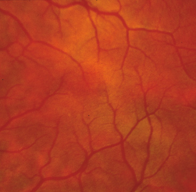
from a temperature-sensitive enzyme [Abstract]. Am J Hum Genet 45(suppl):A8, 1989.

72. King RA, Townsend D, Oetting W, et al: Temperature-sensitive tyrosinase associated with peripheral pigmentation in oculocutaneous albinism. J Clin Invest 87:1046, 1991.

73. Giebel LB, Musarella MA, Spritz RA: A nonsense mutation in the tyrosinase gene of Afghan patients with tyrosinase 
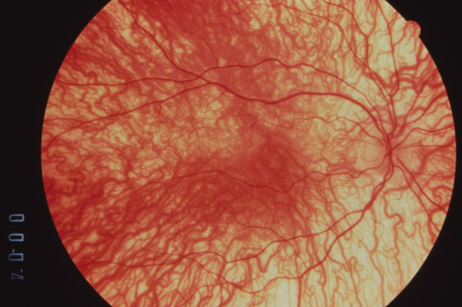
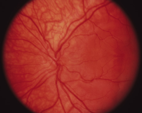
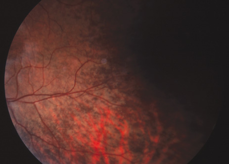
negative (type IA) oculocutaneous albinism. J Med Genet 28:464, 1991.

74. King RA, Creel D, Cervenka J, et al: Albinism in Nigeria with delineation of new recessive oculocutaneous type. Clin Genet 17:259, 1980.

75. King RA, Lewis RA, Townsend D, et al: Brown oculocutaneous albinism. Clinical, ophthalmological, and biochemical characterization. Ophthalmology 92:1496, 1985.

76. King RA, Rich SS: Segregation analysis of brown oculocutaneous albinism. Clin Genet 29:496, 1986.

77. Silvers WK: The coat colors of mice. New York: Springer-Verlag, 1979.

78. Lyon M, Searle AG: Genetic variants and strains of the laboratory mouse. New York: Oxford University Press, 1989.

79. Jackson IJ: A cDNA encoding tyrosinase-related protein maps to the brown locus in mouse. Proc Natl Acad Sci U S A 85:4392, 1988; erratum in Proc Natl Acad Sci U S A 86:997, 1989.

80. Chintamaneni CD, Ramsay M, Colman MA, et al: Mapping the human CAS2 gene, the homologue of the mouse brown (b) locus to human chromosome 9p22-pter. Biochem Biophys Res Commun 178:227, 1991.

81. Murty VV, Bouchard B, Mathew S, et al: Assignment of the human TYRP (brown) locus to chromosome region 9p23 by nonradioactive in situ hybridization. Genomics 13:227, 1992.

82. Wagstaff J: A translocation-associated deletion defines a critical region for the 9p syndrome [Abstract]. Am J Hum Genet 53(suppl):619, 1993.

83. Boissy RE, Zhao H, Austin LM, et al: Melanocytes from an individual with brown oculocutaneous albinism lack expression of TRP-1, the product of the human homologue of the murine brown locus [Abstract]. Am J Hum Genet 53(suppl):160, 1993.

84. Walsh RJ: A distinctive pigment of skin in New Guinea indigenes. Ann Hum Genet 34:379, 1971.

85. O'Donnell FE Jr, Green WR, Fleischman JA, et al: X-linked ocular albinism in blacks: Ocular albinism cum pigmento. Arch Ophthalmol 96:1189, 1978.

86. Falls HF: Sex-linked ocular albinism displaying typical fundus changes in the female 
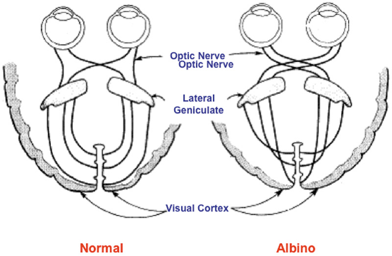
heterozygote. Am J Ophthalmol 34:41, 1951.

87. Gillespie FP, Covelli B: Carriers of ocular albinism with and without ocular changes. Arch Ophthalmol 70:209, 1963.

88. Lang GE, Rott H-D, Pfeiffer RA: X-linked ocular albinism: Characteristic pattern of affection in female carriers. Ophthalmic Paediatr Genet 11:265, 1990.

89. Pearce WG, Johnson GL, Gillan JG: Nystagmus in a female carrier of ocular albinism. J Med Genet 9:126, 1972.

90. Maguire AM, Maumenee IH: Iris pigment mosaicism in carriers of X-linked ocular albinism cum pigmento. Am J Ophthalmol 107:298, 1989.

91. Charles SJ, Moore AT, Grant JW, et al: Genetic counseling in X-linked ocular albinism: Clinical features of the 
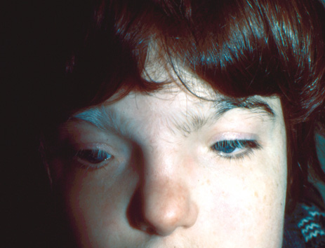
carrier state. Eye 6:75, 1992.

92. Breathnach AS, Wyllie L: Ultrastructure of retinal pigment epithelium of the human fetus. J Ultrastruct Res 16:584, 1966.

93. Mund ML, Rodrigues MM, Fine BS: Light and electronmicroscopic observations on the pigmented layers of the developing human eye. Am J Ophthalmol 73:167, 1972.

94. Wong L, O'Donnell FE Jr, Green WR: Giant pigment granules in the retinal pigment epithelium of a fetus with X-linked ocular albinism. Ophthalmic Paediatr Genet 2:47, 1983.

95. Schnur RE, Wick PA, Bailey C, et al: Phenotypic variability in X-linked ocular albinism: Relationship to linkage genotypes. Am J Hum Genet 55:484, 1994.

96. Benedict PH, Szabo G, Fitzpatrick TB, et al: Melanotic macules in Albright's syndrome and in neurofibromatosis. JAMA 205:618, 1968.

97. Jimbow K, Szabo G, Fitzpatrick TB: Ultrastructure of giant pigment granules (macromelanosomes) in the cutaneous pigmented macules of neurofibromatosis. J Invest Dermatol 61:300, 1973.

98. Guerrier CJ, Lutzner MA, Devico V: An electronmicroscopical study of the skin in 18 cases of xeroderma pigmentosum. Dermatologica 146:211, 1973.

99. Nakagawa H, Hori Y, Sato S, et al: The nature and origin of the melanin macroglobule. J Invest Dermatol 83:134, 1984.

100. Cortin P, Tremblay M, Lemagne JM: X-linked ocular albinism: Relative value of skin biopsy, iris transillumination and funduscopy in identifying affected males and carriers. Can J Ophthalmol 16:121, 1981.

101. Lyon MF: Sex chromatin and gene action in the mammalian X-chromosome. Am J Hum Genet 14:135, 1962.

102. O'Donnell FE, King RA, Green WR, et al: Autosomal recessively inherited ocular albinism: A new form of ocular albinism affecting women as severely as men. Arch Ophthalmol 96:1621, 1978.

103. Morland AB, Hoffmann MB, Neveu M, Holder GE: Abnormal visual projection in a human albino studied with functional magnetic resonance imaging and visual evoked potentials. J Neurol Neurosurg Psychiatry 72:523–526, 2002.

104. Wacks MA, Peachey NS, Fishman GA: Electroretinographic findings in human oculocutaneous albinism. Ophthalmology 96:1778–1785, 1989.

105. Hermansky F, Pudlak P: Albinism associated with hemorrhagic diathesis and unusual pigmented reticular cells in the bone marrow: Report of two cases with histochemical 
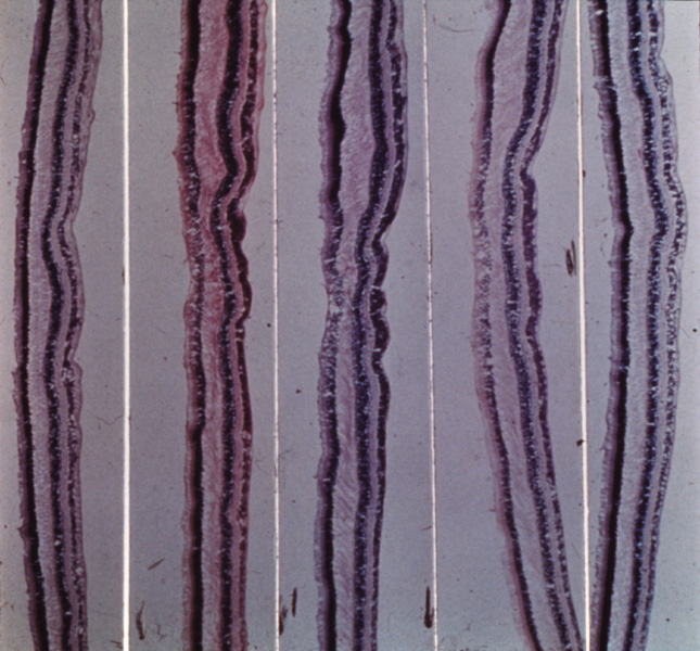
studies. Blood 14:162, 1959.

106. Witkop CJ, Almodavar C, Pineiro B, et al: Hermansky-Pudlak syndrome (HPS). An epidemiologic study. Ophthalmic Paediatr Genet 11:245, 1990.

107. Summers CG, Knobloch WH, Witkop CJ Jr, et al: Hermansky-Pudlak syndrome: Ophthalmic findings. Ophthalmology 95:545, 1988.

108. Scheinfeld NS: Syndromic albinism: a review of genetics and phenotypes. Dermatol Online J 9:5, 2003.

109. Beguez Cesar A: Neutropenia cronica maligna familar con granulaciones atipicas de los leucocitos. Biol Soc Cub Ped 15:900, 1943.

110. Tanaka T: Chédiak-Higashi syndrome: Abnormal lysosomal enzyme levels in granulocytes of patients and family members. Pediatr Res 14:901, 1980.

111. De Beer HA, Anderson R, Findlay GH: Chédiak-Higashi syndrome in a “black” child: Clinical features, immunological studies, and optics of the hair and skin. S Afr Med J 60:108, 1981.

112. Blume RS, Wolff SM: The Chédiak-Higashi syndrome: Studies in four patients and a review of the literature. Medicine 51:247, 1972.

113. Spencer WH, Hogan MJ: Ocular manifestations of Chédiak-Higashi syndrome. Am J Ophthalmol 50:1197, 1960.

114. Johnson DL, Jacobson LW, Toyama R, et al: Histopathology of eyes in Chédiak-Higashi syndrome. Arch Ophthalmol 75:84, 1966.

115. Windhorst DB, Zelickson AS, Good RA: A human pigmentary dilution based on heritable subcellular structural defect: The Chédiak-Higashi syndrome. J Invest Dermatol 50:9, 1966.

116. Bedoya V, Grimley PM, Duque O: Chédiak-Higashi syndrome. Arch Pathol 88:340, 1969.

117. Rausch PDG, Pryzwansky KB, Spitznagel JK: Immunohistochemical identification of azurophilic and specific granule markers in the granules of Chédiak-Higashi neutrophils. N Engl J Med 298:693, 1978.

118. Windhorst DB, Zelickson AS, Good RA: Chédiak-Higashi syndrome: Hereditary gigantism of cytoplasmic organelles. Science 151:81, 1966.

119. Zhao H, Boissy YL, Abdel-Malek Z, et al: On the analysis of the pathophysiology of Chédiak-Higashi syndrome. Defects expressed by cultured melanocytes. Lab Invest 71:25, 1994.

120. Boxer LA, Watanabe AM, Rister M, et al: Correction of leukocyte function in Chédiak-Higashi syndrome by ascorbate. N Engl J Med 295:1041, 1976.

121. Introne W, Boissy RE, Gahl WA: Clinical, molecular, and cell biological aspects of Chediak-Higashi syndrome. Mol Genet Metab 68:283–303, 1999.

122. Karim MA, Suzuki K, Fukai K, et al: Apparent genotype-phenotype correlation in childhood, adolescent, and adult Chediak-Higashi syndrome. Am J Med Genet 2002;108:16–22.

123. Cross HE, McKusick VA, Breen W: A new oculocerebral syndrome with hypopigmentation. J Pediatr 70:398, 1967.

124. Preus M, Fraser FC, Wiglesworth FW: An oculocerebral hypopigmentation syndrome. J Genet Hum 31:323–328, 1983.

125. Patton MA, Baraitser M, Heagerty AH, Eady RA: An oculocerebral hypopigmentation syndrome: a case report with clinical, histochemical, and ultrastructural findings. J Med Genet 24:118–122, 1987.

126. Lerone M, Pessagno A, Taccone A, et al: Oculocerebral syndrome with hypopigmentation (Cross syndrome): Report of a new case. Clin Genet 41:87–89, 1992.

127. Tezcan I, Demir E, Asan E, et al: A new case of oculocerebral hypopigmentation syndrome (Cross syndrome) with additional findings. Clin Genet 51:118–121, 1997.

128. Ozkan H, Unsal E, Kose G: Oculocerebral hypopigmentation syndrome (Cross syndrome). Turk J Pediatr 33:247–252, 1991.

129. De Jong G, Fryns JP: Oculocerebral syndrome with hypopigmentation (Cross syndrome): The mixed pattern of hair pigmentation as an important diagnostic sign. Genet Couns 2:151–155, 1991.

130. Fryns JP, Dereymaeker AM, Heremans G, et al: Oculocerebral syndrome with hypopigmentation (Cross syndrome). Report of two siblings born to consanguineous parents. Clin Genet 34:81–84, 1988.

131. Courtens W, Broeckx W, Ledoux M, Vamosa E: Oculocerebral hypopigmentation syndrome (Cross syndrome) in a gipsy child. Acta Paediatr Scand 78:806–810, 1989.

132. Castle DJ, Jenkins T, Shawinsky AA: The oculocerebral syndrome in association with generalised hypopigmentation. A case report. S Afr Med J 1;76:35–36, 1989.

133. Alfadley A, Parkes RM: Two siblings born preterm with large ears and hypopigmented hair who developed palmoplantar keratoderma and frontal skull bossing: A new syndrome? Pediatr Dermatol 19:224–228, 2002.

134. White CP, Waldron M, Jan JE, Carter JE. Oculocerebral hypopigmentation syndrome associated with Bartter syndrome. Am J Med Genet 46:592–596, 1993.

135. Winship IM, Babaya M, Ramesar RS: X-linked ocular albinism and sensorineural deafness: Linkage to Xp22.3. Genomics 18:444, 1993.

136. Lewis RA: Ocular albinism and deafness [Abstract]. Am J Hum Genet 30:57, 1978.

137. Bard LA: Heterogeneity in Waardenburg's syndrome. Arch Ophthalmol 96:1193, 1978.

138. Donaldson DD: Transillumination of the iris. Trans Am Ophthalmol Soc 72:89, 1974.

139. Bergsma DR, Kaiser-Kupfer M: A new form of albinism. Am J Ophthalmol 77:837, 1974.

140. Margolis S, Siegel IM, Choy A, et al: Oculocutaneous albinism associated with Apert's syndrome. Am J Ophthalmol 84:830, 1974.

141. Mosher DB, Fitzpatrick TB: Piebaldism. Arch Dermatol 124:364, 1988.

142. Hayashibe K, Mishima Y: Tyrosinase-positive melanocyte distribution and induction of pigmentation in human piebald skin. Arch Dermatol 124:381, 1988.

143. Woolf CM, Dolowitz DA, Aldous HE: Congenital deafness associated with piebaldness. Arch Otolaryngol 82:244, 1965.

144. Telfer MA, Sugar M, Jaeger EA, et al: Dominant piebald trait (white forelock and leukoderma) with neurological impairment. Am J Hum Genet 23:383, 1971.

145. Rozycki DL, Ruben RJ, Rapen I, et al: Autosomal recessive deafness associated with short stature, vitiligo, muscle wasting and achalasia. Arch Otolaryngol 93:194, 1971.

146. Waardenburg PJ: New syndrome combining developmental anomalies of the eyelids, eyebrows, and nose root with pigmentary defects of iris and head hair and with congenital deafness. Am J Hum Genet 3:195, 1951.

147. Arias S: Genetic heterogeneity in the Waardenburg syndrome. Birth Defects 7:87, 1971.

148. Foy C, Newton VE, Wellesley D, et al: Assignment of WS1 locus to human 2q37 and possible homology between Waardenburg syndrome and the Splotch mouse. Am J Hum Genet 46:1017, 1990.

149. Tassabehji M, Read AP, Newton VE, et al: Waardenburg syndrome patients have mutations in the human homologue of the PAX-3 paired box gene. Nature 355:635, 1993.

150. Tassabehji M, Read AP, Newton VE, et al: Mutations in the PAX3 gene causing Waardenburg syndrome type 1 and type 2. Nat Genet 3:26, 1993.

151. Baldwin CT, Hoth CF, Amos JA, et al: An exonic mutation in the HuP2 paired domain gene causes Waardenburg's syndrome. Nature 355:637, 1993.

152. Hoth CF, Milunsky A, Lipsky N, et al: Mutations in the paired domain of the human PAX3 gene cause Klein-Waardenburg syndrome (WS-III) as well as Waardenburg syndrome type I (WS-I). Am J Hum Genet 52:455, 1993.

153. Hughes AE, Newton VE, Liu XZ, et al: A gene for Waarburg syndrome type 2 maps close to the human homologue of the microphthalmia gene at chromosome 3p12–p14.1. Nat Genet 7:509, 1994.

154. Tassabehji M, Newton VE, Read AP: Waardenburg syndrome type 2 caused by mutations in the human microphthalmia (MITF) gene. Nat Genet 8:251, 1994.

155. Griscelli C, Durandy A, Guy-Grand D, et al: A syndrome associating partial albinism and immunodeficiency. Am J Med 65:691, 1976.

156. Mancini AJ, Chan LS, Paller AS: Partial albinism with immunodeficiency: Griscelli syndrome: report of a case and review of the literature. J Am Acad Dermatol 38:295–300, 1998.

157. Menasche G, Ho CH, Sanal O, et al: Griscelli syndrome restricted to hypopigmentation results from a melanophilin defect (GS3) or a MYO5A F-exon deletion (GS1). J Clin Invest 112:450–456, 2003.

158. Chen Y, Samaraweera P, Sun TT, et al: Rab27b association with melanosomes: dominant negative mutants disrupt melanosomal movement. J Invest Dermatol 118:933–940, 2002.

159. Sanal O, Ersoy F, Tezcan I, et al: Griscelli disease: genotype-phenotype correlation in an array of clinical heterogeneity. J Clin Immunol 22:237–243, 2002.

160. Fukuda M, Kuroda TS, Mikoshiba K: Slac2-a/melanophilin, the missing link between Rab27 and myosin Va: implications of a tripartite protein complex for melanosome transport. J Biol Chem 277:12432–12436, 2002.

161. Aksu G, Kutukculer N, Genel F, et al: Griscelli syndrome without hemophagocytosis in an eleven-year-old girl: Expanding the phenotypic spectrum of Rab27A mutations in humans. Am J Med Genet 116A:329–333, 2003.

162. Duran-McKinster C, Rodriguez-Jurado R, Ridaura C, et al: Elejalde syndrome—A melanolysosomal neurocutaneous syndrome: clinical and morphological findings in 7 patients. Arch Dermatol 135:182–186, 1999.

163. Bahadoran P, Ortonne JP, Ballotti R, De Saint-Basile G. Comment on Elejalde syndrome and relationship with Griscelli syndrome. Am J Med Genet 116A:408–409, 2003.

164. Perry PK, Silverberg NB: Cutaneous malignancy in albinism. Cutis 67:427–430, 2001.

165. Collins B, Silver J: Recent experiences in the management of visual impairment in albinism. Ophthalmic Pediatr Genet 11:225–228, 1990.

166. Scheinfield NS: Syndromic albinism: a review of genetics and phenotypes. Dermatol Online J 9:5, 2003.

Back to Top