Chapter 35
Keratocentesis and Vitreous Biopsy
JOSEPH B. MICHELSON, MITCHELL H. FRIEDLANDER and G. RICHARD O'CONNOR
Main Menu   Table Of Contents

Search

TECHNIQUE OF KERATOCENTESIS
SPECIAL CONSIDERATIONS
CYTOLOGIC EXAMINATION OF THE AQUEOUS AND VITREOUS
SEROLOGIC EXAMINATION OF AQUEOUS AND VITREOUS HUMOR
ENZYME-LINKED IMMUNOSORBENT ASSAY
SURGICAL SAMPLING
MOLECULAR GENETIC TECHNIQUES FOR CLINICAL ANALYSIS OF THE IMMUNE SYSTEM
REFERENCES

The dazzling panorama of new diagnostic laboratory techniques allows for the identification and characterization of cells, proteins, and histopathologic specimens and even for ultrastructural analysis of very small samples obtained by paracentesis. Diagnostic paracentesis of the eye (keratocentesis of the anterior chamber fluid) and vitreous biopsy (paracentesis of the vitreous fluid in the posterior segment of the eye) have definite value in the following scenarios:
  1. Diagnosing the presence of specific microbial pathogens that are the likely cause of infectious disease in the eye.
  2. Detecting a predominance of certain cell types (e.g., eosinophils, macrophages, epithelial ingrowth, ghost erythrocytes, phacolytic cells) that may provide a clue as to the etiology of an inflammatory disease, which may be autoimmune or allergic in nature.
  3. Identifying:
    1. Specific antibodies in the aqueous humor or vitreous aspirate that are suggestive of infection (e.g., Toxocara, Toxoplasma, herpesvirus, syphilis).
    2. Proteins (e.g., lens proteins, angiotensin-converting enzyme) that are suggestive of granulomatous inflammation, such as sarcoidosis.

Immune complexes and antibodies associated with Behçet's disease may be found. Polymerase chain reaction (PCR) analysis has suggested the presence of DNA from the infection. Tumor cells may be identified when a malignant infiltration of the eye (e.g., large cell lymphoma, leukemia, retinoblastoma, malignant melanoma) masquerades as a uveitis, or by the presence of tumor cell enzymes and antigens (Table 1). 1,2

TABLE 1. Diagnostic Paracentesis


Finding Condition or Disease Indicated
Aqueous 
BacteriaEndophthalmitis
FungiCandida, Aspergillus, etc.
Tumor cellsRetinoblastoma, malignant melanoma, reticulum cell sarcoma, leukemia, metastatic cancer
EosinophilsToxocara canis
MacrophagesPhacolytic glaucoma
Antibodies (ELISA)Toxoplasma gondii, T. canis, reticular cell sarcoma, Behçet's disease, syphilis
Immune complexesBehçet's disease
Other proteins 
Angiotensin-converting enzymesSarcoid
Lactate dehydrogenase isoenzymesRetinoblastoma
Lens fragmentsPhagocytolytic glaucoma
Ghost erythrocytesHemorrhagic glaucoma
Metastatic cancer cellsMetastatic cancer
JXGJXG
Mesenchymal fibrous cellsPHPV
AmyloidAmyloid
Epithelial cellsEpithelial ingrowth
Vitreous 
BacteriaEndophthalmitis
FungiCandida, Aspergillus sp., etc. (e.g., cryptovirus)
Tumor cellsRetinoblastoma, malignant melanoma, reticular cell sarcoma, leukemia, metastatic cancer
EosinophilsT. canis
AntibodiesT. gondii, reticular cell sarcoma, Behçet's disease, syphilis (and immune complexes)
MacrophagesSympathetic ophthalmia, severe retinitis
AmyloidAmyloid
Calcium soapsAsteroid hyalosis
Polymerase chain reaction of viral DNACMV retinitis1,2
Interleukin-10 (ELISA)RCS3
Monoclonal antibodies to Behçet's disease (long)Behçet's disease (clinical activity, numbers of cells)4
?HLA DR and DRYVKH (equally prevalent among Hispanics and Japanese)5

CMV = cytomegalovirus; ELISA = enzyme-linked immunosorbent assay; HLA = human leukocyte antigen.


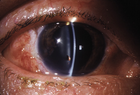
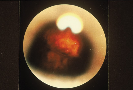
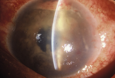
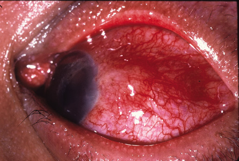
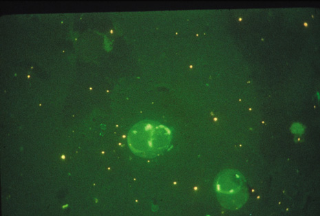
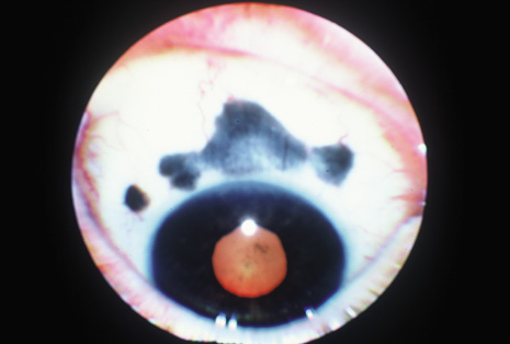
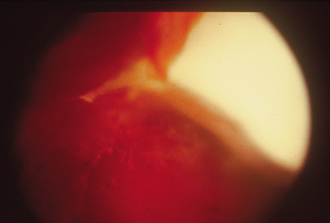
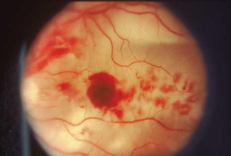
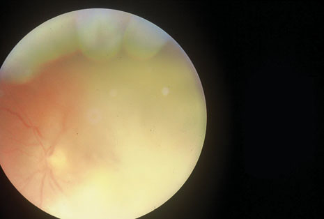
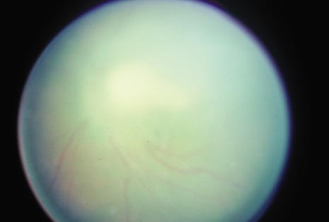
Although keratocentesis had been advocated historically as a treatment for active uveitis, it lost the attention of ophthalmologists until 1919, when Bruckner3 first examined the aqueous humor for diagnostic purposes. Laboratory techniques were revolutionized in the 20th century in areas such as: (a) evaluating very small aliquots of fluid (0.2 to 0.3 mL of aqueous or vitreous), and (b) identifying specific microbial organisms and the predominance of other cell types, antibodies, and proteins in these fluids (Figs. 1, 2, 3, 4, 5, and 6). These advancements have led to the development of diagnostic paracentesis for sight-threatening ocular inflammations that are difficult to diagnose. Witmer4 and O'Connor5 have provided strong evidence that samples of the aqueous humor reflect the antibody-producing capabilities of the iris and ciliary body, particularly when more specific antibody per unit of gamma globulin can be found on the aqueous humor than in the blood of the same patient.6–8 These determinations may be highly significant when one considers the fact that diseased tissue is being bathed in an antibody-containing fluid that is elaborated locally. For instance, in the case shown in Figure 1, the immunofluorescent antibody titer to toxoplasmosis is four times greater in the vitreous aspirate at the time of vitrectomy for repair of retinal detachment than in the plasma. These same considerations have long been recognized in syphilis of the central nervous system, wherein specific antibodies may be present in the cerebrospinal fluid but not in the blood. This is also the case with an unusual presentation of ocular coccidioidomycosis9 or toxocariasis.

Fig. 1. Fundus photograph of a patient with toxoplasmosis who, after vitrectomy and scleral buckling, demonstrates the etiologic chorioretinal lesion at the 11:30 position on the buckle, which is now inactive. The patient's serum indirect fluorescent antibody toxoplasmosis titer was 1:1024 in the plasma: the vitrectomy fluid yielded a titer of 1:4096.

Fig. 2. Typical gram-positive cocci occurring in clusters as isolated from the aqueous of a patient with staphylococcal endophthalmitis.

Fig. 3. Chains of gram-positive cocci obtained from aqueous fluid in a patient with streptococcal endophthalmitis.

Fig. 4. Gram-negative rods obtained from the vitreous of a patient with endophthalmitis.

Fig. 5. Aqueous specimen demonstrating acid-fast bacilli of acute lepromatous leprosy in the aqueous aspiration of a patient with a profound anterior segment inflammation that presented as a diagnostic dilemma.

Fig. 6. Scleral nodule specimen showing a profuse number of acid-fast bacilli from a patient with lepromatous uveitis.

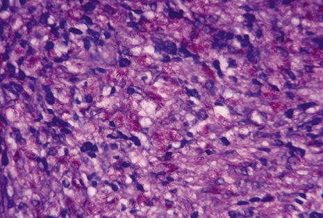
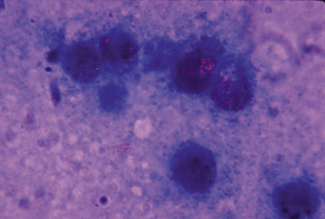
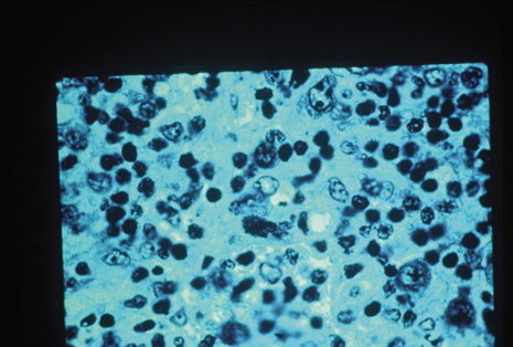
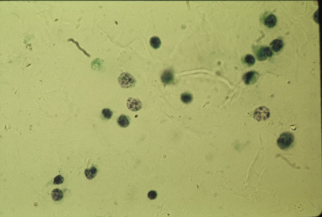
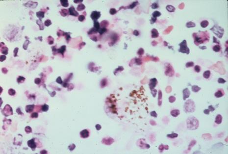
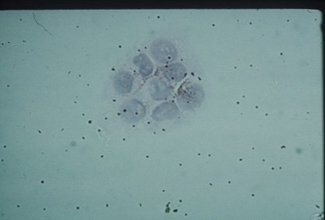
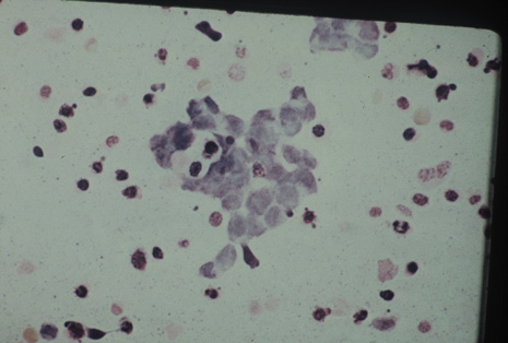
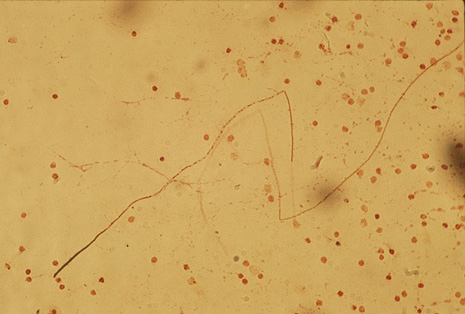
Many forms of uveitis are characterized by specific types of inflammatory cells. Usually, however, one encounters mixtures of cell types in any given specimen, with the relative percentages of lymphocytes and polymorphonuclear leukocytes varying. There may be unusual numbers of eosinophils, or macrophages laden with lens material may be present. Thus, an enumeration of the cells and a careful analysis of their structure can be useful as a diagnostic aid (Figs. 7, 8, 9, 10, 11, 12, 13, 14, 15, 16, 17, 18, 19, and 20). Figure 15 demonstrates eosinophils that were aspirated from the anterior chamber of a patient with Toxocara canis endophthalmitis. Figure 12 demonstrates malignant cell infiltrate from the vitreous, showing the stained presence of monoclonal light chains being elaborated in the cytoplasm. Interleukin-10, detectable in the vitreous of intraocular lymphoma patients, is also directly indicative of both the clinical activity and the number of malignant cells as observed by cytopathology.

Fig. 7. Anterior segment photograph showing a petechiae-speckled hypopyon in a patient with profound anterior segment inflammation resulting from acute lepromatous leprosy.

Fig. 8. Inflamed scleral nodule removed from the same patient as in Figure 7, who had acute lepromatous uveitis.

Fig. 9. Trophozoites of toxoplasmosis in an aspirate from mouse peritoneum, which incubated acute material from a patient with acute acquired systemic toxoplasmosis with retinitis.

Fig. 10. Axillary lymph node of a patient with acquired systemic toxoplasmosis demonstrating trophozoites and a toxoplasma cyst.

Fig. 11. Vitreous aspirate demonstrating large cell lymphoma infiltration in a patient with vitreitis and masquerade uveitis of reticulum cell sarcoma or large cell lymphoma.

Fig. 12. Large cell lymphoma aspirate of vitreous. Immunofluorescence demonstrates a monoclonal infiltrate of lambda light chains on the B cells.

Fig. 13. Fundus photograph demonstrating blunted retinal details resulting from overlying vitreitis of malignant infiltration of large cell lymphoma. Note white subretinal infiltration of tumor cells. The vitreous aspirate had a high cellular content, facilitating a rapid diagnosis.

Fig. 14. Clinical photograph of synechiae. Exuberant aqueous infiltration of cells produced a plastic iritis.

Fig. 15. Aqueous aspirate of the same patient as in Figure 14 shows an abundance of malignant melanoma cells, which demonstrate the etiology of the masquerade inflammation in this patient who had metastatic malignant melanoma from the back to the iris in several foci.

Fig. 16. Ring melanoma of the ciliary body in a patient with dense anterior chamber inflammation, as noted by background illumination in the anterior chamber.

Fig. 17. Aqueous aspirate of the same patient as in Figure 16, showing a very densely cellular panorama of malignant melanoma cells.

Fig. 18. Aqueous aspiration from a 2-year-old child thought to have endogenous endophthalmitis reveals retinoblastoma cells amid cellular debris and an occasional polymorphonuclear leukocyte.

Fig. 19. Pathologic specimen of eyeball demonstrates a diffuse endophytic retinoblastoma filling almost the entire vitreous cavity with tumor material, some with liquefaction and necrosis, which presented as a clinical diagnostic dilemma until paracentesis of the aqueous revealed the malignant retinoblastoma cells.

Fig. 20. Multinucleated eosinophils in aqueous specimen of patient with Toxocara canis endophthalmitis.

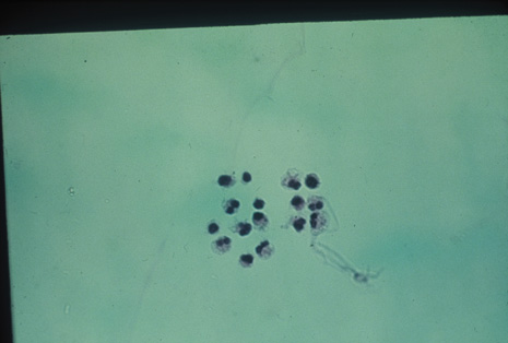
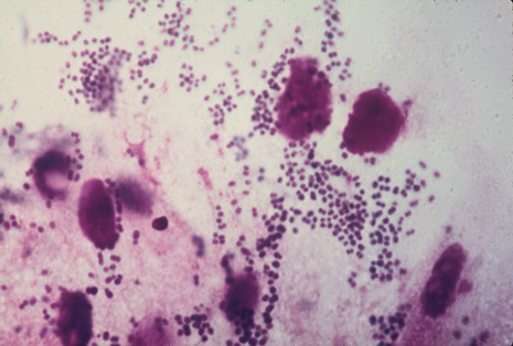
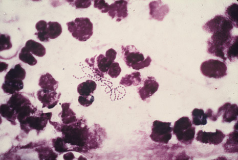
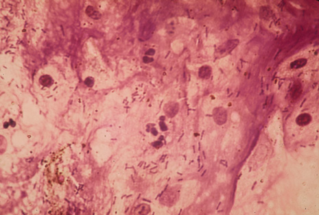
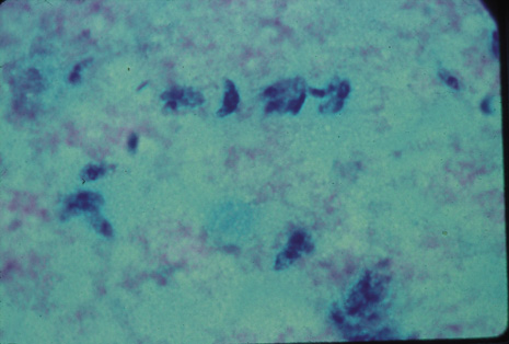
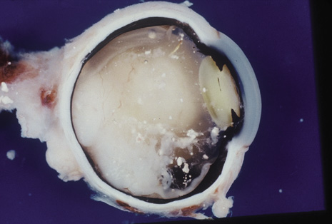
Precise identification and culture of bacterial and fungal pathogens from both the aqueous humor and the vitreous fluid can be obtained. Gram's stain and Giemsa's stain smears of centrifuged specimens from the aqueous humor and the vitreous humor frequently demonstrate the bacterial or fungal causative agent. Attempts to isolate bacteria and fungi and to identify them on Gram's stain or Giemsa's stain smears have been most rewarding in the following cases: (a) postoperative endophthalmitis, (b) infection after a penetrating injury of the eye, (c) drug abuse patients with endogenous endophthalmitis (Figs. 21, 22, 23, 24, and 25), (d) patients receiving hyperalimentation, and (4) patients who are immunocompromised as a result of exogenous immunosuppressive agents.

Fig. 21. Clinical photograph of Toxocara abscess in the peripheral retina with its fibrous band leading back to the posterior pole, a clinical finding thought to be pathognomonic of Toxocara infection. Note the hazy vitreous, whose inflammation is obscuring much of the retina detail below.

Fig. 22. Septate hyphae amid polymorphonuclear leukocytes and cellular debris of vitreous specimen isolated from a patient with Aspergillus endophthalmitis owing to intravenous drug abuse.

Fig. 23. Profound retinal hemorrhage and inflammation in a 23-year-old drug abuse patient. The subretinal exudation was so dense that it formed a meniscus as its superior edge. Vitreous aspiration in this patient proved diagnostic and enabled early therapeutic intervention that resulted in the salvaging of a useful eye.

Fig. 24. Profound intravitreal inflammation obscuring most of the retinal detail except for a vague view of the optic nerve head and a small surrounding patch of retina in a patient with Aspergillus endophthalmitis. The patient underwent early vitrectomy as a consequence of retrieval of hyphae on vitreous sampling.

Fig. 25. Classic presentation of Candida endophthalmitis demonstrating vitreous “fluff balls” and vitreal inflammation obscuring the retina in a 25-year-old drug abuse patient in whom Candida albicans was easily identified and cultured from the vitreous.

Studies have demonstrated the usefulness of ocular paracentesis for the identification of ocular infections in order to implement sight-saving treatment.10–16 Even acid-fast bacilli and viruses may be diagnosed in this fashion when emergency dictates (see Fig. 5).17 It is recommended that diagnostic paracentesis be performed in all cases of postoperative endophthalmitis, and it is safe to perform the postoperative procedure in the operating room with the safety of vitrectomy surgery. Further, any patient older than 65 who presents with a deteriorating uveitis (usually with vitreitis as the predominant infiltrate) of undetermined etiology should undergo paracentesis of the vitreous to rule out reticulum cell sarcoma (large cell lymphoma).18 Similarly, any patient suspected of being an intravenous drug abuser who presents with an endogenous endophthalmitis or uveitis should undergo diagnostic paracentesis to avoid allowing an intraocular infection to be borne by the bloodstream.19,20

Back to Top
TECHNIQUE OF KERATOCENTESIS
Many techniques have been described for paracentesis of the eye. Most authorities agree that aspiration of the aqueous fluid (keratocentesis) is best accomplished with the following technique:
  1. Perform a beveled keratotomy through the peripheral clear limbal cornea with a Ziegler knife, razor blade, or super blade, deeply incising the cornea but not entering the anterior chamber.
  2. Use a 27-gauge needle, attached to a 1-mL tuberculin syringe or a 3-mL syringe, to enter the anterior chamber via this incision, using a rolling technique. Avoid touching corneal endothelium and particularly the lens in phakic patients. Be careful to stay over the peripheral iris at all times. The needle tip should not be aimed toward the center of the pupil.
  3. Obtain a 0.1- to 0.3-mL yield of aqueous and then immediately inoculate the fluid onto blood agar, brain–heart infusion, chocolate agar, and thioglycolate liquid (maintained at body temperature) and onto Sabouraud agar, blood agar, and brain–heart infusion with gentamicin (maintained at room temperature for fungal isolation). Be careful to place the drops of aspirate away from the edges of the plate. The mouths of the tubes of the liquid media should have been flamed before use and after inoculation. Very tiny drops may be left on the tip of the syringe after inoculation of the media to prepare for Gram's, Giemsa's, and Methamine silver stain. Several drops per media plate may be allowed, and one drop is allowed to flow several centimeters on the surface. Roll the plate between your fingers to ensure that it has adequate coverage in the center of the media plate.
  4. In aphakic patients, fit a second tuberculin syringe to a 22-gauge needle and pass it through a slightly larger keratotomy incision and into the vitreous. Manipulate the syringe until you are able to aspirate 0.2 to 0.3 mL of vitreous.
  5. In phakic patients suspected of having endophthalmitis complicating either filtering bleb or trauma or a metastatic cancer etiology, aspirate the vitreous through a sclerotomy at the pars plana 3.5 to 4.5 mm posterior to the limbus on the temporal side, with or without an accompanying anterior chamber paracentesis. Incise the conjunctiva over this area down to clean sclera so that there is no obstruction when you make the sclerotomy stab.
  6. If you find that the vitreous specimen is inadequate, or you suspect a fungal etiology, enlarge the pars plana incision to 2.5 mm and introduce a larger-bore needle to ensure that an adequate amount of liquid vitreous is aspirated.
  7. If you are sure of an infectious etiology and suspect that a therapeutic vitrectomy will have to be undertaken in any event, you may want to perform a vitreous aspiration with a vitreous suction-cutter machine such as an Ocutome, a Peyman unit, or a Visc, which remove formed vitreous.21
Back to Top
SPECIAL CONSIDERATIONS
Suction of the vitreous in a patient with endophthalmitis is very tricky. The vitreous tends to put traction on the retina, leading to unanticipated tearing and splitting of the retina and ultimately retinal detachment. To minimize traction on the retina caused by vitreous condensation and fibrous tissue formations, it is best to aspirate vitreous in the operating room under sterile conditions and under the controlled suction and cutting of a vitreous instrument. This is highly preferable to the unstable “sucking only” of a large-bore needle aspiration at the pars plana. The cutting rate of the vitrectomy instrument should be turned up higher than 300 cuts per minute, which further serves to minimize tractional forces. Too much cutting of the vitreous, however, disturbs, disrupts, contorts, and destroys whatever cellular elements and hyphae one may wish to obtain from the vitreous specimen. The optimal degree of vitreous surgery should include aspiration with a minimum of cutting of cellular elements, but with enough rapid cutting so as not to prolong tractional forces. Such a vitreous sample diluted by the irrigating solution is then passed through a disposable membrane filter system. Adequate sterile technique must be maintained. The procedure for obtaining the vitreous includes a stop-cock assembly somewhere in the line before the machine receptacle is encountered by the sterile line. This varies with each machine used, but some forethought to the system allows for sterile maintenance of the fluid.

Outpatient keratocentesis can be performed carefully at the slit-lamp with a minimum of complications. The same manipulative criteria must be observed as listed under Techniques (i.e., one should avoid the corneal endothelium and avoid rupturing the anterior lens capsule in phakic patients). Although the ideal conditions for vitreous aspiration are absolute sterility of the operating room and the more optimal aspiration/cutting ability of the vitreous instrument, in emergency situations it may be necessary to perform a pars plana paracentesis in the office setting. Even in such an emergency situation, however, it is almost always necessary to delay treatment until the patient can be taken to the operating room for a more controlled surgical evacuation of vitreous material. It is of both diagnostic and therapeutic benefit to the patient to achieve a more complete core vitrectomy, especially when cellular elements (e.g., large cell lymphoma, fungal endophthalmitis) are required to determine the diagnosis.

Back to Top
CYTOLOGIC EXAMINATION OF THE AQUEOUS AND VITREOUS
Cytologic examination of the aqueous and vitreous obtained with paracentesis may be performed by a number of different methods. The simplest method is the single-drop preparation, which consists of placing a small drop of fluid from the aspirating needle directly onto the glass slide. To avoid dispersing the cells too widely, the drop should not be spread. The drop should merely be allowed to air dry. When dry, the preparation is fixed in absolute methanol for 10 minutes and once again allowed to dry. The slide is finally placed in dilute buffered Giemsa solution and allowed to stain for 1 hour. It is then quickly rinsed with 95% ethanol and allowed to dry. A cover slip is mounted with Canada balsam and the preparation examined microscopically.

The relative numbers of lymphocytes, polymorphonuclear leukocytes, and macrophages can be determined immediately. In viral infections and chronic hypersensitivity reactions, the cell type is predominantly mononuclear. In acute uveitis reactions, especially those involving the binding of complement to immune complexes (as in Behçet's syndrome), an abundance of neutrophils may be expected, and aqueous and vitreous can be examined by the Raji immune complex assay to determine the amount of immune complex in those fluids. In lens-induced uveitis, one may expect to see many macrophages as well as some neutrophils. In parasitic infections, numerous eosinophils may be seen (see Fig. 19). Bacteria or other organisms can be detected by the same examination. If bacteria are seen on the Giemsa preparation, a Gram's stain also should be performed on another single-drop preparation.

Other methods of cytologic examination include techniques for concentrating the cellular elements. The simplest of these consists of passing the entire aspirate through a Millipore filter (mean pore size 0.45 γm). The filter disk may be stained and cleared with xylene before it is examined microscopically. Various centrifuges have been designed for the concentration of aqueous cells. Immediately upon aspiration, the cells should be fixed in a glutaraldehyde-paraformaldehyde mixture and postfixed in osmium tetroxide. They are centrifuged onto a thin sheet of araldite before sectioning. This technique results in the preservation of excellent cytologic detail. The secretory granules of the various types of polymorphonuclear leukocytes are easily identifiable, and organelles such as the Golgi apparatus can be seen readily. This technique also provides a method for examining viral inclusions in certain cells.

Wet fixation of cells via a modification of the Papanicolaou technique may provide an ideal means of identifying intranuclear intracytoplasmic inclusions in affected cells. Studies have indicated that this method is far superior to the Giemsa technique for this purpose.

Darkfield microscopic examinations of aqueous humor specimens have sparked much interest. Smith22 has identified mobile Treponema in the aqueous humor of patients suspected of having syphilitic uveitis. Correlative tests using fluorescent antibody methods seemed first to confirm the pathogenic T. pallidum in the anterior chamber of these patients, but subsequent examinations with adequately absorbed antisera seemed to indicate that some of the spirochetal forms previously observed were nonpathogenic treponemes (e.g., T. microdentium). Other difficulties with darkfield microscopy include the length of time necessary for observation and the frequent appearance of artifacts.

Back to Top
SEROLOGIC EXAMINATION OF AQUEOUS AND VITREOUS HUMOR
The use of paracentesis for the procurement of diagnostic samples for serologic testing is increasing. Microtiter methods for virtually all the serologic tests used in the laboratory are being perfected for microtiter specimens (Table 2). These are available for testing aqueous humor and liquid vitreous samples and are particularly useful when tests of the aqueous or vitreous are performed in conjunction with simultaneous tests on the patient's serum. These include the new techniques of enzyme-linked immunosorbent assay (ELISA) and PCR.

TABLE 2. Intraocular Inflammation Serology Measured In Microtiter Quantity


Condition Findings
CytomegalovirusAb: CF, IgG and IgM EIA, IgM Ab IFA CMV, mRNA, cDNA probe/ISU
Herpes simplex virusAb: CF, IgG-EIA, IgM-IFA; polymerase chain reaction (PCR)
HistoplasmaAb: CF, Ag-EIA, Ab-CIE
HTLV-III/LAVAb: EIA, IFA, Ag:EIA
FilariaAb: CIE, EIA, IFA Ag:Giemsa
Treponema pallidumVDRL, RPR, FSA-ABS, FTA-ABS-IgM, MHA-TP, Ab-Ay4D, EIA
SarcoidosisACE, V serum lysozyoid
LeptospiraAb: CF, IHA, IgG and IgM Ab, EIA
MumpsAb: soluble Ag, CF IgM and IgG Ag; viral Ag, CF, IgM and IgG Ab, EIA: PCR
Mycobacterium lepraeAb and IgM and IgG Ab, EIA
Mycobacterium tuberculosisAb: IgG, EIA
Pneumocystis cariniiAb: IFA, EIA, Ag CIE
PseudomonasAb: CF, IHA, CIE
Strongyloides stercoralisIgG and IgE Ab, EIA
Subacute sclerosing panencephalitisMeasles antibody index
Toxocara canisIgG, IgM, IgA Ab EIA, ELISA
Toxoplasma gondiiIgG, IgM Ab EIA, ELISA Ab: CF, IHA Total and IgM Ab:IFA
Trypanosoma cruziAb: IHA, CF, IFA Total (cerebrospinal fluid) IgM: EIA
Varicella zosterAb: CF, IgG and IgM, Ab, EIA: PCR
Cryptococcus neoformansAg:LPA Ab: IPA
Coccidioides immitisAb CF, EIA, ID Ag RIA/EIA

Ab = antibody; ACE = angiotensin-converting enzyme; Ag = antigen; CF = complement fraction; CIE = countercurrent immunoelectrophoresis; EIA = enzymal immunoassay; ELISA = enzyme-linked immunosorbent assay; FTA = fluorescent treponemal antibody; ID = immunodiffusion; IFA = indirect fluorescent antibody; IHA = indirect hemagglutination; PCR = polymerase chain reaction; RIA = radioimmunoassay; RPR = rapid plasma reagin: VDRL = Venereal Disease Research Laboratory (test for syphilis).


The ciliary body and iris may act as local factories for the production of antibodies. It is diagnostically significant whenever it can be established that the specific antibodies detected in the aqueous or vitreous are actually being made in the eye. One also must remember that the malignant B cell seen in the eye in large cell lymphoma (reticulum cell sarcoma) may be identified by the presence of antibodies on its surface (see Fig. 12) and cell-specific markers. One can demonstrate kappa or lambda light chains exclusively that are being produced on these cells as a manifestation of monoclonal infiltration. This is quite different from what is seen in a mixture of both kappa and lambda light chains, as manifested in a pure inflammation.

Numerous qualitative reactions also are available for detection of specific antibodies in the aqueous and vitreous, and these have been adapted to microtiter quantitation by serial dilutions. These include paths of hemagglutination reactions for tuberculosis and, more frequently, ELISA, which have been developed for toxoplasmosis, toxocariasis, and herpesvirus and are available for most infectious pathogens.

Back to Top
ENZYME-LINKED IMMUNOSORBENT ASSAY
The development of solid-phase immunoassays during the past 10 years has added greatly to our ability to detect small amounts of circulating antibodies. One of the first assays to be developed was a radioimmunosorbent technique in which fixation of a patient's antibodies to a solid-phase antigen was detected by the use of radioactive iodine-labeled antihuman globulin. This technique required maintenance of radioactive materials in the laboratory as well as other potentially dangerous procedures.

The development of the ELISA test by Engvall and Perlmann23 provided a much safer method of fixation of antibodies to a solid-phase antigen. ELISA is used to detect antibodies to any antigen, and it screens for a wide number of bacterial, viral, and parasitic antigens, especially Toxocara, Toxoplasma, gonococcus, herpesvirus, and cytomegalovirus. The ELISA method is as follows:

  1. Protein antigens are linked to the walls of disposable polystyrene test tubes.
  2. The patient's serum (aqueous humor or vitreous specimen) is put into the test tube and allowed to react with the antigen, and any unreached material is carefully washed out.
  3. Goat antihuman globulin, conjugated with horseradish peroxidase, is then added to the tube and allowed to react with the antigen–antibody complex that is fixed to the wall. Again, all unbound products are carefully washed out of the tube after a suitable time has been allowed for the reaction to take place.
  4. A “substrate” is added to the tube, consisting of a mixture of 5-aminosalicylic acid and hydrogen peroxidase. As the peroxide is acted on by the peroxidase, a brownish color is imparted to the solution.
  5. This color reaction is quantitated on a spectrophotometer or read grossly, as is done in the microadaptation of the ELISA test for toxoplasmosis.24

Angiotensin-converting enzyme, which is diagnostic of granulomatous inflammation in general and suggestive of sarcoidosis in particular, can now be measured in such small amounts as aqueous specimens as well.

Back to Top
SURGICAL SAMPLING
No chapter on paracentesis of the eye would be complete without mentioning “sampling” of the uveal tissue to rule out certain malignancies. Although nodules of the iris may be benign in nature, and a consequence of inflammation as well as metaplasia or neoplasia, these lesions are more readily accessible for biopsy than choroidal or ciliary body lesions.25–30 Pioneering techniques of sophisticated biopsy surgery, initiated by Peyman and colleagues,30 have lent themselves to “eye wall biopsy,” leaving a candidate for possible enucleation for histopathologic evaluation intact. These specific surgical techniques have included iridocyclectomy (Fig. 26), iridochoroidectomy, eye wall resection, eye wall biopsy, endoretinal biopsy, and ab interno retinochoroidectomy.31 These techniques are especially valuable in the difficult diagnosis of large cell lymphoma infiltrate, atypical malignant melanoma of the choroid, acute retinal necrosis, viral retinitis, nematodes, therapeutic removal of malignant melanoma of the choroid or ciliary body, and metastatic cancer of the choroid.

Fig. 26. Inferior cyclectomy in a patient with atypical ciliary body melanoma, in whom an advanced surgical technique employing a Peyman eye basket for stability of the globe allowed salvaging of a useful eye.

Back to Top
MOLECULAR GENETIC TECHNIQUES FOR CLINICAL ANALYSIS OF THE IMMUNE SYSTEM
During the past two decades, advances in nucleic acid chemistry and recombinant DNA technology have made it possible to analyze individual genes rapidly and precisely. These techniques are now commonly used in research and clinical laboratories. DNA is a remarkably sturdy molecule that is easy to obtain and work with.32 It also provides access to information that allows us to investigate and diagnose a variety of disease processes at a very fundamental level.

These techniques include nucleic acid probes, short strands of bases with known sequences that can detect complementary base sequences. Hybridization assays can be used to evaluate cellular DNA by denaturing it and binding it to membranes, where it can be analyzed with probes to detect certain sequences. Examples of hybridization assays include the Southern blot assay, which determines the clonality of lymphoid cell populations.

PCR is a powerful enzymatic technique that can exponentially replicate specific DNA sequences in the test tube. With this technique, it is now possible to assay vanishingly small samples that initially contain fewer than 10 copies of the sequence of interest. PCR is particularly useful for detecting viruses and other microorganisms in tissue specimens, because such organisms often can be recognized by their unique RNA or DNA sequences much more quickly and inexpensively than in culture. PCR has already assumed an important role in microbiologic diagnosis, and it seems likely that this role will expand in the future. Important organisms that can be assayed with this technique include Epstein-Barr virus, cytomegalovirus,33 human immunodeficiency viruses, and human T-cell leukemia viruses.

DNA-based assays also are very useful for detecting large-scale chromosomal deletions or rearrangements characteristic of several hematologic malignancies.

Back to Top
REFERENCES

1. Michelson JB, Chisari FV, Kansu J: Antibodies to oral mucosa in patients with ocular Behçet's disease. Ophthalmology 92:1277, 1985

2. Novey HS, Wells ID: Allergic bronchopulmonary aspergillosis. West J Med 130:1, 1979

3. Bruckner A: Cytologische Studien am Auge. Graefes Arch Clin Exp Ophthalmol 100:179, 1919

4. Witmer R: Clinical implications of aqueous humor studies in uveitis. Am J Ophthalmol 86:39, 1978

5. O'Connor GR: Antitoxoplasma precipitins in aqueous humor: New application of the agar diffusion technique. Arch Ophthalmol 57:52, 1957

6. Witmer R: Experimental leptospiral uveitis in rabbits. Arch Ophthalmol 53:547, 1955

7. O'Connor GR: Precipitating antibody to toxoplasma: Follow-up study on findings in the blood and aqueous humor. Am J Ophthalmol 44:75, 1957

8. Benitez del Castillo JM, Herreros G, Guillen JL, et al: Bilateral ocular toxocariasis demonstrated by aqueous humor enzyme-linked immunosorbent assay. Am J Ophthalmol 119:514, 1996

9. Michelson JB, Belmont JB, Higginbottom P: Juxtapapillary choroiditis associated with chronic meningitis due to Coccidioides immitis. Ann Ophthalmol 15:666, 1983

10. Allansmith MR, Skaggs C, Kimura SJ: Anterior chamber paracentesis: Diagnostic value in postoperative endophthalmitis. Arch Ophthalmol 84:745, 1970

11. Foster RK: Etiology and diagnosis of bacterial postoperative endophthalmitis. Ophthalmology 85:320, 1978

12. Engel H, de la Cruz ZC, Jimenenz-Abalahin LD, et al: Cytopreparatory techniques for eye fluid specimens obtained by vitrectomy. Acta Cytol 26(4):551, 1982

13. Brucker AJ, Michels RG, Green WR: Pars plana vitrectomy in the management of blood-induced glaucoma with vitreous hemorrhage. Ann Ophthalmol 10:1427, 1978

14. Bruner WE, Stark WJ, Green WR: Juvenile xanthogranuloma of the iris and ciliary body in an adult. Arch Ophthalmol 100(3):457, 1982

15. Stark WJ, Rosenthal AR, Mullins GM, et al: Simultaneous bilateral uveal melanomas responding to BCNU therapy. Trans Am Acad Ophthalmol Otolaryngol 75:70, 1971

16. Piro P, Erozan YS, Michels RG, et al: Metastatic breast carcinoma to the vitreous diagnosed by study of pars plana vitrectomy specimen. Ann Ophthalmol 27:84, 1977

17. Michelson JB, Whitaker JP, Wilson S, O'Connor GR: Possible foreign body granuloma of the retina associated with intravenous cocaine addiction. Am J Ophthalmol 887:278, 1979

18. Michelson JB, Freedman SD, Boyden DG: Aspergillus endophthalmitis in a drug abuser. Ann Ophthalmol 14:1051, 1982

19. Michelson JB, Roth AM, Waring GO: Lepromatous iridocyclitis diagnosed by anterior chamber paracentesis. Am J Ophthalmol 88:674, 1979

20. Michelson JB, Felberg NT, Shields JA, Folberg R: Subretinal fluid examination of LDH, PGI, and CEA in a case of metastatic bronchogenic carcinoma of the choroid. CA CCR 41:2301, 1978

21. Freeman WR: Application of vitreoretinal surgery to inflammatory and infectious disease of the posterior segment: New approaches to vitreoretinal surgery. Int Ophthalmol Clin 18, 1992

22. Smith JL: The presence of treponemes in aqueous humor in human uveitis. In Clinical Methods in Uveitis. St. Louis: CV Mosby, 1968:159

23. Engvall E, Perlmann P: Enzyme-linked immunosorbent assay (ELISA): Quantitative assay of immunoglobulin G. Immunochemistry 8:871, 1971

24. Walls KW, Bullock SL, English DK: Use of the enzyme-linked immunosorbent assay (ELISA) and its microadaptation for the serodiagnosis of toxoplasmosis. J Clin Microbiol 5:273, 1977

25. Morgan WE III, Malmgren RA, Albert DM: Metastatic carcinoma of the ciliary body simulating uveitis: Diagnosis by cytologic examination of aqueous humor. Arch Ophthalmol 83:54, 1970

26. Scholz R, Green WR, Baranano EC, et al: Metastatic carcinoma to the iris: Diagnosis by aqueous paracentesis and response to irradiation and chemotherapy. Ophthalmology 90:1524, 1983

27. Takahashi T, Oda Y, Chiba T, et al: Metastatic carcinoma of the iris and ciliary body simulating iridocyclitis. Ophthalmologica 188:266, 1984

28. Talegaonkar SK: Anterior uveal tract metastasis as the presenting feature of bronchial carcinoma. Br J Ophthalmol 53:123, 1969

29. Woog JJ, Chess J, Albert DM, et al: Metastatic carcinoma of the iris simulating iridocyclitis. Br J Ophthalmol 68:167, 1984

30. Peyman GA, Raichand M, Schulman S: Diagnosis and therapeutic surgery of the uvea: Part I. Surgical technique. Ophthalmic Surg 17:822, 1987

31. Denslow GT, Kielar RA: Metastatic adenocarcinoma to the anterior uvea and increased carcinoembryonic antigen levels. Am J Ophthalmol 85:363, 1978

32. Parslow TG, Stites DP, Terr AI: Basic and clinical immunology, 8th ed. Norwalk, CT: Appleton and Lange, 1994

33. Holland GN: Cytomegalovirus diseases. In Ocular Infection and Immunity. St. Louis: CV Mosby, 1996:1110

Back to Top