Chapter 34
Immunology of Uveitis
MITRA MAYBODI, MEHMET L. GULER and HENRY J. KAPLAN
Main Menu   Table Of Contents

Search

OVERVIEW OF THE IMMUNE RESPONSE
OCULAR IMMUNE PRIVILEGE
CLINICAL IMMUNOLOGY: HYPERSENSITIVITY REACTIONS
IMMUNOLOGY OF UVEITIS
CONCLUSION
ACKNOWLEDGMENT
REFERENCES

During the past two decades, important advances have been made in understanding the immunopathologic basis of ocular diseases. This has resulted largely from developments in the field of immunology and molecular biology. Immunology has progressed from a simple dichotomy of Metchnikoff's theory of cellular immunity and Ehrlich's theory of antibody-mediated immunity to a better understanding of their complex relation in both generating and regulating the immune response. We are in an era in which many fundamental immunologic observations are being described not only at the cellular but also at the molecular level. These advances promise to provide an understanding of the pathogenesis of many systemic inflammatory disorders and to direct the search for therapeutics. In this chapter, we discuss the generation and regulation of the immune response, present specific clinical applications in the context of the different types of hypersensitivity, and address additional immune mechanisms in uveitis.
Back to Top
OVERVIEW OF THE IMMUNE RESPONSE
When a host encounters a protein or other immunogen that it recognizes as non-self (i.e. an antigen), a complex series of responses ensue that are designed to protect the host from possible harmful effects of the protein. In the initial phase, components of natural immunity (also known as native, innate, or nonspecific immunity) intervene. Natural immunity (1) is present before the introduction of infectious microbes and other foreign macromolecules, (2) is not enhanced by prior exposure, and (3) protects the host immediately on contact with a pathogen.

While natural immunity is containing the antigen, acquired immunity (also known as specific immunity) is mounted to destroy the foreign substance. Acquired immunity is stimulated by exposure to foreign substances, is extremely specific for particular macromolecules, and increases in magnitude with each successive exposure.1 Extensive interaction occurs between natural and acquired immunity. Often, the type of acquired immunity developed by a host—critical in successful clearance of a pathogen—is directly influenced by signals from the innate immune system.

As our knowledge of the immune response increases, its complexity becomes more apparent. After generating an immune response to an antigen, the next important task is regulating that response. Although the immune response usually functions effectively, it occasionally goes awry and leads to autoimmunity.

GENERATION OF THE IMMUNE RESPONSE

A complex series of events is initiated when the immune system responds to an antigen. The first line of defense is natural immunity, which is present before exposure to antigen. Components of this branch of immunity include physical barriers, such as skin and mucosal membranes; cells, such as neutrophils, eosinophils, basophils, macrophages and natural killer (NK) cells; and soluble plasma proteins constituting the complement system.

Neutrophils, the major cell population in acute inflammation, and tissue macrophages respond rapidly to chemotactic stimuli and subsequently phagocytose and destroy foreign particles nonspecifically. Tissue mast cells and circulating basophils contain inflammation-inducing granules, the release of which is responsible for immediate hypersensitivity (allergic) reactions. Eosinophils, which also contain potent inflammatory granules, are known to function mainly in parasitic and helminthic infections. Additionally, the presence of eosinophils has been noted at sites of chronic allergy and inflammation. NK cells are large granular lymphocytes that kill target cells by osmotically lysing target cells, with exocytosed granules containing perforin and granzymes, and by inducing apoptosis (programmed cell death); this killing is not specific for any antigen and is categorized as part of natural immunity.1

While natural immunity initially “keeps the enemy at bay,” it directs the acquired immunity to deliver the definitive attack against the foreign invader. Furthermore, the acquired immune response focuses and directs natural immune mechanisms to eliminate the antigen. The generation of acquired immunity entails (1) the production and secretion of antibody (immunoglobulin) into the blood and other body fluids (i.e., humoral immunity), and (2) the generation of sensitized lymphocytes (i.e., cellular immunity).

Humoral immunity involves soluble and circulatory factors in the immune system, such as antibodies and complement proteins. Whereas antibodies are generally considered part of acquired immunity, the complement cascade participates in host defense by two different pathways. One pathway is activated by antigen-antibody complexes (classical) and the other is nonspecific (alternative), thus contributing to both the acquired and natural immune responses. Activation of either pathway leads to covalent binding of complement proteins to the microbial cell surfaces. Complement activation on the surface of a microbial cell promotes adherence of the microbe to a phagocytic cell that is competent at phagocytosing and killing the microbe. This process of “tagging” a cell with antibody or complement factors to facilitate phagocytosis is referred to as opsonization.

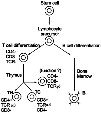
Cellular immunity is based on the function of the lymphocyte.2 Although lymphocytes are morphologically identical, they are functionally heterogeneous. They express an array of cell-surface molecules (markers), which define two major populations: T cells, B cells, and their subsets (Fig. 1).3

Fig. 1. Lymphoid differentiation. Stem cells from the bone marrow migrate to the thymus, where hormonal factors stimulate differentiation into T cells. T cells subsequently differentiate further into αβ TCR CD4+ , αβ TCR CD8+ , and γδ TCR T cells. B lymphocytes are also derived from stem cells and differentiate in the bone marrow. Their cell surfaces express membrane-bound antibodies. TCR = T-cell receptor; Y = surface immunoglobulin; α, β, γ, δ = T-cell receptor subunits.

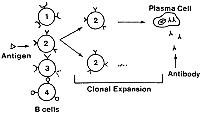
Lymphocytes interact with each other and with macrophages to allow selective expansion or suppression of specific populations (clones) of lymphocytes. Each lymphocyte clone has a unique receptor on its cell surface, which allows it to recognize and combine optimally with one antigen. Combination of this protein receptor with antigen results in the transmission of a signal across the cell membrane that activates the lymphocyte and results in selective proliferation and expansion of that lymphocyte clone (Fig. 2). Although numerous cellular interactions are involved in the generation of an immune response to antigen, our focus is on three distinct types: (1) antigen recognition by macrophages (phagocytic cells that play a key role in cellular immunity) and presentation to T cells (macrophage-T-cell interaction); (2) antibody production in response to antigen (T-cell-B-cell interaction); and (3) cytokine-mediated augmentation or inhibition of T-cell differentiation and proliferation (T-cell-T-cell interaction).

Fig. 2. Theory of clonal expansion. The receptor on the cell surface of each lymphocyte clone is a unique protein that allows it to recognize and optimally combine with a specific antigen. After the combination of this receptor with the appropriate antigen, proliferation and expansion of that lymphocyte occur. Thus, the immune response can selectively focus its attention on the antigen or antigens being recognized.

Macrophage-T-Cell Interaction

T-cell antigen recognition and activation depends on presentation of peptide antigen by specialized antigen-presenting cells (APCs). APCs include macrophages, B cells, and dendritic cells, which are accessory cells such as Langerhans cells in the skin or follicular dendritic cells in lymph nodes. All APCs digest proteins into peptide fragments and present these peptides within the clefts of special surface proteins coded by the major histocompatibility complex (MHC).

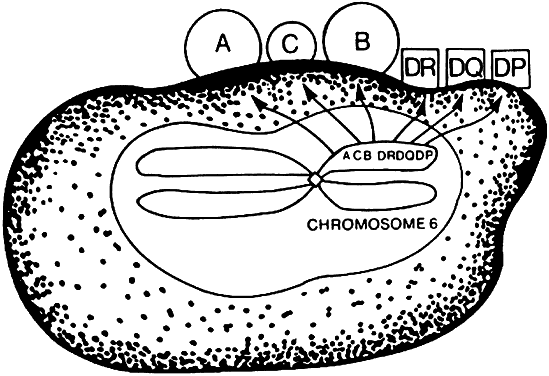
The MHC is a genetic segment on the short arm of chromosome 6 that codes for class I molecules (e.g., human leukocyte antigen (HLA)-A, HLA-B, and HLA-C) and class II molecules (HLA-DR, HLA-DP, HLA-DQ; Fig. 3) in addition to complement proteins and cytokines. Class I and II molecules were originally discovered as triggering T-cell responses leading to transplant rejection, hence the origin of the term histocompatibility.

Fig. 3. Major histocompatibility complex (MHC). Human chromosome 6 contains a genetic segment that codes for the class I MHC (HLA-A, HLA-B, and HLA-C) and class II MHC molecules (HLA-DR, HLA-DQ, and HLA-DP). These cell-surface molecules are glycoproteins present on many of the cells that are intimately involved in the recognition, activation, and effector phases of the immune response. HLA = human leukocyte antigen.

The class II region, first identified in mice as the I region,4 codes for protein molecules expressed only on the cell membrane of macrophages, B cells, and dendritic cells. Class II-associated antigen presentation requires phagocytosis of extracellular antigens (e.g., bacterial proteins), followed by intracellular processing of the antigens. Activation of a major subset of T cells termed CD4+ helper T cells requires class II-associated antigen presentation; therefore, CD4+ T cells are called class II-restricted. (CD—cluster of differentiation—molecules are lymphocyte cell-surface markers; CD4+ designates helper T cells, and CD8+ identifies cytolytic T cells).

Class I molecules are expressed on almost all cells and are involved in presentation of endogenous antigens, such as tumor antigens and viral proteins, of which the latter are not phagocytosed but enter the cell by viral infection. CD8+ cytolytic T lymphocytes (CTL) are class I-restricted and on activation destroy the entire host cell harboring the foreign antigen.

The manner in which MHC molecules interact with antigen or T cells has only been elucidated in the past decade. It has been shown that the MHC molecule binds antigen within its clefts and that the T-cell receptor (TCR) has regions that recognize the MHC complex and other regions that contact the foreign peptide (Fig. 4).5 The MHC-antigen complex must bind the antigen-specific TCR to achieve T-cell activation. Costimulatory molecules on APCs also bind with other T-cell membrane receptors during this APC-T-cell binding and may be responsible for the development of either activation or tolerance.1

Fig. 4. T-cell-macrophage interaction. T-cell activation requires recognition of antigen in conjunction with a class II MHC molecule, processed and presented by the macrophage. The antigen and class II major histocompatibility complex molecule are recognized by a specific T-cell receptor. Activated antigen-presenting cell (APC) express costimulatory molecules, which are required for T-cell activation. IL-2 is released by activated T cells, assisting in the subsequent clonal expansion and differentiation of T cells and also in the activation of resting APCs. IL-2 = interleukin-2.

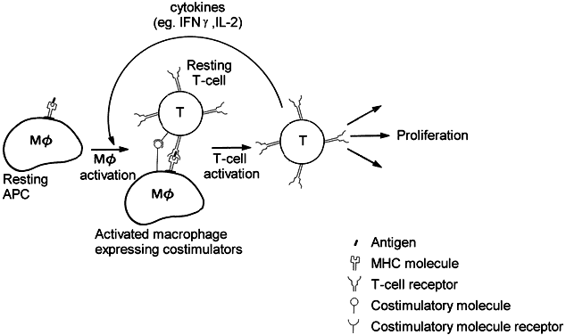
Antigen recognition is followed by clonal proliferation and expansion of those T lymphocytes that are directed at the involved antigen. Several antigen-specific memory T cells are also generated during this process, enabling the host to mount a greater and faster response on future exposure to the same antigen. Upon activation, the T cell elaborates many soluble factors called cytokines, including interleukin-2 (IL-2). IL-2 is the major T-cell growth factor, acting both in an autocrine and in a paracrine fashion, stimulating growth of not only other CD4+ and CD8+ T cells but also B cells and NK cells. The potent immunosuppressant drug cyclosporin inhibits the production of IL-2 by activated T cells, thereby blocking proliferation and differentiation of immune cells. Cytokines are not unique to T cells but are secreted by many different types of cells, enabling the immune system to orchestrate inflammatory responses.

In particular, interferon-gamma (IFN-γ) and tumor necrosis factor (TNF) secreted by T cells activate macrophages by setting off a cascade of upregulatory events, rendering the macrophage a more powerful and efficient killer of intracellular organisms. For example, on activation, the macrophage is induced to produce large amounts of nitric oxide, which is thought to inhibit bacterial and viral replication. While the proliferation of microorganisms is inhibited, potent proteases are released within the phagolysosomes, thereby destroying the pathogens more effectively. Also, angiogenic factors are released, such as fibroblast growth factor, which initiates endothelial cell migration and proliferation and thus tissue repair.

T-Cell-B-Cell Interaction

B-cell and helper T-cell cooperation entails activation and proliferation of both subsets of lymphocytes. As described above, T-cell activation requires antigen presentation, and B cells can serve well as APCs. Once antigen is recognized by a surface immunoglobulin on a B cell, the B cell is activated. The antigen is subsequently internalized, processed, and presented by the B cell in the context of a class II MHC molecule. The advantage of B cells over other APCs is that they are able to present antigen at a 104- to 106-fold lower concentration.1

Binding of a T cell to an antigen-presenting B cell leads to T-cell activation and release of IL-2, among other cytokines, promoting antigen-specific B-cell and T-cell proliferation. Binding of CD40, a B-cell surface receptor, to its complementary CD40 ligand on the activated T cell is also necessary for B-cell activation, differentiation, and survival. Other cytokines elaborated as a result of T-cell activation (e.g., IL-4, IL-5, transforming growth factor beta [TGF-β], and IFN-γ) serve to enhance antibody secretion and isotype switching.1

The production of antibody against most antigens requires this synergistic cooperation of helper T cells and B cells. The omission of either population results in limited or no antibody production. This is true in both the primary (i.e., on initial antigen exposure) and secondary (i.e., on subsequent antigen exposure) antibody responses.

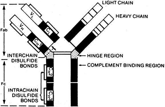
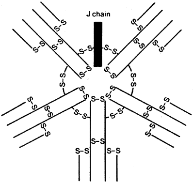
B cells respond to a wider variety of antigens than T cells, including polysaccharides, lipids, and proteins. The selective activation of B cells is followed by differentiation into immunoglobulinsecreting cells (i.e., plasma cells; Fig. 5). Memory B cells are also produced in this process of differentiation for a response of greater magnitude and speed on future encounter with the same antigen. Immunoglobulins are an important component of the response of the immune system to invasive pathogens. The basic structure of an immunoglobulin molecule consists of two heavy and two light chains, each composed of specific amino acid sequences (Fig. 6). There are five different classes (or isotypes) of immunoglobulin molecules based on the amino acid sequence in the constant region of their heavy chain (IgG, IgM, IgA, IgE, and IgD). The IgG molecule has the structure depicted in Fig. 6. In contrast, the IgM molecule is a pentamer, consisting of five such units linked by a joining (J) chain (Fig. 7). The IgA molecule is most frequently a dimer, also joined by the J chain.

Fig. 5. B-cell proliferation and differentiation. After B-cell activation by a specific antigen, clonal expansion occurs. The expanded population of B cells then differentiates into plasma cells, which secrete the immunoglobulin directed at the initially recognized antigen.

Fig. 6. Immunoglobulin structure. The basic immunoglobulin molecule consists of two heavy and two light chains composed of specific amino acid sequences. The Fab (antigen-binding portion of the immunoglobulin molecule) contains the region of greatest variability in amino acid sequence (VL and VH, variable light- and heavy-chain domains) and a region of relatively constant amino acid sequence (CH1, constant heavy-chain 1; CL, constant light-chain domains). The Fc portion consists of only constant regions (CH2 and CH3) and contains the Fc receptor and complement-binding sites.

Fig. 7. IgM. The IgM molecule is a pentamer consisting of five basic Ig units linked by disulfide bonds with a J chain. The biologic properties of the various immunoglobulin classes are distinctive and frequently determined by their physical properties. The IgM molecule does not cross the placenta and therefore is helpful in the diagnosis of congenital ocular toxoplasmosis.

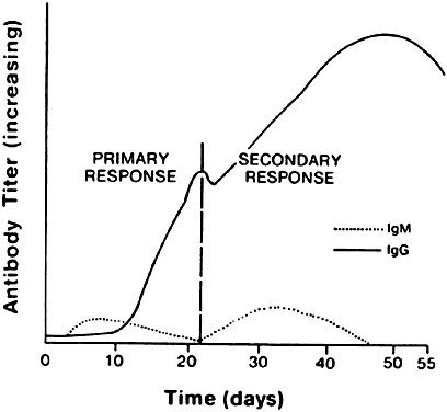
The isotype of antibody that is produced can be important diagnostically. After initial contact with antigen, the immune response produces antibody in what is called the primary response (Fig. 8). The first class of antibody produced is IgM. Only a low titer of IgM antibody is produced, however, and it rapidly declines to undetectable serum levels. During the second week of the immune response, an IgM to an IgA, IgE, or IgG isotype switch occurs, depending on the cytokine produced by the helper T cell, and high titers of the new isotype antibody are produced in the plasma. These titers persist long after the onset of the immune response. When the immune response again encounters the same antigen, a secondary antibody response occurs. The main antibody produced by the memory B cells is either IgA, IgE, or IgG, which reaches even higher serum levels and persists even longer. In contrast, IgM antibody titers appear transiently, remain much lower, and rapidly return to undetectable levels.

Fig. 8. Primary and secondary antibody responses. In this example, the primary antibody response to an antigen is characterized by a switch from IgM to IgG. Subsequent reexposure (i.e., the secondary antibody response) to this antigen preferentially produces IgG antibody.

T-Cell-T-Cell Interaction

The interplay between subsets of T cells is achieved through secretion of cytokines, which modulate the immune response. For example, the immune response to antigen can result in the generation of CD8+ CTLs with the ability to kill virus-infected host cells or allogeneic transplanted cells (i.e., transplanted cells from genetically different individuals of the same species). Optimal generation of CTLs from their precursors (pre-CTLs) requires cytokine release from helper T cells.

Initially, pre-CTLs bind peptide antigen expressed in the context of class I MHC molecules on the surface of target cells and are thus activated. Additional ligand-receptor interactions between pre-CTLs and target cells, in the presence of the cytokines IL-2 and IFN-γ, subsequently lead to full activation and differentiation of pre-CTLs to CTLs capable of killing target cells. IL-2 and IFN-γ, both of which are necessary for the full activation and differentiation of CTLs, are produced mainly by neighboring helper T cells, which are concurrently activated in response to the viral infection or allogeneic graft. Helper T cells, however, are activated by a different peptide sequence that binds to class II MHC molecules on APCs. Dendritic cells especially have been implicated in presenting foreign proteins expressed by viruses or allogeneic grafts by endocytosing fragments of apoptotic cells that express these proteins. Therefore, an intimate cooperation between class I-restricted CD8+ T cells (i.e., pre-CTLs) and class II-restricted CD4+ helper T cells leads to the development of CTLs capable of recognizing and killing target cells that express foreign proteins.

This accessory ligand-receptor conjugation between the target cell and the antigen-specific CTL is required for efficient killing. Once this conjugate is formed, CTLs deliver their lethal hit mainly by granule exocytosis (e.g., perforin, a membrane pore-forming protein; and granzymes, enzymes containing reactive serines at their active site), leading to apoptosis and osmotic lysis.

Many apoptotic mechanisms in the immune system have been linked to the interaction of Fas ligand, a cell-surface protein on the killer cell, with its receptor Fas (also known as CD95) on the target cell. The binding of this receptor to its ligand triggers a cascade of intracellular protein-protein interactions and proteolytic activities, culminating in apoptosis of the target cell. This pathway is thought to be more important in immune regulation—that is, for controlling excessive lymphocyte activation especially against self-antigens—than for CTL functions.1,6

REGULATION OF THE IMMUNE RESPONSE

Because multiple cell-cell interactions occur during the generation of an immune response, it is necessary to regulate these interactions so that the net effect is appropriate, minimizing the inadvertent destruction of innocent host bystander cells.

Multiple homeostatic controls exist to achieve immunoregulation, of which we briefly describe the principal mechanisms. So far, clonal expansion has been considered in the context of selection by antigen. As such, the regulation of clonal expansion can be achieved by antigen removal by secreted antibody or T cells. The depletion of antigen for lymphocyte receptors serves as negative feedback for antigen-driven clonal expansion. Thus, only memory cells remain, which require subsequent activation by antigen for regeneration of a full response.

Immunologic tolerance is an important mechanism for immune regulation, applicable to both B and T lymphocytes. Studies have shown that tolerance is antigen-specific and thus likely achieved by either deletion or inactivation of specific B- or T-cell clones (i.e., clonal deletion or clonal anergy). Immature lymphocytes are more susceptible to induction of tolerance than mature, functional cells. In the thymus, immature T cells are thought to be exposed to self-antigens; if a T cell recognizes any self-antigen, it is eliminated by apoptosis. This mechanism of tolerance is referred to as clonal deletion because it involves cell death. The purpose of this central tolerance (central because it occurs in the thymus) is the prevention of autoimmunity.1

Under special conditions, tolerance may be induced in mature lymphocytes, away from the central lymphoid tissues (peripheral tolerance). For example, a large concentration of foreign polysaccharides injected intravenously does not stimulate a B-cell response; even subsequent exposure to immunogenic doses of the antigen fails to activate B cells. Peripheral CD4+ T-cell tolerance has also been achieved in various ways, including high-dose intravenous administration of proteins without adjuvant (high-dose tolerance), repeated low-dose exposure to proteins without adjuvant (low-dose tolerance), and oral administration of proteins (oral tolerance). T-cell tolerance is generally induced at lower concentrations of protein than is B-cell tolerance and lasts longer. Moreover, without CD4+ T-cell help, the B-cell response to low levels of protein antigen is less. The mechanisms involved in peripheral tolerance are clonal anergy as well as clonal deletion—the former referring to functional inactivation without cell death.1

Because the antigen receptor on lymphocyte cell membranes is the only clonally distributed structure, it is likely involved in cell regulation. This can occur through either cell-receptor interaction with antigen or with factors directed at the antigen receptor itself (i.e., idiotype-anti-idiotype recognition). The precise details of the idiotypic-anti-idiotypic networks of immunoregulation are not discussed here because it has a more theoretic basis with insufficient experimental evidence.

Some evidence has been provided for immune regulation mediated by suppressor T cells. Despite repeated studies and attempts to purify these cells, however, not much advancement has been made. Therefore, one thought is that perhaps a series of suppressor mechanisms exist instead of a discrete class of suppressor T cells.1 For example, transforming growth factor-beta (TGF-β) effectively inhibits T-cell and B-cell proliferation; therefore, cells that secrete significant amounts of this cytokine may function as suppressor cells.

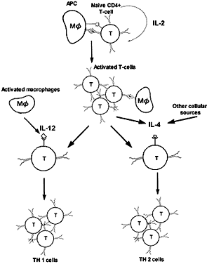
Similarly, different types of immune responses may be suppressed. CD4+ T cells have been further subclassified as T helper 1 (TH1) and T helper 2 (TH2) cells. TH1 cells direct the immune reaction toward a cell-mediated response through expression of IFN-γ, IL-2, and TNF-α; TH2 cells elaborate IL-4, IL-5, and IL-10, leading to IgE-mediated humoral immunity. Interestingly, IFN-γ inhibits the IL-4-mediated B-cell isotype switch to IgE; conversely, IL-4 inhibits IFN-γ-mediated macrophage activation. The trigger for the TH1 response has been shown to be IL-12 secreted by the activated macrophage (Fig. 9).1

Fig. 9. Induction of TH1 and TH2 cells. Cytokines produced by the innate immune response to pathogens or during early phases of specific immunity can influence the differentiation of naive CD4+ T cells into TH1 or TH2 cells. IL-12, made by activated macrophages or dendritic cells, is a strong inducer of TH1-cell development. IL-4, which may be secreted during the initial activation of naive T cells, favors induction of TH2 cells. (Adapted from Abbas AK, Lichtman AH, Pober JS: Cellular and Molecular Immunology, 3rd ed, p 274. Philadelphia, WB Saunders, 1997.)

In certain conditions, however, the desirable immune response becomes suppressed, leading to a detrimental outcome. For example, a TH2 response in leprosy is associated with chronicity and greater host-tissue destruction, whereas a TH1 response theoretically would be more effective. One explanation for the tendency toward a TH2 response may be as follows. IL-4 is initially produced in small amounts by the T cell at the time of its initial activation, and its concentration increases when high concentrations of antigen persist. If this is combined with the absence of IL-12 secretion, a TH2 response will predominate. Genetic factors have also been implicated in the predominance of one type of response over the other.1

Back to Top
OCULAR IMMUNE PRIVILEGE
The eye is considered to be an immunologically privileged organ. This allows for the existence of immune defense mechanisms within the eye but with control of intraocular inflammation, thus achieving preservation of organ function and integrity. Experimental evidence for ocular immune privilege has been reported in several separate studies7–9 showing that tissue grafts or tumor cells placed in the anterior chamber are not rejected, whereas they are rejected rapidly when placed elsewhere, such as in subcutaneous tissue. The high success rate of allogeneic orthotopic corneal grafts in mouse and human eyes even in the presence of gross histoincompatibility is another example of ocular immune privilege.10

Features that confer immune privilege in the eye include a blood-tissue barrier, deficient lymphatic drainage, intraocular fluid drainage into the circulatory system, and reduced expression of MHC class I and II molecules on parenchymal cells.11 Other mechanisms exist that contribute to the control of the immune response within the eye:

  1. Complement inhibition. Several complement regulatory proteins, including membrane cofactor protein (MCP), decay-accelerating factor (DAF), and CD59, have been identified in ocular tissue. The putative role of these molecules is protection of intraocular tissues from complement-mediated destruction.12
  2. Clonal deletion. Fas ligand is constitutively expressed in the iris, ciliary body, cornea, and retina. Evidence from tissue grafts in vivo and tissue cultures in vitro support the view that constitutive expression of Fas ligand in a tissue leads to deletion of the Fas+ T cells that enter that tissue and recognize antigen.13
  3. Clonal anergy. Aqueous humor contains many immunomodulatory substances, including TGF-β2 (a powerful inhibitor of T-cell and B-cell proliferation), free cortisol, and IL-1 receptor antagonist. Cultures of primed T cell that encounter antigen in the presence of aqueous humor failed to undergo proliferation or to produce expected cytokines such as IFN-γ. Interestingly, these cultured T cells did not revert to their normal capacity to produce IFN-γ and actually inhibited other primed T cells from producing IFN-γ. When soluble TGF-β2 receptor was used to “quench” the TGF-β2 in the culture medium, IFN-γ production by the primed T cells was restored.14
  4. Immune deviation. Investigations by Kaplan and Streilein7 demonstrated that histoincompatible cells (lymphoid or tumor) placed in the anterior chamber evoke a systemic immune response that deviates from the usual pattern. Mice injected with allogeneic tumor cells into the anterior chamber lost the ability to reject skin grafts from the allogeneic donor; delayed-type hypersensitivity reactions against allogeneic tissue was abrogated. This phenomenon is termed ACAID, or anterior chamber-associated immune deviation.
    In subsequent studies, foreign protein injection into the anterior chamber revealed a predilection of the ocular immune system toward a TH2 or humoral immune response. A potentially important application of this phenomenon was demonstrated in the setting of experimental autoimmune uveoretinitis (EAU), in which injection of the autoantigen into the anterior chamber before the uveitogenic regimen inhibited uveitis.14
  5. T-cell suppression. Spleens of mice in which ACAID has been induced contain T cells that are capable of conferring ACAID when adoptively transferred into naive recipients. Thus, similar to the donors, these naive recipients become unable to mount a delayed-type hypersensitivity response to the antigen.15 Experimental evidence supports the view that either a soluble signal from the eye or antigen-bound APCs from the anterior chamber enter the bloodstream and migrate to the spleen. In the spleen, ACAID-mediating T cells (CD8+ cells) are generated. Some of the responding CD8+ T cells secrete TGF-β, further suppressing CD4+ T-cell activation, both locally and eventually systemically.14 The active role of both the eye and the spleen in ACAID is highlighted by the finding that removal of the spleen or the eye within the first 3 days after exposure to antigen results in the typical immune response.16,17
Back to Top
CLINICAL IMMUNOLOGY: HYPERSENSITIVITY REACTIONS
When an immune response occurs and results in gross tissue damage, it is referred to as a hypersensitivity reaction. Coombs and Gell18 classified hypersensitivity into four classic types, and a fifth is sometimes added. Type I, II, III, and V involve antigen-antibody reactions, whereas type IV results from cell-mediated immunity. In this section, we review these hypersensitivity reactions and their relevance to ocular inflammation.

TYPE I: IMMEDIATE HYPERSENSITIVITY

Type I hypersensitivity is mediated by IgE antibody. Initial contact with antigen usually occurs in the respiratory and gastrointestinal mucosa, where IgE is produced by plasma cells. The antibody molecules bind to a high-affinity Fc receptor (FcεRI) on the surface of mast cells and basophils. Subsequent antigen exposure and binding to mast cell-bound IgE results in cross-linking of the IgE molecules, leading to mast cell degranulation and release of the mediators of immediate hypersensitivity.

The primary mediators consist of (1) biogenic amines—particularly histamine and serotonin; (2) arachidonic acid-derived lipids such as prostaglandin D2, leukotrienes (of which LTC4, LTD4, and LTE4 were formerly known as the slow-reacting substance of anaphylaxis [SRS-A]), and platelet-activating factor (PAF); (3) cytokines, of which IL-5 acts as eosinophil-activating factor and IL-4 upregulates an eosinophil-binding adhesion molecule on the surface of endothelial cells; and (4) proteins and proteoglycans, including serine proteases and heparin.1

These mediators effect endothelial cell retraction, vascular smooth muscle relaxation, bronchial smooth muscle constriction, and chemotaxis of inflammatory cells, including eosinophils and TH2 cells. TH2 cells, eosinophils, and basophils express a chemotactic receptor called the eatoxin receptor (CCR3), through which all of these cells can be recruited to inflamed tissue. The attraction of TH2 cells by eotaxin, produced by epithelial and phagocytic cells, may represent a key mechanism in allergic reactions because it promotes the allergen-driven production of IL-4 and IL-5 necessary to activate basophils and eosinophils.19 Clinically, this series of events manifests as edema, erythema, bronchospasm, and local tissue destruction.1,20

IgE-mediated disorders are frequently associated with elevated levels of IgE in the blood or tears. Allergen-specific IgE may be detected in vitro by mast cell degranulation or by the radioallergosorbent test.21

Penicillin allergy and atopy are systemic examples of type I hypersensitivity reactions. In the eye, hay fever, atopic, and giant papillary conjunctivitis are type I reactions. In hay fever conjunctivitis, the antigens are airborne pollens, whereas giant papillary conjunctivitis is thought to be due to allergens associated with foreign bodies such as contact lenses. Atopic keratoconjunctivitis may be due to several environmental antigens. Although type I hypersensitivity results in primarily external disease, the occurrence of “seasonal iritis” and retinal edema has been described in severe cases of hay fever.22–25 Instances of drug and food allergy responsible for episodes of acute iritis have been reported.26,27 Although most reports lack definitive documentation, the sudden onset and course of many cases of acute iritis are consistent with type I-mediated disease. In EAU, the early detection of both IgE specific for the uveitogenic antigen and mast cell degranulation support the view that a type I hypersensitivity mechanism is involved in the initial trigger of uveitic response.28

Treatment of type I reactions may be either symptomatic, using antihistamines or steroids, or prophylactic, using cromolyn sodium or olopatadine to stabilize mast cell membranes and prevent degranulation. Attempts can also be made at desensitization through repeated immunizations with the allergen. Although the mechanism is not clear, these repeated immunizations preferentially induce IgG antibodies against the offending antigen, and IgE titers eventually decrease.

TYPE II: CYTOTOXIC ANTIBODY

In type II hypersensitivity, antibodies bind to antigen on the surface of target cells, resulting in cell death. This can be mediated by (1) phagocytosis of the cell, with the help of complement-binding or opsonic antibody adherence to the Fc receptor (at the binding site depicted in Fig. 6)—the receptor that binds the carboxyterminal end of the immunoglobulin constant region; (2) cell lysis mediated by the complement cascade; or (3) direct cytotoxicity by NK cells with receptors for the Fc portion of immunoglobulin (the latter is called antibodydependent cell-mediated cytotoxicity [ADCC]).

Type II hypersensitivity may be detected by the demonstration of complement-fixing antibody in the serum or increased killer cell activity in vitro. Examples of type II hypersensitivity include transfusion reactions; rhesus (Rh) incompatibility (hemolytic disease of the newborn); autoimmune hemolytic anemia; and hyperacute graft rejection. Drugs may bind to cell surfaces and form new antigenic determinants to which cytotoxic antibody is produced. This can cause a drug-induced hemolytic anemia or thrombocytopenic purpura. In the eye, type II reactions have been implicated in Mooren's ulcer, Vogt-Koyanagi-Harada's (VKH) disease, sympathetic ophthalmia, and corneal graft rejection.29–31

In sympathetic ophthalmia and VKH disease, antimelanin autoantibodies and melanin-sensitized lymphocytes have been demonstrated in the peripheral blood,32 and serum antibodies to retinal antigens have been detected.33 This does not establish a causal relation, however. Although some investigators have found antibodies to uveal extracts in patients with endogenous uveitis, similar antibodies were also found in patients with inflammatory eye disease that spared the uvea and in nonuveitis patients with retinal disease.34,35

TYPE III: IMMUNE COMPLEXES

The formation of antigen-antibody complexes (immune complexes) is a mechanism for removing antigens from the circulation. Under certain conditions—such as the appropriate antigen and antibody charge, size, and concentration—these immune complexes may form locally or be deposited in the walls of blood vessels. They may cause the fixation of complement and the release of anaphylatoxins (C3a, C5a), mast cell mediators, and leukocytic chemotactic factors. These immune complexes may also cause platelet aggregation, resulting in the release of vasoactive amines. Tissue damage results from microthrombi, hemorrhage, and ischemia.

The Arthus reaction is an example of local immune complex deposition. The intradermal injection of soluble antigen in patients with circulating antibodies results in local vasculitis and tissue reaction, peaking in 3 to 8 hours.

Circulating and local deposition or formation of immune complexes has been demonstrated in numerous diseases (Table 1). These immune complexes have been detected in patients with Behçet's disease, retinal vasculitis, scleritis, sarcoid uveitis, and chronic idiopathic uveitis.29,36–38


TABLE 1. Diseases Associated With Immune Complexes

  Circulating Immune Complexes
  Serum sickness
  Glomerulonephritis (membranous and poststreptococcal)
  Systemic lupus erythematosus
  Polyarteritis nodosa
  Dengue virus hemorrhagic shock syndrome
  Penicillin allergy
  Sarcoidosis
  Retinal vasculitis
  Chronic idiopathic uveitis
  Scleritis
  Behçet's syndrome
  Rheumatoid arthritis
  Wegener's granulomatosis
  Local Immune Complex Deposition*
  Allergic alveolitis or Farmer's lung (actinomycetes)
  Cheese washer's disease (Penicillium casei)
  Furrier's lung (fox fur)
  Elephantiasis (Wuchereria bancrofti)
  Jarisch-Herxheimer reaction (Treponema pallidum)
  Rheumatoid arthritis (IgG)


*The antigen in the immune complex is in parentheses.


Char and coworkers39 demonstrated immune complexes in the circulation of patients with chronic idiopathic iridocyclitis and panuveitis. In the patients with iridocyclitis, they demonstrated a correlation between increased immune complex levels and disease activity. The immunoglobulin in these complexes was IgG but the antigen was not identified.

Immune complexes have been demonstrated in iritis patients with serum sickness and sarcoidosis.36,40 Retinal vasculitis caused by immune complex deposition has been documented by immunofluorescent techniques in lupus retinopathy.41

Dernouchamps and Michiels42 have detected antigen-antibody (Ag-Ab) complexes in the aqueous humor or serum of patients with VKH, Behçet's disease, uveitis associated with ankylosing spondylitis and scleroderma, idiopathic uveitis, and Fuchs' syndrome. Although not all patients with these inflammatory diseases demonstrated Ag-Ab complexes, they were detected in many patients in the aqueous humor only or in the aqueous humor in higher concentrations than in the serum, suggesting a local origin. It should be emphasized that the presence of immune complexes in any disease may be secondary to tissue damage rather than the etiologic factor.

Alternatively, immune complexes may play a role in suppressing disease, as has been proposed in studies on retinal vasculitis. High titers of immune complexes have been reported in the serum of patients with mild retinal vasculitis, whereas low titers have been correlated with severe inflammation.43,44 Similarly, in Behçet's disease, circulating immune complexes have been shown to increase in response to cyclosporin A, a potent inhibitor of cell-mediated immunity. This increase in circulating immune complexes may be associated with the disease but is not necessarily involved in its pathogenesis.45

TYPE IV: DELAYED-TYPE HYPERSENSITIVITY

Unlike hypersensitivity types I, II, and III, delayed-type hypersensitivity is mediated by sensitized T cells rather than by antibodies. The T lymphocytes that possess antigen-specific receptors may be stimulated either to produce lymphokines (mediators) or to differentiate into killer T cells after contact with macrophage-bound antigen. The tuberculin skin test is the classic example of delayed-type hypersensitivity. The intradermal injection of antigen results in erythema and induration, peaking at 24 to 48 hours. Histologically, the lesion at this peak time is composed of perivascular fibrin and infiltrates of lymphocytes and macrophages.

Several methods are available for in vitro measurement of cell-mediated hypersensitivity. Sensitized T cells, when stimulated with antigen, produce migration-inhibition factor (MIF), which causes macrophages to accumulate. An assay for migration-inhibition factor can then be used as an indirect measurement of the presence of antigen-specific T cells. Alternatively, sensitized T cells proliferate in the presence of antigen, and “blast transformation” can be detected by measurement of incorporation of radiolabeled thymidine into newly synthesized DNA.

Systemically, cell-mediated immunity has long been recognized as playing an important role in (1) immunity against certain bacterial (e.g., tuberculosis) and viral infections; (2) contact dermatitis; (3) tumor surveillance; and (4) acute and chronic graft rejection, including corneal transplants. In the eye, type IV hypersensitivity also has been implicated as a mechanism in interstitial keratitis and phlyctenulosis.

Sugiura and associates46 used a leukocyte migration-inhibition test to study patients with Harada's disease and Behçet's disease. In Harada's disease, two groups of patients were identified. Those patients with a positive cell-mediated immunity test had chronic recurrent uveitis associated with systemic depigmentation; those with a negative cell-mediated immunity had a short course of uveitis responsive to steroids. This suggests a possible role for cell-mediated immunity as a way to identify patients with a better prognosis in Harada's disease. Perhaps more importantly, the immune response in Harada's disease might be therapeutically modulated to suppress cell-mediated immunity.

Sugiura and colleagues' results suggested that in Behçet's disease, cell-mediated immunity is a result of the disease rather than its pathologic mechanism. We now know, however, that cyclosporin A—a potent IL-2 inhibitor and therefore a potent inhibitor of cell-mediated immunity—can be therapeutically effective in Behçet's disease.45,47,48 Among other uveitides, sympathetic ophthalmia, pars planitis, and birdshot retinochoroidopathy are also thought to be cell-mediated in mechanism.49

TYPE V: STIMULATORY ANTIBODY

In type V hypersensitivity (sometimes added to the four classic types), antibodies directed at certain cell-surface receptors may functionally stimulate the target cell. The classic example of this abnormality is long-acting thyroid-stimulating antibody, which is thought to cause Grave's disease. This autoantibody is directed against an antigen on the thyroid-stimulating hormone receptor of thyroid cells and produces the same changes in cell metabolism as thyroid-stimulating hormone. There is no clinical example of type V hypersensitivity in ocular inflammatory disease.

Back to Top
IMMUNOLOGY OF UVEITIS
The choroid, ciliary body, and iris comprise the uvea. Uveitis, however, is generally described as inflammation of not only the uvea but also the vitreous and sclera. The etiology of this inflammation may be either infectious (syphilis and tuberculosis being the classic examples) or noninfectious, as are most cases in the Western world. The eye loses its immune privilege in uveitis as inflammatory cells infiltrate the intraocular space and tissue. In addition to the mechanisms of uveitis already discussed in the context of hypersensitivity reactions, other key points warrant mention regarding this immune invasion of the eye.

The mechanistic understanding of uveitis has been afforded by animal models. One model has been EAU, described in the rat, rabbit, guinea pig, and nonhuman primate. It produces primarily an experimental form of retinochoroiditis, similar to human birdshot choroidopathy or sympathetic ophthalmia. Many retinal antigens have been identified and used to induce EAU: (1) retinal S-antigen, also known as “arrestin,” found in the photoreceptor outer segments and in the pineal gland of some species; it competes with transducin for rhodopsin in its photoexcited and phosphorylated state; (2) interphotoreceptor retinoid-binding protein (IRBP), a major soluble protein in the interphotoreceptor matrix; (3) rhodopsin, a well-defined transmembrane protein of rod outer segments, participating in visual transduction; and (4) recoverin, a calcium-binding protein that selectively localizes to the retina and pineal gland, associated with cancer-associated retinopathy.49,50

Ten to fourteen days after immunization with the antigen, clinical disease develops, characterized by variable degrees of vitritis, choroidal and retinal infiltrates, and retinal vasculitis. Predominantly T cells but also macrophages and mast cells infiltrate the eye.50

More recently, insoluble uveal antigens derived from bovine retinal pigmented epithelial (RPE) cells51 or from bovine iris and ciliary body52 have been shown to cause experimental acute anterior uveitis (EAAU), which more closely resembles acute anterior uveitis in humans.

SPECIFIC AND NONSPECIFIC IMMUNE MECHANISMS IN UVEITIS

An antigen-specific immune-mediated inflammatory response and a nonspecific inflammatory response may be important in the production of uveitis. The nonspecific (also termed natural or innate) immune response recruits inflammatory cells to the eye by the action of mediators such as prostaglandins, leukotrienes, and cytokines, leading to nonspecific damage from the release of cellular enzymes and free radicals. A role for nitric oxide, a potent free radical, has been suggested in another animal model called endotoxin-induced uveitis, which may resemble Reiter's syndrome.53 Intraperitoneal injections of a specific nitric oxide synthase inhibitor reduced clinical signs of uveitis in this model.54 In addition, high levels of nitrite, the immediate metabolite of the unstable radical, were demonstrated in the aqueous humor and vitreous of rats at the time of maximal ocular inflammation.55 The role of nitric oxide may be vasodilatory by a cGMP pathway56 and cytotoxic through peroxynitrite (generated after reaction with superoxide anion), causing oxidative damage.57,58

The antigen-specific (or acquired) immune-mediated mechanisms in the eye involve both humoral and cellular pathways. In the eye, RPE cells may serve as antigen processing and presenting cells59 in addition to resident macrophages.60

Humoral Immunity in Uveitis

Antibody production plays a controversial role in uveitis. As discussed, immune complexes may or may not be significant mediators in the pathogenesis of uveitis. Conversely, detection of IgE antibody specific for S-antigen in the serum early in EAU and of mast cell degranulation before the onset of EAU61 suggest type I hypersensitivity as a mechanism for initial trigger of the inflammatory response, allowing for vasopermeability and the penetration of inflammatory factors and cells.28

High titers of circulating antibodies are induced after a single immunization with retinal autoantigens in the experimental setting. The injection of hyperimmune serum to rod outer segments, either into the eye or systemically in a naive animal, induces moderate EAU.62 Adoptive transfer of T cells from an animal with EAU to a naive animal more effectively causes EAU, however. Therefore, antibodies are not the major mediators of this disease.63,64

New evidence suggests a role for antibody-mediated degeneration of photoreceptors. In patients with cancer-associated retinopathy—a condition associated with systemic cancer, particularly small cell cancer of the lung—antirecoverin antibody is frequently detected in the serum and has been linked to progressive irreversible retinopathy. The evidence for this is the apoptosis of photoreceptor cells in culture induced by antirecoverin antibody. The exact mechanism is not fully understood but it involves specific uptake of antirecoverin antibody followed by apoptosis of the photoreceptor cells.65

In contrast, exogenous antibodies can play an immunosuppressant role in particular instances. Pooled IgG from human donors (IVIg, Sandoglobulin, Central Laboratories of Swiss Red Cross, Bern, Switzerland) has been reported to protect against EAU in the rat.66 Many mechanisms have been proposed for the action of IVIg, including the ability of IVIg to suppress numerous cytokines, such as IFN-γ and IL-2.28,67

Cellular Immunity in Uveitis

Experimental autoimmune uveoretinitis is thought to be a TH1 cell-dependent autoimmune disease. Increased production of IL-2, IL-4, and TNF in EAU has been implied by increased mRNA production, though the amount of expressed protein was not studied; IFN-γ upregulation in EAU has been demonstrated by increased mRNA production and immunohistochemical staining for the protein.68,69 If EAU is a TH1 cell-mediated disease, however, an active role for IL-4 would seem contradictory.

EAAU is considered also to be cell-mediated. The adoptive transfer of T cells70 (or more specifically, primed CD4+ T cells71) but not serum72 from uveitic animals induced the disease in naive recipients.

Because uveitides in humans such as VKH, sympathetic ophthalmia and Behçet's disease are also thought to be T-cell-mediated, suppression of T-cell activation would be a logical treatment alternative. Indeed, inhibition of IL-2 production by cyclosporin inhibits EAU in 100% of animals.73 Cyclosporin also effectively inhibits the development of EAAU.74 Initial clinical trial results of cyclosporin on patients with sight-threatening uveitis, including Behçet's disease, also have been favorable.47,48

Oral tolerance has been shown in experimental animal models of other autoimmune diseases to be protective. This is achieved by feeding the animal the antigen that is putatively triggering the autoimmune disorder. One such example is experimental autoimmune encephalomyelitis, a model for multiple sclerosis, in which the animal is fed myelin basic protein.75,76 It is thought that low-dose oral tolerance is mediated by TH2 cells that secrete IL-4, IL-10, and TGF-β, leading to active suppression, and high-dose oral tolerance is mediated by clonal deletion or anergy of helper T cells.49,77 An initial clinical trial of oral tolerance using retinal antigens in the setting of various uveitides was inconclusive, however, showing statistically insignificant improvement in the course of uveitis treated orally with retinal S antigen, when compared with other retinal protein mixtures or placebo.78

HUMAN LEUKOCYTE ANTIGEN ASSOCIATIONS

Associations have been made between various uveitides and HLA antigens, such as Reiter's syndrome (HLA-B27), Behçet's disease (HLA-B5), and birdshot choroidopathy (HLA-A29). One explanation for this phenomenon may be molecular mimicry, whereby a microbial pathogen expresses antigens similar to normal tissue proteins in the host. As the host immune response is mounted against the pathogen, an autoimmune response may ensue if self-antigens are also attacked by the immune system. The evidence for this hypothesis in the setting of uveitis has been debated in various studies.

The presence of Klebsiella species in the stool was reported to be increased during attacks of anterior uveitis in patients with79 and without80 ankylosing spondylitis; however, this has been disputed.81 The incidence of Klebsiella was thought to be highest in patients with HLA-B27 or HLA-B7—which cross-reacts with B27 (B7, Bw22, BW40, and Bw42).80,82

Other studies found that regardless of HLA-B27 status, patients with uveitis did not have any greater incidence of Klebsiella infection83,84 or any higher antibody titers to Klebsiella than a control population.85 Moreover, cell-mediated immunity to Klebsiella has not been associated with uveitis.82,86

Other organisms have also been implicated as the possible etiology of HLA-B27-associated uveitis. Antibodies to Yersinia species have been detected in patients with anterior uveitis without reactive arthritis,87,88 and one study suggested that their incidence may be higher in HLA-B27-positive patients.88 Cell-mediated immunity to Chlamydia trachomatis was recorded in two-thirds of unselected HLA-B27-positive patients with anterior uveitis, but not in HLA-B27-negative controls.82,89

Birdshot choroidopathy is a disease associated with HLA-A29 in about 95% of cases—one of the strongest associations between HLA and disease reported. The relative risk of an individual with HLA-A29 developing birdshot choroidopathy is between 5090 and 244.82,91 This strong association suggests that HLA-A29 may be directly implicated in the pathogenesis of this disease.

Several mechanisms have been postulated to explain the association of autoimmune diseases with the inheritance of particular MHC sequences. One is molecular mimicry of host antigens by a pathogen, as described. Another is that regulatory T cells, whose normal function is to prevent autoimmunity, may be activated by class II MHC (HLA-D) molecules. For example, in a murine diabetes model, disease-producing T cells react with a self-peptide presented in association with a class II MHC molecule that is lacking aspartic acid at a particular position. T cells that prevent tissue injury react with a complex of the self-peptide and the class II MHC molecule containing aspartic acid in that position. This murine class II MHC molecule is homologous to an HLA-DQ molecule in the human. Therefore, this evidence may explain why inheritance of a DQ allele with aspartic acid in a particular position in humans protects against type I diabetes mellitus. Last, the actual disease-associated gene may be separate from but genetically linked to the HLA allele.1

CELL-ADHESION MOLECULES

The infiltration of the intraocular space and tissue by inflammatory cells can be explained by the upregulation of cell-adhesion molecules in the setting of uveitis. Infiltration of leukocytes occurs as a result of homing and migration of these cells into the eye. Selectins, integrins, and members of the immunoglobulin supergene family are considered to be the three main groups of cell-adhesion molecules involved in uveitis. These cell-surface molecules on the vascular endothelium allow leukocyte recruitment to the eye by adherence to the vascular wall, followed by transendothelial migration into the eye.49

Monoclonal antibodies against specific celladhesion molecules such as intercellular adhesion molecule-1 (ICAM-1) were shown to markedly inhibit EAU in mice immunized with interphotoreceptor retinoid-binding protein.92 In another study, normal eye-bank eyes were found to not express ICAM-1, whereas eye-bank eyes with uveitis93 and failed human corneal grafts94 were found to express ICAM-1.

In EAAU, cell-adhesion molecules, including ICAM-1, were found to be upregulated in the epithelial cells of the uveal tract and RPE at the onset of intraocular inflammation.72 These data suggest a possible role for blocking of cell-adhesion molecules in the treatment of uveitis.

FAS-FAS LIGAND

Examination of lesions in Behçet's disease suggested the presence of apoptotic cells in the aphthous ulcer and erythema nodosum-like lesion of corticosteroid-treated patients.95,96 When the peripheral blood T cells of Behçet patients were examined, Fas ligand was found to be insufficiently expressed on CD4+ T cells, whereas it was highly expressed on CD8+ T cells.97 Another study on T cells in the aqueous humor of uveitis patients revealed an increase in the number of memory T cells expressing Fas ligand in the aqueous humor.98

Fas and Fas ligand expression was studied in patients with posterior uveitis, compared with normal patients. Constitutive expression of both Fas and Fas ligand was found in the normal retina, but they were expressed less in the choroid. In chorioretinal scars and chorioretinal granulomas of uveitic patients, Fas and Fas ligand were found to be increased. In biopsy specimens with acute retinal necrosis, however, little Fas or Fas ligand was noted on infiltrating lymphocytes.99 This evidence suggests that dysregulation of Fas-Fas ligand interactions could lead to a predisposition to autoimmunity.100–102

Back to Top
CONCLUSION
The immune system consists of a complex network of cells and soluble factors whose interplay is directed toward protection against foreign pathogens. To achieve this task, the innate and antigen-specific immune responses complement each other. Macrophages act as antigen-presenting cells, processing and presenting antigen on their surface for T cells and thus inducing their activation. Upon B- or T-cell activation, clonal expansion and proliferation ensues. Activated B cells also act as APCs for T cells; on antigen recognition by a T cell, the T cell is activated and both B- and T-cell clones differentiate and proliferate. Thus, the B cell can differentiate into an antibody-secreting plasma cell. In this differentiation process, memory B and T cells are also generated to expedite the immune response on future encounters with the same antigen.

T cells differentiate in the thymus into two main subsets: helper (CD4+ ) and cytolytic (CD8+ ) T cells. CD4+ T cells recognize exogenous antigens in the context of MHC class II molecules and subsequently are activated and proliferate. CD8+ T cells recognize endogenous or intracellulary processed antigens in the context of MHC class I molecules and are thus activated. Upon activation, helper T cells secrete cytokines such as IL-2, inducing proliferation of many immune cells, including NK cells, CTLs, B cells, and other helper T cells. Through signals derived from the innate immune system, helper T cells may also differentiate into TH1 cells, which coordinate cell-mediated immunity through secretion of the cytokines IL-2, TNF-α, and IFN-γ. Once CD8+ T cells or CTLs are activated, specific cells are destroyed in an antigen-specific manner. The result is programmed cell death by osmotic lysis and apoptosis of the target cell. In certain circumstances, however, the TH1 response can become suppressed by factors such as TGF-β and IL-4, leading the immune response toward a TH2 or humoral response with elaboration of IL-4 and IL-5 instead.

Another immunoregulatory mechanism is tolerance, which is designed to assure no reaction against self-antigens. Tolerogenic mechanisms include clonal deletion, in which immune cells reactive to an antigen are killed, and clonal anergy, in which the immune cells remain viable but are rendered nonfunctional. Tolerogenic methods are being investigated to treat autoimmune diseases such as uveitis.

The eye is an immunologically privileged organ because of anatomic characteristics (e.g., the nonspecific reasons such as a blood-retina barrier) and active mechanisms, including complement inhibition, clonal anergy, clonal deletion, immune deviation, and T-cell suppression. ACAID refers to the phenomenon of an altered immune response to an antigen induced by the introduction of that antigen into the anterior chamber. This presentation of antigen into the anterior chamber induces the immune system to generate a response that favors antibody production while inhibiting delayed-type hypersensitivity. This property can be conferred to a naive animal by adoptive transfer of T cells that are immunosuppressant, perhaps by TGF-β production.

Classification of the immune response into five types of hypersensitivity reactions allows the application of our understanding of the immune system to clinical entities. All types of hypersensitivity are antibody-mediated except type IV, which is cell-mediated; type III hypersensitivity may also be complement-mediated. This is an artificial division imposed on a complex biologic system, and there may be multiple mechanisms involved in any disease.

In uveitis, ocular immune privilege is compromised as inflammatory cells infiltrate the ocular tissue and cause damage. The homing and migration of leukocytes through cell-adhesion molecules such as ICAM-1 have been shown to play an important role in this invasion. Once a critical number of inflammatory cells have entered the eye, the naturally occurring immune suppressing and deviant mechanisms may be overcome, and tissue damage ensues. Another factor that may render these eyes more susceptible to uveitis may be dysfunctional or unbalanced immunoregulatory mechanisms by Fas-Fas ligand interaction.

It is often recurrent uveitic attacks that cause progressive tissue destruction, interfering with vision. Further understanding of autoimmune disorders will advance knowledge of immune regulation and direct treatments to target the specific disease mechanisms. By effectively altering the chronicity of uveitis, the ocular tissues may be better protected and thus sight better preserved.

Back to Top
ACKNOWLEDGMENT
We thank Tongalp Tezel, MD, for his kind assistance in graphic illustrations.
Back to Top
REFERENCES

1. Abbas AK, Lichtman AH, Pober JS: Cellular and Molecular Immunology, 3rd ed. Philadelphia, WB Saunders, 1997

2. Golub ES: Cellular Basis of the Immune Response, 2nd ed, pp 13–68, 235–275. Sunderland, MA, Sinauer Associates, 1981

3. Reinherz EL, Kung PC, Goldstein G et al: Discrete stages of human intrathymic differentiation: analysis of normal thymocytes and leukemic lymphoblasts of T cell lineage. Proc Natl Acad Sci USA 77:1588, 1980

4. McDevitt HO, Chinitz A: Genetic control of the antibody response: relationship between immune response and histocompatibility (H-2) type. Science 163:1207, 1969

5. Allen PM, Matsueda GR, Evans RJ et al: Identification of the T-cell and Ia contact residues of a T-cell antigenic epitope. Nature 327:713, 1987

6. Berke G: The Fas-based mechanism of lymphocytotoxicity. Hum Immunol 54:1, 1997

7. Kaplan HJ, Streilein JW: Immune response to immunization via the anterior chamber of the eye. I. F1 lymphocyte-induced immune deviation.J Immunol 188:809, 1977

8. Niederkorn JY, Steilein JW, Shadduck JA: Deviant immune responses to allogeneic tumors injected intracamerally and subcutaneously in mice. Invest Ophthalmol Vis Sci 20:355, 1981

9. Medawar PB: Immunity to homologous grafted skin. III. The fate of skin homografts transplanted to the brain, to subcutaneous tissue, and to the anterior chamber of the eye.Br J Exp Pathol 29:58, 1948

10. Sonoda Y, Streilein JW: Orthotopic corneal transplantation in mice—evidence that the immunogenetic rules of rejection do not apply. Transplantation 54:694, 1992

11. Streilein JW: Regulation of ocular immune responses. Eye 11:171, 1997

12. Bora NS, Gobleman CL, Atkinson JP, Pepose JS, Kaplan HJ: Differential expression of the complement regulatory proteins in the human eye. Invest Ophthalmol Vis Sci 5:428, 1993

13. Griffith TS, Brunner T, Fletcher SM et al: Fas ligand-induced apoptosis as a mechanism of immune privilege. Science 270:1189, 1995

14. Streilein JW, Takeuchi M, Taylor AW: Immune privilege, T-cell tolerance, and tissue-restricted autoimmunity. Hum Immunol 52:138, 1997

15. Streilein JW, Niederkorn JY: Characterization of the suppressor cell(s) responsible for anterior chamber associated immune deviation (ACAID) in BALB/c mice by P815 cells. J Immunol 134:1381, 1985

16. Streilein JW, Niederkorn JY: Induction of anterior chamber associated immune deviation requires an intact, functional spleen. J Exp Med 153:1058, 1981

17. Ferguson TA, Griffith TS: A vision of cell death: insights into immune privilege. Immunol Rev 156:167, 1997

18. Coombs RRA, Gell PGH: Classification of allergic reactions responsible for clinical hypersensitivity and disease. In Gell PGH, Coombs RRA, Lachmann PJ (eds): Clinical Aspects of Immunology, 3rd ed, p 762. Oxford, Blackwell Scientific Publications, 1975

19. Sallusto F, Mackay CR, Lanzavecchia A: Selective expression of eotaxin receptor CCR3 by human T helper 2 cells. Science 277:2005, 1997

20. Allansmith MR, Ross RN: Ocular allergy and mast cell stabilizers. Surv Ophthalmol 30:229, 1986

21. Rahi AHS, Holborow EJ, Perkins ES et al: Immunological investigations in uveitis. Trans Ophthalmol Soc UK 96: 113, 1976

22. Allansmith MR, O'Connor GR: Immunoglobulins: structure, function and relation to the eye. Surv Ophthalmol 14:367, 1970

23. Peshkin M: Personal communication. Cited in Theodore FH, Schlossman (eds): Ocular Allergy, p 38. Baltimore, Williams & Wilkins, 1958

24. Ruedemann AD: Ocular manifestations of allergy. In Thomas JW (ed): Allergy in Clinical Practice, pp 256–274. Philadelphia, JB Lippincott, 1961

25. Sherman W: Personal communication. Cited in Theodore FH, Schlossman A (eds): Ocular Allergy, p 38. Baltimore, Williams & Wilkins, 1958

26. Parry TGW: Ocular reaction to foreign protein: account of slightly unusual case. Br Med J 2:396, 1939

27. Woods AC: Endogenous Inflammations of the Uveal Tract. Baltimore, Williams & Wilkins, 1961

28. De Kozak Y: Antibody response in uveitis. Eye 11:194, 1997

29. James DG, Graham E, Hamblin A: Immunology of multisystem ocular disease. Surv Ophthalmol 30:155, 1985

30. Hamblin AS: Immunological tests for the ophthalmologist. Trans Ophthalmol Soc UK 97:465, 1977

31. Tagawa Y, Sugiura S: Antibody-dependent cell-mediated cytotoxicity (ADCC) against melanocyte antigen in Vogt-Koyanagi-Harada disease. Nippon Ganka Gakkai Zasshai 81:1101, 1977

32. Hammer H: Lymphocyte transformation test in sympathetic ophthalmitis and the Vogt-Koyanagi-Harada syndrome. Br J Ophthalmol 55:850, 1971

33. Chan CC, Palestine AG, Nussenblatt RB et al: Anti-retinal auto-antibodies in Vogt-Koyanagi-Harada syndrome, Behçet's disease and sympathetic ophthalmia. Ophthalmology 92:1025, 1985

34. Aronson SB, Yamamoto E, Goodner EK, O'Connor GR: The occurrence of autoantiuveal antibody in human uveitis. Arch Ophthalmol 72:621, 1964

35. Hallett JW, Wolkwicz MI, Leopold IH et al: Autoimmune complement fixation test in endogenous uveitis. Arch Ophthalmol 68:268, 1962

36. James DG, Neville E, Langley DA: Ocular sarcoidosis. Trans Ophthalmol Soc UK 96:133, 1976

37. Levinsky RJ: Circulating soluble immune complexes in recurrent oral ulceration and Behçet's syndrome. Clin Exp Immunol 32:193, 1978

38. O'Connor GR: Factors related to the initiation and recurrences of uveitis. Am J Ophthalmol 96:577, 1983

39. Char DH, Stein P, Masi R, Christensen M: Immune complexes in uveitis. Am J Ophthalmol 87:678, 1979

40. Theodore FH, Lewson AC: Bilateral iritis complicating serum sickness. Arch Ophthalmol 21:828, 1939

41. Aronson AJ, Ordonez NG, Diddie KR, Ernest JT: Immune-complex deposition in the eye in systemic lupus erythematosus. Arch Intern Med 139:1312, 1979

42. Dernouchamps JP, Michiels J: Circulating antigen-antibody complexes in the aqueous humor. In Silverstein AM, O'Connor RG (eds): Immunology of the Eye, pp 40–50. New York, Mason Publishing, 1979

43. Dumonde DC, Kasp-Grochowska E, Graham E et al: Anti-retinal autoimmunity and circulating immune complexes in patients with retinal vasculitis. Lancet 2:787, 1982

44. Stanford MR: A longitudinal study of clinical and immunological findings in two patients with retinal vasculitis. Br J Ophthalmol 72:442, 1988

45. Nussenblatt RB, Palestine AG, Chan C-C et al: Effectiveness of cyclosporin therapy for Behçet's disease. Arthritis Rheum 28:671, 1985

46. Sugiura S, Sanefuji M, Ohno S: Immunological studies on Behçet's and Harada's diseases. I. Cell mediated immunity in Behçet's and Harada's diseases. II. HLA antigen associated with Behçet's disease.Mod Probl Ophthalmol 16: 267, 1976

47. Nussenblatt RB, Palestine AG, Rook AH et al: Cyclosporine therapy in the treatment of intraocular inflammatory disease resistant to systemic corticosteroids or cytotoxic agents. Am J Ophthalmol 96:275, 1983

48. Masuda K, Nakajima A, Urayama A et al: Double-masked trial of cyclosporin versus colchicine and long-term open study of cyclosporin in Behçet's disease. Lancet 1:1093, 1989

49. Whitcup SM, Nussenblatt RB: Immunologic mechanisms of uveitis: new targets for immunomodulation. Arch Ophthalmol 115:520, 1997

50. Nussenblatt RB, Gery I: Experimental autoimmune uveitis and its relationship to clinical ocular inflammatory disease. J Autoimmun 9:575, 1996

51. Broekhuyse RM, Kuhlmann ED, Winkens HJ et al: Experimental autoimmune anterior uveitis (EAAU), a new form of experimental uveitis. I. Induction by a detergent-insoluble, intrinsic protein fraction of the retinal pigment epithelium.Exp Eye Res 52:465, 1991

52. Bora NS, Kim MC, Kabeer NH: Experimental autoimmune anterior uveitis. Induction with melanin-associated antigen from the iris and ciliary body.Invest Ophthalmol Vis Sci 36:1056, 1995

53. Rosenbaum JT, Mac Devitt HO, Guss RB et al: Endotoxin-induced uveitis in rats as a model for human disease. Nature 286:611, 1980

54. Parks DJ, Cheung MK, Chan CC et al: The role of nitric oxide in uveitis. Arch Ophthalmol 112:544, 1994

55. Goureau O, Bellot J, Thillaye B et al: Increased nitric oxide production in endotoxin-induced uveitis: reduction of uveitis by an inhibitor of nitric oxide synthase. J Immunol 154:6518, 1995

56. Schmidt HHHW, Lohmann SM, Walter U: The nitric oxide and cGMP signal transduction system: regulation and mechanism of action. Biochim Biophys Acta 1178:153, 1993

57. Stamler JS: Redox signaling: nitrosylation and related target interactions of nitric oxide. Cell 78:931, 1994

58. Becquet F, Courtois Y, Goureau O: Nitric oxide in the eye: multifaceted roles and diverse outcomes. Surv Ophthalmol 42:71, 1997

59. Percopo CM, Hooks JJ, Shinohara T et al: Cytokinemediated activation of neuronal retinal resident cell provokes antigen presentation. J Immunol 145:4101, 1990

60. McMenamin PG: The distribution of immune cells in the uveal tract of the normal eye. Eye 11:183, 1997

61. de Kozak Y, Sainte-Laudy J, Benveniste J et al: Evidence for immediate hypersensitivity phenomena in experimental autoimmune uveoretinitis. Eur J Immunol 11:612, 1981

62. de Kozak Y, Yuan WS, Bogossian M et al: Humoral and cellular immunity to retinal antigens in guinea pigs. Mod Probl Ophthalmol 16:51, 1976

63. Caspi RR, Roberge FG, McAllister CC et al: T cell lines mediating experimental autoimmune uveoretinitis (EAU) in rat. J Immunol 1986;136:928, 1986

64. Mochizuki M, Kuwabara T, McAllister C et al: Adoptive transfer of experimental autoimmune uveoretinitis in rats: immunopathogenic mechanisms and histological features. Invest Opthalmol Vis Sci 26:1, 1985

65. Adamus G, Machnicki M, Seigel GM: Apoptotic retinal cell death induced by antirecoverin autoantibodies of cancer-associated retinopathy. Invest Ophthalmol Vis Sci 38:283, 1997

66. Abdelhadi S, Hurez V, de Kozak Y et al: Human immunoglobulin preparations for intravenous use prevent experimental autoimmune uveoretinitis. J Immunol 5:1559, 1993

67. Andersson UG, Bjork L, Skansen SU et al: Down-regulation of cytokine production and interleukin-2 receptor expression by pooled IgG. Immunology 79:211, 1993

68. Charteris DG, Lightman SL: Interferon-gamma (IFN-γ) production in vivo in experimental autoimmune uveoretinitis.Immunology 75:463, 1992

69. Charteris DG, Lightman SL: In vivo lymphokine production in experimental autoimmune uveoretinitis. Immunology 78:387, 1993

70. Broekhuyse RM, Kuhlmann ED, Winkens HJ: Experimental autoimmune anterior uveitis (EAAU). II. Dose-dependent induction and adoptive transfer using a melanin-bound antigen of the retinal pigmented epithelium.Exp Eye Res 55:401, 1992

71. Bora NS, Woon MD, Tandhasetti MT et al: Induction of experimental autoimmune anterior uveitis by a self-antigen: melanin complex without adjuvant. Invest Ophthalmol Vis Sci 38:2171, 1997

72. Kim MC, Kabeer NH, Tandhasetti MT et al: Immunohistochemical studies on melanin associated antigen (MAA) induced experimental autoimmune anterior uveitis (EAAU). Curr Eye Res 14:703, 1995

73. Nussenblatt RB, Rodrigues MM, Wacker WB et al: Cyclosporin A: inhibition of experimental autoimmune uveitis in Lewis rats. J Clin Invest 67:1228, 1981

74. Broekhuyse RM, Kuhlmann ED, Winkens HJ: Experimental autoimmune anterior uveitis (EAAU): induction by melanin antigen and suppression by various treatments. Pigment Cell Res 6:1, 1993

75. Whitacre CC, Gienapp IE, Orosz CG et al: Oral tolerization in experimental autoimmune encephalomyelitis, III: evidence for clonal anergy. J Immunol 147:2155, 1991

76. Chen Y, Kuchroo VK, Inobe J et al: Regulatory T cell clones induced by oral tolerance: suppression of autoimmune encephalomyelitis. Science 265:1237, 1994

77. Weiner HL, Friedman A, Miller A et al: Oral tolerance: immunologic mechanisms and treatment of animal and human organ-specific autoimmune diseases by oral administration of autoantigens. Ann Rev Immunol 12:809, 1994

78. Nussenblatt RB, Gery I, Weiner HL et al: Treatment of uveitis by oral administration of retinal antigens: results of a phase I/II randomized masked trial. Am J Ophthalmol 123:583, 1997

79. Ebringer R, Cawdell D, Ebringer A: Klebsiella pneumoniae and acute anterior uveitis in ankylosing spondylitis. Br Med J 1:383, 1979

80. White L, McCoy R, Tait B et al: A search for gram-negative enteric micro-organisms in acute anterior uveitis: association of Klebsiella with recent onset of disease, HLA-B27, and B27 CREG. Br J Ophthalmol 68:750, 1984

81. Warren RE, Brewerton D: Klebsiella, spondylitis, and uveitis. Letter. Br Med J 1:889, 1979

82. Meyer PAR: Immunological mechanisms in the eye. In Lachmann PJ, Peters Sir K, Rosen FS, Walport MJ (eds): Clinical Aspects of Immunology, 5th ed, pp 2113–2136. Boston, Blackwell Scientific Publications, 1993

83. Willshaw HE: Acute anterior uveitis and Klebsiella aerogenes: a causal relationship? Br J Ophthalmol 65:796, 1981

84. Beckingsale AB, Williams D, Gibson JM: Klebsiella and acute anterior uveitis. Br J Ophthalmol 68:866, 1984

85. Kjilstra A, Luyendijk L, van der Gaag: IgG and IgA immune response against Klebsiella in HLA-B27 associated anterior uveitis. Br J Ophthalmol 70:85, 1986

86. Holland EJ, Loren AB, O'Donnell MJ: Klebsiella pneumoniae, and the relation to acute anterior uveitis. Invest Ophthalmol Vis Sci 22:213, 1982

87. Mattila L, Granfors K, Toivanen A: Acute anterior uveitis after Yersinia infection. Br J Ophthalmol 66:209, 1982

88. Wakefield D, Stahlberg TH, Toivanen A et al: Serologic evidence of Yersinia infection in patients with anterior uveitis. Arch Ophthalmol 108:219, 1990

89. Wakefield D, Penny R: Cell-mediated immune response to chlamydia in anterior uveitis: role of HLA B27. Clin Exp Immunol 51:191, 1983

90. Nussenblatt RB, Mittal KK, Ryan S et al: Birdshot choroidopathy associated with HLA-A29 antigen and immune responsiveness to retinal S antigen. Am J Ophthalmol 94:147, 1982

91. Priem HA, Kjilstra A, Nones L et al: HLA typing in birdshot choroidopathy. Am J Ophthalmol 105:182, 1988

92. Whitcup SM, DeBarge LR, Caspi RR et al: Monoclonal antibodies against ICAM-1 (CD54) and LFA-1 (CD11a/CD18) inhibit experimental autoimmune uveitis. Clin Immunol Immunopathol 67:143, 1993

93. Whitcup SM, Chan CC, Li Q et al: Expression of cell adhesion molecules in posterior uveitis. Arch Ophthalmol 110:662, 1992

94. Whitcup SM, Nussenblatt RB, Price FW Jr et al:Expression of cell adhesion molecules in corneal graft failure. Cornea 12:475, 1993

95. Honma T, Saito T, Fujioka Y: Possible role of apoptotic cells of the oral epithelium in the pathogenesis of aphtous ulceration. Oral Surg Med Pathol 59:379, 1985

96. Honma T, Bang D, Saito T et al: Assessment of apoptosis of infiltrating lymphocytes in erythema nodosum-like lesions of corticosteroid-treated patients with Behçet's syndrome. J Submicrosc Cytol Pathol 21:691, 1989

97. Nakamura S, Sugita M, Matoba H et al: Insufficient expression of Fas antigen on helper T cells in Behçet's disease. Br J Ophthalmol 80:174, 1996

98. Ohta K, Norose K, Wang XC et al: Apoptosis-related fas antigen on memory T cells in aqueous humor of uveitis patients. Curr Eye Res 15:299, 1996

99. Chan CC, Matteson DM, Li Q et al: Apoptosis in patients with posterior uveitis. Arch Ophthalmol 115:1559, 1997

100. Carson D, Ribeiro JM: Apoptosis and disease. Lancet 341:1251, 1993

101. Lynch DH, Ramsdell F, Alderson MR: Fas and FasL in the homeostatic regulation of immune responses. Immunol Today 16:569, 1995

102. Liu C-C, Young LHY, Young JD-E: Lymphocyte-mediated cytolysis and disease. N Engl J Med 335:1651, 1996

Back to Top