Chapter 29
Complications of Refractive Surgery
JAY C. RUDD and RANDALL J. OLSON
Main Menu   Table Of Contents

Search

INCISIONAL KERATOTOMY
HEXAGONAL KERATOTOMY
LAMELLAR REFRACTIVE SURGERY
PHOTOREFRACTIVE KERATECTOMY
LASER IN SITU KERATOMILEUSIS
THERMOKERATOPLASTY
CORNEAL IMPLANTS
INTRASTROMAL CORNEAL RING SEGMENTS
INTRAOCULAR SURGERY
CLEAR LENS EXTRACTION
CONCLUSION
REFERENCES

Refractive surgery continues to grow in both popularity and acceptance in the ophthalmology community. In 1999, it was estimated that nearly 1 million laser-assisted in situ keratomileusis (LASIK) procedures were performed in the United States. By 2005, prognosticators predict more than 1.5 million laser vision correction procedures will be performed per year.1 This popularity has burgeoned not only because of the tremendous results offered by each refractive procedure, but also by their limited side effects. Patients and ophthalmologists are not satisfied with almost perfect results when they are dealing with excellent preoperative visual acuity. For this reason, the emphasis for researchers in the field of refractive surgery is the identification and limitation of vision-threatening complications.

The history of refractive surgery is full of procedures that failed as a result of unacceptably high complication rates, despite good refractive results. In fact, refractive surgery innovations are aimed at eliminating the problems of previous techniques. Without a thorough understanding of the problems seen with early surgeries, it is difficult to develop new strategies for eliminating refractive errors. This chapter will identify several techniques and their associated complications in an effort to help ophthalmologists understand the evolution of refractive surgery.

Back to Top
INCISIONAL KERATOTOMY

RADIAL KERATOTOMY

In 1938, Kokott described a circular ligament of the cornea2 that when cut via radial corneal incisions would cause bulging of the peripheral cornea. This peripheral bulge would induce a central flattening of the cornea, reducing its dioptric power and eliminating myopia. The principle of radial keratotomy (RK) was based on these findings. A Dutch ophthalmologist, Lendert J. Lans, is credited with developing the idea of partial-thickness incisional keratotomy.3 He constructed detailed drawings of the various forces acting on the cornea after incisional keratotomy. His work resulted in the understanding that the depth of a corneal incision was important in determining the extent of corneal curvature change. Despite Lans's significant advances, practical use of incisional keratotomy was not developed until the late 1930s.

Tutomo Sato experimented with posterior and anterior corneal incisions to induce corneal alterations in keratoconus patients. In his report of 281 eyes, Sato and colleagues showed a 3.00-diopter (D) reduction of corneal power using incisions created in the posterior corneal surface.4 It was not until 20 years later, with the discovery of the importance of the corneal endothelium, that the complications associated with his posterior incisions were discovered. A follow-up study of Dr Sato's patients found that 75% had developed significant corneal edema by 20 years after surgery.5

Because of the Sato experience, many ophthalmologists were skeptical of the popular incisional keratotomy procedures first developed by Svyatoslov Fyodorov in Moscow. Fyodorov reported excellent results but produced few published reports.6 When Leo Bores performed the first RK procedure in the United States in 1978, a strong public desire for the procedure ensued. Despite the lack of adequate peer-reviewed literature, RK became the first popular refractive surgery procedure. However, US physicians remained skeptical, demanding to see peer-reviewed data before accepting the newest cure for myopia.

Subsequently, the National Eye Institute sponsored the Prospective Evaluation of Radial Keratotomy (PERK) Study beginning in 1980 to evaluate the effectiveness and safety of RK. The PERK study was a multicenter, controlled study of patients who underwent RK. In the 19 years since its inception, the PERK study has served as the authority on the long-term results of RK and serves as a model for future prospective evaluations of refractive surgery. As the follow-up of the PERK study patients continued, the principal investigators identified several complications. We have divided these complications associated with radial keratotomy into intraoperative, postoperative, refractive, and anatomic.

INTRAOPERATIVE COMPLICATIONS

The initial work of Sato, Kotoff, and Fyodorov revealed that the incisions created for incisional keratotomy must be deep enough to induce maximum curvature change. Without the aid of accurate determination of the thickness of the corneal stroma and the depth of each radial incision, surgeons would inadvertently enter the anterior chamber. Two classes of perforations were then identified.7 Microperforations were classified as those that did not compromise the anterior chamber. Macroperforations, in contrast, led to a flat anterior chamber and required urgent repair. Early studies reported perforation rates of nearly 40%, but all were microperforations.8 Theoretically, microperforations increase the risk of endophthalmitis and endothelial cell damage.

To decrease the risk of both micro- and macroperforations, surgeons began to use diamond blades with adjustable blade lengths. The depth of the blade can be accurately set using a microscope mounted on a gauge block. In addition, ultrasound pachymetry led to more accurate measurements of corneal thickness. Surgeons could measure the corneal thickness, set the appropriate depth for the incisions, and be confident that the blade would not penetrate the anterior chamber. In the PERK study, four measurements of central corneal thickness were obtained. The diamond blades were set to 100% of the thinnest measurement, and the incisions were not redeepened with a second pass of the blade.

Perforations were also more prevalent with certain incision techniques. The original technique (Russian) described by Fyodorov used a centripetal (limbus to clear zone) pass of the blade. This technique would create its deepest point near the corneal apex, the thinnest portion of the cornea. In the PERK study, the American technique was used, creating radial incisions with a centrifugal (optical zone to limbus) stroke. The American-style incision is believed to have less chance of creating a perforation, because the central portion of the incision was the shallowest. Because of these refinements, the rate of microperforation in the PERK study was 2.27%,9 and there were no perforations that required surgical repair.

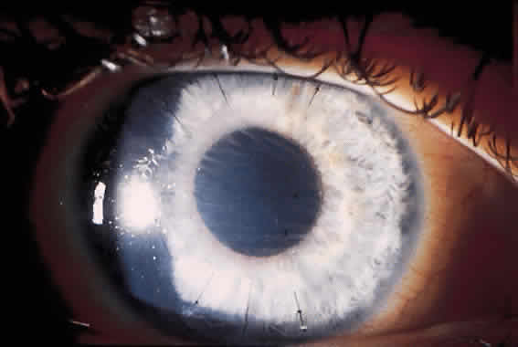
Although microperforations were the most common incisional complication encountered during RK, encroachment of the central clear zone was often the most visually disabling. Patients would complain of significant glare and halos around lights. Also, the resultant irregular astigmatism and corneal opacities often reduced the best spectacle-corrected visual acuity (BSCVA). Occasionally, patients would need corneal transplantation.

The original Russian incisions had a higher risk of inadvertent encroachment of the visual axis. Using this technique, the surgeon was required to terminate the incision manually at the marked clear zone. Unexpected movements by either the surgeon or the patient often caused the blade to cross the visual axis.

Irregular RK incisions are additional sources of subjective halos and glare. All RK incisions are created freehand. Thus, the regularity, linearity, and quality of the incisions are largely surgeon-dependent. Surgeons have created markers for the cornea to guide the incisions radially. However, angulation of the blade can create a beveled incision, reducing depth and thus the effect of the incision.

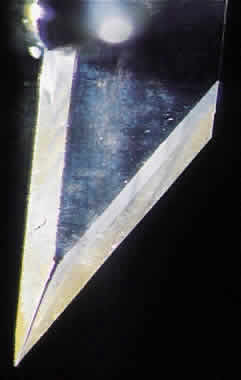
Current techniques have evolved to eliminate most of these complications. The diamond blades used today have two cutting edges. The main cutting edge is sharp for the entire length of the blade, whereas the anterior surface has a cutting surface for only 250 μm (Fig. 1). The blunt portion of the blade helps prevent inadvertent encroachment of the visual axis. Also, the new blades have a housing that will glide along the corneal surface to help keep the incisions perpendicular.

Fig. 1. Duo-Trak radial keratotomy blade (× 40). Note the oblique cutting edge and reverse cutting edge. The reverse cutting edge is sharp for only 200 μm from the blade tip; the remainder is dull.

POSTOPERATIVE COMPLICATIONS

The advances made in instrument design and surgical technique have greatly limited the incidence of intraoperative complications. However, postoperative complications continue to cause significant problems. Immediately after the procedure, the patient often feels a foreign body sensation for 1 to 3 days because of the breached epithelium. Topical nonsteroidal anti-inflammatory medications (NSAIDs) have been shown to be successful in reducing postoperative pain10 and are widely used following RK, photorefractive keratectomy (PRK), and LASIK. The epithelium is generally healed after 48 to 72 hours, with the resolution of symptoms.

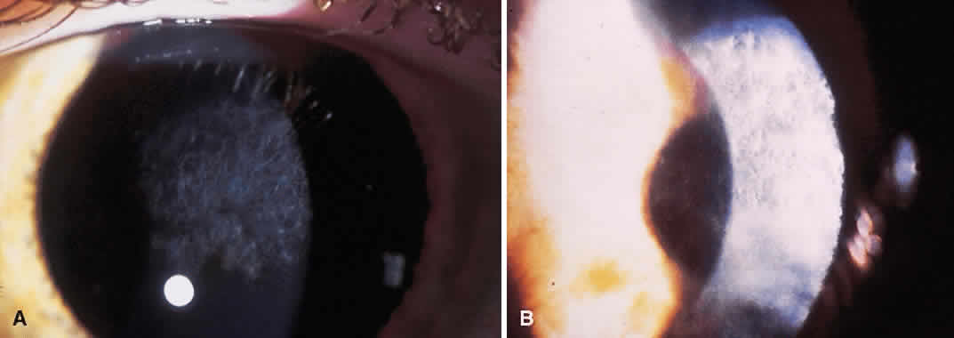
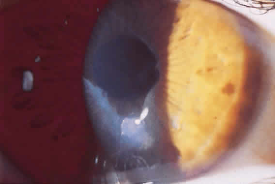
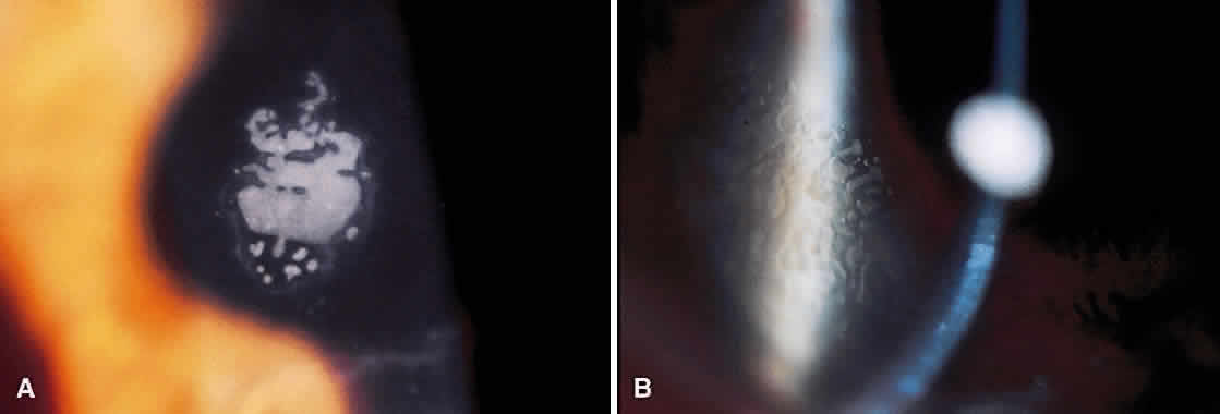
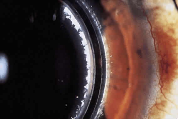
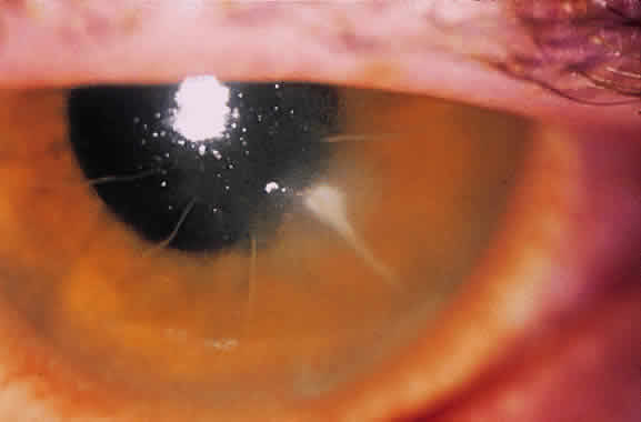
Until the epithelium completely heals, the risk of postoperative infection is greatest. Post-RK bacterial keratitis is primarily the result of the break in epithelial barrier created by the incisions. Adherence of causative organisms to the denuded epithelium over radial incisions can lead to infectious keratitis (Fig. 2). Recently, Panda and colleagues11 highlighted the severity of bacterial keratitis, despite its low incidence. They reported nine cases of post-RK bacterial keratitis at a tertiary-care cornea service during a 9-year period. Four of the nine cases followed primary RK and five followed enhancement procedures. Therapeutic penetrating keratoplasty was performed in one patient. Four of the nine eyes required subsequent penetrating keratoplasty, and one required lamellar keratoplasty. All but one eye retained 20/60 vision or better. These results are discouraging, but the actual incidence of bacterial keratitis lies somewhere between 0% and 0.4%.9,12–14 The PERK study found only 3 cases of bacterial keratitis in 793 eyes (0.38%); all occurred more than 7 months after surgery. All cases occurred in inferior incisions, and only one was spontaneous (one case with contact lens use and another with ocular trauma). None of the three eyes lost BSCVA. Hoffer and associates14 reported only one case of corneal ulcer after RK in 134 procedures performed at Jules Stein.

Fig. 2. Bacterial keratitis after radial keratotomy. Patient presented 3 days after surgery with increased pain and photophobia. Cultures were positive for Staphylococcus epidermidis. Aggressive antibiotic therapy cleared the infection, with final uncorrected visual acuity of 20/25. Note the more common inferior location and central segment of incision.

Post-RK bacterial keratitis usually occurs within 2 weeks of the procedure and is caused by virulent bacteria. The most common causative organisms are listed in Table 1. Infiltrates most commonly arise in the RK incision. Culture and Gram stain should be performed, and frequent fortified topical antibiotics that target the most common organisms should be used until the acute infection is under control. These cases of bacterial keratitis can result in significant vision loss, but if treated appropriately these infections can often be controlled with minimal reduction in vision.11,15 If the site of keratitis coincides with an area of microperforation or macroperforation, aggressive therapy and follow-up are required to avoid bacterial endophthalmitis.16–19


TABLE 1. Etiologic Agents Involved in Infectious Keratitis Following Radial Keratotomy

  Acute keratitis (1 day to 2 weeks)

  Staphylococcus aureus
  Staphylococcus epidermidis
  Stretpcococus pneumoniae
  Pseudomonas species
  Serratia marcescens


  Delayed keratitis (>1 month)

  Staphylococcus epidermidis
  Pseudomonas aeruginosa
  Moraxella
  Propionibacterium acnes
  Serratia liquefaciens
  Enterobacter gergoviae


  Atypical keratitis

  Candida parapsilosis
  Mycobacterium chelonae
  Herpes simplex virus


An entity unique to RK is a delayed keratitis, seen up to 40 months after surgery.20,21 The cause is unclear, because each case was associated with positive bacterial cultures. Several cases were associated with contact lens use, inducing microtrauma to the corneal epithelium. Aside from an infectious cause, some theorize that epithelial plugs in RK incisions contain cysts that rupture, leading to sterile inflammation. These cases of sterile keratitis respond to topical corticosteroids. If postoperative keratitis develops, however, therapy should be aimed at infectious causes before the use of topical steroids.

REFRACTIVE COMPLICATIONS

Early results of RK were encouraging: nearly 85% of patients gained uncorrected visual acuity (UCVA) of 20/40 or better at 1 year.8,9 Creation of nomograms for the number and location of incisions allowed more accurate treatments, but overcorrections and undercorrections continued to be a problem. Undercorrection is often a desirable result of refractive surgery, especially in presbyopic patients.

Attempts to treat overcorrection induced by RK were often unsuccessful. However, surgeons now have many options available to treat overcorrection with variable success. Before the use of the excimer laser, laser thermokeratoplasty, keratomileusis, lamellar keratoplasty, and various suture techniques were used. The most popular suture technique is the lasso technique, where one 10-0 nylon suture is passed through the radial incisions at a 7-mm optical zone. Results are initially promising, but some surgeons report regression of effect over time.22 More recently, LASIK has shown promise in reversing overcorrection.23

Undercorrection after RK is easier to correct. Enhancement rates varied from 0.6%7 to 42%,24 with 9% of the PERK patients having an enhancement procedure in the first year.9 An enhancement involved either deepening existing incisions to obtain full refractive effect or creating additional incisions. Despite the large rate of enhancements in some studies, the success rate of enhancements alone has not been addressed in the peer-reviewed literature.

During the long-term follow-up of RK patients, a hyperopic shift was noted in up to 54% of eyes.25,26 The rate of hyperopic shift seen in the PERK study was most pronounced in the first 2 years (+ 0.21 D per year) and continued up to 10 years after surgery (+ 0.06 D per year). When the authors looked at factors that influenced the development of a hyperopic shift, only the size of central clear zone and corneal diameter (and thus the length of incision) were correlated. Smaller clear zones and larger corneal diameters were weakly associated with a greater hyperopic refraction at 10 years. Lindstrom27 addressed this issue with the development of mini-RK, in which the incisions were 3 to 4 mm long. Lindstrom demonstrated that the peripheral extent of the incisions did not significantly affect the amount of central flattening and was able to obtain similar refractive results while theoretically limiting the postoperative hyperopic shift.

The refractive instability was not the only chronic problem: daily fluctuations in visual acuity were not uncommon. Some patients needed to take a nap to restore their vision. In addition, patients reported changes in vision when the barometric pressure changes. Intrigued by this phenomenon, Winkle and associates28 demonstrated that the hyperopic shift seen at high altitude was a result of corneal hypoxia, not the alteration of barometric pressure.

ANATOMIC COMPLICATIONS

Along with refractive instability, 90% corneal thickness incisions decrease the integrity of the cornea. Patients with RK incisions are at greater risk of serious eye injury from trauma. Histologic analysis of the healing corneal incisions reveals that healing is not completed for more than 5 years after RK.29 These healing wounds may rupture from direct ocular trauma or during intraocular surgery.30 Even after the healing process is completed, the structural integrity of the cornea remains diminished over its preoperative state.31 Other structural abnormalities were discovered after RK. Nelson and colleagues32 described map-dot-fingerprint changes in the corneal epithelium after RK. These patients did not develop recurrent erosions, but the changes were visually significant.

Controversy still exists as to the effect of RK on the corneal endothelium. A recent report states that although there is an acute loss of endothelial cells, the total rate of cell loss was not significant after 7 years compared with contact lens-wearing controls.33 However, there have been several studies demonstrating a significant loss of endothelial cells and resultant loss of function.34–36 With the incidence of microperforations nearing 40% in some studies, it is likely that many incisions approach Descemet's membrane. This may directly alter the interaction of the corneal endothelium and overlying stroma.

LOSS OF VISUAL ACUITY

The most crucial statistic reported for refractive surgery is loss of BSCVA. The PERK study reported that only 3% of patients lost more than two lines of BSCVA at 10 years.25 Arrowsmith and Marks12 found that nearly 15% of patients had a similar loss; however, only two of their patients had postoperative BSCVA of worse than 20/40. From this standpoint, RK was a safe procedure.

CONCLUSIONS

Complications of RK are well reported in the medical literature because it was the first popular procedure to correct myopia. Subsequently, the ophthalmologic community demanded controlled studies to evaluate its success and complications. Just as with Sato's original posterior incisional keratotomy, the complications associated with the procedure did not become evident until the long-term studies were published. Thus, the popularity of RK has diminished to give way to more predictable, safer surgeries. The refractive and structural instability has kept ophthalmologists from recommending RK except in certain circumstances. RK still has a place in the refractive surgeon's armamentarium, but it is now reserved for very low myopes and patients who are not candidates for the laser refractive procedures.

Many surgeons continue to perform incisional keratotomy to correct corneal astigmatism. Whether performed during cataract surgery or as an adjunct to laser refractive surgery, astigmatic keratotomy (AK) carries the same risks as RK. Infectious keratitis, corneal perforation, and refractive fluctuations can complicate even minor astigmatic correction. Therefore, strict sterility precautions and guarded diamond blades must be used with astigmatic keratectomy just as with RK.

Back to Top
HEXAGONAL KERATOTOMY
After the success of RK, surgeons tried to develop an incisional procedure that would correct hyperopia. The idea was to design an incision that would secondarily steepen the cornea. The Japanese were the first to design incisions in a hexagonal pattern, and Gilbert37 modified their technique by adding paracentral incisions at the apices of the hexagon incision. The T-hex pattern would induce a great amount of steepening, but there were many reports of complications. Casebeer and Phillips38 reported that 15% of eyes lost two lines of BSCVA. Subsequently, Basuk and colleagues39 reported on 15 eyes that experienced complications after hexagonal keratotomy. Complications included uncontrolled steepening, displacement of the central cornea, and irregular astigmatism. They concluded that the procedure was not safe, and that it should be “abandoned until further study.” In their study, 8 of the 15 eyes had lost BSCVA and 3 eyes required penetrating keratoplasty. This procedure is no longer performed to correct hyperopia.
Back to Top
LAMELLAR REFRACTIVE SURGERY
Lamellar refractive surgery involves the lamellar dissection of corneal tissue followed by removal of corneal stroma, which leads to a change in the refractive status of the eye. Drs Castroviejo and Barraquer performed the first work with lamellar refractive surgery. Initially, they would perform keratomileusis (cryolathed anterior stromal cap) and keratophakia (implantation of a crafted allogeneic lenticule), which in their skilled hands produced acceptable results.40 However, not many surgeons could reproduce their results because of the complexity of the surgery. Over the years, several modifications of the original lamellar procedures have been developed to limit complications and improve refractive results. However, surgeons still encounter complications inherent to lamellar refractive surgery.

The technique pioneered by Barraquer was keratomileusis for both myopia and hyperopia and involved a lamellar corneal dissection with a device known as a microkeratome. Complications associated with the microkeratome are inherent to all lamellar refractive procedures. These complications range from decentration to anterior chamber penetration (Table 2). Early designs of the microkeratome were cumbersome and difficult to assemble. The depth of lamellar dissection is determined by the placement of a footplate on the undersurface of the keratome. In the early models, if the footplate was not assembled correctly, the anterior chamber could be entered during the keratectomy. Because of the high intraocular pressure needed to perform a keratectomy, intraocular contents would be incarcerated in the wound and destroyed, potentially leading to permanent vision loss. This disastrous complication was reported in up to 2% of cases.41,42


TABLE 2. Flap Complications Associated with Lamellar Refractive Surgery


Complication%
Intraoperative 
 Irregular keratectomy0.9
 Incomplete keratectomy0.3
 Free cap1.0
 Total2.2
Postoperative 
 Folds1.1
 Displaced flap2.0
 Epithelial ingrowth removal2.2
 Diffuse lamellar keratitis1.8
 Infectious keratitis0.1

N = 1019 eyes, 490 myopic keratomileusis eyes, 529 laser in situ keratomileusis eyes.
From Lin RT, Maloney RK: Flap complications associated with lamellar refractive surgery. Am J Ophthalmol 1999;127:129---136).


With the improvement in design of the microkeratome, the incidence of inadvertent entry into the anterior chamber has greatly decreased. In a recent report by Stulting and associates,43 there was not one case of perforating injury during keratectomy in 1062 eyes. Their team of surgeons used the Automated Corneal Shaper (Chiron Vision Corp, Irvine, CA), which still required assembly of the footplate. The newer microkeratomes (Hansatome, Bausch and Lomb Surgical, Irvine, CA; Corrazio-Barraquer, Moria Instruments, Lansdowne, PA) have factory-installed footplates, which should prevent inadvertent perforation.

The first keratomes were manually driven across the cornea to create the lamellar cuts. This often resulted in irregular cuts and uneven stromal beds due to multiple variables. The irregularities could be in the form of blade chatter, depth fluctuations, or incomplete cuts. The modern keratomes are motor-driven and the blades oscillate rapidly in an attempt to create smooth lamellar dissections. These complications were readily evident in automated lamellar keratoplasty (ALK), in which two keratectomies were performed, with the second lenticule of corneal tissue discarded to flatten the cornea. The refractive keratectomy must be as regular as possible to limit optical distortions created by the irregular cut surface. Once the excimer laser was used to create the refractive keratectomy, smooth ablations were achieved, thus improving final visual outcomes.

Although all lamellar refractive surgical procedures have the same types of risks associated with the creation of the lamellar flap and refractive keratectomy, advances in technology have greatly reduced the incidence of vision-threatening complications.

KERATOMILEUSIS

Myopic and hyperopic keratomileusis were technically difficult procedures. The refractive modification was performed on the corneal cap, which posed several problems. First, surgeons had to be able to make a reproducible refractive cut on the corneal cap. Barraquer used a cryolathe on which the cap was frozen and the posterior surface was cut based on complex mathematical calculations. Several reports have documented alteration in stromal architecture after freezing, raising questions as to the accuracy that could be achieved using the cryolathe.44,45 To eliminate the uncertainty raised by freezing, Barraquer-Krumeich and Swinger developed the BKS system, which alters the anterior surface of the cornea using a suction device and creates a lamellar cut on the posterior surface of the corneal cap. This technique offered theoretical advantages over the cryolathe, but controlling tissue rigidity was always a concern. Subsequently, initial studies showed a 31% loss of Snellen acuity.46

As with all lamellar procedures, the depth of the keratectomy must be accurate to avoid corneal ectasia or perforation. The depth of lamellar dissection was initially determined by the amount of stroma required to prevent folds in the surgically augmented cap. It was believed that the residual cap thickness should not be less than 100 μm. For this reason, the caps were created up to 300 μm thick. With the average central corneal thickness of 500 μm, the creation of a corneal cap for myopic keratomileusis left a residual bed of 200 μm. Recent reports of corneal ectasia after myopic keratomileusis and LASIK when thin residual beds remain have led to the recommendation that 250 μm be the minimum amount of stromal bed remaining after lamellar surgery.47

Clinical studies reporting the results and complications of myopic and hyperopic keratomileusis are limited. In fact, the American Academy of Ophthalmology Ophthalmic Procedure Assessment of Keratomileusis noted that there was only one prospective study of myopic keratomileusis, in which there was significant visual loss and insufficient follow-up to assess clinical safety.48 Because of the cumbersome technique and variable results, myopic and hyperopic keratomileusis were slowly abandoned for the newer technique of ALK.

AUTOMATED LAMELLAR KERATOPLASTY

As the name states, the ALK procedure used an automated microkeratome to create the lamellar dissection and refractive cut in the residual stromal bed. The advent of the automated microkeratome was instrumental in eliminating many of the interface irregularities encountered with the hand-driven devices. ALK was the first refractive procedure to use automated instruments to create a keratectomy. In fact, with ALK, the keratome was used to fashion two keratectomies. The first cut was to create a flap; the second was the refractive cut made in the residual corneal bed. Because the refractive changes were made in the residual stroma and not the corneal cap, this procedure was named keratomileusis in situ.

INTRAOPERATIVE COMPLICATIONS

The diameter and thickness of the resected corneal tissue determined the amount of refractive change caused by the second keratectomy in ALK. Therefore, the second pass of the keratome was the most crucial portion of the procedure. The calculations involved in creating the refractive keratectomy were complex, and the reproducibility of refractive cuts was poor. Studies performed on cadaver eyes showed that resected corneal tissue had diameters that were 6% to 25% off predicted values, depending on the type of keratome used.49 They also demonstrated that the range of deviation achieved by the same microkeratomes was 8 to 24 μm. Arenas-Archila and colleagues50 confirmed these data on human eyes and found 28 of 32 cuts that were deemed “inaccurate.” Because of the unpredictability of keratectomies, refractive outcomes were variable. Manche and Maloney51 reported that fewer than 50% of patients had UCVA of 20/40 or better at 3 months, and only 23% achieved this vision at 6 months. Some authors, however, documented much better success: Lyle and Jin52 showed 86% of their patients to be 20/40 or better. To achieve these results, however, nearly 77% of their patients had enhancements, with 27% having three or more.

In addition to the unpredictability of the refractive cuts, the regularity of the resected cornea was a significant problem. Early keratomes with low blade-oscillation rates and slow translational velocities often produced ridges on the stromal bed. This washboard-like appearance was called blade chatter. Lyle and Jin52 experienced this phenomenon in 5.5% of cases. This irregularity was believed to cause disabling glare and significant astigmatism. The problem of blade chatter was magnified when a manual keratome was used.

Intraoperative complications associated with the corneal cap/flap were infrequent. Price and coworkers53 had only 3 dislocated corneal caps in 144 cases (2.1%), whereas Manche and Maloney51 encountered only 4 in 135 cases (3%). When a free corneal cap was present, the surgeon could either replace it with or without sutures, or leave the stromal bed bare.54 If the stromal bed were left bare, the refractive result was determined by the extent of epithelial hyperplasia over the stromal defect. If there was a robust epithelial response, the patient would become more myopic than if the epithelium returned to its normal configuration. All attempts were made to reattach the free corneal cap. Unfortunately, sometimes it was difficult to identify the epithelial versus the stromal surfaces. If the epithelial surface were sutured toward the stromal bed, a significant anterior stromal scar would develop, reducing BSCVA.

More devastating complications associated with ALK were rare. As with all lamellar procedures, there was a risk of corneal perforation. In the three large reviews, there were no cases of inadvertent corneal perforation. One can extrapolate that the risk of corneal perforation during ALK is no higher than 1 in 400 cases.

POSTOPERATIVE COMPLICATIONS

The refractive results seen with ALK would not be acceptable by today's standards. As noted above, the keratomes were not accurate when resecting the corneal lenticule. Price found that the ratio of diameter to depth of the resected cornea accounted for nearly 40% of the refractive outcome.48 In Manche's group, the variability of outcome measured was found to have a standard deviation of 1.50 D. This means that 92% of patients would be within 3.00 D of intended correction.46 Because of this uncertainty, Price recommended undercorrecting patients and correcting the remainder of the myopia and astigmatism with RK/AK. It was clear from these studies that ALK was designed for high myopia, where undercorrection of 3.00 D would be a more acceptable result.

Postoperative astigmatism also appeared to be a problem in Lyle's patients, in whom a 1.00-D increase in astigmatism was found 4% of the time. Manche noted that the mean magnitude of induced astigmatism was 1.30 D. Both investigators could not explain the reason for induced astigmatism, because neither sutured the flaps to the stromal bed. It is possible that the irregularity of the refractive cuts made with the microkeratome could induce cylinder. In addition, suturing of a free lenticule to the stromal bed would likely increase the probability and amount of induced astigmatism.

Visually disturbing glare, multiplopia, and distortion of vision were reported as a result of ALK.55 Again, these complications were most likely attributable to the irregular cutting of the early microkeratomes. Some patients can be corrected with hard contact lenses, but a significant number continue to have disabling glare. Penetrating keratoplasty has occasionally been necessary to relieve severe symptoms.56

Isolated reports of postoperative cap complications can be found in the literature.57–59 In the early days of lamellar surgery, a corneal cap was created and then sutured to the residual bed. Surgeons then allowed the caps to seal to the corneal bed without sutures. A bandage contact lens would often be used during the first few postoperative days. Occasionally, the corneal cap would become dislodged and adhere to the contact lens. Inadvertent trauma during the first postoperative week could also dislocate the cap. Surgeons would attempt to resuture the cap to the corneal bed, being careful to orient the cap epithelial surface up. In an effort to reduce the complications associated with the free cap, surgeons began creating a hinged flap rather than a free cap.60 This greatly facilitated replacement of the lenticule and is the preferred method of keratectomy today.

Infectious keratitis can develop in ALK patients acutely or long after surgery. The incidence of bacterial infection after ALK is difficult to determine because of the lack of published long-term data. Friedman and coworkers59 published the only report on bacterial infection after ALK. The causative organism was an Acanthamoeba organism, seen after therapeutic penetrating keratoplasty. Final visual acuity was counting fingers.

As in other forms of lamellar refractive surgery, epithelial cells can seed in the interface between the corneal cap and the residual stroma. The nests of epithelial cells do not usually induce inflammation, nor are they visually disturbing, unless they are in the visual axis. In Manche's 135 cases, only 2 (1.5%) had epithelial ingrowth significant enough to warrant débridement. Both of these eyes recovered 20/20 BSCVA by 3 months after surgery. Similarly, Lyle removed epithelial nests from only 3 of 128 eyes (2.3%). Epithelial nests did not significantly affect the results for ALK.

The most important statistic for refractive surgeons when evaluating the safety of a new procedure is the rate of loss of BSCVA. ALK had a high rate of loss of two lines of BSCVA. Manche reported 10% of his patients lost two or more lines of vision at 3 months, but that number improved to only 2% at 6 months. This trend may be significant, but he was able to evaluate fewer than 40% of his original 135 patients at 6 months. Both Lyle and Price found approximately 6% of patients lost two or more lines of BSCVA.

HYPEROPIC AUTOMATED LAMELLAR KERATOPLASTY

The concept of the correction of myopia with lamellar refractive surgery was simple: flattening of the central cornea was achieved by removing tissue from the corneal stroma. However, when one attempts to correct hyperopia, one must add tissue to the central cornea to create a steeper refractive surface. During the development of ALK for myopia, Dr Antonio Ruiz developed the hyperopic ALK procedure for the correction of hyperopia. His procedure involved the creation of a “controlled ectasia” of the central cornea. The microkeratome depth plate was set for 65% to 70% of central corneal thickness. After creation of the keratectomy, the flap would be replaced without additional treatment. As a result of the thin residual corneal tissue, Ruiz postulated that the intraocular pressure would induce slow and predictable ectasia of the central cornea.

There were few published reports on the results and complications of hyperopic ALK. In 1995, Kerizian and Gremillion reported on 85 eyes that were hypermetropic up to 5.00 D.61 Their refractive predictability was similar to the myopic ALK (76% 20/40 or better), but 13% of the eyes lost one to three lines of BSCVA.

ALK enjoyed a short-lived popularity. The problems inherent to ALK were the irregularity and unpredictability of the refractive cuts made by the microkeratomes. Although the automated keratomes made marked improvements, the excimer laser soon replaced the microkeratome as the refractive device. Keratomileusis in situ was still the method of choice to cure moderate to high myopia, but now the laser would be used to perform the refractive keratectomy with the precision needed to improve results and decrease complications.

Back to Top
PHOTOREFRACTIVE KERATECTOMY
Stephen Trokel was the first to describe the ability of the excimer (excited dimer) laser with a wavelength of 193 nm to ablate corneal tissue with remarkable accuracy.62 He postulated that the excimer would be ideal for refractive keratectomy, replacing the cryolathe, microkeratome, and BKS lathe. However, the first use of the excimer laser was for PRK, which treated the corneal surface after epithelial débridement. The US Food and Drug Administration (FDA) approved the excimer laser for PRK in 1995, when Summit Technologies lasers were allowed to treat up to 7 D of myopia. Shortly thereafter, VISX Corp. received approval for their laser to correct myopia alone. Subsequent approvals for the treatment of myopia with astigmatism and simple hyperopia followed. As of today, the excimer laser is approved to treat simple myopia (up to 14.0 D), myopia with astigmatism (up to 4.00 D), and hyperopia (up to 6.00 D).

The results of PRK have been well documented. In a recent Ophthalmic Procedure Preliminary Assessment report, PRK was found to be safe and effective for low to moderate myopes.63 Long-term follow-up of Korean patients undergoing PRK revealed that 62.4% had UCVA of 20/25 or better and 70.8% had 20/40 or better.64 Stephenson and colleagues65 also demonstrated the accuracy of refractive effect after 6 years, with 43% to 91% of patients within 1.00 D of intended refraction with no regression after 6 to 12 months.

Thousands of articles have been published on the results and complications of PRK in the peer-reviewed literature. The most difficult aspect in evaluating these papers is that the technology has evolved so dramatically since the laser was first used that the articles quickly become outdated. Many of the advances have evolved in response to complications encountered early in the study of the excimer (e.g., enlarging the ablation zone to decrease refractive aberrations). This review will therefore concentrate on the complications associated with the procedure as it is performed today.

INTRAOPERATIVE COMPLICATIONS

Intraoperative complications associated with PRK are extremely rare. Epithelial removal can be performed manually with a Kimura spatula or a rotary brush, or by laser-scrape technology. The entire area to be treated needs to be devoid of epithelium before treatment to ensure accurate ablation depths. There have been no reported complications associated with this portion of the procedure, although incomplete removal of the epithelium may be a cause of undercorrection.

During laser ablation, patient fixation is imperative. Decentered ablation patterns can cause significant optical aberrations (diplopia, halos, glare) that can be visually disabling (Fig. 3). In Seiler and associates'66 report, 5.2% of the operations were complicated by patient body or eye movements. The ablation was stopped until the patient could reorient his or her fixation. Only one quarter of these patients had significant decentration (more than 1 mm). BSCVA was not diminished under scotopic conditions, but one patient lost three lines of BSCVA “under glare conditions.” They concluded that small decentrations do not influence BSCVA under normal scotopic conditions, but BSCVA was affected more commonly under glare situations. When interpreting these results, one must consider that all treatment zones were 4.5 to 5 mm in diameter. Now that nearly all surgeons use at least 6-mm zones, the effect of a 1-mm decentration should be less noticeable.

Fig. 3. Decentered excimer laser photoablation after laser in situ keratomileusis. Patient complained of diplopia, glare, halos, and decreased visual acuity. Symptoms were more pronounced under scotopic conditions.

Summit and Autonomous Technologies have introduced a new eye-tracking system that will eliminate all small-amplitude patient movements, greatly improving refractive results. McDonald and colleagues67 recently reported the initial results of the eye-tracker system. Their refractive results were remarkable for over 98% 20/40 or better, and 72% 20/20 or better. Theoretically, the tracker systems should eliminate decentrations and the resultant loss of BSCVA.

Intraoperative complications associated with the function of the laser are often out of the surgeon's control. A laser malfunction can result in an inhomogeneous ablation, overcorrection, or undercorrection. The termination of laser treatment prematurely poses a difficult problem. Broad-beam lasers perform their ablation using an expanding diaphragm. The initial treatment is over a small diameter, which gradually increases during the treatment. Therefore, an incomplete treatment will consist of a small ablation zone. The surgeon must therefore resume the treatment from the same point at which it stopped. This will create the smoothest ablation profile possible.

POSTOPERATIVE COMPLICATIONS

Early postoperative complications of PRK are often related to epithelial healing. Re-epithelialization usually occurs in 2 to 4 days. Because the removal of epithelium and laser treatment significantly reduces corneal sensitivity,68 re-epithelialization is important. Most surgeons use a bandage soft contact lens after surgery until the epithelium is healed. Not only does this speed healing, but it also decreases discomfort.69 However, one should exercise caution when using a bandage lens after surgery because it may increase the risk of bacterial keratitis. Aside from this risk, delayed re-epithelialization can cause stromal haze and may result in myopic regression.70

As with any nonhealing epithelial defect, anesthetic abuse should be suspected if healing is not complete in 1 to 2 weeks. Postoperative pain is a significant problem after PRK. Aside from the bandage lens used, topical NSAIDs are effective in reducing pain. Several surgeons have begun prescribing dilute topical anesthetic for post-PRK pain control. This practice is very dangerous, risking severe keratitis and permanent corneal scarring. One such case resulted in a penetrating keratoplasty and counting fingers vision.71

After PRK, as well as with LASIK, many patients will experience symptomatic dry eyes. In an effort to explain the increased ocular irritation, özdamar and colleagues72 evaluated Schirmer's test and tear break-up time of treated eyes versus contralateral untreated eyes. They found a significant decrease in both Schirmer's and break-up time in the treated eyes at 6 weeks and related this to a decreased corneal sensitivity. It is unclear whether this phenomenon persists beyond the 6 weeks because of the restoration of corneal sensation during the first 6 to 8 months.73 Regardless, perioperative superficial punctate keratopathy after excimer laser therapy should be aggressively addressed to reduce the risk of infection and severe epitheliopathy.

With the regeneration of epithelium after PRK comes the possibility of duplicated epithelial basement membrane and recurrent erosions. Alio and associates74 found a few patients who had recurrent erosion syndrome after PRK. Combined with the decreased tear flow and corneal sensation, recurrent erosions can reduce BSCVA and increase the risk of infectious keratitis.

Any breach of the barrier function of the corneal epithelium predisposes the patient to infectious keratitis. Thus, bacterial keratitis should be a significant problem facing all PRK surgeons, but the exact incidence is not known. There have been few reported cases of bacterial keratitis after PRK.75–78 Etiologic agents include Pseudomonas aeruginosa, Staphylococcus epidermidis, Streptococcus pneumoniae, and Mycobacterium chelonae. All patients had typical keratitis symptoms, pain, photophobia, and redness. Clinical examination revealed stromal infiltrates with anterior chamber reaction with or without hypopyon. Laboratory cultures and immediate use of broad-spectrum antibiotics are warranted.

Keratitis after PRK is not always infectious. Sterile keratitis can present as subepithelial infiltrates that are associated with the use of topical NSAIDs or antibiotics.79 Because bacterial keratitis is the most dangerous complication seen after PRK, all infiltrates seen after surgery should be treated aggressively with topical antibiotics and cultures if warranted. If a noninfectious cause is suspected, discontinuation of NSAIDs and initiation of topical steroids are warranted. Probst and Machat80 have halved their postoperative NSAID regimen in an attempt to eliminate this complication.

Another infrequent perioperative complication seen after PRK is a steroid-induced intraocular pressure spike. Between postoperative week 4 and 6, 3% to 32% of patients experience a rise in intraocular pressure.81,82 Most, if not all, respond to topical antihypertensives and discontinuation of topical steroids. Regardless, patients with pre-existing glaucoma should be monitored closely because measurements of intraocular pressure with the Goldmann applanation tonometer read slightly lower over the treated cornea than the paracentral regions.83

REFRACTIVE COMPLICATIONS

Once the initial epithelial restoration is complete, most complications seen with PRK are refractive in nature. Refractive complications consist of overcorrection and undercorrection, induction of astigmatism, optical aberrations (halos, glare), central island formation, and loss of BSCVA. The incidence of these complications varies depending on the ablation profile. For example, enlarging the ablation diameter has decreased the incidence of visual aberrations, improved refractive predictability, and reduced the number of overcorrections.84 These results have prompted the use of a 6.0-mm ablation zone for all spherical ablations with both the VISX and Summit lasers. Recently, VISX has made software changes to its lasers to allow an even larger ablation zone (6.5 mm). Despite the widespread use of larger ablation zones, most of the studies reporting complications with PRK were performed using ablation diameters up to 5 mm. These articles will be discussed, but one must use caution when extrapolating these results to treatments with a 6-mm ablation zone.

Overcorrection after myopic PRK renders the patient dependent on glasses, whereas undercorrection leaves room for enhancement. In addition, successful correction of the iatrogenic hyperopia is difficult. Therefore, most surgeons err on the side of undercorrection for their initial treatment. Hersh and colleagues85 showed that age and the amount of intended correction affect the predictability of PRK. Seiler and coworkers66 also showed that patients treated for low myopia (2.4%) were less likely to experience significant overcorrection or undercorrection (±1.00 D) than patients treated for middle (8.2%), high (55.6%), or highest (75%) myopia. Most of these deviations were undercorrections, with only 2 of 176 eyes having more than 1.00 D of overcorrection at 2 years of follow-up. More recent data using 6-mm ablation zones revealed a 1% to 5% risk of significant overcorrection.86 Using the newer eye-tracker system from Summit/Autonomous and a 6-mm ablation zone, McDonald and colleagues67 had nearly 90% of eyes within 1.00 D of intended refraction.

Spherical ablations with a broad-beam laser should perform purely spherical ablation profiles, but several patients have experienced surgically induced astigmatism after spherical PRK. Such astigmatism can either improve the refractive state of the eye by eliminating preoperative astigmatism or increase the astigmatism, reducing UCVA. In the review of complications by Seiler and colleagues,66 5.6% of eyes had an increase in astigmatism between 0.75 and 1.50 D at 1 year. Only 30% of those eyes had an increase in astigmatism between 0.75 and 1.00 D at 2 years. In no case was there a change in cylindrical axis from the preoperative value, although a change in axis is possible.

Visual aberrations after PRK are usually described as glare and halos around lights, with the peak of symptoms occurring at dusk. Most commonly, glare and halos are created by either spherical aberration or irregular refraction created by the transition zone. As with spherical aberration, symptoms are more prevalent for patients with large pupils, or under scotopic conditions. The location of the transition zone is determined by both the diameter of ablation and the centration of treatment. In Gartry and colleagues'82 report using 4-mm ablations, 78% of patients complained of night halos. Studies using larger ablation zones found lower rates of optical aberrations.84 In addition, software programs are now designed to eliminate abrupt changes at the transition zones to reduce visual disturbances.

Another source of decreased visual acuity after PRK is central island formation. A central island is an area of steepening of more than 3.00 D, more than 1.5 mm in diameter (Fig. 4).87 The incidence of central islands ranged from 26% to 80% before the implementation of software alterations.87–89 Clinical symptoms related to central islands include blurred vision, monocular diplopia, and glare. Several theories have been proposed as to the cause of central islands. One theory identifies the accumulation of fluid in the central cornea, which then interferes with ablation centrally. Another theory claims that the plume created by each laser burst, if not evacuated before the application of the next pulse, will interfere with subsequent laser treatment.90 In an effort to eliminate islands, one laser manufacturer has devised a plume evacuator; another attempted blowing nitrogen gas over the cornea to dry its surface. Both of these innovations, along with software developments, have greatly reduced the incidence of central island formation.91 Islands can still form, however, and are more likely to develop with larger attempted corrections and larger ablation zones.87 Förster and associates91 recommend using a plume evacuator and wiping excess fluid from the central cornea after every 25 pulses.

Fig. 4. Videokeratographic illustration of central island after excimer laser treatment for 9.00 D of myopia. There is a paracentral elevation of more than 3.00 D. Uncorrected visual acuity was 20/60, with severe glare disability.

Another technique used to reduce island frequency is scanning laser technology. The scanning laser uses a spot or slit beam with small-diameter (1 to 2 mm) laser pulses delivered in a random fashion. This random pattern eliminates the need for a plume evacuator because the plume will have dissipated by the time a subsequent pulse is delivered to the same location. Further studies with this type of laser are needed to understand their impact on island formation.

Refractive stability is achieved after PRK by 3 months in most patients, with minimal regression after 6 months.63 Myopic regression before 6 months is a significant problem for refractive surgeons. The cause of regression is thought to be epithelial hyperplasia92 or stromal remodeling (haze). Rapid proliferation of epithelial cells can reduce the initial overcorrection created after PRK. Epithelial hyperplasia fills the area of ablated tissue to decrease the amount of corneal flattening. Corneal haze is a result of aggressive remodeling and likely increases the refractive effect of the cornea. Subsequently, stromal remodeling and haze correlate with the development of regression.92 Therefore, when evaluating a patient for regression, one should assess the amount of corneal haze present. If there is not significant haze, epithelial hyperplasia is probably the main cause of the regression. Patients with regression from epithelial hyperplasia are less likely to have a favorable response to enhancement, because they will probably have a similar epithelial reaction, negating much of the retreatment.

An interesting phenomenon occurs in women who undergo PRK and then become pregnant. Although there is no consensus as to the role of pregnancy on corneal wound healing, Sharif93 found that 66% of eyes of patients who became pregnant developed myopic regression, 83% of which developed 1 to 2+ corneal haze. After delivery, 50% of the patients experienced resolution of the haze and myopia. Hefetz and colleagues,94 in contrast, found no influence on outcomes in pregnant patients. The truth probably lies somewhere in between. This uncertainty emphasizes the importance of obtaining thorough informed consent with women of childbearing age, informing them that their results are not as predictable because of possible hormonal fluctuations.

Fortunately, most undercorrection and myopic regression can be addressed with further laser therapy. Enhancement rates vary depending on several factors (percentage of high myopes treated, patient tolerance of small refractive error, physician willingness to operate for very low myopia). Most enhancement rates for PRK are 10% to 20%. Rozsíval and Feuermannová95 reported that PRK retreatments are safe and effective for patients with low myopia (82.6% with 20/40 or better UCVA). However, patients who have regressed beyond -3.50 D and have more than 2+ haze are at significant risk of further regression, haze, and loss of vision.92 The importance of topical corticosteroids in preventing corneal haze, and thus regression, is debatable. There have not been any studies looking at the effectiveness of topical corticosteroids in preventing regression, but they are still used as part of routine postoperative care.

When operating on eyes with 20/20 vision, success is measured by both the postoperative UCVA and BSCVA. Refractive surgery complications can result in reduced BSCVA, but the loss of two lines of BSCVA is unacceptable. PRK has shown a very low percentage of patients with a loss of two lines of BSCVA. In Seiler's review of complications, only 1.1% of patients lost two or more lines of BSCVA, and both were at least 20/30. Lowenstein81 and Kim96 and their colleagues reported that fewer than 1% of patients lose two or more lines. However, when treating the higher myopes (more than -5), the incidence increases to nearly 13%.97 Possible reasons for the loss of BSCVA include central islands, decentered ablations, and corneal haze.

Corneal haze is a significant problem after PRK (Fig. 5). Although the vast majority of cases resolve and cause no visual sequelae, the possibility of regression and loss of BSCVA makes haze a great concern for PRK surgeons. The cause of the haze is likely an abundant proliferation of fibroblasts in response to the ablation performed. The grading of haze has been standardized in the literature (Table 3),98 with a grade of 2+ indicating clinical significance. Fortunately, the incidence of clinically significant haze after PRK is low, ranging from 0% to 6.7%.63 Several factors are thought to increase the likelihood of developing haze. The most important factor is the amount of attempted correction.99 Allergic conjunctivitis,100 when untreated, appears to be another important factor. However, keloid formers do not appear to have an increased risk of corneal haze.101


TABLE 3. Grades of Corneal Haze


GradeDescription
0Clear, no haze
0.5+Barely detectable or trace
1+Mild, not affecting refraction
1.5+Mildly affecting refraction
2+Moderate, refraction possible but difficult
3+Opacity prevents refraction, anterior chamber easily visualized
4+View of anterior chamber difficult
5+Unable to view anterior chamber
(Braunstein RE, Jain S, McCally RL et al: Objective measurement of corneal light scattering after excimer laser keratectomy. Ophthalmology 1996;103:439---443)


Fig. 5. Slit-lamp photographs of corneal haze after photorefractive keratectomy. A. Grade 1+ haze 3 months after surgery with minimal effect on best corrected visual acuity (20/25). B. Grade 2+ haze 4 months after surgery with significant decrease in best correct visual acuity (20/50).

In an effort to reduce the risk of corneal haze, Stein and colleagues102 focused on the increased temperature of the cornea during PRK as an important etiologic factor. They then attempted to cool the cornea during surgery with cooled balanced salt solution and noted a significant decrease in the amount of haze in the eyes that was cooled. Other agents have been tried in an effort to decrease haze, including interferon α-2b103 and antitransforming growth factor β.104 Although both showed some promise in the laboratory, their use clinically is limited.

PRK continues to be a viable option for refractive surgery candidates with low to moderate myopia. However, patients requiring larger corrections are at increased risk of haze, regression, and loss of BSCVA. Therefore, patients with myopia less than 6.00 D are excellent candidates for PRK.

Back to Top
LASER IN SITU KERATOMILEUSIS
For myopia up to -12.00 D, LASIK has quickly become the refractive surgery of choice. In this procedure, the laser replaces the second cut performed in ALK. It combines the rapid recovery of ALK with the accuracy of PRK. In 1999, the FDA approved both the Summit and VISX lasers for LASIK. However, surgeons have been performing LASIK as an off-label procedure since 1996, when the excimer laser was approved for PRK. The excitement over LASIK stemmed from the excellent refractive results and the minimal incidence of complications. There have been many reports on the efficacy of LASIK to treat myopia.105–107 However, the literature on LASIK complications is sparse. Recently, Stulting and colleagues43 outlined their experience with LASIK complications. They reported a complication rate of 4.4%, with less than half occurring during surgery. Only 3 of 1062 eyes (0.28%) lost greater than two lines of BSCVA to worse than 20/50. The complication rates decreased with increasing surgeon experience and improvements in technology. Although the complication rates have dropped, the potential for severe visual loss still exists. The complications encountered in modern LASIK can be broken down into preoperative, intraoperative, postoperative, and refractive.

PREOPERATIVE COMPLICATIONS

Proper preoperative evaluation is the most important step in laser refractive surgery. Accurate manifest and cycloplegic refractions must be obtained. Many surgeons elect to perform only manifest refractions before surgery, but with young patients, the cycloplegic refraction can help prevent overcorrection.

Other important steps performed before surgery are corneal pachymetry and topographic analysis. Corneal pachymetry is performed to evaluate the candidacy of a patient. Early work with myopic keratomileusis demonstrated that inadequate corneal tissue in the stromal bed could lead to iatrogenic corneal ectasia. In fact, Ruiz used this phenomenon to his advantage in hyperopic keratomileusis, where the keratectomy was performed at greater than 65% depth. Using these experiences, surgeons developed a recommendation that at least 250 μm of stroma should remain after LASIK to prevent unwanted ectasia. Wang and associates108 appeared to confirm the relation between a thin residual bed (less than 250 μm) and an increased likelihood of posterior corneal bulge. There is some controversy with this study relating to the method of measurement. However, there still appears to be a trend to posterior bulge with thinner stromal beds. Therefore, the preoperative pachymetry is essential to determine whether there is sufficient tissue to perform the desired correction.

Corneal topography is also essential to the preoperative evaluation. Corneal mapping may identify clinically undetectable keratoconus (Fig. 6). Also, one can identify irregular astigmatism that can be corrected only by, as of yet unavailable, “topo-link” technology. This technology will allow surgeons to develop custom ablations based on corneal topography and wave-front analysis. They will be able to sculpt corneas with ablations that may actually enhance the resolution efficiency of the eye. Topographic analysis can also identify flat corneas (less than 40.00 D), which when treated may develop more flap irregularities, leading to a decreased quality of vision.109

Fig. 6. Orbscan (Bausch and Lomb) evaluation of refractive surgery screening patient demonstrating keratoconus-like topography (A). Evaluation of the posterior surface elevation (B) shows a significant gradient (more than 0.1-mm change between flat and steep curves) suggestive of keratoconus.

There has also been a report of rhegmatogenous retinal detachment after LASIK.43 The patient was a -22.50 D myope with a significantly increased risk of detachment with or without surgery. Although there is controversy regarding the cause-and-effect relation between LASIK and retinal detachment, a thorough examination of the peripheral retina should be performed in an effort to identify risk factors for retinal tears (e.g., lattice degeneration). If extensive lattice is found, it may be advisable to perform prophylactic laser or cryotherapy before surgery.

INTRAOPERATIVE COMPLICATIONS

Complications during LASIK are rare but potentially vision-threatening. Microkeratome complications range from inability to apply the suction ring to inadvertent anterior chamber penetration. Complications associated with the application of laser to the stromal tissue are related to interface debris (e.g., heme, linen fibers) and laser decentration.

In patients with small interpalpebral fissures, either vertically or horizontally, it may be difficult to insert the suction ring to apply adequate suction. Gimbel and colleagues110 looked at the incidence of complications by an inexperienced surgeon and found that inadequate suction was encountered only 0.5% of the time. With more experienced surgeons, this complication occurs less frequently. When surgeons have difficulty with suction, they may perform a lateral canthotomy to permit proper apposition. Others will remove the lid speculum before applying the suction ring. This maneuver increases the likelihood of complications associated with the lids or lashes, but often allows the surgeon to perform the keratectomy. If it is impossible to insert the suction ring adequately, conversion to PRK is advised.

Proper application of the suction ring is critical to the creation of an adequate flap. Inadequate suction can lead to thin flaps and even doughnut-shaped flaps.111 The doughnut-shaped flaps are formed when the suction is low and the keratome surfaces and then re-enters the cornea. If the suction breaks during the pass of the microkeratome, an incomplete flap or a free cap can result. Newer microkeratomes have a built-in safety mechanism that will not allow the keratome to advance when the suction is inadequate. However, when conjunctiva alone occludes the suction port, the keratome senses that the suction is adequate. Therefore, it is important to check intraocular pressure with a tonometer before initiating the keratectomy.

Flap problems can occur despite adequate suction. One of the more common intraoperative flap complications is the buttonhole. There are conflicting theories regarding the cause of the buttonhole flaps. One theory is that a steep cornea protrudes through the suction ring more than a flat cornea, buckling the central cornea (Fig. 7). The theory that flat corneas are more prone to buttonholes is not as compelling. Unfortunately, there is no published report indicating the exact cause of the buttonhole.

Fig. 7. Slit-lamp photograph of buttonhole flap. The full-thickness hole was noted at the time of surgery, and photoablation was postponed. Three months later, photorefractive keratectomy was performed successfully. Residual astigmatism was addressed at 6 months after surgery with astigmatic keratectomy. Final uncorrected visual acuity was 20/40, with best corrected visual acuity of 20/20. Preoperative keratometry was 46 D.

Centration of the keratectomy is important to the success of LASIK. If the flap is decentered, the optical zone of the laser treatment may extend beyond the edge of the flap. This situation is encountered when using smaller suction rings and larger treatment optical zones. An important rule learned early in the development of the LASIK procedure is to delay treatment if the initial keratectomy is inadequate for proper laser correction. When one encounters a problem with the keratectomy, the laser treatment should be delayed. The flap should be repositioned and re-evaluated in 3 months. If a visually significant scar has not developed, the flap can be recut and the laser applied at this time. When a surgeon attempts a laser treatment under an inadequate flap, irregular astigmatism is the usual result.

Although laser ablation is relatively uncomplicated, it cannot be taken lightly by the surgeon or the patient. Patients must maintain fixation to ensure centered ablations. Minute fluctuations in fixation will not significantly affect the refractive outcome, but large ocular excursions can lead to a decentered ablation and decreased visual acuity. To help patients maintain fixation, their globe can be fixated with a scleral fixation ring. Asymmetric pressure applied to the globe during ablation can distort the central cornea and result in an irregular ablation.

Another problem seen in patients with peripheral corneal vascularization is erythrocyte extravasation onto the stromal bed during ablation. Davidorf and associates112 experienced this problem in 3% of their patients. If this occurs, the laser treatment should be halted until the debris can be removed. Surgeons can remove the hemorrhage with a sponge and apply phenylephrine to the bleeding vessels. Once the stromal bed is cleared, the treatment can resume. MacRae and coworkers113 described an inflammatory reaction in the interface after intraoperative hemorrhage. Even though the visible erythrocytes were cleared on reapposition of the flap, a visually significant interface inflammation occurred similar to diffuse lamellar keratitis. The inflammation resolved with topical corticosteroid therapy alone.

POSTOPERATIVE COMPLICATIONS

Once the flap is repositioned on the stromal bed, the endothelial pump action serves to create a capillary-type reaction and adhesion between the flap and the stroma. Experts disagree as to the ideal drying time after repositioning of the flap. The most conservative surgeons wait up to 5 minutes before allowing blinking over the operated eye, but others remove the speculum immediately on completion of the procedure. The purpose of drying time is to allow adequate adhesion between the flap and stroma to prevent dislodging of the flap. Displacement of the flap most commonly occurs in the first 24 hours after surgery.111 Examination reveals striae of the flap (Fig. 8), best observed under retroillumination. One should inspect the flap at the laser, shortly after the procedure, and at 24 hours. Once the epithelium has healed over the keratectomy, the likelihood of displacement is greatly decreased. some surgeons place a bandage contact lens to limit postoperative flap misalignment.

Fig. 8. Slit-lamp photographs of visually significant (uncorrected visual acuity 20/60) flap folds 1 day after laser in situ keratomileusis. The flap was repositioned and sutured. Uncorrected visual acuity returned to 20/20.

Once a flap becomes dislodged, it must be replaced as quickly as possible to reduce the amount of flap folds. The flap is lifted at the operating microscope and profusely irrigated to decrease the folds or wrinkles. One may use a contact lens to secure the flap in position, or suture the flap to the stroma if repeated repositioning attempts have failed. Postoperative irregular astigmatism is commonly encountered in eyes with flap folds. Repositioning of the flap, especially if performed several hours after dislocation, may not eliminate all the corneal folds. This irregular astigmatism may limit BSCVA and is often correctable with rigid gas-permeable contact lenses. In a series of 4500 cases at the University of Utah's John A. Moran Eye Center, only 10 flap complications were noted.114 Of these patient, 80% could see 20/20 with contact lens correction, and all were able to see 20/40 with correction. In general, flap complications will limit UCVA because of the induced astigmatism but may limit BSCVA if not realigned quickly.

Another complication noted after surgery is epithelial ingrowth (Fig. 9). Controversy exists as to the exact cause, and thus terminology, of epithelial nests in the LASIK interface. One theory supports the idea that epithelium is caught on the keratectomy blade and planted in the interface (epithelial growth). Another possible explanation is that the epithelial cells migrate from the junction of flap (ingrowth).115 Whichever the case, the epithelial nests can proliferate, causing decreased vision secondary to obscuration of the visual axis or induction of irregular astigmatism.

Fig. 9. Slit-lamp photograph of epithelial nests in the corneal interface after laser in situ keratomileusis enhancement. A. This nest of epithelial cells were observed and exhibited no growth and no reduction of BSCVA. B. These nests demonstrated progression toward the visual axis and several attempts were made to remove them. Finally, after absolute alcohol treatment and scraping, the nests resolved. Final uncorrected visual acuity was 20/200, and best corrected acuity was 20/20 with a refraction of + 2.75 + 1.75 × 110.

Rates of epithelial growth in the interface have ranged from 1% to 10%.43,116–118 Both Perez-Santonja118,119 and Stulting43 and their associates demonstrated a low incidence of epithelium in the interface after primary treatment. They also saw an increased rate of ingrowth (up to 32%) after a retreatment. Removal of the epithelial cells is indicated if there is significant vision loss due to irregular astigmatism or obstruction of the visual axis. The epithelial cells can be removed by lifting the flap and scraping the cells from the interface. Recalcitrant cases often need repeated scrapings, sometimes with the administration of absolute alcohol or excimer laser to kill the epithelial cells. Often, peripheral epithelial nests can be observed, but they may cause flap necrosis directly over the nests.119 Any sign of growth toward the visual axis should precipitate surgical intervention.

Infectious keratitis after LASIK is potentially vision-threatening. The incidence of infectious keratitis is extremely low. Machat120 reported the incidence to be 1 in 5000. In fact, this is so rare that there are only case reports of culture-proven bacterial infection after LASIK.121–126 In all cases, the patient did not lose more than two lines of BSCVA, and most were correctable to 20/20 or better. When bacterial keratitis is suspected, one should treat it as an infected corneal ulcer. Corneal scraping, with or without lifting the flap, and aggressive topical antibiotic therapy are warranted.

Herpetic reactivation is theoretically possible,127 making previous herpetic eye disease a relative contraindication for surgery. However, the effect of the excimer laser on active viral particles has been studied extensively. It appears that herpes simplex virus is not destroyed by the laser energy, but may actually be dispersed in the resultant plume. Thus, previous infection with herpes simplex virus, varicella zoster virus, or other viral infections may pose a health risk to office staff more than to the patient.

Postoperative inflammation after LASIK can be either infectious or noninfectious. Acute bacterial keratitis typically presents with pain and a focus of inflammation with surrounding edema. When the inflammation is diffuse, the distinction between infectious and inflammatory reaction is difficult. Diffuse lamellar keratitis is unique to lamellar refractive surgery and presents a toxic or allergic reaction to an unknown antigen. The syndrome (“sands of Sahara”) was originally described by Smith and Maloney128 as a nonspecific inflammation in the lamellar interface. Cultures are negative, and the syndrome is usually self-limited, resolving in 1 to 2 weeks. Symptoms include pain or photophobia, which usually present 2 to 6 days after surgery. On examination, the inflammation is confined to the interface, with no extension into the anterior or posterior stroma. The anterior chamber is usually quiet but may have a trace cellular reaction. Treatment consists of frequent topical steroids, but the inflammation typically resolves on its own. Recalcitrant cases may require irrigation of the interface when topical steroids are ineffective.

Retinal complications associated with LASIK are rare. There have been isolated reports of macular hemorrhage after LASIK. In one case, a bilateral hemorrhage occurred.129 Postoperative retinal detachment has also been reported,43 but the cause-and-effect relationship is still in question. Patients undergoing LASIK are generally myopic and at greater risk for retinal tears and detachment even before surgery. Further studies must be performed to identify the exact role of LASIK in retinal complications.

REFRACTIVE COMPLICATIONS

The LASIK procedure is performed with an excimer laser that ablates corneal tissue. When the size or shape of the laser ablation varies from the intended target, visual symptoms develop. The three most common reasons for refractive complications are over- or undercorrection, irregular ablations, and central islands.

After years of excimer laser experience with photorefractive keratectomy, adjustments were needed when programming the laser for treatment under a corneal flap. Currently, the lasers are using PRK programs to treat during LASIK. When programming in the desired laser treatment, the surgeon must use a “fudge factor” to adjust the program. Some surgeons cut a certain percentage of treatment for all patients, but a customized nomogram based on age and spherical equivalent would probably yield more accurate results.

Despite the experience with the laser, over- and undercorrections do occur. In early articles, the accuracy of LASIK was not precise. In 1994, Brint and colleagues130 found only 46% of patients within 1.00 D of intended correction. However, as surgeons gained more experience, nearly all patients were within 1.00 D.110 Despite this success, the rate of enhancement is roughly 15%.118 Enhancement can be performed for both overcorrections and undercorrections. As Perez-Santonja and coworkers118 demonstrated, enhancements carry a higher risk of epithelium in the interface (31%) and flap melting (10.9%). Others found that retreating low levels of myopia was very successful (96.2% 20/40 or better), with very few complications (e.g., no interface epithelium). Although these complications were more common with enhancement, vision difficulties were eliminated in a significant number of patients. When performing enhancements, careful calculations of the residual stromal bed thickness must ensure at least 250 μm of tissue to prevent iatrogenic ectasia.

Irregular ablation patterns can cause visually significant glare and aberrations. One of the more common irregular patterns is the decentered ablation. If during laser treatment the patient loses fixation and the surgeon does not correct the deviation, a decentered ablation results. Because the treatment was planned for the center of the patient's pupil, a decentered ablation often results in the transition zone of ablation intersecting the pupil. When this occurs, diffraction of light through the transition zone will cause significant glare. Treatment of decentered ablations requires a custom ablation plan, which is currently experimental. Decentered ablations are not the only type of irregular ablation. Interface debris at the time of surgery and fluid accumulation in the periphery of the treatment zone can both lead to an irregular ablation. Whatever the cause, the treatment is the same: custom corneal ablations assisted by topography.

As previously discussed, central islands are a result of several factors, most important of which are the laser plume and fluid accumulation in the center of treatment. Central islands are defined as an area on topography that shows at least 3.00 D of steepening at least 1.5 mm in diameter.87 Manche and colleagues131 successfully treated central islands by adjusting the treatment diameter for individual patients. Using Munnerlyn's formula, the amount of tissue removal needed was calculated. In all patients after retreatment, the BSCVA was 20/25 or better.

LASIK has proven to be a very effective means of correcting refractive errors. For the correction of myopia, surgeons can offer excellent results with a very low risk of significant vision loss. New laser technology (flying spot, topography link, tracking system) along with improved means of creating a lamellar flap (femtosecond laser, waterjet) will improve the results and decrease the complication rates. Thus, for a new technology to supplant LASIK as the refractive surgery of choice, it will require near-perfect results and almost no complications.

Back to Top
THERMOKERATOPLASTY
Thermal treatment of the cornea was introduced by Lans more than 100 years ago.3 Different modalities to deliver heat to the corneal stroma have been studied. Several modalities were successful in creating the desired thermal effect, stromal shrinkage. However, when used to create a change in corneal curvature to cure refractive errors, the effect was short-lived, and significant scarring developed.133

Currently under investigation, holmium laser thermokeratoplasty (Ho:LTK) is showing promise as the modality of choice for thermokeratoplasty. The Ho:LTK system, designed by Sunrise Technologies, (Fremont, CA) delivers energy to the cornea without contacting the surface epithelium while creating sufficient stromal effect. This noncontact modality eliminates the epithelial defects created by the contact treatments.

Refractive instability was the major complication seen with refractive thermokeratoplasty. Using the noncontact Ho:YAG modality, Koch and coworkers134 demonstrated a regression of 50% at the 6-month follow-up. After 6 months, the refraction remained stable. Day 1 effect was nearly 4.00 D for the two-ring treatment group, which regressed to 1.92 D at 6 months. Other investigators also found a regression of 50% during a 2-year follow-up.135 The mechanism of regression remains unclear and under investigation.

In addition to regression, LTK has not shown a predictable refractive effect. In Koch's patients, 25% of the patients in the two-ring group returned to within 0.75 D of their preoperative refraction. In the one-ring group, more than 40% had an “unchanged” refractive error (within 0.25 D) at 1 year. Tutton and Cherry135 also found only 25% of patients within 1.00 D of desired effect.

Neither Koch or Tutton found a loss of BSCVA in any patient undergoing Ho:YAG LTK. In addition, no significant loss of endothelial density, no corneal edema, and no increase in intraocular pressure were noted with the noncontact LTK. Thus, the procedure appears to be safe but continues to have difficulty with refractive stability. The Ho:YAG LTK is under review by the FDA and should be approved if the problem with stability is addressed.

Back to Top
CORNEAL IMPLANTS
In 1964, Jose Barraquer described his first cases of keratophakia in which he introduced an alloplastic lenticule into the cornea, altering its refractive state. The lenticule was lathed from either fresh or frozen corneal tissue and sutured beneath a cap created in the patient's cornea. Keratophakia, like myopic keratomileusis, was surgically challenging, even for the most experienced surgeons. Initial studies in the United States showed poor refractive results, with an error in refractive change of up to 28%.136,137 Refinements in the calculations used to create the lenticule improved the surgical accuracy, but there was still nearly 6% variation in the refractive results.

In 1977, Kelly, Swinger, and Troutman performed the first keratophakia in the United States. A 1981 article by Troutman and coworkers136 reported that the accuracy of the procedure was improving steadily, but they were “still unable to ensure the exact correction of the refractive error.” With the routine implantation of intraocular lenses (IOLs) after phacoemulsification, the incidence of aphakia is becoming very low. Keratophakia continues to be a viable option for patients not able to have intraocular surgery. However, the precision of the refractive effect continues to be the limiting factor. Prelathed corneal tissue has been proposed as an answer to the refractive uncertainty. Unfortunately, the effect of storage on the lenticule's refractive power has not been elucidated.

In an effort to improve the predictability of keratophakia, several investigators have worked with synthetic materials that could be manufactured to precise dioptric powers. The synthetic material must be water-permeable and able to maintain its optical clarity in vivo. Despite the promising work with hydrogel intracorneal implants,138 numerous complications were still encountered. Beekhius and associates138a experienced complications associated with lamellar dissection (9.4%), implantation of debris in the interface (100%), keratitis or vascularization of the implant (15.6%), and significant over- or undercorrection (28.1%). Although not all of these complications were visually significant, they highlight the difficulty of implanting synthetic material in the cornea. In an attempt to find more suitable materials for implantation, McCarey and colleagues139 studied the effects of polymethylmethacrylate (PMMA), polysulfone, and hydrogel materials on the refractive state of primate corneas. The refractive change induced by the three materials correlated with the refractive indices without significantly affecting the corneal curvature.

Another modality used to correct aphakia was epikeratophakia, described by Kaufman in 1980.140 Prelathed lenticules were sutured to a de-epithelialized cornea to induce a myopic shift. Unfortunately, refractive accuracy was not precise. When studied for the correction of myopia, only 35% were corrected to within 10% of emmetropia, and only 33% achieved UCVA of 20/40.141 Aside from the refractive complications, interface opacities,142 delayed epithelialization,143 stromal opacification,144 and induced astigmatism136 have been reported. Therefore, epikeratoplasty is reserved for patients with limited options for correction.

Excitement about corneal lens implants has abated as a result of the decreasing number of patients left aphakic and the numerous options for correction. Many surgeons will suture a posterior chamber lens or place a new-generation anterior chamber lens before attempting keratophakia or epikeratophakia. However, the experience with synthetic materials in keratophakia has led to the newest intracorneal refractive device, the intrastromal corneal ring segments (ICRSs).

Back to Top
INTRASTROMAL CORNEAL RING SEGMENTS
The ICRS (Intacs, KeraVision, Fremont, CA) procedure involves inserting two 150-degree arc segments made of PMMA into the corneal stroma. The lamellar channels are created at two-thirds corneal depth. The PMMA segments are inserted in these peripheral channels, producing a secondary flattening of the central cornea and thus reducing its refractive power. The visual results presented to the FDA are very encouraging, but there continue to be complications associated with the procedure.

INTRAOPERATIVE COMPLICATIONS

Intraoperative complications are rare. In data presented to the FDA, 451 of 454 eyes had the ICRSs placed successfully. Two (0.4%) of the cases were unsuccessful because of anterior chamber perforation during the creation of a radial groove incision. This occurred because of either improper calibration of the diamond blade or overaggressive use of the blade. In any case of anterior chamber perforation, the rings should not be implanted. There has not been a case of anterior chamber perforation during the creation of the channels. However, in a phase II study,145 there was one case of an anterior chamber perforation with introduction of the ring segment into the anterior chamber. This segment was removed and there was no loss of BSCVA.

The other unsuccessful implantation was due to shallow placement of the ring segment. The shallow segment was subsequently removed. Possible explanation for the shallow placement of the ring segment includes poor initial dissection, creation of a second (false) passage through the anterior third of the cornea, or loss of suction. When a segment is implanted in the anterior cornea, it must be removed. If the segment, or any impermeable synthetic device, is located in the anterior third of the cornea, nutrients cannot diffuse through the cornea to support an active epithelium.146 The patient will often develop an epithelial defect that will be difficult to cure. The defect can progress to a melt and the implant may extrude. Therefore, all segments implanted at less than 33% depth should be replaced.

POSTOPERATIVE COMPLICATIONS

Problems arising during the immediate postoperative period include epithelial defects, infectious keratitis, and segment migration. Postoperative epithelial defects are healed by day 3 in 96% of patients and by day 7 in 100% of patients. More experienced surgeons recommend a bandage contact lens be placed over an epithelial defect of more than 2 mm. Once the epithelium is intact, the risk of infection is significantly reduced.

Although there is no clinical evidence for its effectiveness, antibiotic prophylaxis is recommended to decrease the risk of postoperative infection. Culture-proven infectious keratitis has been described in the peer-reviewed literature. Pain out of proportion to the clinical appearance is indicative of infectious keratitis. An associated anterior chamber reaction may be present. If infectious keratitis is present after insertion of an ICRS, immediate removal of the offending segment or segments is recommended. Aggressive topical antibiotic therapy should be initiated using fortified agents (e.g., cefazolin 50 mg/ml and tobramycin 14 mg/ml). There is still some debate about the effectiveness of channel irrigation with antimicrobial agents, with no data to support the need for such therapy.

Postoperative migration of individual segments is relatively common. Although migration may be aesthetically unappealing to the surgeon, the clinical effect is minimal. When a segment migrates to a location directly under the incision, however, the wound may not heal adequately, and the segment must be rotated into position. Therefore, the ICRSs were designed with the superior segments 20 degrees to either side of the incision. It is estimated that the channels in which the segments rest will seal around the segment in the first month, making migration unlikely.

When the channels are created in the corneal stroma, they are made larger than the width needed to place the segment. Therefore, when the segment is placed in the channel, there is a space that is not filled with synthetic material. The cornea will respond to this empty space by forming debris and deposits in the channels (Fig. 10). The deposits and debris are not visually significant but often can be seen with the naked eye. Surgeons who have removed the segments when the deposits become severe note that the deposits dissolve once the segment is removed.

Fig. 10. Slit lamp-photograph after intracorneal ring segment implantation demonstrating grade 4+ channel deposits. Visual acuity was not affected. (Courtesy of KeraVision, Fremont, CA.)

Less common complications noted were a reduction of central corneal sensation (5.5%), neovascularization (2.7%), iritis (0.2%), and noninfectious infiltrate (0.5%). These complications were not associated with visual disturbances and are expected to improve over time.

REFRACTIVE COMPLICATIONS

The refractive results achieved with the ICRSs are very impressive. Nearly 97% of patients achieved 20/40 UCVA at 12 months, and 74% were 20/20 or better. An astounding 53% of patients were 20/16 or better. Refractive stability was achieved at 3 months after surgery. Despite these encouraging results, complications associated with refractive changes induced by the ring segments have been prominent. Night vision difficulties were classified as “always” present and “severe” in 4.8% of patients. When this symptom was broken down according to ring size, 15% of the patients with the largest (0.35 mm) segments had problems continuously. The larger segments also were associated with fluctuating distance vision 4.5% of the time. These symptoms can be visually disabling, necessitating explantation of the segments.

The ICRSs are technically easy to remove. When the segments are explanted, the visual symptoms improve in most patients. However, 11% of the patients who underwent explantation for glare, night vision difficulties, or diplopia continued to have these symptoms at greater frequency than before surgery. Explanting the segments also returns the cornea to nearly the same refractive state as it was before surgery. In the phase II trial,145 only two patients requested explantation. Both of these patients' refractive errors returned to within 0.75 D of their preoperative values after removal of the segments.

The ICRSs are a promising new refractive surgery tool. The effectiveness data reveal that a significant number of patients will enjoy an increase in UCVA over their preoperative BSCVA. However, the visually disabling refractive symptoms continue to be a significant problem in a few patients.

Back to Top
INTRAOCULAR SURGERY

PHAKIC INTRAOCULAR LENSES

Refractive surgery has historically been aimed at altering the refractive power of the cornea. These procedures have shown great promise for the correction of low to moderate refractive errors. However, the surgical correction of high ametropes with corneal surgery carries a high risk of complications. Because of the limitations of corneal refractive surgery in these patients, several authors are implanting IOLs in phakic eyes. This technology, still in its infancy, has evolved from its original description by Barraquer in the 1950s to posterior chamber IOLs made of porcine collagen/HEMA copolymer (collamer), able to be implanted through an incision smaller than 3 mm.

The original phakic IOLs were angle-supported anterior chamber IOLs designed and implanted by Baikoff.147 These first phakic IOLs caused corneal decompensation and pupil distortion,148 which prompted a change in design. The new design had less vault and endothelial touch, but it still induced unacceptable corneal damage and edema.149

In an effort to limit corneal complications, Fyodorov designed the first posterior chamber phakic IOL in 1991.150 However, the intimate relation between the phakic IOL and crystalline lens induced a significant number of cataracts (81.9%),151 prompting the discontinuation of this lens. An additional posterior chamber phakic IOL has been developed with sufficient vault to avoid contact with the lens capsule centrally. The Staar Implantable Contact Lens is currently in phase III FDA trials for the correction of myopia and hyperopia. The implantable lens is made of collamer and can be inserted through a 2.8-mm clear corneal incision. The results of the phase I study were promising,152 with all patients within 1.00 D of intended correction. Fifty percent were 20/20 or better and all were 20/40 or better without correction. No complications were reported at the 6-month follow-up.

Several complications have arisen since the phase I report. The lens is sulcus-fixated and when implanted causes a shallowing of the anterior chamber with contact between the lens and IOL and the iris and IOL.153 The most common complication is pupillary block,154,155 which is relieved by peripheral iridotomy. One patient developed intractable glaucoma with subsequent “retrociliary secretion of aqueous” requiring lensectomy and vitrectomy.156 Final visual acuity results for this patient were not specified. Rosen and Gore156 also noted pigmentary deposits on the implantable lens surface, which were not visually significant. These pigment deposits were thought to be from lens-iris touch, as demonstrated by ultrasound biomicroscopy.157

An additional lens under investigation is the iris clip anterior chamber lens first designed by Worst and Fechner. The original Worst-Fechner lens caused endothelial decompensation and increased anterior chamber flare.158 The new Artisan lens, modeled similar to the Worst lens, is under investigation. There are still concerns that this lens will cause similar anterior chamber inflammation and possibly corneal failure.

Despite the differences between the many phakic IOLs, the risk associated with intraocular surgery remains. There has been one reported case of endophthalmitis,159 with resultant light perception acuity, out of 355 implanted, an incidence of 0.28%. Also, the risk of retinal detachment appears to increase after implanting a phakic IOL. The rate reported by Ruiz-Moreno and colleagues160 (4.8%) is slightly higher than that of all myopes more than -6.00 D (3.2%). They postulate that the transient hypotony during surgery may cause an imbalance in the vitreous, leading to premature traction and subsequent retinal tears. Further studies must be performed to identify the exact relation between phakic IOLs and retinal detachments.

Phakic IOLs are becoming the most exciting advance in refractive surgery. However, until the incidence of cataract formation, glaucoma, and retinal detachment is well documented to be sufficiently low, the phakic IOL will be reserved for cases of severe myopia.

Back to Top
CLEAR LENS EXTRACTION
Another option in refractive surgery for high myopes and hyperopes is clear lens extraction with IOL implantation. This technique is controversial and carries all of the risks associated with cataract surgery: endophthalmitis, posterior capsular rupture, cystoid macular edema, and retinal detachment. Clear lens extraction is an important option for hyperopes more so than for high myopes because of the increased risk of retinal detachment in high myopes.161–163
Back to Top
CONCLUSION
Refractive surgery complications are very uncommon, but the procedures are not without risk. This chapter has attempted to outline the complications associated with refractive surgery procedures and to discuss how innovations have helped limit their prevalence. Not all complications were addressed, but an understanding of the most common complications is important for refractive surgeons. Despite the diligent investigation of each modality, there continue to be limitations on our ability to alter the natural state of the eye. For this reason, there will continue to be innovations in the area of refractive surgery until we have developed a procedure that eliminates all complications and delivers unsurpassed visual results.
Back to Top
REFERENCES

1. Lindstrom RL, Maller BS: Market trends in refractive surgery. J Cat Refract Surg 25:1408–1411, 1999

2. Kokott W: Über mechanisch-funktionelle Struktturen des Auges. Arch Ophth 138:424, 1938

3. Lans LJ: Experimentelle Untersuchungen über Entstehung von Astigmatism durch nicht-perforirende Corneawunden. Archiv Ophthalmologie 45:117–152, 1898

4. Sato T, Akiyama R, Shibata H: A new surgical approach to myopia. Am J Ophthalmol 36:823–829, 1953

5. Yamaguchi T et al: Bullous keratopathy after anterior-posterior radial keratotomy for myopia and myopic astigmatism. Am J Ophthalmol 93:600–606, 1983

6. Fyodorov SN, Durnev VV: Operation of dosaged dissection of corneal circular ligament in cases of myopia of mild degree. Ann Ophthalmol 11:1885–1890, 1979

7. Waring GO: Refractive Keratotomy for Myopia and Astigmatism. St Louis: Mosby, 1992

8. Deitz MR, Sanders DR, Marks RG: Radial keratotomy: An overview of the Kansas City study. Ophthalmology 91:467–478, 1984

9. Waring GO, Lynn MJ, Gelender H et al: Results of the prospective evaluation of radial keratotomy (PERK) study one year after surgery. Ophthalmology 92:177–198, 1985

10. Yee RW et al: Analgesic efficacy and safety of nonpreserved ketorolac tromethamine ophthalmic solution following radial keratotomy. Am J Ophthalmol 125:472–480, 1998

11. Panda A, Das GK, Vanathi M, Kumar A: Corneal infection after radial keratotomy. J Cat Refract Surg 24:331–334, 1998

12. Arrowsmith PN, Marks RG: Visual, refractive, and keratometric results of radial keratotomy: Five-year follow-up. Arch Ophthalmol 107:506–511, 1989

13. Waring GO, Lynn MJ, Culbertson W et al: Three-year results of the prospective evaluation of radial keratotomy (PERK) study. Ophthalmology 94:1339–1354, 1987

14. Hoffer KJ, Darin JJ, Pettit TH: Three years' experience with radial keratotomy: The UCLA study. Ophthalmology 90:627–636, 1983

15. Wilhelmus KR, Hamburg S: Bacterial keratitis following radial keratotomy. Cornea 2:143–146, 1983

16. Gelender H, Flynn HW, Mandelbaum SH: Bacterial endophthalmitis resulting from radial keratotomy. Am J Ophthalmol 93:323–326, 1982

17. O'Day D, Feman S, Elliot JH: Visual impairment following radial keratotomy: A cluster of cases. Ophthalmology 93:319–326, 1986

18. Heidemann DG, Dunn SP, Haimann M: Endophthalmitis after radial keratotomy enhancement. J Cat Refract Surg 23:951–953, 1997

19. McLeod SD, Flowers CW, Lopez PF et al: Endophthalmitis and orbital cellulitis after radial keratotomy. Ophthalmology 102:1902–1907, 1995

20. Shivitz IA, Arrowsmith PN: Delayed keratitis after radial keratotomy. Arch Ophthalmol 104:1152–1155, 1986

21. Mandelbaum S, Waring GO, Forster RK et al: Late development of ulcerative keratitis in radial keratotomy scars. Arch Ophthalmol 104:1156–1160, 1986

22. Miyashiro MJ, Yee RW, Patel G et al: Lasso procedure to revise overcorrection with radial keratotomy. Am J Ophthalmol 126:825–827, 1998

23. Suarez E, Torres F, Duplessie M: LASIK for correction of hyperopia and hyperopia with astigmatism. Int Ophthalm Clin 36:65–72, 1996

24. Charpentier DY, Garcia P, Grunewald F et al: Refractive results of radial keratotomy after ten years. J Refract Surg 14:646–648, 1998

25. Waring GO, Lynn MJ, McDonnell PJ et al: Results of the prospective evaluation of radial keratotomy (PERK) study 10 years after surgery. Arch Ophthalmol 112:1298–1308, 1994

26. Deitz MR, Sanders DR, Raanan MG, DeLuca M: Long-term (5- to 12-year) follow-up of metal blade radial keratotomy procedures. Arch Ophthalmol 112:614–620, 1994

27. Lindstrom RL: Minimally invasive radial keratotomy: Mini-RK. J Cat Refract Surg 21:27–34, 1995

28. Winkle KA, Mader TH, Parmley VC et al: The etiology of refractive changes at high altitude after radial keratotomy: Hypoxia versus hyperbaria. Ophthalmology 105:282–286, 1998

29. Binder PS, Nayak SK, Deg JK et al: An ultrastructural and histochemical study of long-term wound healing after radial keratotomy. Am J Ophthalmol 103:432–440, 1987

30. Budak K, Friedman NJ, Koch DD: Dehiscence of a radial keratotomy incision during clear corneal cataract surgery. J Cat Refract Surg 24:278–280, 1998

31. Peacock LW, Slade SG, Martiz J et al: Ocular integrity after refractive procedures. Ophthalmology 104:1079–1083, 1997

32. Nelson JD, Williams P, Lindstrom RL, Doughman DJ: Map-fingerprint-dot changes in the corneal epithelial basement membrane following radial keratotomy. Ophthalmology 92:199–205, 1985

33. MacRae SM, Rich LF: Long-term effects of radial keratotomy on the corneal endothelium. J Refract Surg 14:49–52, 1998

34. Chiba K, Tsubota K, Oak SS: Morphometric analysis of corneal endothelium following radial keratotomy. J Catt Refract Surg 13:263–267, 1987

35. Salz J: Progressive endothelial cell loss following radial keratotomy: A case report. Ophthalmic Surg 13:997–999, 1982

36. MacRae SM, Matsuda M, Rich LF: The effect of radial keratotomy on the corneal endothelium. Am J Ophthalmol 100:538–542, 1985

37. Gilbert ML, Friedlander M, Granet N: Corneal steepening in human eye bank eyes by combined hexagonal and transverse keratotomy. Refract Corneal Surg 6:126, 1990

38. Casebeer JC, Phillips SG: Hexagonal keratotomy: An historical review and assessment of 46 cases. Ophthalmol Clin North America 5:727, 1992

39. Basuk WL, Zisman M, Waring GO et al: Complications of hexagonal keratotomy. Am J Ophthalmol 117:37–49, 1994

40. American Academy of Ophthalmology: Keratophakia and keratomileusis: Safety and effectiveness. Ophthalmic Procedures Assessment. Ophthalmology 99:1332–1341, 1992

41. Barraquer JI: Keratomileusis for the correction of myopia. Ann Inst Barrauer 5:209–229, 1964

42. Barraquer C, Gutierrez AM, Espinosa A: Myopic keratomileusis: Short-term results. J Refract Surg 5:307–313, 1989

43. Stulting RD, Carr JD, Thompson KP et al: Complications of laser in situ keratomileusis for the correction of myopia. Ophthalmology 106:13–20, 1999

44. Schanzlin DJ, Jester JV, Kay E: Cryolathe corneal injury. Cornea 2:57–68, 1983

45. Koch P et al: Ultrastructure of human lenticules in keratophakia. Arch Ophthalmol 99:1634–1639, 1981

46. Colin J et al: The surgical treatment of high myopia: Comparison of epikeratoplasty, keratomileusis and minus power anterior chamber lenses. Refract Corn Surg 6:245–251, 1990

47. Wang Z, Chen J, Yang B: Posterior corneal surface topographic changes after laser in situ keratomileusis are related to residual corneal bed thickness. Ophthalmology 106:406–410, 1999

48. American Academy of Ophthalmology: Keratophakia and keratomileusis: Safety and effectiveness. Ophthalmology 99:1332–1341, 1992

49. Hoffman RF, Bechara SJ: An independent evaluation of second-generation suction microkeratomes. Refract Corneal Surg 8:348–354, 1992

50. Arenas-Archila E, Sanchez-Thorin JC, Naranjo-Uribe JP et al: Myopic keratomileusis in situ: A preliminary report. J Cataract Refract Surg 17:424–435, 1991

51. Manche EE, Maloney RK: Keratomileusis in situ for high myopia. J Cat Refract Surg 22:1443–1450, 1996

52. Lyle AW, Jin GJC: Initial results of automated lamellar keratoplasty for correction of myopia: One-year follow-up. J Cat Refract Surg 22:31–43, 1996

53. Price FW, Whitson WE, Gonzales JS et al: Automated lamellar keratomileusis in situ for myopia. J Refract Surg 12:29–35, 1996

54. Lam DSC, Leung ATS, Wu JT et al: Management of severe flap wrinkling or dislodgment after laser in situ keratomileusis. J Cat Refract Surg 25:1441–1447, 1999

55. Belmont S: Night halos after automated lamellar keratoplasty. J Refract Surg 12:809–812, 1996

56. Crews KR, Mifflin MD, Olson RJ: Complications of automated lamellar keratoplasty. Arch Ophthalmol 112:1514–1515, 1994

57. Steinmann TL, Denton NC, Brown MF: Corneal lenticular wrinkling after automated lamellar keratoplasty. Am J Ophthalmol 126:588–590, 1998

58. Hoffman CJ, Rapuano CJ, Cohen EJ, Laibson PR: Displacement of corneal lenticule after automated lamellar keratoplasty. Am J Ophthalmol 118:109–111, 1994

59. Friedman RF, Chodosh J, Wolf TC: Catastrophic complications of automated lamellar keratoplasty. Arch Ophthalmol 115:925–926, 1997

60. Pallikaris IG, Papatzanaki ME, Siganos DS, Tsilimbaris MK: A corneal flap technique for laser in situ keratomileusis: Human studies. Arch Ophthalmol 109:1699–1702, 1991

61. Kerizian GM, Gremillion CM: Automated lamellar keratoplasty for the correction of hyperopia. J Cat Refract Surg 21:386–392, 1995

62. Trokel SL, Srinivasan R, Braren B: Excimer laser surgery of the cornea. Am J Ophthalmol 96:710–715, 1983

63. American Academy of Ophthalmology: Excimer laser photorefractive keratectomy (PRK) for myopia and astigmatism. Ophthalmology 106:422–37, 1999

64. Kim JH, Kim MS, Hahn TW et al: Five-year results of photorefractive keratectomy for myopia. J Cat Refract Surg 23:731–735, 1997

65. Stephenson CG, Gartry DS, O'Brart DP et al: Photorefractive keratectomy: A six-year follow-up study. Ophthalmology 105:273–281, 1998

66. Seiler T, Holschbach A, Derse M et al: Complications of myopic photorefractive keratectomy with the excimer laser. Ophthalmology 101:153–160, 1994

67. McDonald MB, Deitz MR, Frantz JM et al: Photorefractive keratectomy for low-to-moderate myopia and astigmatism with a small beam, tracker-directed excimer laser. Ophthalmology 106:1481–1489, 1999

68. Murphy PJ, Corbett MC, O'Brart DPS et al: Loss and recovery of corneal sensitivity following photorefractive keratectomy for myopia. J Refract Surg 15:38–45, 1999

69. Lim-Bon-Siong R, Valluri S, Gordon ME, Pepos JS: Efficacy and safety of the ProTek (Vifilcon A) therapeutic soft contact lens after photorefractive keratectomy. Am J Ophthalmol 125:169–176, 1998

70. Ditzen K, Anschütz T, Schröder E: Photorefractive keratectomy to treat low, medium, and high myopia: A multicenter study. J Cat Refract Surg 20:234–238, 1994

71. Kim JY, Choi YS, Lee JH: Keratitis from corneal anesthetic abuse after photorefractive keratectomy. J Cat Refract Surg 23:447–449, 1997

72. özdamar A, Aras C, Karakas N et al: Changes in tear flow and tear film stability after photorefractive keratectomy. Cornea 18:437–439, 1999

73. Kauffman T, Bodanowitz S, Hesse L et al: Corneal reinnervation after photorefractive keratectomy and laser in situ keratomileusis: An in vivo study with a confocal videomicroscopy. Ger J Ophthalmol 5:508–512, 1996

74. Alio JL, Artola A, Claramonte PJ et al: Complications of photorefractive keratectomy for myopia: Two-year follow-up of 3000 cases. J Cat Refract Surg 24:619–626, 1998

75. Sampath R, Ridgeway AE, Leatherbarrow B: Bacterial keratitis following excimer laser photorefractive keratectomy: A case report. Eye 8:481–482, 1994

76. Amayem A, Ali AT, Waring GO III, Ibrahim O: Bacterial keratitis after photorefractive keratectomy. J Refract Surg 12:642–644, 1996

77. Wee WR, Kim JY, Choi YS, Lee JH: Bacterial keratitis after photorefractive keratectomy in a young, healthy man. J Cat Refract Surg 23:954–956, 1997

78. Brancato R, Carones F, Venturi E et al: Mycobacterium chelonae keratitis after excimer laser photorefractive keratectomy. Arch Ophthalmol 115:1316–1318, 1997

79. Teal P, Breslin C, Arshinoff S, Edmison D: Corneal subepithelial infiltrates following excimer laser photorefractive keratectomy. J Cat Refract Surg 21:516–518, 1995

80. Probst LE, Machat JJ: Corneal subepithelial infiltrates following photorefractive keratectomy. J Cat Refract Surg 22:281, 1996

81. Lowenstein A, Lipshitz I, Varssano D, Lazar M: Complications of excimer laser photorefractive keratectomy for myopia. J Cat Refract Surg 23:1174–1176, 1997

82. Gartry DS, Muir MGK, Marshall J: Excimer laser photorefractive keratectomy: 18-month follow-up. Ophthalmology 99:1209–1219, 1992

83. Fournier AV, Podtetenev M, Lemire J et al: Intraocular pressure changes measured by Goldmann tonometry after laser in situ keratomileusis. J Cat Refract Surg 24:905–910, 1998

84. O'Brart DPS, Corbett MC, Lohmann CP et al: The effects of ablation diameter on the outcome of excimer laser photorefractive keratectomy: A prospective, randomized, double-blind study. Arch Ophthalmol 113:438–443, 1995

85. Hersh PS, Schein OD, Steinert R et al: Characteristics influencing outcomes of excimer laser photorefractive keratectomy. Ophthalmology 103:1962–1969, 1996

86. Indications, contraindications, warnings, precautions and adverse events in VISX PRK professional use information manual. VISX Corp, 1996

87. Krueger RR, Saedy NF, McDonnell PJ: Clinical analysis of steep central islands after laser photorefractive keratectomy. Arch Ophthalmol 114:377–381, 1996

88. Levin S, Carson CA, Garrett SK, Taylor HR: Prevalence of central islands after excimer laser refractive surgery. J Cat Refract Surg 21:21–26, 1995

89. Lin DTC: Corneal topographic analysis after excimer laser photorefractive keratectomy. Ophthalmology 101:1432–1439, 1994

90. Noack J, Tönnies R, Hohla K et al: Influence of ablation plume dynamics on the formation of central islands in excimer laser photorefractive keratectomy. Ophthalmology 104:823–830, 1994

91. Förster W, Clemens S, Brüning S et al: Steep central islands after myopic photorefractive keratectomy. J Cat Refract Surg 24:899–904, 1998

92. Gartry DS, Larkin FP, Hill AR et al: Retreatment for significant regression after excimer laser photorefractive keratectomy. Ophthalmology 105:131–141, 1998

93. Sharif K: Regression of myopia induced by pregnancy after photorefractive keratectomy. J Refract Surg 13(Suppl): S445–446, 1997

94. Hefetz L, Gershevich A, Haviv D et al: Influence of pregnancy and labor on outcome of photorefractive keratectomy. J Refract Surg 12:512–513, 1996

95. Rozsíval P, Feuermannová A: Retreatment after photorefractive keratectomy for low myopia. Ophthalmology 105:1189–1192, 1998

96. Kim JH, Sah WJ, Hahn TW, Lee YC: Some problems after photorefractive keratectomy. J Refract Corneal Surg 10(Suppl):S226–230, 1994

97. Carson CA, Taylor HR: Excimer laser treatment for high and extreme myopia. Arch Ophthal 113:431–436, 1995

98. Braunstein RE, Jain S, McCally RL et al: Objective measurement of corneal light scattering after excimer laser keratectomy. Ophthalmology 103:439–443, 1996

99. Kremer I, Kaplan A, Novikov I, Blumenthal M: Patterns of late corneal scarring after photorefractive keratectomy in high and severe myopia. Ophthalmology 106:467–473, 1999

100. Yang HY, Fujishima H, Toda I et al: Allergic conjunctivitis as a risk factor for regression and haze after photorefractive keratectomy. Am J Ophthalmol 125:54–58, 1998

101. Tanzer DJ, Isfahani A, Schallhorn SC et al: Photorefractive keratectomy in African Americans including those with known dermatologic keloid formation. Am J Ophthalmol 126(5):625–9, 1998

102. Stein HA, Salim AG, Stein RM, Cheskes A: Corneal cooling and rehydration during photorefractive keratectomy to reduce postoperative corneal haze. J Refract Surg 15(Suppl):S232–233, 1999

103. Gillies MC, Garrett SK, Shina SM et al: Topical interferon alpha 2b for corneal haze after excimer laser photorefractive keratectomy. J Refract Surg 22:891–900, 1996

104. Thom SB, Myers JS, Rapuano CJ et al: Effect of topical anti-transforming growth factor-beta on corneal stromal haze after photorefractive keratectomy in rabbits. J Cat Refract Surg 23:1324–1330, 1997

105. Pallikaris IG, Papatzanaki ME, Stathi EZ et al: Laser in situ keratomileusis. Lasers Surg Med 10:463–468, 1990

106. Salah T, Waring GO III, el-Maghraby A et al: Excimer laser in-situ keratomileusis (LASIK) under a corneal flap for myopia of 2 to 20 D. Am J Ophthalmol 121:143–155, 1996

107. Güell JL, Muller A: Laser in situ keratomileusis (LASIK) for myopia from -7 to -18 diopters. J Refract Surg 12:222–228, 1996

108. Wang Z, Chen J, Yang B: Posterior corneal surface topographic changes after laser in situ keratomileusis are related to residual corneal bed thickness. Ophthalmology 106:406–410, 1999

109. Moshirfar M, Rudd JC: The effect of corneal curvature on LASIK outcomes. Presented at the International Society of Refractive Surgery Conference Oct 21–23, 1999, Orlando, FL

110. Gimbel HV, Penno EEA, van Westenbrugge JA et al: Incidence and management of intraoperative and early postoperative complications in 1000 consecutive laser in situ keratomileusis cases. Ophthalmology 105:1839–1848, 1998

111. Wilson SE: LASIK: Management of common complications. Cornea 17:439–467, 1998

112. Davidorf JM, Zaldivar R, Oscherow S: Results and complications of laser in situ keratomileusis by experienced surgeons. J Refract Surg 14:114–122, 1998

113. MacRae S, Macaluso DC, Rich LF: Sterile interface keratitis associated with micropannus hemorrhage after laser in situ keratomileusis. J Cat Refract Surg 25:1679–1681, 1999

114. Moshirfar M, Rudd JC: Flap complications in 4500 LASIK cases 1999

115. Helena MC, Meisler D, Wilson SE: Epithelial growth within the lamellar interface after laser in situ keratomileusis. Cornea 16:300–305, 1997

116. Montes M, Chayet A, Gomez L et al: Laser in situ keratomileusis for myopia of -1.50 to -6.00 diopters. J Refract Surg 15:106–110, 1999

117. Güell J, Muller A: Laser in situ keratomileusis (LASIK) for myopia from -7 to -18 diopters. J Refract Surg 12:222–228, 1996

118. Perez-Santonja JJ, Ayala MJ, Sakla HF et al: Retreatment after laser in situ keratomileusis. Ophthalmology 106:21–28, 1999

119. Perez-Santonja JJ, Bellot J, Claramonte P et al: Laser in-situ keratomileusis to correct high myopia. J Cat Refract Surg 23:372–385, 1997

120. Machat J: LASIK complications and their management. In Machat J: Excimer Laser Refractive Surgery: Practice and Principles, pp 359–400. Thorofare, NJ: Slack, 1996

121. Webber SK, Lawless MA, Sutton GL, Rogers CM: Staphylococcal infection under a LASIK flap. Cornea 18:361–365, 1999

122. Rudd JC, Moshirfar M: Methicillin-resistant Staphylococcus aureus (MRSA) following LASIK. (Submitted for publication)

123. Watanabe H Sto S, Maeda N et al: Bilateral corneal infection as a complication of laser in situ keratomileusis. Arch Ophthalmol 115:1593–1594, 1997

124. Perez-Santonja JJ, Sakla HF, Abad JL et al: Nocardial keratitis after laser in situ keratomileusis. J Refract Surg 13:314–317, 1997

125. Reviglio V, Rodriguez ML, Picotti GS et al: Mycobacterium chelonae keratitis following laser in situ keratomileusis. J Refract Surg 14:357–360, 1998

126. Mulhern MG, Condon PI, O'Keefe M: Endophthalmitis after astigmatic myopic laser in situ keratomileusis. J Cat Refract Surg 23:948–950, 1997

127. Davidorf JM: Herpes simplex keratitis after LASIK. J Refract Surg 14:667, 1998

128. Smith RJ, Maloney RK: Diffuse lamellar keratitis: A new syndrome in lamellar refractive surgery. Ophthalmology 105:1721–1726, 1998

129. Luna JD, Reviglio VE, Juarez CP: Bilateral macular hemorrhage after laser in situ keratomileusis. Graefes Arch Clin Exp Ophthalmol 237:611–613, 1999

130. Brint SF, Ostrick DM, Fisher C et al: Six-month results of the multicenter phase I study of excimer laser myopic keratomileusis. J Cat Refract Surg 20:610–615, 1994

131. Manche EE, Maloney RK, Smith RJ: Treatment of topographic central islands following refractive surgery. J Cat Refract Surg 24:464–470, 1998

132. Lans LJ: Experimentelle Untersuchungen über Entstehung von astigmatismus durch nicht-perforirende Corneawunden. Graefes Arch Clin Exp Ophthalmol 45:117–152, 1898

133. Feldman ST, Ellis W, Frucht-Pery J et al: Experimental radial thermokeratoplasty in rabbits. Arch Ophthalmol 108:997–1000, 1990

134. Koch DD, Kohnen T, McDonnell PJ et al: Hyperopia correction by noncontact holmium:YAG laser thermokeratoplasty: United States phase IIA clinical study with a 1-year follow-up. Ophthalmology 103:1525–1536, 1996

135. Tutton MK, Cherry PMH: Holmium:YAG laser thermokeratoplasty to correct hyperopia: Two years follow-up. Ophthalmol Surg Lasers 27:S521–S524, 1996

136. Troutman RC, Swinger C, Goldstein M: Keratophakia update. Ophthalmology 88:36–38, 1981

137. Swinger CA, Barraquer JI: Keratophakia and keratomileusis: Clinical results. Ophthalmology 88:709–715, 1981

138. McCarey BE, Andrews DM: Refractive keratoplasty with intrastromal hydrogel implants. Invest Ophthalmol Vis Sci 21:107–115, 1981

138a. Beekhuis WH, McCarey BE, vanRij G, Waring GO: Complications of hydrogel intracorneal lenses in monkeys. Arch Ophthalmol 105:116–122, 1987

139. McCarey BE, Waring GO, Street DA: Refractive keratoplasty in monkeys using intracorneal lenses of various refractive indexes. Arch Ophthalmol 105:123–126, 1987

140. Kaufman HE: The correction of aphakia. Am J Ophthalmol 89:1–10, 1980

141. McDonald MB, Kaufman HE, Aquavella JV: The nationwide study of epikeratophakia for myopia. Am J Ophthalmol 103:375–383, 1987

142. Morgan KS, Beuerman RW: Interface opacities in epikeratophakia. Arch Ophthalmol 104:1505–1508, 1986

143. Martel J, Martel J: Intraepikeratophakia. Ann Ophthalmol 19:287–292, 1985

144. Frangieh GT, Kenyon KR, Wagoner MD et al: Epithelial abnormalities and sterile ulceration of epikeratoplasty grafts. Ophthalmology 95:213–227, 1988

145. Schanzlin DJ, Asbell PA, Burris TE, Durrie DS: The intrastromal corneal ring segments: Phase II results for the correction of myopia. Ophthalmology 104:1067–1078, 1997

146. Brown ST, Mishima S: The effect of intralamellar water-impermeable membranes on corneal hydration. Arch Ophthalmol 76:702–708, 1966

147. Baikoff G, Joly P: Correction chirurgicale de la myopie forte parun implant de chambre anterieure dans l'oeil phake. Bull Soc Belge Ophtol 233:109–125, 1989

148. Baikoff G, Colin J: Damage to the corneal endothelium using anterior chamber intraocular lenses for myopia [letter]. Refract Corneal Surg 6:383, 1990

149. Mimouni F, Colin J, Koffi V, Bonnet P: Damage to the corneal endothelium from anterior chamber intraocular lenses in phakic myopic eyes. Refract Corneal Surg 7:277–281, 1991

150. Fyodorov SN, Zuev VY, Aznabayev BM: Intraocular correction of high myopia with negative posterior chamber lens. Ophthalmosurgery 3:57–58, 1991

151. Brauweiler PH, Wehler T, Busin M: High incidence of cataract formation after implantation of a silicone posterior chamber lens in phakic, highly myopic eyes. Ophthalmology 106:1651–1655, 1999

152. Sanders DR, Brown DC, Martin RG et al: Implantable contact lens for moderate to high myopia: Phase I FDA clinical study with 6 month follow-up. J Cat Refract Surg 24:607–611, 1998

153. Trindade F, Pereira F, Cronemberger S: Ultrasound biomicroscopic imaging of posterior chamber phakic intraocular lens. J Refract Surg 14:497–503, 1998

154. Davidorf JM, Zaldivar R, Oscherow S: Posterior chamber phakic intraocular lens for hyperopia of + 4 to + 11 diopters. J Refract Surg 14:306–311, 1998

155. Zaldivar R, Davidorf JM, Oscherow S: Posterior chamber phakic intraocular lens for myopia of -8 to -19 diopters. J Refract Surg 14:294–305, 1998

156. Rosen E, Gore C: Staar collamer posterior chamber phakic intraocular lens to correct myopia and hyperopia. J Cat Refract Surg 24:596–606, 1998

157. Trindade F, Pereira F: Cataract formation after posterior chamber phakic intraocular lens implantation. J Cat Refract Surg 24:1661–1663, 1998

158. Perez-Santonja JJ, Bueno JL, Zato MA: Surgical correction of high myopia in phakic eyes with Worst-Fechner myopia intraocular lenses. J Refract Surg 13:268–284, 1997

159. Perez-Santonja JJ, Ruiz-Moreno JM, de la Hoz F: Endophthalmitis after phakic intraocular lens implantation to correct high myopia. J Cat Refract Surg 25:1295–1298, 1999

160. Ruiz-Moreno JM, Alio JL, Perez-Santonja JJ, de la Hoz F: Retinal detachment in phakic eyes with anterior chamber intraocular lenses to correct severe myopia. Am J Ophthalmol 127:270–275, 1999

161. Colin J, Robinet A, Cochener B: Retinal detachment after clear lens extraction for high myopia: Seven-year follow-up. Ophthalmology 106:2281–2285, 1999

162. Goldberg MF: Clear lens extraction for axial myopia. An appraisal. Ophthalmology 94:571–582, 1987

163. Barraquer C, Cavalier C, Mejia LF: Incidence of retinal detachment following clear lens extraction in myopic patients. Arch Ophthalmol 112:336–339, 1994

Back to Top