Chapter 28
Chemical Injuries of The Eye
ROBERT A. RALPH
Main Menu   Table Of Contents

Search

EPIDEMIOLOGY
ALKALI VERSUS ACID INJURIES
PATHOPHYSIOLOGY OF CHEMICAL INJURIES
PREVENTION OF CORNEAL ULCERATION
CLASSIFICATION OF CHEMICAL BURNS
CLINICAL APPEARANCE
DETERMINATION OF EXTENT OF DAMAGE
CLINICAL COURSE OF UNTREATED CHEMICAL INJURIES
GOALS OF TREATMENT
TREATMENT
REFERENCES

Chemical injuries are among the most urgent of ocular emergencies, often resulting in a dramatic decrease in visual acuity or loss of an eye. The prognosis for a burned eye depends not only on the severity of the injury but also on the rapidity with which therapy is initiated.
Back to Top
EPIDEMIOLOGY
Ocular chemical injuries can occur under diverse circumstances and in such varied locations as the home, the workplace, and school. These injuries are common in industrial chemical laboratories, in machine factories, in agriculture, and among laborers and construction workers. They also are frequently reported from fabric mills, automotive repair facilities, and cleaning and sanitizing crews. Chemical burns of the eyes occur most often among the age group from 20 to 40 years, with young men at greatest risk. Injuries caused by caustic chemicals are among the most severe; the collective prognosis for satisfactory recovery from these injuries is extremely poor.1

In a retrospective study on the incidence and prevalence of ocular chemical burns, 171 consecutive patients were studied during an interval of one year. Industrial accidents caused 61% of these burns; 37% occurred in the home. The remainder were of unknown origin. The authors reasoned that household chemical accidents are among the most difficult to prevent because of the paucity of safety rules.2 Most of the eye injuries at home result from the use of lime and drain cleaners.1

Automotive battery acid burns have become increasingly more common, and can be especially devastating when combined with the shrapnel resulting from explosions.3 These accidents typically occur in the colder months, almost always after dark, and usually involve young men. During recharging of a lead acid storage battery, which contains up to 25% sulfuric acid, hydrogen and oxygen produced by electrolysis form a highly explosive gaseous mixture.4 The most common causes of storage battery explosions are lit matches (used to see battery cells) and the incorrect use of jumper cables. The National Safety Council has frequently advised the public regarding safe use of jumper cables.5

The deployment of automobile air bags involves the conversion of sodium azide to nitrogen gas, and is accompanied by the sudden release of an alkaline gas and powder.6 Ocular chemical injuries from air bag inflation must be considered dangerous and as great a threat to vision as other caustic injuries.7

Back to Top
ALKALI VERSUS ACID INJURIES
In general, alkali injuries are more damaging to the eye than those caused by acids. Alkalies are water-soluble substances that release hydroxyl ions and have a basic pH in solution. On the ocular surface, they saponify cell membranes and intercellular bridges, which facilitates rapid penetration into the deeper layers and into the aqueous and vitreous compartments.8 Cell damage from alkaline agents depends on both the concentration of the alkali and the duration of exposure. The higher the pH, the greater the damage to the eye, with the most significant injuries occurring at a pH of 11 or higher.9 As the pH rises, destruction of the epithelial barrier to penetration becomes progressively more extreme. In the corneal stroma, alkali cations cause damage and necrosis by binding to the mucopolysaccharides and to collagen.10

The most common alkalies involved in ocular injury are calcium hydroxide (lime), potassium hydroxide (potash), sodium hydroxide (lye), and ammonium hydroxide (ammonia). Of these, calcium hydroxide penetrates corneal tissue slowest because the calcium soaps it forms are relatively insoluble, impeding further penetration of the agent. Potassium hydroxide penetrates corneal tissue a bit faster. Sodium hydroxide penetrates even faster, but ammonia's transit across the cornea is fastest. If the epithelial barrier is removed, rates of penetration increase until they are nearly equal for all alkalies except ammonium hydroxide, which remains the fastest.9

Ammonium hydroxide passes through the cornea most rapidly because it destroys the epithelial barrier by saponification of the lipoidal cell walls and because it diffuses fastest through the stroma. Ammonia is fat soluble but most other alkalies are not. In addition, other alkalies lack the high mobility of ammonia necessary to permeate layers of cells.9

Acids dissociate in water to form hydrogen ions. The strongest acids have the highest concentration of hydrogen ions in solution. Free hydrogen ions cause cellular necrosis. Weak mineral acids generally cause less severe ocular damage than alkalies, although tissue destruction from strong acids (such as sulfuric, nitric, and hydrochloric acids) may be as severe as that from ammonia. The intact epithelium offers moderate protection against penetration of dilute or weak acids, allowing little damage unless the pH is 2.5 or less. However, severe tissue destruction can occur at a pH greater than 2.5 after the epithelium has been removed.9

Acids quickly denature proteins in the corneal stroma, forming precipitates that retard additional penetration. Overall, the rates of penetration for acids at equivalent concentrations and pH vary widely, with sulfurous acid penetrating more rapidly than hydrochloric, phosphoric, or sulfuric acids.9

Corneal tissue has an inherent buffering capacity that tends to equilibrate local pH to physiological levels, but severe chemical injuries exhaust the cellular and extracellular resources, allowing extremes of pH that are incompatible with tissue survival.11

Hydrofluoric acid is a strong inorganic acid (used in industry for cleaning and etching) that is particularly toxic because it has a complex mode of tissue injury. Along with necrosis from a high concentration of hydrogen ions, this agent causes cellular death because the fluoride anion binds calcium more quickly than the body can mobilize calcium from bone to equilibrate the loss.12 In addition, the fluoride blocks the Na-K ATPase enzyme of cell membranes, resulting in a fatal loss of potassium from the cells.13

Back to Top
PATHOPHYSIOLOGY OF CHEMICAL INJURIES

ROLE OF THE CORNEAL AND CONJUNCTIVAL EPITHELIUM

The corneal and conjunctival epithelia merge with each other at the limbus, where the subtle transition from the nonkeratinized, stratified squamous epithelium of the cornea to the nonkeratinized, stratified columnar epithelium of the conjunctiva occurs. These two epithelial surfaces together cover the exposed portions of the eye and function as a barrier to chemical insults and to invasion of microorganisms.

The Corneal Epithelium

The corneal epithelium serves the unique function of providing a smooth optical surface and the transparency necessary to transmit images with minimal distortion. The chemically-injured corneal epithelium may desquamate, or it may become irregular and lose its clarity. When the integrity of the epithelium is compromised, exposure of the underlying stroma may result in an alteration of hydration that further compromises corneal transparency.

The epithelial cell membranes are composed of lipid, which renders these cells hydrophobic. Adjacent cells are linked by secure junctional complexes, which impede the penetration of caustic substances through the epithelium. Strong alkalies pass rapidly through this relative barrier, however, because they saponify and liquefy the lipoidal cell membranes and junctional complexes.

Epithelial cells can be vigorous contributors to various phases of the corneal immune response.14,15 Intermingled with corneal epithelial cells are sparse Langerhans cells,16,17 which appear during local or remote corneal inflammation18 and which also participate in the corneal immune reaction.19 Epithelial cells secrete cytokines, including those that can inhibit the production of type I collagenase (capable of digesting corneal stroma) by underlying keratocytes.20 Curiously, epithelial cytokines can also stimulate the same keratocytes to produce type I collagenase.21 In stromal ulcerative disorders, for example after severe chemical injuries, regulatory participation of regenerating epithelium can help to tip the balance toward either further stromal degradation or re-establishment of corneal integrity. Epithelial cells themselves can produce a collagenase, although it is type V collagenase (gelatinase), which digests a substrate of denatured collagen (Fig. 1).22 Epithelial cells also can release prostaglandins in response to inflammation.14

Fig. 1. Digestion of collagen gel surrounding an explant from an ulcerating alkali-burned cornea.

Source of Regenerating Corneal Epithelium

The undisturbed corneal epithelium consists of approximately five layers of cells overlying the basal epithelial cells, which are affixed to their underlying basement membrane (Bowman's layer) through a group of adhesional components, primarily the hemidesmosomal anchoring plaques. The presence of an intact extracellular matrix, with fibronectin, laminin, glycosaminoglycans, and collagen is essential for secure binding of the basal epithelial cells.23

Epithelial cells arising from multipotential stem cells at the corneoscleral limbus migrate continuously in a centripetal fashion toward the corneal center. They replace the epithelial cells that have moved toward the surface during their normal maturation and have desquamated from the cornea.24 In the uninjured cornea, complete replacement of epithelial cells occurs every 5 to 7 days.25 The rate at which migrating cells move over the corneal surface increases after a traumatic loss of corneal cells,26 for example after chemical injuries resulting in epithelial desquamation. In the ideal situation, sheets of advancing corneal epithelium impinge on the epithelial defect in convex waves that eventually meet in healing ridges resembling the arms of a “Y”. These ridges later disappear as the healing cells readjust their positions to restore the normal contour of the injured cornea.

Within the first few hours after corneal epithelial injury, the surviving margin of intact epithelium sends fingerlike extensions forward into the injured zone. Fibronectin and other proteins from the tear film are deposited on the bare stroma or intact Bowman's layer. By the sixth hour, basal epithelial cells from the margin of the wound have lost their hemidesmosomal attachments and have become mobile. They migrate centripetally into the denuded zone, dragging with them one or two overlying layers of epithelial cells. At first, individual cells become thin, increasing their surface area to facilitate migration over the defect. Later, their numbers increase as mitosis occurs a few millimeters behind the advancing edge. Only after the defect is completely closed do the healing epithelial cells establish secure attachments to the underlying basement membrane and extracellular matrix. Next, they synthesize the proteins and intercellular bridges that render an intact epithelial surface resistant to penetration by infectious agents and noxious chemicals.25–27

The Conjunctival Epithelium

The conjunctiva is a vascularized mucous membrane that lines the entire exposed surface of the eye posterior to the cornea. It is reflected through the conjunctival fornices onto the posterior aspect of the lids. Its epithelium provides a moist, smooth surface over which the lids can pass. It participates with the corneal epithelium in establishing a relative barrier to the passage of microorganisms and noxious chemical agents, and it is active in local immune reactions.28 Its goblet cells produce mucin, which adsorbs to the glycoproteins coating the microvilli of corneal and conjunctival epithelial cells. This precorneal and preconjunctival mucin layer merges gradually with the overlying aqueous tear film to ensure complete wetting of the ocular surface.

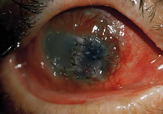
The conjunctival epithelium provides a source of cells to repopulate the corneal surface when the entire corneal epithelium has been denuded and the limbal stem cells have been destroyed, as in severe chemical injuries.29,30 After complete re-epithelialization of the cornea with conjunctival epithelium, a phenotypic change in the structure of these cells takes place by the process of transdifferentiation.31 Gradually the new epithelial surface begins to resemble that of corneal epithelium. The pseudostratified, columnar conjunctival epithelium becomes pseudostratified squamous, and there is an attrition of goblet cells. After several weeks, the biochemical functions of the healing cells begin to resemble more closely those of corneal epithelium. After transdifferentiation in cases of severe chemical injury, however, vascularization of the healing tissue inevitably occurs, with a return of goblet cells and conjunctivalization of the corneal surface.32

ROLE OF THE CORNEAL STROMA

Ninety percent of the cornea is stroma, consisting of approximately 200 layers of mostly type I collagen.33 Resting on the most superficial portion of the stroma is Bowman's layer, a meshwork of filaments derived from collagen types I, III, V, VI and possibly IV.34 The stroma itself is relatively acellular, with only 2% occupied by keratocytes, which are the corneal equivalent of fibroblasts. These keratocytes produce collagen, which accounts for more than 70% of the stroma by weight. The keratocytes secrete into the extracellular space a procollagen triple helix, from which collagen molecules form. These coalesce into fibrils, which in turn assemble into collagen fibers. Collagen fibers are uniform in diameter, with a constant spacing, factors which favor the transparency of corneal stroma.35

Keratocytes also synthesize glycosaminoglycans, which help to regulate water metabolism in the stroma as well as maintain the stroma's transparency. Most of the water in the intercellular stroma is bound to glycosaminoglycan molecules, which maintain uniformity of collagen fibril spacing and the cornea's transparency.35

Keratocytes also fabricate matrix metalloproteinases (MMPs),36 otherwise collectively known as matrixins or collagenases. These proteases regulate the synthesis and degradation of extracellular matrices, and participate vigorously in corneal wound healing. The MMPs consist of nine types, of which MMP-1 and MMP-8 are true collagenases, MMP-2 and MMP-9 are gelatinases (which utilize a substrate of degraded collagen), and the remaining varieties (MMPs 3, 7, 10, 11, and 12) are stromelysins.36 MMPs are critical components of the process by which injured extracellular matrix is digested and later reformed. MMPs are regulated in vivo by tissue inhibitors of metalloproteinases (TIMPs) and other inhibitors.36 Imbalance between MMPs and TIMPs can lead to excessive fibrosis (when the inhibitor exceeds the enzyme) or to excessive tissue melting (when the enzyme exceeds the inhibitor).36 The major proteinase inhibitors of the extracellular matrix are α2-macroglobulin,36 which also constitutes most of the anticollagenase activity in plasma,37 and the TIMPs.

Among the other participants in corneal wound healing are the various growth factors, including epidermal growth factor (EGF) and transforming growth factors (TGFs) α and β. These are mitogens and chemotactic agents.38 In cornea, there is a constant interaction between epithelium and stroma in the repair of stromal injury.39 For example, TGF beta-2 from epithelial cells in culture inhibits collagenase synthesis by cultured corneal stromal cells; other epithelial cytokines stimulate stromal keratocytes to produce collagenase.39

After a chemical burn or similar injury, keratocytes increase in number by mitosis, and new ones migrate into the region of damage. Migration of keratocytes occurs only under an intact epithelium.34 EGF and fibroblast growth factors both help to regulate the influx of new keratocytes.40 The energized keratocytes produce new collagen and proteoglycans.34 Although the new collagen is type I,41 the diameter of the resulting fibers is larger and the spacing is irregular,42 decreasing transmission of light through the resulting scar. Meanwhile, the new proteoglycans bind water more avidly, resulting in excess hydration of the scar, which further insures irregular spacing (with lack of transparency) of the new collagen.34 In addition, stromal keratocytes develop intracytoplasmic contractile elements that cause contraction of the scar and irregular astigmatism.34

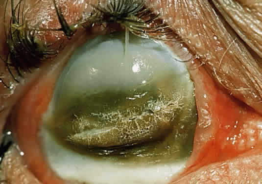
In the chemically-injured cornea, these complex mechanisms by which the stroma responds typically lead to minimal scarring and loss of transparency when an overlying epithelial defect heals rapidly and the damage to the stroma is superficial. In injuries of great severity, with long chemical contact time and maximal variances from physiological pH, the overwhelming stromal response is that of degradation or melting. These are situations in which the intrinsic control mechanisms fail to maintain homeostasis.

ROLE OF THE POLYMORPHONUCLEAR NEUTROPHIL

Collagenase activity has been documented in polymorphonuclear neutrophils (PMNs).43 Before they are released from the bone marrow, PMNs synthesize proteases, including gelatinase B and collagenase 2.44,45 Matsuda and Smelser observed numerous PMNs at sites of stromal ulceration after ocular chemical injuries.46 Pfister and colleagues observed that after alkali injury of the cornea, the tripeptide degradation products of stromal collagen are among the earliest biochemical factors stimulating PMN influx into the cornea.47,48 The gathering PMNs themselves release leukotrienes, which are chemotactic agents for the additional influx of neutrophils.49 The alkali-injured collagen also liberates a cytokine that triggers a respiratory burst among the accumulating PMNs. These cells release not only proteolytic enzymes, but also superoxide radicals, both of which aid in further collagen degradation and resultant corneal ulceration.49

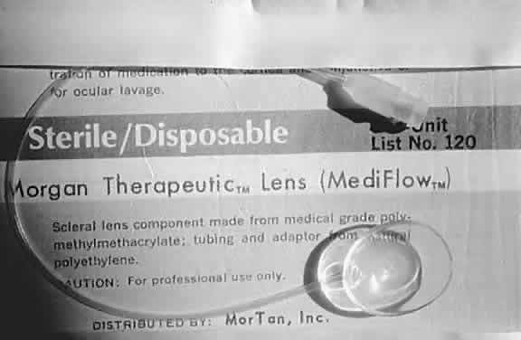
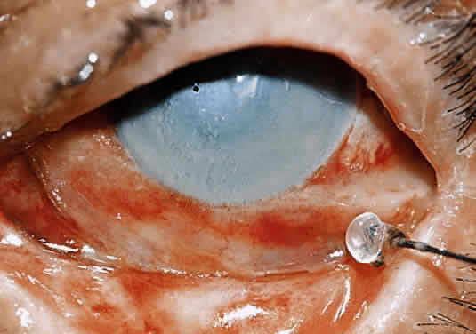
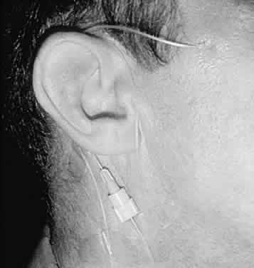
Kenyon and colleagues demonstrated that in rabbit eyes, after burns with sodium hydroxide, when both epithelium and PMNs were excluded from the exposed stroma by gluing a contact lens in place two days after the burn, only 10% of corneas ulcerated.50 In the control eyes without corneal protection, ulceration occurred in 2 weeks. If the lenses were removed at day 14, 80% of corneas ulcerated within the next week. When rings (exposing the corneas centrally) were glued onto the denuded stroma of 15 burned eyes, epithelium was excluded but PMNs migrated to the exposed central zone through the tear film. Eleven of the corneas developed ulcerations, with PMNs adhering to the digested tissue. In non-alkali-burned controls, with or without glued-on contact lenses or rings, no ulcerations occurred despite the presence of many PMNs.50

Evidence suggests that 10% sodium citrate applied hourly to the alkali-burned eye may offer some relative protection against stromal ulceration51 by inhibition of selective PMN activities. The PMNs must adhere to the endothelium of vessels before their movement through the vascular wall and chemotaxis can cause a tissue infiltrate. Citrate inhibition of PMNs may be through a mechanism that keeps them circulating and prevents them from adhering to the endothelium.52

Back to Top
PREVENTION OF CORNEAL ULCERATION

INHIBITORS OF COLLAGENASE

Endogenous inhibitors of collagenase and related proteinases participate vigorously in embryogenesis and tissue maturation, and in the remodeling of connective tissues that occurs during wound healing, bone resorption, and various types of periodontal disease.37,53 TIMPs located in proximity to connective tissue cells are important regulators of the status of the extracellular matrix. Humoral inhibitors, primarily α2-macroglobulin, control the activity of metalloproteinases in body fluids.37 Binding of α2-macroglobulin to MMPs is extremely tenacious, rendering this circulating agent one of the strongest known inhibitors of MMPs.37 Berman and associates used α1-antitrypsin, another anticollagenolytic factor derived from human serum, in an effort to inhibit corneal ulceration.54

The search for safe, effective exogenous inhibitors of collagenase progressed during most of the 1960s. Eisen and coworkers used cysteine to prevent collagenolytic activity in human skin.55 Both the disodium and calcium derivatives of ethylenediaminetetraacetic acid (EDTA) inhibited lysis in concentrations as low as 10-4 M. The calcium EDTA appeared safer, because it did not inhibit cell growth in tissue culture in concentrations as high as 10-2 M.56,57

Hook and coworkers believe that EDTA chelated the calcium necessary for collagenase activity. Because calcium is replaced by tissue fluids, EDTA would have to be used almost continuously to maintain anticollagenolytic activity. Cysteine theoretically could be used less frequently because it has a double action: not only does it chelate calcium, it also irreversibly reduces a critical disulfide bond in the collagenase molecule.58

Slansky and associates reported that along with 0.2 M l-cysteine and EDTA, 1.2 M N-acetyl-l-cysteine was also effective in vivo against collagenase and was more stable than cysteine. Initial trials on ulcerating rabbit corneas were performed with N-acetylcysteine (Mucomyst), as a 10% or 20% solution. Later, the studies were reconfirmed with a pure acetylcysteine solution when it was determined that Mucomyst contains 10-3 M EDTA (itself a known collagenase inhibitor) as a stabilizer. When treatment with this agent was stopped on the 21st day, four of six eyes ulcerated within 48 hours, but none of the eyes still receiving the drug ulcerated.59

TETRACYCLINE

Tetracyclines are a family of broad-spectrum antibiotics that exhibit antiinflammatory and anticollagenolytic activity independent of their antimicrobial properties.60–62 Windsor and colleagues reported that after subjects received oral tetracyclines for four days, white blood cell phagocytic activity diminished, and their ascorbic acid levels decreased by at least 50%.60 Forsgren and coworkers demonstrated both in vitro and in vivo that in the presence of tetracycline or doxycycline, human PMNs displayed a marked decrease in their phagocytosis.61 In 1985, Perry and Golub described the healing of a noninfected corneal ulcer after the patient began taking 250 mg oral tetracycline four times a day.63

The inhibition of MMPs in periodontal disease and reactive arthritis, inflammatory conditions resembling ocular collagenolytic disorders, has been demonstrated using tetracycline, chemically-modified tetracycline (without antibiotic properties), doxycycline, and minocycline.64–66

Numerous studies have documented the benefits of topical and systemic tetracycline in the management of ocular chemical injuries. In alkali-burned rabbit eyes treated with topical 1% or 5% tetracycline hourly for 12 hours a day, 54% of the control eyes ulcerated, but only 8% of the treated eyes ulcerated.67 Intramuscular tetracycline also inhibited corneal ulcerations in alkali-burned rabbit eyes. Eyes that ulcerated showed inflammatory infiltrates with PMNs, but there were no inflammatory cells in nonulcerating eyes.68 When oral tetracycline was added to the therapy of eighteen patients with nonhealing corneal epithelial defects, the erosions of fourteen patients healed.69

Although tetracycline, doxycycline, and minocycline all can inhibit mammalian collagenases, antibiotics such as penicillin and cefazolin are ineffective. Collagenases and the other MMPs can function only in the presence of divalent calcium and zinc cations. When these ions are chelated, the activity of collagenase decreases. Tetracycline binds to collagenase by a calcium bridge, inactivating the enzyme unless additional calcium is added.69 Tetracycline also appears to decrease collagenase activity by altering the gene expression responsible for synthesis of the MMPs.70 Tetracycline has both direct and indirect effects on PMNs. It decreases their ascorbic acid levels,60 and it reduces PMN infiltrates in injured corneal stroma by decreasing collagen lysis, the products of which are chemotactic for PMNs.68

SYNTHETIC INHIBITORS OF COLLAGENASE

Among the many synthetic inhibitors of collagenase is the hydroxymate-containing dipeptide, Galardin.71 This agent appears to prevent the corneal ulceration of alkali injury by diminishing the release from PMNs of MMPs that digest capillary walls to allow extravasation of the PMNs.72 Any decrease in the number of locally-extravasated PMNs decreases the potential pool of MMPs at the site of injury. Galardin further reduces inflammation by preventing release of tumor necrosis factor-α (TNF-α), a cytokine that activates PMNs, from producer cells such as macrophages and activated T cells.25 Galardin also may block MMPs released from inflammatory cells, corneal fibroblasts, and epithelial cells.25

Other synthetic inhibitors of MMPs include mercaptan (thiol)-containing compounds resembling the drug captopril, which inhibits the metalloproteinase angiotensin-converting enzyme in the treatment of heart failure and hypertension.73 One of the MMP inhibitors derived in this manner, HSCH2CH[CH2CH(CH3)2]CO-Phe-Ala-NH2 (SIMP), is effective in inhibiting corneal ulceration in the alkali-burned rabbit eye when the therapy is initiated shortly after onset of ulceration.74,75

ROLE OF STEROIDS

Koob and colleagues reported in 1974 that dexamethasone inhibited collagenase production in tissue cultures of human skin,76 but it also predisposed to perforation the alkali-burned rabbit cornea, possibly by decreasing collagen synthesis.77 François and Feher demonstrated that topical corticosteroids retarded the fibroblastic repopulation of acellular stroma,78 and that corticosteroids given during the second and third weeks after an alkali burn enhanced the severity of ulceration.79 This enhancement of ulceration appeared to result from an inhibition of repair processes rather than an increase in collagen lysis. Thus the suppression of collagen repair in a severely burned cornea could result in perforation even when collagenase production was simultaneously reduced.

Jeffrey and coworkers80 and Halme and associates81 observed progesterone blocked collagenase formation in cultures of postpartum rat and rabbit uterus. Newsome and Gross in 1977 reported that in alkali-burned rabbit corneas, topical, intramuscular, or subconjunctival application of medroxyprogesterone was effective in preventing corneal ulceration.82 Because medroxyprogesterone is not known to be an inhibitor of collagenase itself, the data from these rabbit trials were consistent with a blockage of collagenase production. In 1981, however, Lass and associates were not successful in reducing ulceration using topical or subconjunctival medroxyprogesterone in alkali-burned rabbit eyes.83

ROLES OF ASCORBATE AND CITRATE

Both ascorbate and citrate have been studied extensively as possible therapeutic modalities in the prevention and treatment of corneal ulceration after ocular chemical injury.

Experimental alkali burns of rabbit eyes release ascorbic acid from injured tissues,84 suggesting that a possible therapeutic approach might include supplementation with ascorbic acid. Ascorbic acid is required for hydroxylation of the proline and lysine that are used by fibroblasts in the synthesis of a collagen peptide chain. When hydroxylation is imperfect, as in ascorbic acid deficiency, the result is the formation of unstable collagen molecules vulnerable to proteolytic enzymes.85 Because ascorbic acid is closely involved in the biosynthesis and maintenance of collagen,86 it might prove useful in the treatment or prevention of corneal ulceration in corneal chemical injuries. Ascorbic acid is not an inhibitor of collagenase, however, because collagenolytic activity in skin wound healing is not affected by altered ascorbic acid levels.87

Ascorbic acid levels in aqueous may be an indicator of alkali damage to the ciliary body,88 because ascorbic acid is actively transported by the ciliary body into the aqueous. After severe ocular chemical burns in rabbits, aqueous ascorbic acid concentrations drop markedly, remaining low in eyes that ulcerate. When the aqueous ascorbic acid level is artificially maintained at a level greater than 15 mg/dl, corneal ulceration can be prevented or significantly reduced.89 The large demand for the small amount of ascorbic acid in the aqueous after alkali burning helps explain corneal ulcers and perforation on the basis of insufficient collagen production while the denatured collagen is being resorbed.89

Citrate is effective in decreasing corneal ulceration after alkali burns of rabbit eyes.51 It inhibits the collagenolysis promoted by PMNs by chelating the extracellular calcium required for PMN activities and by preventing the PMNs from adhering to the endothelium of vessels near the injured site. PMNs must adhere to endothelium before their movement through a vascular wall can cause a tissue infiltrate.52

Back to Top
CLASSIFICATION OF CHEMICAL BURNS
Hughes originally classified ocular chemical burns according to their clinical findings during the acute phase90,91:
  1. Mild
    1. Erosion of corneal epithelium.
    2. Faint haziness of cornea.
    3. No ischemic necrosis of conjunctiva or sclera

  2. Moderately severe.
    1. Corneal opacity blurring iris details.
    2. Minimal ischemic necrosis of conjunctiva and sclera

  3. Very severe
    1. Blurring of pupillary outline
    2. Blanching of conjunctival and scleral vessels

Ballen,92 and later Roper-Hall,83 revised the Hughes classification, expanding it to include four stages. They emphasized the importance of limbal hyperemia and blanching in determining a prognosis.

Back to Top
CLINICAL APPEARANCE

MILD BURNS

Mild alkali and acid burns have similar clinical manifestations. Focal areas of conjunctival hyperemia and chemosis are common. Small conjunctival ecchymoses may be scattered around the perilimbal sclera, or they may be confluent with larger subconjunctival hemorrhages. No significant patches of perilimbal ischemia are present, and there is no interruption of blood flow through vessels of the conjunctiva and episclera. In burns of least severity, there is only slight haziness of the intact corneal epithelium, with possible scattered superficial erosions. Occasionally, larger denuded areas or sloughing of the entire corneal epithelium is apparent. Even in the latter cases, although the surface of the cornea may be dull and may stain with fluorescein, the stroma remains clear or only slightly edematous. The anterior chamber depth is normal, with clear aqueous or only minimal cells and flare. The lens is clear. No change in intraocular pressure is detected.

MODERATE BURNS

Moderately severe chemical burns are often associated with periocular dermal injury. Some degree of chemosis is typical. There is scattered blanching of the perilimbal conjunctival and episcleral vessels (Fig. 2), which exhibit no flow of blood through the thrombosed zones. The burned eye with an intact Bell phenomenon usually exhibits the most damage along the lower limbus. As the eye rolls upward in avoidance, the inferior portion remains exposed and in most prolonged contact with the noxious substance. The corneal epithelium is generally completely denuded, with moderately dense edema and opacities in the unprotected stroma. Details of the iris surface and pupillary margin are clearly visualized or only partially obscured. An anterior chamber reaction is common, as is a temporary elevation of intraocular pressure. Although the lens is clear initially, opacification may occur later.

Fig. 2. Acute alkali burn of moderate degree. Scattered blanching of perilimbal vessels can be seen. Corneal epithelium is denuded. Mottled corneal edema partly obscures anterior chamber details.

SEVERE BURNS

The most severe ocular chemical injuries may be accompanied by involvement of the lids, forehead, cheeks, and nose (Fig. 3), where tissue damage may resemble that seen in second and third degree thermal burns. Generalized chemosis and several clock hours of perilimbal blanching is the rule. Slit lamp examination fails to reveal any patent blood vessels near the limbus. The edematous cornea is markedly thickened; it may be entirely opaque or barely translucent, precluding a view of iris details, pupil, or lens (Figs. 4 to 6). The anterior chamber reaction is consistent with iridocyclitis; however, because of the corneal opacities it may be nearly impossible to visualize cells and flare. An elevation of intraocular pressure is the rule. Several days after major alkali damage, the thrombosed vessels may release small hemorrhages into the previously pale, ischemic episclera and into the corneal periphery. Penetration of hydroxyl ions through the injured sclera can cause an underlying necrotic retinopathy.94

Fig. 3. Acute alkali burn of great severity. Marked involvement of facial skin is apparent.

Fig. 4. Acute alkali burn of marked severity, with perilimbal blanching and translucent cornea.

Fig. 5. Acute alkali burn of greatest severity. Perilimbal blanching, chemosis, and corneal opacification are evident.

Fig. 6. Acute alkali burn of severe degree. The eye rolled upward in avoidance (Bell phenomenon), exposing the lowest aspect of the cornea to the greatest damage.

After severe acid burns, corneal and conjunctival epithelium may rapidly become opaque and white. With nitric or chromic acids, these tissues may turn yellow or brown. Necrotic epithelium desquamates within the first few days, leaving a clear corneal stroma and a chemotic, hyperemic, hemorrhagic conjunctiva. Often the exposed stroma has a grayish, ground-glass appearance. Even when the cornea remains clear for the first few days, it may eventually opacify or perforate (Fig. 7). In these cases the prognosis is poor if the peripheral cornea is invaded rapidly by vessels and inflammatory cells. The most severe acid injuries are frequently indicated by complete corneal anesthesia, perilimbal pallor, and a florid iridocyclitis.

Fig. 7. Severe alkali burn. A. Two weeks after injury: pannus begins to invade the opaque cornea from above. B. Three weeks after injury: pannus grows as the cornea begins to thin and clear. C. Seven weeks after injury: collagenolytic erosion and descemetocele in advance of the pannus. D. Eight weeks after injury: frank perforation of the cornea.

ALTERATIONS IN INTRAOCULAR PRESSURE

Elevated intraocular pressure after severe alkali injury is a biphasic response.95–98 While the initial response results from contraction of collagen, with resultant shrinkage of the outer coats of the eye, the second is caused by the intraocular release of prostaglandins99 and is more sustained. In experimentally-induced burns, the latter response can be reduced to some extent by pretreatment with inhibitors of prostaglandins, such as the nonsteroidal antiinflammatory agents. The increase in intraocular pressure after an acid burn also is biphasic and in every respect similar to that which occurs after an alkali injury.

Back to Top
DETERMINATION OF EXTENT OF DAMAGE
The extent of damage and prognosis for recovery after chemical injury of the eye depend on a multiplicity of factors, including the type of agent involved, the amount of that substance, the pH of the agent, and the duration of contact. The clinical appearance of the perilimbal area, however, provides the most reliable information regarding the subsequent course of the injured eye.

In moderate to severe burns, vessels of the conjunctiva and episclera are often thrombosed, precluding passage of blood. Eyes with a greater number of clock hours of perilimbal blanching are more likely to develop corneal ulceration and perforation than are those with very little blanching. Any delay in re-epithelialization can favor corneal melting, because it increases the exposure time to proteolytic agents. Damage to the deep limbal crypts and their reserves of basal epithelial stem cells may destroy the most important source for regeneration of corneal epithelial cells,100 and may lead to either corneal ulceration or conjunctivalization of the corneal surface with an unstable, vascularized membrane.

Injured eyes with total circumferential perilimbal blanching carry the worst prognosis, especially if the surrounding conjunctiva is also necrotic. In these cases, the probable complete destruction of limbal stem cells prevents normal re-epithelialization and favors almost certain loss of the eye through corneal perforation.

Back to Top
CLINICAL COURSE OF UNTREATED CHEMICAL INJURIES
The immediate or acute period of chemical injury, characterized by damage or necrosis of ocular tissue, an acute inflammatory response, and possible fluctuations in intraocular pressure, lasts several days to a week. Cases of mild involvement may resolve completely, with resurfacing of injured corneal epithelium and clarification of mildy edematous stroma. Small conjunctival hemorrhages and patches of chemosis disappear spontaneously.

In chemical injuries of moderate severity the denuded cornea re-epithelializes sluggishly, especially in quadrants with blanching of the perilimbal and episcleral vessels. The cornea remains edematous and hazy, and anterior uveitis lingers unabated despite topical corticosteroids and cycloplegics. These burns progress to the intermediate or subacute period, which begins by the end of the first week and persists for weeks to months. The predominant feature during this period is active inflammatory destruction of the eye, with proteolytic breakdown, neovascularization, and opacification of corneal tissue. Secondary glaucoma resulting from peripheral anterior synechiae and scarring of the trabecular meshwork may become the limiting factor in retention of vision. Often progressive obliteration of the conjunctival fornices by symblephara begins in the intermediate period.

In moderately to severely burned eyes that survive the intermediate period without corneal perforation, the late or chronic period eventually supervenes. For months to years, any persistent inflammatory response becomes quiet or settles into a barely perceptible smoldering mode. Further scarring gradually occurs, with contraction of bulbar and adnexal tissues, limitation in lid and globe movement, and frequent development of a mucus-deficient dry eye.101 The cornea may become opaque with dense neovascularization, and secondary glaucoma may worsen significantly.

Back to Top
GOALS OF TREATMENT
In the ideal situation therapy for an ocular chemical injury leads to complete resolution, with retention of a functional eye. Realistic expectations, however, often fall short of these goals, except in cases of least involvement.

Re-establishment of epithelial integrity is critical. Unless this goal is achieved within a reasonable interval, the exposed stroma remains easy prey to proteolytic attack. If an adequate tear film can be maintained, with acceptable lid-eye congruity, protection of the ocular surface is facilitated. Control of ocular inflammation is essential in order to minimize the influx of PMNs laden with proteases and inflammatory effectors. Preservation of corneal clarity by suppression of anterior uveitis, endothelial decompensation, and neovascularization may be a gargantuan challenge, especially if it is hampered by difficulty in controlling intraocular pressure. The sometimes impossible task of preventing corneal ulceration may be an overriding concern in the most severe chemical injuries, and it may require a triad of therapeutic modalities: suppression of collagenase formation, inhibition of collagenase activity, and potentiation of collagen reformation.

The following sections describe possible therapeutic approaches to chemical eye injuries, with a summary of suggested actions for the acute, intermediate, and chronic stages. These suggestions are derived from my personal experience and from developments reported in the literature; most of the modalities restricted to experimental use or involving clinically unavailable drugs are omitted. These are basic therapeutic pathways that may require adaptations, additions, or deletions depending on unique findings in specific cases.

Back to Top
TREATMENT

ACUTE (IMMEDIATE) PERIOD

Irrigation

The initial treatment of every chemical injury should be immediate flushing with water; this may be the most important determinant in the ultimate prognosis of the injury. No significant added therapeutic benefit results from the use of balanced salt or buffered phosphate solutions, so a delay to obtain irrigants other than water is not warranted. Every moment that passes without treatment allows longer contact time with undiluted chemical and increases the risk of more serious injury.102 Irrigation should be continued for at least 1 to 2 hours,49 or until litmus paper indicates neutrality of the fornices.103 In calcium hydroxide (lime) burns, there may be some benefit in perfusing the eye with 0.024 M disodium EDTA (prepared by diluting 0.5 M disodium edetate with 20 parts normal saline), which chelates the calcium and helps loosen particles lodged in the fornices.

Once the patient has arrived in the emergency department or the physician's office, the easiest way to continue irrigation is through an intravenous delivery tube, which can be directed by an assistant onto the injured eye. More effective irrigation may be facilitated by instillation of 0.5% proparacaine solution and use of a lid speculum. A polymethylmethacrylate scleral lens with an attached perfusion tube (Medi-flow or Morgan Therapeutic Lens) has been designed for ocular irrigation with an intravenous delivery apparatus104 (Fig. 8). There is also a perforated silicone tube105 (Oklahoma Eye Irrigating Tube) shaped to fit the conjunctival fornices and adaptable to an intravenous delivery system.103 Another method of irrigation, perhaps better suited for prolonged continuous perfusion, is a thin (PE 20) polyethylene tube inserted percutaneously into the conjunctival fornix and attached to either an intravenous drip apparatus or a mobile ocular perfusion pump.106,107

Fig. 8. Polymethylmethacrylate scleral lens for irrigation. Attached tube couples to intravenous delivery apparatus.

Debridement

Once irrigation has been initiated, an exhaustive search of the fornices is necessary to locate and remove sequestered particles of caustic material. If allowed to remain, these particles dissolve slowly, allowing additional toxic substances to leach into surrounding tissues. The search must include double eversion of the lids after application of 0.5% proparacaine solution and deep swabbing of the conjunctival recesses using moistened cotton-tipped applicators. Careful attention must be directed to those regions where extreme chemosis is likely to hide particulate matter in crypts and folds.

Paracentesis

The relative importance of irrigation is diminished slightly by findings that external perfusion of alkali-burned animal eyes, although vital in reducing surface pH, may be incapable of lowering aqueous pH by more than 1.5 units.108 A further decrease in pH by 1.5 units can be achieved by removing aqueous by paracentesis, using a 25- or 27-gauge needle inserted at the limbus under slit lamp visualization. If buffered phosphate solution is then used to refill the anterior chamber, a greater reduction in pH (another 1.5 units) is possible.

Early Assessment

During the first hour or two of emergency treatment with irrigation, debridement, and possibly paracentesis, critical evaluation of the severity of injury dictates the nature of further therapy.

Antibiotics

Topical antibiotics are essential in the period immediately after chemical injury of the eye. It may be impossible to sterilize the ocular surfaces completely, but at least the risk of secondarily infecting an inflamed eye with surface defects and necrotic, avascular conjunctiva is diminished. Even in the absence of any additional therapy, the burned eye should be treated with topical antibiotics as soon as irrigation, debridement, and paracentesis are completed. Adequate coverage usually can be achieved using a fluoroquinolone agent such as ofloxacin or ciprofloxacin four times a day. An aminoglycoside such as gentamicin or tobramycin four times a day also may suffice, but may be more irritating to the damaged ocular surface because of its higher concentration of preservative.

Cycloplegics

Cycloplegics are essential for all but the most insignificant chemical injuries. Within the first few hours of any burn resulting in corneal epithelial loss or tissue necrosis, the appearance of iridocyclitis is common. Posterior synechiae may form later, altering the anterior segment architecture and possibly impeding transfer of aqueous fluid between the posterior and anterior chambers. A cycloplegic such as scopolamine 0.25% solution four times a day should be included in the initial regimen. The mydriatic agent phenylephrine, which is also a vasoconstrictor, should be avoided in cases in which perilimbal ischemia is already a prominent factor.103

Corticosteroids

Control of iridocyclitis in the first few days after an ocular chemical injury may necessitate the use of topical corticosteroids, which not only reduce inflammation but also inhibit healing and the reformation of new collagen. The enhancement of collagenase by corticosteroids is greatest during the second and third weeks after an alkali burn but insignificant during the first six days or in the fourth or fifth weeks.79 Judicious use of topical corticosteroids under close observation during the first six days is probably safe, and may be beneficial in its reduction of inflammation. Beyond that point it is inadvisable to continue these agents unless the patient is hospitalized or can be examined daily as an outpatient.

Ocular Hypotensive Agents

Although severe postburn iridocyclitis can decrease aqueous fluid production, outflow through the trabecular meshwork may be obstructed by inflammatory debris, resulting in an increase in intraocular pressure. Oral carbonic anhydrase inhibitors, such as acetazolamide and methazolamide, may decrease aqueous secretion so that production can more closely balance output, allowing better control of intraocular pressure. Topical agents such as dorzolamide, another carbonic anhydrase inhibitor, and the beta-adrenergic blockers, such as timolol and betaxolol, also reduce intraocular pressure by decreasing aqueous production. It is not clear whether the α-adrenergic receptor agonists such as brimonidine, which reduce intraocular pressure in part by increasing uveoscleral outflow, have any benefit in treating the acute rise in intraocular pressure from collagen shrinkage95 often seen immediately after a pronounced burn.

Ascorbate and Citrate

In moderate to severe chemical injuries, the likelihood of corneal ulceration is great. Low levels of aqueous ascorbic acid result in insufficient production of new collagen during periods when injured collagen is being degraded by proteases. Using topical sodium ascorbate 10% solution hourly and 500 to 1000 mg oral ascorbic acid four times a day may maintain aqueous ascorbic acid at levels high enough to prevent or reduce corneal ulceration. Sodium citrate 10% solution, which inhibits movement of PMNs into the injured area and their release of proteases,51,109 also can be applied hourly.

Tetracycline

In moderate to severe burns in which corneal ulceration can be expected, the use of oral tetracycline (250 mg four times a day), doxycycline (100 mg twice a day), or minocycline (100 mg twice a day) may be valuable in inhibiting collagenase. Topical tetracycline preparations (1% suspension; 3% ointment) also may be effective, and can be used four times daily in conjunction with oral tetracycline preparations and other topical medications.

Hydrophilic and Collagen Bandage Lenses

Hydrophilic bandage contact lenses may be of value in chemical injuries of mild to moderate severity when the principal concern is efficient re-epithelialization. When the corneal stroma is clear or only minimally edematous, subtotal epithelial defects may close faster when protected from the moving lids by a therapeutic soft contact lens. Antibiotic coverage and close observation are necessary, especially if the patient is also receiving topical corticosteroids. The presence of a therapeutic soft lens does not preclude the development of stromal melting from collagenase.

Although a collagen bandage lens may be useful in delivering topical medications such as antibiotics,110,111 it does not seem to facilitate the rate of healing of epithelial defects.112 In alkali-burned rabbit eyes, corneas treated with collagen shields ulcerated earlier than those of the control eyes.113

Glued-On Contact Lens

For chemical injuries of moderate to great severity, this treatment may be worth consideration. Its purpose is to protect the denuded stroma from collagenase-containing epithelium, PMNs, and tears.50 For optimum effectiveness, the lens must be secured to the de-epithelialized stroma by a continuous ring of butyl-2-cyanoacrylate adhesive in the first few days after injury, before the release of significant levels of collagenase and gelatinase by PMNs and epithelium (Fig. 9). The glued-on contact lens is a long-term commitment of at least a year.114 Its removal while inflammation remains active is likely to promote collagenolysis of the stroma.

Fig. 9. Contact lens glued with cyanoacrylate adhesive to chemically-burned cornea.

Summary of Suggested Actions During the Immediate (Acute) Period

  1. Irrigation is critical. Use water or saline for at least 1 to 2 hours. Check pH of fornices with litmus paper.
  2. Debridement is essential to remove residual caustic particles. Use speculum and topical anesthetic.
  3. Paracentesis helps to normalize the anterior chamber pH more quickly.
  4. Antibiotics are essential. Use singly or in combination. Those with less preservative are preferred.
  5. Cycloplegics are necessary if there is an anterior chamber inflammatory reaction.
  6. Corticosteroids (such as dexamethasone 0.1% solution four times a day) are helpful in reducing inflammation during the first week. Danger from corticosteroid use increases after the first week.
  7. Ocular hypotensives, such as carteolol 1% solution every 12 hours with or without 50 mg oral methazolamide every 8 hours, are necessary to reduce elevated intraocular pressure.
  8. Sodium ascorbate 10% solution every hour and 500 to 1000 mg ascorbic acid four times a day helps to maintain aqueous ascorbate levels in moderate to severe burns.
  9. Sodium citrate 10% solution applied hourly inhibits PMN facilitation of ulceration in moderate to severe burns.
  10. 250 mg tetracycline four times daily or 100 mg doxycycline every 12 hours may be useful in inhibiting collagenase in moderate to severe burns.
  11. A therapeutic soft contact lens may facilitate corneal re-epithelialization in burns of minimal grade.

SUBACUTE (INTERMEDIATE) PERIOD

Topical Antibiotics, Cycloplegics, and Corticosteroids

As the first week of treatment draws to a close, continued assessment of the risk of infection is essential. Persistent epithelial defects, necrotic corneal stroma, and corneal melting all facilitate infection and therefore necessitate the continued use of topical antibiotics. Long-term use of topical antibiotics, however, can lead to development of bacterial resistance or corneal toxicity from preservatives.

The continued use of a cycloplegic is based on therapeutic need. If the cornea is clear enough to allow confirmation of an anterior chamber inflammatory reaction, the agent should not yet be withdrawn. If the cornea is opaque or necrotic, and if there is still considerable external ocular inflammation, there is very little risk in maintaining cycloplegic therapy.

The case against continued use of topical corticosteroids after the first week is a strong one, especially if damaged stroma remains unprotected by epithelium. High levels of MMPs begin to favor corneal stromal ulceration at this time, and corticosteroids enhance this degradation by decreasing synthesis of new collagen.76–79 As the corticosteroid is withdrawn, antiinflammatory activity can be maintained by phasing in a topical nonsteroidal antiinflammatory agent such as diclofenac or ketorolac. These do not seem to favor corneal ulceration. Although evidence suggests that medroxyprogesterone administered topically, intramuscularly, or subconjunctivally can block collagenase production and possibly prevent corneal ulceration,82 a safe and effective dosage has not yet been established.

Ocular Hypotensive Agents

Persistent anterior segment inflammation or fibrosis of the trabecular meshwork may continue to compromise aqueous outflow pathways, necessitating continued use of ocular hypotensives.

Ascorbate, Citrate, and Tetracycline

Because these agents tend to reduce corneal ulceration after chemical injuries, they should be continued in those cases in which re-epithelialization is incomplete.

Glued-on Contact Lens

Once the lens is in place, it must be left on the cornea as long as possible. The decision to place this lens during the first few days after chemical injury is predicated on the assumption that burns of moderate to great severity are likely to ulcerate the cornea. Evidence for the clinical efficacy of the glued-on contact lens is strong, but the bond between cornea and lens must remain intact, with an unbroken ring of cyanoacrylate glue as the adhesive. The patient must be followed closely enough so that spontaneous loosening or dislodgement of the lens can be treated as soon as possible by regluing the lens or by applying a new one.

Cyanoacrylate Adhesives for Covering Ulcers and Sealing Perforations

Once stromal ulceration has begun, it is not possible to glue a contact lens to the cornea because the bond will be imperfect. The cyanoacrylate adhesive may be useful, however, because it can be applied over an ulcer bed and covered with a therapeutic soft contact lens.115 If the glue displaces spontaneously from the cornea in a few weeks, it can be reapplied to achieve continued stromal protection.

When a cornea has already perforated, or if there is a descemetocele, cyanoacrylate adhesive may still be helpful in preserving ocular integrity. A drop of adhesive is placed on a small polyethylene disk, which is held on the end of an applicator stick by a bit of ophthalmic ointment. The drop is then pressed against the completely dried perforation site for a few seconds, allowing the glue to polymerize and adhere to the cornea. Under ideal circumstances, the glue should remain until displaced by regrowth of epithelium and fibroblasts.116,117 In actual practice the bond is temporary, simply preserving an anterior chamber while plans for more definitive surgery (for example, keratoplasty) are finalized.

When the perforation is close to the corneal center, a slight variation in technique is helpful. A thin polyethylene tube, cut obliquely, is used to streak the material rapidly back and forth over the site. This avoids possible injury to the crystalline lens from a spicule of polymerized glue forced into the anterior chamber by pressure of the applicator.

Even when protected with a therapeutic soft lens, cyanoacrylate adhesive tends to displace from the cornea spontaneously, remaining in place an average of 50 days.117 Patients should be observed closely because recurrent perforations are common.118 Of possible interest is a bacteriostatic effect that cyanoacrylate adhesive has in vivo against Staphylococcus aureus and Streptococcus pneumoniae.119

Collagenase Inhibitors

Most of the newest collagenase inhibitors (such as the thiol peptides and Galardin) are not yet available for general clinical use, but acetylcysteine and EDTA are both easy to obtain. N-Acetylcysteine (Mucomyst) 10% solution can be applied as often as every hour without significant toxicity, aside from an occasional transient superficial stromal haze.120 The pharmacist should be asked to supply the acetylcysteine in a dark dropper bottle because of its instability in light. When continuous perfusion is employed for delivery of acetylcysteine, a dilution of 1% to 2% with balanced salt solution has proved effective in preventing ulcerations.106 Disodium EDTA (Endrate) and calcium disodium EDTA (calcium disodium Versenate) inhibit collagenase through chelation.56,121–123 Evidence of local irritation has been documented.124 Guidelines for effective doses are not available, but 0.2 M solution has been used safely.121

Homologous or Autologous Serum

The α2-macroglobulin of serum is a powerful inhibitor of collagenase and the other MMPs.37 α1-Antitrypsin is considerably less effective against collagenase, but it too has been documented to prevent corneal ulceration after ocular chemical injuries.54 Blood drawn into dry, sterile containers containing no anticoagulants clots and yields serum that can be separated and refrigerated until needed. If autologous blood is available, its therapeutic use need not be delayed by testing for human immunodeficiency virus or hepatitis. Gentamicin sulfate sufficient to achieve a concentration of 0.003% can be added to the serum before its administration by drops or by continuous perfusion.106

Continuous Perfusion

Continuous delivery of therapeutic fluids to the ocular surface has been achieved by a direct drip from an intravenous delivery system, from a variety of perfusing devices inserted into the conjunctival recesses,104,105,125 and through percutaneous tubes exiting in the conjunctival fornices106 (Figs. 10 and 11). One portable method involves a small electrolytic pump carried in a bag suspended around the neck that delivers fluid through a percutaneous (PE 20) polyethylene tube terminating in a flared end in the lower conjunctival fornix.106,107,126 In the few cases of severe alkali injuries treated by this mobile ocular perfusion pump, corneal ulceration ceased and no perforations occurred unless perfusion of collagenase inhibitors or homologous serum was discontinued (Fig. 12).106

Fig. 10. Continuous perfusion of a chemically-injured eye through a flared PE20 polyethylene tube passed percutaneously into the lower fornix. Suture aids in positioning the tube.

Fig. 11. Continuous perfusion. Percutaneous passage of PE 20 polyethylene tube.

Fig. 12. Same eye as seen in Figure 6. Rapid desiccation of cornea just before perforation in an alkali-burned eye. Continuous perfusion with collagenase inhibitors was discontinued several days earlier.

Maintenance of the Conjunctival Fornices and Ocular Mobility

Transudation of intravascular fluid from an ocular chemical injury often results in bridges of fibrin that threaten to obliterate the conjunctival fornices. In a matter of days fibrovascular buds begin to replace these tenuous structures with true symblephara and ankyloblephara. Dividing these adhesions at the earliest stage can be beneficial because otherwise their inexorable progression may lead to an immobile eye with nonfunctional lids in the late cicatricial stages (Fig. 13). A glass rod greased with an antibiotic ointment can be used to sweep the fornices daily to lyse early adhesions.103 A flush-fitting scleral shell or acrylic ring can be employed for the almost impossible task of preventing symblepharon progression, but continued maturation of symblephara can expel or crack these devices.

Fig. 13. Severe ocular chemical burn. Immobile eye in late cicatricial stage.

Limbal Stem Cell Transplantation

If no significant epithelialization has taken place over a denuded cornea by the third to sixth week after a severe chemical injury, eventual conjunctivalization with vascularization will probably occur unless the eye also has suffered profound loss of conjunctiva. The various characteristics of conjunctival tissue, including its vasculature and goblet cells, are slowly lost as the conjunctivalized cornea undergoes transdifferentiation to a metabolically-imperfect corneal epithelium. Because of its instability and its tendency to vascularize after minor trauma, this new epithelial covering derived from conjunctiva is less desirable than true corneal epithelium.

To re-establish corneal epithelium over the exposed stroma after a severe chemical injury, it may be necessary to consider a limbal stem cell autograft or homograft.127,128 A patient with a monocular chemical burn is a candidate for an autograft, but homologous tissue must be used if both eyes have sustained significant damage.129,130 The clarity, degree of adherence, and stability of the epithelial layer that results from limbal stem cell transplantation cannot be matched by any other current method of re-establishing tissue protection over denuded stroma.

Lamellar Keratoplasty

Progression of stromal ulceration to the descemetocele stage may threaten to perforate the cornea. The almost invariable presence of an anterior chamber inflammatory reaction makes the eye a poor candidate for penetrating keratoplasty, but lamellar keratoplasty may be the procedure of choice to re-establish the architectural integrity of the eye. A lamellar graft that opacifies or vascularizes may be ideal, because at least it heals securely. Restoration of vision by penetrating keratoplasty can be attempted at a later date. Collagenase inhibitors should be continued after a lamellar graft for descemetocele, but topical corticosteroids are withheld whenever possible. Protection of the grafted cornea with a therapeutic soft lens may aid in healing.

Blowout Patch and Penetrating Keratoplasty

Once frank perforation has occurred, a lamellar transplant is difficult to perform and is less likely to seal the perforation. Additional dissection around the site of perforation is almost impossible because it increases the risk of injury to intraocular contents. A full-thickness blowout patch can be cut freehand from corneal donor material (Fig. 14), or a standard trephine can be used to excise a circular button for penetrating keratoplasty. In either case, a metal scleral expander should be sutured to the eye before further manipulation, and a viscoelastic substance may be helpful in reforming a flat anterior chamber before placing the graft. Suturing may be difficult if the donor material has to be tapered to fit an ulcer crater, but wound apposition is facilitated by close observation under the operating microscope. Interrupted 10-0 nylon is the suture of choice, with all knots buried if possible. A therapeutic soft lens is placed on the eye postoperatively. No topical corticosteroids are employed, but collagenase inhibitors should be continued.

Fig. 14. Same eye as seen in Figure 7. Blowout patch on perforated, alkali-burned cornea; healing occurs with scarring.

With both the blowout patch and the penetrating keratoplasty, the primary goal is to restore the anterior chamber and preserve the eye. Success is thus achieved, assuming that the wound heals, even if the graft fails to remain clear. The possible need for a repeat penetrating keratoplasty of optical quality can be reassessed months or years later.

Summary of Suggested Actions During the Intermediate (Subacute) Period

  1. Topical antibiotics should be continued.
  2. Topical cycloplegics should be continued if there is an anterior chamber inflammatory reaction.
  3. Topical steroids should be discontinued unless the patient is observed daily.
  4. Ocular hypotensives should be continued if the intraocular pressure remains high.
  5. Ascorbate, citrate, and tetracycline should be continued.
  6. A glued-on contact lens may be considered in severe burns with complete denudation of the cornea and no corneal melting. Gluing these securely is somewhat difficult, but use of these lenses effectively thwarts central corneal ulceration.
  7. Cyanoacrylate adhesives for sealing small perforations are easy to use after practice, but they may not be readily available.
  8. N-acetylcysteine (Mucomyst) 10% solution should be applied as often as every hour.
  9. Autologous or homologous serum can be applied dropwise, although it works best by continuous perfusion, which makes it less practical. Continuous perfusion is easier to perform with an intravenous delivery apparatus than with a portable pump, but being tethered to an intravenous line restricts the patient's freedom of movement.
  10. Lysis of adhesions by glass rod and maintenance of separation by scleral lenses or rings may be helpful.
  11. Limbal stem cell transplantation requires specialized surgical technique (review literature); however, it may be the only way to re-establish a protective covering of true corneal epithelium over a denuded stroma that has not yet ulcerated.
  12. Lamellar keratoplasty may be helpful in some cases of extreme thinning, but special training is essential.
  13. A blowout patch and penetrating keratoplasty are last resorts for a major perforation.

LATE (CHRONIC) PERIOD

General Concepts

Although it is satisfying to guide the patient with severe ocular chemical injury through the acute and intermediate periods of recovery, many of the greatest challenges still lie ahead. The overall goal of treatment in the late or chronic period is restoration of ocular function and appearance, but the obstacles that lie in the way may be nearly insurmountable.

As the inflammatory process subsides, cicatricial changes limiting movement of the lids, decreasing aqueous and mucoid tear production, and compromising protection of the ocular surface may further complicate problems such as corneal vascularization and secondary glaucoma. Major goals of therapy in the chronic phase include reestablishment of lid motility with lid-corneal congruity, supplementation of a deficient tear film, and restoration of a clear visual axis.

Lysis and Control of Symblephara and Ankyloblephara

Progressive cicatrization in the late phase of a severe ocular chemical injury may fuse the lids to each other or to the globe, impairing blinking and preventing adequate surfacing of the tear film. Before penetrating keratoplasty can be performed, it is essential to restore the conjunctival fornices and to re-establish functional lid motility. Unfortunately, lysis of symblephara often results in only transient relief because the fibrous bands almost always form again, especially if the raw edges of apposing mucosal surfaces are allowed to contact each other before healing is complete.

Division of symblephara may be followed by a mucosal graft from the upper conjunctival fornix of an unaffected fellow eye or from buccal mucosa. The graft should be secured deep in the fornix by double-armed mattress sutures that first engage the periosteum of the orbital margin and then pass through the lid to be tied over a square of 0.005-inch silicone rubber sheet.103 An interim prosthesis, such as an acrylic shell or ring, must be used to separate the lids from the globe, or symblephara rapidly recurs. If there is bilateral injury or if it is not possible to use a mucosal graft, larger sheets of the very flexible 0.005-inch silicone rubber can be fashioned to line the exposed subconjunctival tissue in the deepened fornix (Fig. 15). It is possible to use similarly a microthin polyvinyl plastic film of the type used for food wrap in the kitchen; this is easy to obtain and readily sterilizable with heat. These prosthetic sheets must be sutured securely to the periosteum of the orbital margin, after which a scleral shell is inserted.131 Although conjunctiva grows over these dissected surfaces, preservation of the deepened fornices remains a major challenge because regrowth of symblephara is almost the rule. As the cicatricial bands form once again, retention of a scleral shell or silicone rubber sheets becomes increasingly difficult. In an attempt to inhibit reformation of lysed symblephara, beta-irradiation has been applied after excision of the scar tissue.

Fig. 15. Reconstruction of contracted fornices several months after severe alkali burn. After lysis of symblephara, sheets of silicone rubber were sutured deep into the fornices. A scleral shell was inserted as a conformer.

Management of Cicatricial Entropion and Trichiasis

In the chronic phase of severe chemical eye injuries, fibrotic changes bind the lids to the globe in symblephara and fuse the lids together in ankyloblephara, preventing effective lid-globe apposition. Cicatricial entropion and trichiasis also may hinder uniform surfacing of the tear film and may cause further corneal scarring. In cicatricial entropion, the lid shortens and its margin rotates inward, often directing the lashes toward the corneal surface, where they can cause repetitive trauma and stimulate neovascularization. In pure trichiasis, the lid margin is in its proper position but the lashes are misdirected backward against the cornea.

Surgical management of entropion and trichiasis includes procedures using either full-thickness or partial-thickness transverse lid incisions, with placement of sutures designed to evert the lid margins to normal position.

For lower lid entropion,132 a skin incision is made 3 mm below the lid margin, excising a central triangle of skin with its base on the incision and its apex pointing downward. To enhance the eversion, a 5-mm strip of orbicularis muscle also may be excised the length of the wound. After undermining the skin on each side, the vertical (triangular) and horizontal incisions are closed separately with appropriate suture material.

For upper lid entropion, the full thickness of the lid is incised 4 mm above the lashes. Three double-armed 4-0 silk sutures are passed through the tarsus from the conjunctival side of the superior lid portion, exiting the cut surface and engaging the loose 4-mm marginal strip near its anterior surface. The sutures are pulled up securely on the skin side, rotating the lid margin upward and away from the cornea as they are secured over bolsters. The unsutured cleft posteriorly on the tarsoconjunctival surface is allowed to fill with granulation tissue or can be grafted with buccal mucous membrane or autologous conjunctiva from an uninjured fellow eye.132

Variations in technique using autologous tarsoconjunctiva or even scleral tissue have been adapted to these basic procedures in attempts to decrease the likelihood of recurrence.

To achieve simple lid eversion for the treatment of trichiasis, the lid border is split in the gray line to a depth of 3 mm. A 3-mm strip of tarsoconjunctiva, tapering at its ends, is taken from the attached tarsal border of the same or opposite lid and sutured in place using 6-0 silk. To evert the ciliary margin further, a 3-mm strip of skin can be resected from the lid 2 mm from the cilia.132

A simple suture technique not requiring an incision uses two or three double-armed, nonresorbable sutures passed from the conjunctival surface along the inferior border of the tarsus, horizontally or very slightly superiorly through the full thickness of the lid. These sutures are tied over bolsters. Eversion sutures of this type are ideal for use in reconstruction of the fornices using 0.005-inch silicone rubber sheets.131

Penetrating Keratoplasty

The success of penetrating keratoplasty in the late or chronic period after an ocular chemical injury depends not only on the skill of the surgeon and the quality of the donor material, but also on the extent of inflammatory response from surgical manipulation. Even an eye that has remained quiet for months can develop a sudden surge of neovascularization, a nonhealing epithelial or stromal defect, anterior uveitis, or a retrocorneal membrane (Figs. 16 and 17). Patients who undergo late keratoplasties (two or more years after their injuries) appear to have a better prognosis than those who undergo the procedure earlier.133 Although intraoperative and postoperative complications are often unpredictable, most surgeons agree that when potential problems can be anticipated their control or minimization is easier134; with this in mind, some of the following techniques are helpful to consider before keratoplasty:

Fig. 16. Heavily vascularized cornea with symblepharon several years after severe chemical burn. Poor prognosis is expected for penetrating keratoplasty.

Fig. 17. Opacification of keratoplasty in heavily vascularized cornea.

  1. Re-establishment of obliterated fornices and lid motility before keratoplasty is essential. Otherwise, spreading of tears on the eye may be incomplete and protection of the graft may be inadequate.
  2. Correction of cicatricial entropion and trichiasis is also essential before keratoplasty to avoid repetitive trauma to the graft.
  3. Continuous perfusion of an extremely dry eye must be considered if difficulty maintaining a moist surface with eyedrops alone is anticipated. A small-gauge (PE 20) polyethylene tube with a flared end can be led percutaneously into the lower fornix, but a portable pump must be attached and serviced as needed. Preparation of a sterile fluid supply is a constant problem, and inevitable mechanical failures necessitate availability of both spare parts and technical assistance.
  4. Vitreous pressure should be reduced preoperatively by a bolus of intravenous mannitol and by external pressure-reducing devices such as the Honan cuff.
  5. The inflammatory pannus must be dissected and peeled off the underlying corneal stroma. If trephination is performed through the pannus, a source of vascularization is left in direct approximation to the graft, and the thickness of the recipient stromal tissue may be overestimated, resulting in shallow placement of sutures on the host side.
  6. A Girard scleral expander or Flieringa ring should be attached to the perilimbal sclera with four 7-0 silk sutures in the intermuscular quadrants, even if lens extraction is not anticipated. Silk sutures (4-0) are tied to the ring at the 12- and 6-o'clock positions and are clamped to the drapes to stabilize the eye. If the lens appears clear after the host corneal button is removed, the ring still can help maintain anterior chamber depth and facilitates separation of any iridocorneal adhesions.
  7. Use of vacuum-assisted trephination provides a more regular wound margin in the host and a vertical edge on the donor tissue, assuring more secure apposition during suturing.
  8. The size of the graft should preserve 1.5 to 2 mm of surrounding host corneal rim, so that the new tissue does not impinge directly on the heavily vascularized limbus. The diameter of the donor tissue should exceed that of the host trephination by 0.5 mm,135 yielding greater vaulting and a deeper anterior chamber angle than would result if the donor tissue matched the host wound.
  9. The surgeon must assume that some manipulation within the anterior chamber is necessary, even if the lens is not going to be removed. Every moment spent working within the anterior chamber after excision of the host corneal button increases the risk of suprachoroidal expulsive hemorrhage. A prudent approach is to tilt the trephine slightly superiorly to encourage entry into the anterior chamber at the 12-o'clock position. A 7-0 silk safety suture is placed across the adjacent lips of the wound superiorly and loosely looped to itself so that it can be cinched down instantly if the intraocular pressure suddenly increases. The trephined wound can be opened further with right- and left-curved corneal scissors, leaving a hinge of about one clock hour at the 6-o'clock position. The diseased corneal button can be reflected inferiorly while separation of adhesions or other anterior chamber manipulations are taking place.
  10. Open sky lens removal can be accomplished with the hinged cornea held aside. A suitable capsulorhexis allows access to the nucleus, which can be floated up by hydrodissection or removed by a cryoprobe. Remaining cortical material can be removed by irrigation-aspiration. At that time it can be decided whether it is safe to insert an intraocular lens into the capsular bag.
  11. Sixteen or 24 10-0 monofilament nylon interrupted sutures are used to anchor the graft, with all knots rotated into their respective tracts.
  12. Hydrophilic soft lenses are usually necessary for extended periods to prevent postoperative epithelial erosions in the graft.
  13. The judicious use of topical corticosteroids (such as dexamethasone 0.1% solution every 2 to 4 hours) is essential to curb postoperative inflammation and graft vascularization. Because collagenolytic activity is potentiated by corticosteroids, their dose should be decreased promptly at the first sign of wound melting or deepening of an epithelial defect into the stroma.
  14. Collagenase inhibitors should be started if postoperative melting is observed. Acetylcysteine 10% solution and sodium citrate 10% solution may be applied every 2 hours around the clock, and 100 mg doxycycline can be taken orally every 12 hours.
  15. A topical antibiotic (such as a fluoroquinolone with a low concentration of preservative) should be applied postoperatively to reduce the local bacterial population, particularly if a therapeutic soft lens is in place.
  16. An attempt should be made to maintain normal intraocular pressure with a topical beta-blocker; an oral carbonic anhydrase inhibitor may be used in addition if necessary.

Keratoprosthesis

Keratoprosthesis for corneal reconstruction after chemical injury has been largely unsatisfactory. The greatest limiting factor has been collagenolytic erosion of the interfaces at which corneal tissue adjoins prosthetic material(Fig. 18).

Fig. 18. Keratoprosthesis in chemical injury. Collagenolytic lysis occurs around the central optical post.

Summary of Suggested Actions During the Late (Chronic) Period

  1. The tear film should be augmented when necessary with preservative-free artificial tears.
  2. Lysis of symblephara and reconstruction of the fornices, possibly with mucosal grafts, may be performed. Silicone rubber sheets and an acrylic conformer are useful.
  3. Correction of cicatricial entropion and trichiasis is necessary if keratoplasty is anticipated.
  4. Penetrating keratoplasty, with exquisite attention to the small details favoring success, may be performed.

Back to Top
REFERENCES

1. Kuckelhorn R, Makropoulos W, Kottek A et al: Retrospective study of severe alkali burns of the eyes. Klin Monatsbl Augenheilkd 203:397, 1993

2. Kuckelhorn R, Luft I, Kottek A et al: Chemical and thermal eye burns in the residential area of RWTH Aachen. Analysis of accidents in 1 year using a new automated documentation of findings. Klin Monatsbl Augenheilkd 203:34, 1993

3. Siebert S: Ocular trauma from lead-acid vehicle battery explosions. Aust J Ophthalmol 10:53, 1982

4. Holekamp TLR, Becker B: Ocular injuries from automobile batteries. Trans Am Acad Ophthalmol Otolaryngol 83: 805, 1977

5. Minatoya HY: Eye injuries from exploding car batteries. Arch Ophthalmol 96:477, 1978

6. Smally AJ, Binzer A, Dolin S et al: Alkaline chemical keratitis: Eye injury from airbags. Ann Emerg Med 21:1400, 1992

7. White JE, McClafferty K, Orton RB et al: Ocular alkali burn associated with automobile air-bag activation. Can Med Assoc J 153:933, 1995

8. Arena JM: Treatment of caustic alkali poisoning. Mod Treat 8:613, 1971

9. Grant WM: Toxicology of the Eye, p 88. 2nd ed. Springfield, IL: Thomas, 1974

10. Grant WM, Kern HL: Cations and the cornea. Am J Ophthalmol 42:167, 1956

11. Friedenwald JS, Hughes WF, Herrmann H: Acid-base tolerance of the cornea. Arch Ophthalmol 31:279, 1944

12. Mistry D, Wainwright D: Hydrofluoric acid burns. Am Fam Physician 45:1748, 1992

13. McIvor M: Delayed fatal hyperkalemia in a patient with acute fluoride intoxication. Ann Emerg Med 16:118, 1987

14. Iwata M, Yagihashi A, Roat MI et al: Human leukocyte antigen-class II positive human corneal epithelial cells activate allogeneic T cells. Invest Ophthalmol Vis Sci 35: 3991, 1994

15. Iwata M, Kiritoshi A, Roat MI et al: Regulation of HLA class II antigen expression on cultured corneal epithelium by interferon-gamma. Invest Ophthalmol Vis Sci 33:2714, 1992

16. Seto SK, Gillette TE, Chandler JW: HLA-DR+ /T6—Langerhans cells of the human cornea. Invest Ophthalmol Vis Sci 28:1719, 1987

17. Gillette TE, Chandler JW, Greiner JV: Langerhans cells of the ocular surface. Ophthalmology 89:700, 1982

18. Asbell PA, Kamenar T: The response of Langerhans cells in the cornea to herpetic keratitis. Curr Eye Res 6:179, 1987

19. Williams KA, Mann TS, Lewis M et al: The role of resident accessory cells in corneal allograft rejection in the rabbit. Transplantation 42:667, 1986

20. Johnson-Muller B, Gross J: Regulation of corneal collagenase production: Epithelial-stromal cell interactions. Proc Natl Acad Sci USA 75:4417, 1978

21. Johnson-Wint B, Bauer EA: Stimulation of collagenase synthesis by a 20,000 dalton epithelial cytokine. J Biol Chem 260:2080, 1985

22. Fini ME, Girard MT: Expression of collagenolytic/gelatinolytic metalloproteinases by normal cornea. Invest Ophthalmol Vis Sci 31:1779, 1990

23. Gipson IK, Watanabe H, Zieske JD: Corneal wound healing and fibronectin. Int Ophthalmol Clin 33:149, 1993

24. Thoft RA, Friend J: The X, Y, Z hypothesis of corneal epithelial maintenance. Invest Ophthalmol Vis Sci 24: 1442, 1983

25. Schultz GS: Modulation of corneal wound healing. In Krachmer JH, Mannis MJ, Holland EJ (eds): Cornea: Fundamentals of Cornea and External Disease, p 183. St Louis: Mosby, 1997

26. Kuwabara T, Perkins DG, Cogan DG: Sliding of the epithelium in experimental corneal wounds. Invest Ophthalmol Vis Sci 15:4, 1976

27. Dua HS, Forrester JV: Clinical patterns of corneal epithelial wound healing. Am J Ophthalmol 104:481, 1987

28. Montan PG, Biberfeld PJ, Scheynius A: IgE, IgE receptors, and other immunocytochemical markers in atopic and nonatopic patients with vernal keratoconjunctivitis. Ophthalmology 102:725, 1995

29. Shapiro MS, Friend J, Thoft RA: Corneal re-epithelialization from conjunctiva. Invest Ophthalmol Vis Sci 21:135, 1981

30. Danjo S, Friend J, Thoft RA: Conjunctival epithelium in healing of corneal epithelial wounds. Invest Ophthalmol Vis Sci 28:1445, 1987

31. Kinoshita S, Friend J, Thoft RA: Biphasic cell proliferation in transdifferentiation of conjunctival to corneal epithelium in rabbits. Invest Ophthalmol Vis Sci 24:1008, 1983

32. Tseng SCG, Hirst LW, Farazdaghi M et al: Goblet cell density and vascularization during conjunctival transdifferentiation. Invest Ophthalmol Vis Sci 25:1168, 1984

33. Lee RE, Davison PF, Cintron CE: The healing of linear nonperforating wounds in rabbit corneas of different ages. Invest Ophthalmol Vis Sci 23:660, 1982

34. Cameron JD: Corneal reaction to injury. In Krachmer JH, Mannis MJ, Holland EJ (eds): Cornea: Fundamentals of Cornea and External Disease, p 163. St Louis: Mosby, 1997

35. Nishida T: Cornea. In Krachmer JH, Mannis MJ, Holland FJ (eds): Cornea: Fundamentals of Cornea and External Disease, p 3. St Louis: Mosby, 1997

36. Woessner JF Jr: The family of matrix metalloproteinases. Ann New York Acad Sci 732:11, 1994

37. Nagase H, Itoh Y, Binner S: Interaction of alpha 2-macroglobulin with matrix metalloproteinases and its use for identification of their active forms. Ann NY Acad Sci 732: 294, 1994

38. Grant MB, Khaw PT, Schultz GS et al: Effects of epidermal growth factor, fibroblast growth factor and transforming growth factor-beta on corneal cell chemotaxis. Invest Ophthalmol Vis Sci 33:3292, 1992

39. Strissel KJ, Rinehart WB, Fini ME: A corneal epithelial inhibitor of stromal cell collagenase synthesis identified as TGF-beta 2. Invest Ophthalmol Vis Sci 36:151, 1995

40. Wilson SE, Lloyd SA, He Y-G: EGF, Basic FGF, and FGF beta-1 messenger RNA production by rabbit corneal epithelial cells. Invest Ophthalmol Vis Sci 33:1987, 1992

41. Birk DE, Trelstad RL: Extracellular compartments in matrix morphogenesis: Collagen fibril, bundle and lamellar formation by corneal fibroblasts. J Cell Biol 99:24, 1984

42. Kenyon KR: Morphology and pathologic response of the cornea to disease. In Smolin G, Thoft RA (eds): The Cornea, p 43. Boston: Little Brown, 1987

43. Lazarus GS, Brown RS, Daniels JR et al: Human granulocyte collagenase. Science 159:1483, 1968

44. Pourmotabbed T, Solomon TL, Hasty KA et al: Characteristics of 92kDa type IV collagenase/gelatinase produced by granulocytic leukemia cells: Structure, expression of cDNA in E. coli and enzymic properties. Biochim Biophys Acta 1204:97, 1994

45. Hasty K, Pourmotabbed TF, Goldberg GI et al: Human neutrophil collagenase. A distinct gene product with homology to other matrix metalloproteinases. J Biol Chem 265: 11421, 1990

46. Matsuda H, Smelser GK: Epithelium and stroma in alkali-burned corneas. Arch Ophthalmol 89:396, 1973

47. Pfister RR, Haddox JL, Dodson RW et al: Alkali-burned collagen produces a locomotory and metabolic stimulant to neutrophils. Invest Ophthalmol Vis Sci 28:295, 1987

48. Pfister RR, Haddox JL, Sommers CI et al: Identification and synthesis of chemotactic tripeptides from alkali-degraded whole cornea: A study of N-acetyl-Proline-Glycine-Proline and N-methyl-Proline-Glycine-Proline. Invest Ophthalmol Vis Sci 36:1306, 1995

49. Pfister RR, Pfister DA: Alkali-injuries of the eye. In Krachmer JH, Mannis MJ, Holland EJ (eds): Cornea: Fundamentals of Cornea and External Disease, p 1443. St Louis: Mosby, 19

50. Kenyon KR, Berman MB, Rose J et al: Prevention of stromal ulceration in the alkali-burned rabbit cornea by glued-on contact lens: Evidence for the role of polymorphonuclear leukocytes in collagen degradation. Invest Ophthalmol Vis Sci 18:570, 1979

51. Pfister RR, Nicolaro ML, Paterson CA: Sodium citrate reduces the incidence of corneal ulcerations and perforation in extreme alkali-burned eyes. Acetylcysteine and ascorbate have no favorable effect. Invest Ophthalmol Vis Sci 19:486, 1981

52. Pfister RR, Haddox JL, Snyder TL: Topical citrate inhibits the adherence of neutrophils to postcapillary venules. Cornea 9:238, 1990

53. Overall CM: Regulation of tissue inhibitor of matrix metalloproteinase expression. Ann NY Acad Sci 732:51, 1994

54. Berman MB, Barber JC, Talamo RT et al: Corneal ulceration and the serum antiproteases. I. Alpha-1-antitrypsin. Invest Ophthalmol 12:759, 1973

55. Eisen AZ, Jeffrey JJ, Gross J: Human skin collagenase, isolation, and mechanism of attack on the collagen molecule. Biochim Biophys Acta 151:637, 1968

56. Itoi M, Gnädinger MC, Slansky HH et al: Collagenase in the cornea. Exp Eye Res 8:369, 1969

57. Slansky HH, Gnädinger MC, Itoi M et al: Collagenase in corneal ulceration. Arch Ophthalmol 82:108, 1969

58. Hook CW, Brown SI, Iwanij W et al: Characterization and inhibition of corneal collagenase. Invest Ophthalmol 10: 496, 1971

59. Slansky HH, Berman MB, Dohlman CH et al: Cysteine and acetylcysteine in the prevention of corneal ulcerations. Ann Ophthalmol 2:488, 1970

60. Windsor ACM, Hobbs CB, Treby DA et al: Effect of tetracycline on leukocyte ascorbic acid levels. Br Med J 1:214, 1972

61. Forsgren A, Schmeling D, Quie PG: Effect of tetracycline on the phagocytic function of human leukocytes. J Infect Dis 130:412, 1974

62. Golub LM, Suomalainen K, Sorsa T: Host modulation with tetracyclines and their chemically modified analogues. Curr Opin Dent 2:80, 1992

63. Perry HD, Golub LM: Systemic tetracyclines in the treatment of non-infected corneal ulcers: A case report and proposed new mechanism of action. Ann Ophthalmol 17:742, 1985

64. Karimbux NY, Ramamurthy NS, Golub LM et al: Tissue healing with doxycycline and chemically modified tetracycline treatments in rats with porphyromonas gingivalis-induced periodontitis. Ann NY Acad Sci 732:431, 1994

65. Lauhio A, Konttinen YT, Salo T et al: The in vivo effect of doxycycline treatment on matrix metalloproteinases in reactive arthritis. Ann NY Acad Sci 732:431, 1994

66. Golub LM, Lee HM, Lehrer G et al: Minocycline reduces collagenolytic activity during diabetes: Preliminary observations and a proposed new mechanism of action. J Periodontal Res 18:515, 1983

67. Szmyd L Jr, Spector SM, Perry HD et al: Topical tetracycline in the treatment of alkali-induced corneal ulceration in the rabbit. Invest Ophthalmol Vis Sci (Suppl) 27:208, 1986

68. Seedor JA, Perry HD, McNamara TF et al: Systemic tetracycline treatment of alkali-induced corneal ulceration in rabbits. Arch Ophthalmol 105:268, 1987

69. Perry HD, Kenyon KR, Lamberts DW et al: Systemic tetracycline hydrochloride as adjunctive therapy in the treatment of persistent epithelial defects. Ophthalmology 93:1320, 1986

70. Uitto V-J, Firth JD, Nip L et al: Doxycycline and chemically modified tetracyclines inhibit gelatinase A (MMP-2) gene expression in human skin keratinocytes. Ann NY Acad Sci 732:140, 1994

71. Schultz GS, Strelow S, Stern GA et al: Treatment of alkali-injured rabbit corneas with a synthetic inhibitor of matrix metalloproteinases. Invest Ophthalmol Vis Sci 33:3325, 1992

72. Galardy RE, Cassabonne ME, Giese C et al: Low molecular weight inhibitors in corneal ulceration. Ann NY Acad Sci 732:315, 1994

73. Gray RD, Paterson CA: Application of peptide-based matrix metalloproteinase inhibitors in corneal ulceration. Ann NY Acad Sci 732:206, 1994

74. Burns FR, Stack MS, Gray RD et al: Inhibition of purified collagenase from alkali-burned corneas. Invest Ophthalmol Vis Sci 30:1569, 1989

75. Burns FR, Gray RD, Paterson CA: Inhibition of alkali-induced corneal ulceration and perforation by a thiol peptide. Invest Ophthalmol Vis Sci 31:107, 1990

76. Koob TJ, Jeffrey JJ, Eisen AZ: Regulation of human skin collagenase activity by hydrocortisone and dexamethasone in organ culture. Biochem Biophys Res Commun 61: 1083, 1974

77. Brown SI, Weller CA, Vidrich AM: Effect of corticosteroids on corneal collagenase of rabbits. Am J Ophthalmol 70: 744, 1970

78. François J, Feher J: Collagenolysis and regeneration in corneal burnings. Ophthalmologica 165:137, 1972

79. Donshik PC, Berman MB, Dohlman CH et al: Effect of topical corticosteroids on ulceration in alkali-burned corneas. Arch Ophthalmol 96:2117, 1978

80. Jeffrey JJ, Coffey RJ, Eisen AZ: Studies on uterine collagenase in tissue culture. II. Effect of steroid hormones on enzyme production. Biochim Biophys Acta 252:143, 1971

81. Halme J, Woessner JF Jr: Effect of progesterone on collagen breakdown and tissue collagenolytic activity in the involuting rat uterus. J Endocrinol 66:357, 1975

82. Newsome DA, Gross J: Prevention by medroxyprogesterone of perforation in the alkali-burned rabbit cornea: Inhibition of collagenolytic activity. Invest Ophthalmol Vis Sci 16:21, 1977

83. Lass J, Campbell RC, Rose J et al: Medroxyprogesterone on corneal ulceration: Its effect after alkali burns on rabbits. Arch Ophthalmol 99:673, 1981

84. Blinova LI, Trypin LM, Sheinberg AI: Riboflavin and ascorbic acid levels in the cornea following eye burns. Vestn Oftalmol 74:48, 1961

85. Hurych J, Chvapil M, Tichy M et al: Evidence for a faster degradation of an atypical hydroxyproline and hydroxylysine deficient collagen formed under the effect of 2,2'-dipyridyl. Eur J Biochem 3:242, 1967

86. Gould BS: The role of certain vitamins in collagen formation. In Gould BS (ed): Treatise on Collagen, p 323. London: Academic Press, 1968

87. Grillo HC, Gross J: Collagenolytic activity during mammalian wound repair. Dev Biol 15:300, 1967

88. Levinson RA, Paterson CA, Pfister RR: Ascorbic acid prevents corneal ulceration and perforation following experimental alkali burns. Invest Ophthalmol 15:986, 1976

89. Pfister RR, Paterson CA: Additional clinical and morphological observations on the favorable effect of ascorbate in experimental ocular alkali burns. Invest Ophthalmol Vis Sci 16:478, 1977

90. Hughes WF Jr: Alkali burns of the eye. I. Review of the literature and summary of present knowledge. Arch Ophthalmol 35:423, 1946

91. Hughes WF Jr: Alkali burns of the eye. II. Clinical and pathological course. Arch Ophthalmol 36:189, 1946

92. Ballen PH: Treatment of chemical burns of the eye. Eye Ear Nose Throat Mon 43:57, 1964

93. Roper-Hall T: Thermal and chemical burns. Trans Ophthalmol Soc UK 85:631, 1965

94. Smith RE, Conway B: Alkali retinopathy. Arch Ophthalmol 94:81, 1976

95. Stein MR, Naidoff MA, Dawson CR: Intraocular pressure response to experimental alkali burns. Am J Ophthalmol 75:99, 1973

96. Chiang TS, Moorman LR, Thomas RP: Ocular hypertensive response following acid and alkali burns in rabbits. Invest Ophthalmol 10:279, 1971

97. Paterson CA, Pfister RR: Ocular hypertensive response to alkali burns in the monkey. Exp Eye Res 17:449, 1973

98. Paterson CA, Pfister RR: Intraocular pressure changes after alkali burns. Arch Ophthalmol 91:211, 1974

99. Paterson CA, Eakins KE, Paterson E et al: The ocular hypertensive response following experimental acid burns in the rabbit eye. Invest Ophthalmol Vis Sci 18:67, 1979

100. Puangsricharem V, Tseng SCG: Cytologic evidence of corneal diseases with limbal stem cell deficiency. Ophthalmology 102:1476, 1995

101. Ralph RA: Conjunctival goblet cell density in normal subjects and in dry eye syndromes. Invest Ophthalmol 14: 299, 1975

102. Dohlman CH, Pfister RR: Management of chemical burns of the eye. In: Symposium on the Cornea: Transactions of the New Orleans Academy of Ophthalmology, p 105. St Louis: Mosby, 1972

103. Ralph RA, Slansky HH: Therapy of chemical burns. Int Ophthalmol Clin 14:171, 1974

104. Morgan LB: A new therapeutic scleral lens. Rocky Mt Med J 68:26, 1971

105. Tan BG: Oklahoma eye irrigating tube. Trans Am Acad Ophthalmol Otolaryngol 74:435, 1970

106. Ralph RA, Doane MG, Dohlman CH: Clinical experience with a mobile ocular perfusion pump. Arch Ophthalmol 93:1039, 1975

107. Doane MG: Mechanical devices. Int Ophthalmol Clin 13:239, 1973

108. Paterson CA, Pfister RR, Levinson RA: Aqueous humor pH changes after experimental alkali burns. Am J Ophthalmol 79:414, 1975

109. Haddox JL, Pfister RR, Lank KM: Citrate or ascorbate/citrate treatment of established corneal ulcers in the alkali-injured rabbit eye. Invest Ophthalmol Vis Sci 29:1110, 1988

110. Aquavella JV, del Cerro M, Musco PS et al: The effect of a collagen bandage lens in corneal wound healing: A preliminary report. Ophthalmic Surg 18:570, 1987

111. Poland DE, Kaufman HE: Clinical uses of collagen shields. J Cataract Refract Surg 14:489, 1988

112. Robin JB, Keys CL, Kaminski LA et al: The effect of collagen shields on rabbit corneal epithelialization after chemical debridement. Invest Ophthalmol Vis Sci 31: 1294, 1990

113. Wentworth JS, Paterson CA, Wells JT et al: Collagen shields exacerbate ulceration of alkali-burned rabbit corneas. Arch Ophthalmol 111:389, 1993

114. Kuckelhorn R, Schrage N, Redbrake C et al: Clinical experiences with adhesive hard contact lenses. Klin Monatsbl Augenheilkd 202:520, 1993

115. Fogle JA, Kenyon KR, Foster CS: Tissue adhesive arrests stromal melting in the human cornea. Am J Ophthalmol 89:795, 1980

116. Boruchoff SA, Refojo MF, Slansky HH et al: Clinical applications of adhesives in corneal surgery. Trans Am Acad Ophthalmol Otolaryngol 73:499, 1969

117. Weiss JL, Williams P, Lindstrom RL et al: The use of tissue adhesive in corneal perforations. Ophthalmology 90:610, 1983

118. Hirst LW, Smiddy WE, Stark WJ: Corneal perforations: Changing methods of treatment 1960-1980. Ophthalmology 89:630, 1982

119. Eiferman RA, Snyder JW: Antibacterial effect of cyanoacrylate glue. Arch Ophthalmol 101:958, 1983

120. Sugar A, Waltman SR: Corneal toxicity of collagenase inhibitors. Invest Ophthalmol Vis Sci 12:779, 1973

121. Slansky HH, Dohlman CH, Berman MB: Prevention of corneal ulcers. Trans Am Acad Ophthalmol Otolaryngol 75:1208, 1971

122. Slansky HH, Dohlman CH: Collagenase and the cornea. Surv Ophthalmol 14:402, 1970

123. Brown SI, Hook CW: Treatment of corneal destruction with collagenase inhibitors. Trans Am Acad Ophthalmol Otolaryngol 75:1199, 1971

124. Brown SI, Akiya S, Weller CA: Prevention of ulcers of the alkali-burned cornea: Preliminary studies with collagenase inhibitors. Arch Ophthalmol 82:95, 1969

125. Friedberg ML, Pleyer U, Mondino BJ: Device drug delivery to the eye. Collagen shields, iontophoresis, and pumps. Ophthalmology 98:725, 1991

126. Dohlman CH, Doane MG, Reshmi CS: Mobile infusion pumps for continuous delivery of fluid and therapeutic agents to the eye. Ann Ophthalmol 3:126, 1971

127. Pfister RR: Corneal stem cell disease: Concepts, categorization, and treatment by auto- and homotransplantation of limbal stem cells. CLAO J 20:64, 1994

128. Ronk JF, Ruiz-Esmenjaud S, Osorio M et al: Limbal conjunctival autograft in a subacute alkaline corneal burn. Cornea 13:465, 1994

129. Kenyon KR: Limbal autograft transplantation for chemical and thermal burns. Dev Ophthalmol 18:53, 1989

130. Morgan S, Murray A: Limbal autotransplantation in the acute and chronic phases of severe chemical injuries. Eye 10:349, 1996

131. Ralph RA: Reconstruction of the conjunctival fornices using silicone rubber sheets. Ophthalmic Surg 6:55, 1975

132. Fox SA: Lid Surgery: Current Concepts, p 102. New York: Grune & Stratton, 1972

133. Kramer S: Late numerical grading of alkali burns to determine keratoplasty prognosis. Trans Am Ophthalmol Soc 81:97, 1983

134. Abel R Jr, Binder PS, Polack FM et al: The results of penetrating keratoplasty after chemical burns. Trans Am Acad Ophthalmol Otolaryngol 79:584, 1975

135. Foulks GN, Perry HD, Dohlman CH: Oversize corneal donor grafts in penetrating keratoplasty. Ophthalmology 86:490, 1979

Back to Top