Chapter 16A
Corneal Edema
WOODFORD S. VANMETER, W. BARRY LEE and DOUGLAS G. KATZ
Main Menu   Table Of Contents

Search

CORNEAL PHYSIOLOGY
CLINICAL ENTITIES
TREATMENT OF CORNEAL EDEMA
REFERENCES

Corneal edema may be associated with a wide variety of subjective complaints and objective findings. An assortment of clinical symptoms and etiologic entities present with corneal edema. Cogan1 previously emphasized that two distinct phenomena encompass the term corneal edema: epithelial edema and stromal edema. Both may occur independently, produce different symptoms, and have different causes. To properly manage and treat corneal edema, a basic understanding of corneal anatomy and physiology is required.
Back to Top
CORNEAL PHYSIOLOGY
The cornea is a vital structure of the eye and has an array of essential functions. Some of the critical functions of the cornea include its maintenance of clarity, ocular defense mechanisms, and a powerful converging lens system. The cornea provides two-thirds of the eye's refractive power and must have smooth surfaces and a high degree of transparency to orderly refract light rays with minimal light scattering. Normally, more than 90% of the incident light is transmitted through the cornea.2 This high percentage of transmittance is the result of physical factors such as a smooth anterior surface, uniform and regular arrangement of the epithelial cells, closely packed stromal lamellae of uniform size, and the absence of vasculature. Factors that affect corneal hydration also affect corneal transparency.

PHYSICAL FACTORS

Epithelium

The corneal epithelium is composed of five to seven cell layers with a thickness of approximately 50 μm, accounting for about 10% of the total corneal thickness. The cornea is covered by nonkeratinized, stratified squamous epithelium that is constantly sloughed and regenerated. Limbal stem cells produce epithelial cells that migrate centrally and superficially. The epithelium consists of flat superficial cells, deeper winged cells, and an underlying monolayer of columnar basal cells. The basal cells are responsible for secretion of a basement membrane adjacent to Bowman's layer, which maintains organization of the epithelium and acts as a scaffold on which cells can migrate. One important function of the epithelium is to act as a mechanical barrier to corneal absorption of fluid from tears that may contain instilled topical medications or pathogens residing on the ocular surface. Tight junctions are present in the superficial epithelial cells that help serve a barrier function. Electrical resistance of the cornea predominantly resides in the epithelium, and the resulting impermeability of the epithelium is important in the pharmacokinetics of topical medication penetration.3 The endothelium and stroma have a lower electrical resistance, leading to greater permeability of these layers.4

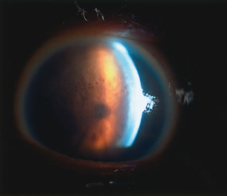
The transparency of the normal epithelium is the result of the homogeneity of the refractive index of cells throughout this cellular layer.5 When epithelial edema occurs, the epithelium loses its homogeneity and the corneal surface becomes irregular. This surface irregularity causes a reduction in vision along with symptoms of glare, photophobia, and halos around light due to light scatter. The effect of epithelial edema on vision depends on ambient lighting conditions. In mesopic conditions, such as the ophthalmologist's examining room, there may be minimal effect on visual acuity. However, in bright light, edematous epithelium creates light scatter, with marked vision. Surface irregularities caused by epithelial edema are more damaging to vision than stromal edema or scarring. Often the influence of the epithelial surface irregularities on visual acuity is underestimated, whereas the role of stromal scarring and stromal edema is overestimated.

Stroma

The stroma makes up approximately 90% of the corneal thickness and has a composition of uniformly arranged collagen fibrils in lamellae. The collagen fibrils are surrounded by a ground substance made up primarily of glycosaminoglycans, which include keratan sulfate, dermatan sulfate, and chondroitin. The stroma is for the most part an extracellular compartment with keratocytes and nerves accounting for only 5% and 0.01% of its volume, respectively. Water makes up approximately 70% of the stromal volume.

The corneal stroma plays a primary role in maintenance of corneal shape and physical strength. Its constituents help preserve transparency in conjunction with the endothelium. Stromal edema may be caused by malfunction of the endothelium and/or epithelium. When the stroma swells, the diameter of the collagen fibrils remains constant. Swelling is caused by an increase in fluid in the ground substance, leading to an increased anteroposterior spatial separation between the lamellae of collagen fibrils.6 Because the collagen lamellae are oriented in the direction of the corneal diameter, the diameter of the cornea does not increase with corneal edema.

Endothelium

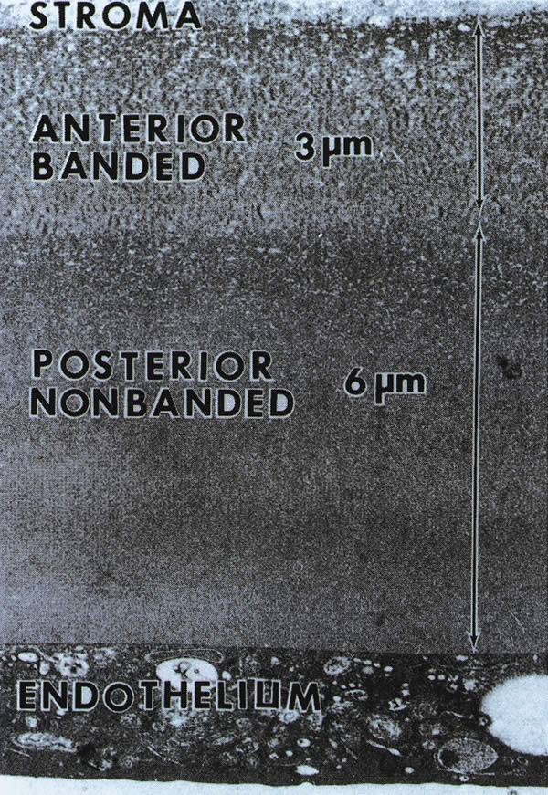
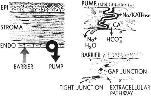
The endothelium is a monolayer of homogeneous, closely packed, polygonal cells approximately 5 μm thick. The endothelial contribution to maintenance of stromal deturgescence and transparency occurs by two mechanisms. The first mechanism is the barrier function of the endothelium between the cornea and aqueous components, a passive barrier that is much less efficient than the epithelial barrier function. The second mechanism involves the pump function of the endothelium through use of active sodium-potassium adenosine triphosphatase (Na-K ATPase) pumps that actively remove fluid that leaks into the stroma from the aqueous compartment.

The normal endothelial cell density is 3000 to 3500 cells/mm2 in the young adult. The number decreases by about two-thirds in elderly patients.7 Even though human corneal endothelial cells have been shown to proliferate in laboratory cell cultures, they have little or no ability for mitosis after birth in vivo.8 When endothelial loss occurs through aging or trauma, the endothelial response is enlargement and sliding of the existing cells to cover the area previously occupied by the lost cells. When cell loss decreases cell density below a critical number (usually 500 cells/mm2, depending on the health of the cells), corneal edema ensues.

CORNEAL HYDRATION

In addition to physical factors, the transparency of the cornea depends on its hydration status. To maintain transparency, the cornea must remain relatively thin and dehydrated. The corneal stroma of most vertebrates, including humans, swells if placed in aqueous solution because the osmotic load of the glycosaminoglycans in the stroma draws fluid into the stroma. Because the cornea swells only in the direction of its thickness, corneal thickness and hydration are linearly related. This linear relation allows measurement of corneal hydration by measurement of corneal thickness. Preservation of adequate corneal dehydration results from the following five factors: stromal swelling pressure (SP), the barrier function of the epithelium and endothelium, the endothelial pump, evaporation from the corneal surface, and the intraocular pressure (IOP). Understanding the mechanisms of corneal hydration is critical in the management of patients with corneal edema.

Stromal Swelling Pressure

An excised cornea is normally 78% hydrated, but hydration can increase up to 98% when the cornea is placed in aqueous media, with a proportional increase in its thickness. The stromal swelling is due to interfibrillary imbibition of water, not swelling of collagen fibrils. If one extracts the glycosaminoglycans from the stroma, a marked reduction in swelling occurs, indicating that glycosaminoglycans are the major cause of this hydration phenomenon.2 Keratan sulfate and chondroitin both have fixed negative charges that repel each other, and electrostatic repulsion between the negative charges appears to be a major force involved in the swelling. Because of the poorly cross-linked configuration of the collagen fibrils, the glycosaminoglycans can expand with very little resistance. This unhindered expansion results in the marked degree to which the stroma can swell.

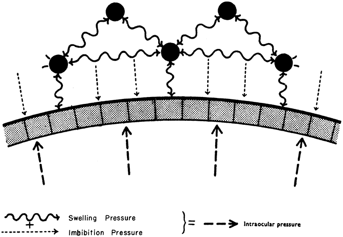
The stroma is normally maintained in a relatively dehydrated state compared with its ability to swell. The potential ability of the stroma to swell decreases as its hydration increases. The SP of the normal excised cornea is 50 mm Hg. SP is caused by the presumed anionic repulsion of the glycosaminoglycans, which expands the tissue and draws fluid into the stroma. The negative pressure drawing fluid into the cornea is called imbibition pressure (IP). In excised corneal tissue, the values of SP and IP are equal. However, in vivo, IP is lower than SP because of the compressive effect of IOP on the stroma. The following relationship establishes imbibition pressure: IP = IOP – SP. The compressing effect of IOP on the endothelium in vivo creates an IP of 30 to 40 mm Hg (Fig. 1). Water flow occurs from inside the eye as aqueous percolates into the corneal stroma and evaporation occurs from the epithelial side.

Fig. 1. Pressure relationships at the endothelium. (Doughman DJ: Corneal physiology. In Peyman GA, Sanders DR, Goldberg MF (eds): Principles and Practices of Ophthalmology. Vol 2. Philadelphia, WB Saunders, 1980. Reprinted with permission.)

Glycosaminoglycan molecules create a high resistance to water flow across the stroma in the normally dehydrated cornea. The resistance is reduced as corneal hydration increases, so that in the more edematous cornea, there is less resistance to increased water flow across the stroma. There is no lateral flow of water in the cornea except at the limbus.9

Barrier Function of Epithelium and Endothelium

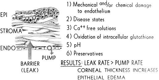
Both the epithelium and endothelium act as semipermeable membranes, creating a barrier to diffusion of electrolytes and to the flow (not diffusion) of water. The epithelium offers twice the resistance to flow of water than the endothelium, and relative resistance to diffusion of electrolytes is 200 times greater in the epithelium than in the endothelium.10 Therefore, the epithelium constitutes a relatively impermeable membrane. The ultrastructural basis for this impermeability comes from the tight junctions known as zonula occludens that seal the intercellular space between the superficial epithelial cells. By contrast, gap junctions bridge the intercellular space at the apical end of the endothelial cells.11 Permeation of small ions and water into the stroma from the aqueous is due to the semipermeable nature of the endothelium and is driven by the IOP and negative IP of the stroma. The interaction of all these forces on the hydration of the corneal stroma is shown in Figure 2. Factors that affect barrier function are listed in Figure 3.

Fig. 2. Location of corneal endothelial metabolic pump and barrier. (Waring GO, Bourne WM, Edelhauser HF, et al: The corneal endothelium. Normal and pathologic structure and function. Ophthalmology 89:546, 1982.)

Fig. 3. Factors that affect barrier functions. (Waring GO, Bourne WM, Edelhauser HF, et al: The corneal endothelium. Normal and pathologic structure and function. Ophthalmology 89:546, 1982.)

Endothelial Pump

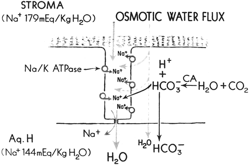
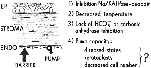
A constant amount of fluid leakage occurs across the endothelium from the anterior chamber, a finding that validates the fact that another mechanism aids in maintaining corneal deturgescence. The cornea has been observed to thicken in cooled enucleated eyes, whereas subsequent warming promotes thinning to the original thickness.12 This “temperature reversal” of hydration requires oxygen and does not occur in the presence of metabolic inhibitors, such as ouabain, which block cation transport.13,14 Deturgescence with warming suggests that there is an active transport of water and/or electrolytes from the stroma in order to maintain normal corneal hydration. Furthermore, after the epithelium has been removed, the temperature reversal effect still occurs, showing that the metabolic pumps exist within the endothelium.13 Evidence now points to ion transporter complexes within the endothelium that facilitate the passive secondary movement of water inflow.

Although the active transport of sodium and potassium ions may play a role in the endothelial pump, the major component of the pump is the active transport of bicarbonate ions into the aqueous humor.15 A negative electrical potential exists on the aqueous side of the endothelium, suggesting the bicarbonate anion, rather than the sodium or potassium cation, is responsible for the fluid transport across the endothelium.16 The endothelial pump is dependent on oxygen, glucose, carbohydrate metabolism, and ATPase (Fig. 4). Dactinomycin, ouabain, and oligomycin are potent inhibitors of this system. Factors that influence the endothelial pump function are listed in Figure 5.

Fig. 4. Corneal endothelial pump mechanism. (Waring GO, Bourne WM, Edelhauser HF, et al: The corneal endothelium. Normal and pathologic structure and function. Ophthalmology 89:546, 1982.)

Fig. 5. Factors that affect pump function. (Waring GO, Bourne WM, Edelhauser HF, et al: The corneal endothelium. Normal and pathologic structure and function. Ophthalmology 89:546, 1982.)

Evaporation From the Corneal Surface

Evaporation was shown to play a major role in the thinning of rabbit cornea.17 However, only 4% of thinning occurs in humans as a result of osmotic extraction of fluid due to tears made hypertonic by evaporation.18 This loss of fluid is readily replaced by aqueous and therefore plays little role in corneal dehydration.

Intraocular Pressure

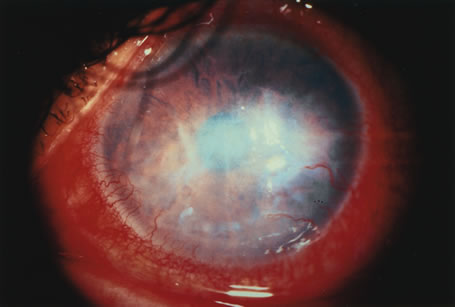
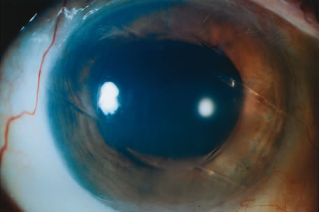
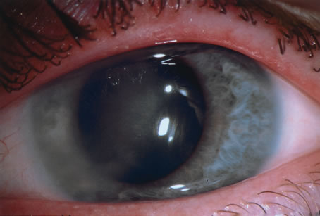
Elevated IOP can cause epithelial edema, but it is not associated with a change in corneal thickness. Ytteborg and Dohlman19,20 showed that when the IP becomes positive—that is, when the IOP exceeds the SP—epithelial edema ensues. Microcystic changes may occur in several settings. One case involves high IOP and normal stromal SP and thickness, as in glaucoma. The other setting occurs with a normal IOP and low stromal SP, such as that seen in the corneal edema of endothelial dystrophies. Stromal edema may follow microcystic epithelial changes.

In summary, maintenance of normal corneal hydration is achieved by the barrier function of both the epithelium and endothelium, preventing excessive imbibition of water from the tears and aqueous. In addition, the active transport of some anions and cations contributes to the normal corneal thickness with obligatory transport of water out of the cornea via the endothelial pump.

Back to Top
CLINICAL ENTITIES
Any insult to the cornea that compromises endothelial cell function may lead to corneal edema. A variety of factors, such as trauma, inflammation, degenerations, or dystrophies, have the potential to disturb the endothelium. Various causes of cornea edema are listed in Table 1 according to etiology. The common denominator for all of these conditions is clinical stromal and/or epithelial edema, which may be caused by endothelial dysfunction or by physiologic situations that exceed the barrier and pump capacity of the endothelium.


Table 1. Clinical Entities of Corneal Edema

Corneal edema with normal endothelium
   Persistent epithelial defect
   Increased intraocular pressure
Primary endothelial disease
   Endothelial dystrophies
      Fuchs' endothelial dystrophy
      Posterior polymorphous dystrophy
      Congenital hereditary endothelial dystrophy
   Iridocorneal endothelial syndrome
Mechanical trauma to the endothelium
   Cataract surgery
      Preexisting endothelial disease
      Surgical trauma
         Intraoperative mechanical trauma
         Postoperative trauma
            Vitreous touch
            Intraocular lens
            Brown-McLean syndrome
   Vitreoretinal surgery
   Refractive surgery
   Corneal trauma/intraocular foreign body
Nonmechanical damage to the endothelium
   Chemical injury
   Inflammation
   Increased intraocular pressure
   Contact lenses


CORNEAL EDEMA WITH NORMAL ENDOTHELIAL FUNCTION

Transient corneal edema may occur with an anatomically correct and physiologically functioning endothelium in transient circumstances. These situations develop because the corneal stroma accumulates more fluid than normal. If an influx of fluid into the stroma overcomes the effective ability of the endothelial pump to deturgesce the stroma, corneal edema may occur and may remain as long as the underlying source of edema persists.

Persistent Epithelial Defect

Persistent epithelial defects represent one way in which corneal edema occurs despite a normally functioning endothelium. Chronic, persistent epithelial defects may initially present with decreased vision from corneal edema localized to the area of the defect, with or without infectious keratitis. It should be noted that infectious keratitis could alter the pH of its local surroundings, exacerbating corneal edema.

Because the epithelium serves as a semipermeable membrane that is 200 times more impermeable than the endothelium, a barrier toward diffusion of electrolytes and to the flow of fluid from the tear film into the stroma exists. If the epithelium is removed and replaced with a glued-on rigid contact lens, no increase in stromal thickness is seen.21 The etiology of corneal stromal edema in persistent epithelial defects is associated with abnormal lamellar structure due to imbibed fluid from the tear film. Later stages may be the result of collagenolysis from tear film enzymes. The loss of the epithelial barrier, pH alterations, and local collagenolysis can retard the normal deturgescent forces of an otherwise normal endothelium.

Increased Intraocular Pressure

Another example of corneal edema in the face of an intact and functional endothelium occurs in acute glaucoma. In this instance, elevated IOP combined with normal stromal SP can create an increase in corneal thickness. With normal endothelial cell function, elevated IOP may initially compress the stroma. However, persistent elevated IOP drives fluid across the endothelium, creating edema of both the epithelium and stroma, as seen in acute angle-closure glaucoma. Although increased IOP in itself does not disrupt the barrier function, a greater pressure gradient forces more fluid across an intact barrier and decreases the efficiency of the endothelial pump.22

PRIMARY ENDOTHELIAL DISEASE

Primary diseases of the endothelium in the absence of trauma can compromise endothelial function by reducing the effectiveness of both the barrier and the endothelial pump function. These conditions may result from a decrease in endothelial cell function, cell number, or both. Entities called primary endothelial diseases are seen in patients with no history of trauma to the cornea and no previous intraocular surgery. Among this group of disorders are the primary corneal endothelial congenital and adult dystrophies and the iridocorneal endothelial (ICE) syndrome.

Fuchs' Dystrophy

HISTORY.

Ernest Fuchs,23 professor and chair of ophthalmology at the University of Vienna, described bilateral central corneal clouding in 13 elderly patients in 1910. This entity he referred to as dystrophia epithelialis corneae has now become known as Fuchs' dystrophy. Fuchs suggested that changes in the endothelium of his patients were similar to those that he had noted in patients following traumatic intraocular surgery or prolonged glaucoma.23 Kraupa24 in 1920 and Vogt25 in 1921 hypothesized that the endothelial changes observed with the slit lamp may precede the epithelial disease. Kirby26 and Gifford27 referred to the disorder as an endothelial dystrophy of the cornea, whereas Stocker28 suggested the name endothelial and epithelial dystrophy of the cornea.

Fuchs' dystrophy is an inherited, bilateral but often asymmetric, noninflammatory disorder in which the corneal endothelial cells develop morphologic and functional abnormalities that lead to varying degrees of epithelial and stromal edema, pain, decreased vision, and corneal guttae. Vogt25 first coined the term corneal gutta in 1921 to describe “droplike excrescences of the posterior surface of the cornea.” Guttae (Latin: [noun] = drop; guttate [adjective] = droplike) represent collective excrescences on the endothelium seen with slit lamp biomicroscopy. Guttae may be present without corneal edema in stage 1 of Fuchs' dystrophy. They may also form secondarily to inflammatory conditions involving the cornea. However, peripheral corneal guttae, known as Hassall-Henle warts, are not associated with Fuchs' dystrophy. They occur without corneal edema or decreased endothelial cell count and are a manifestation of aging.29

CLINICAL MANIFESTATIONS.

Fuchs' endothelial dystrophy can be grouped into four recognized categories.30–32 The four stages usually evolve gradually over a period of 25 years and, like other corneal dystrophies, are bilateral yet often asymmetric.

The first stage is marked by the onset of corneal guttae, usually in the fourth decade of life. Slit lamp biomicroscopy shows central corneal guttae, a variable amount of pigment dusting on the posterior corneal surface, and a thickened, beaten-metal appearance of Descemet's membrane by direct illumination and specular reflection. The excrescences seen on the endothelial layer initially in stage 1 may become more numerous and confluent, so that individual gutta are completely lost in the beaten-metal appearance of the endothelial surface (Fig. 6). As the disease progresses, the guttae spread to the periphery. The first stage is usually only recognized retrospectively because patients have no corneal edema and are asymptomatic.

Fig. 6. Slit lamp photograph of corneal guttata from retroillumination in Fuchs' endothelial dystrophy.

In stage 2, patients have painless decrease in vision and symptoms of glare and halos around lights, which are usually more severe on awakening. These symptoms are primarily caused by varying degrees of epithelial and stromal edema. Epithelial edema can be seen as small droplets (bedewing) on slit lamp retroillumination. Decreased evaporation of tears during sleep causes increased tear osmolarity and promotes increased edema and decreased visual acuity on awakening.

Stage 3 develops after epithelial microcysts coalesce to form bullae. The bullae eventually burst and produce varying amounts of pain, hence the name bullous keratopathy (Fig. 7). Wrinkles in Descemet's membrane known as striae develop as the cornea thickens posteriorly because of stromal swelling. These striae form when the cornea swells as a result of shortening in the arc of Descemet's membrane from limbus to limbus. As the microcystic epithelial vesicles break, foreign-body sensations and severe pain ensue along with more extensive corneal epithelial disruption. Recurrent corneal erosions, microbial ulceration, and persistent pain may occur during this stage.

Fig. 7. Bullous keratopathy in stage 3 Fuchs' endothelial dystrophy.

The fourth and final stage of Fuchs' dystrophy is marked by the development of subepithelial pannus along the epithelial basement membrane (Fig. 8). The degenerative pannus markedly reduces vision, but at the same time, it reduces painful epithelial bullous formation and promotes relief from pain. Although the epithelial edema is reduced, the stromal edema persists. The scar tissue from the pannus stabilizes the epithelial layer and leads to patient comfort and pain relief despite the markedly thickened cornea.

Fig. 8. Slit lamp photograph of extensive fibrovascular pannus from stage 4 Fuchs' endothelial dystrophy.

EPIDEMIOLOGY.

Fuchs' corneal endothelial dystrophy occurs with a 3-to-1 ratio of women to men affected with symptoms. Expressivity of the disorder is more pronounced in female patients.6,32 Although the incidence of Fuchs' endothelial dystrophy has not been reported since Fuchs' original description, a number of studies have determined that 3% to 18% of all patients have asymptomatic corneal guttae.33–35 Lorenzetti and colleagues36 in 1967 studied 2002 eyes in 1016 patients, looking for a relation between guttae and Fuchs' dystrophy. They found that 31% of patients under age 40 had guttae, and 70% of patients over age 40 had corneal guttae. Of this70 %, only 3.8% had greater than stage 1 Fuchs' dystrophy and only 0.1% had epithelial edema or bullae.

Although corneal guttae have been linked with familial inheritance patterns since 1939, recent studies have confirmed the autosomal dominant inheritance of corneal guttae and Fuchs' dystrophy.32,37–41 Krachmer and colleagues42 examined the relatives of 228 patients with corneal guttae and found 38% of patients' relatives over age 40 were affected. Studies by Magovern and associates40 and Rosenblum and colleagues41 concluded that Fuchs' dystrophy is indeed an autosomal dominant disorder with 100% penetrance and variable expressivity.

HISTOPATHOLOGY.

The underlying pathologic mechanism in Fuchs' endothelial dystrophy is dysfunctional and diseased endothelial cells. This dysfunctional state can increase the corneal thickness from the normal average of 520 μm to 1000 μm or more.43 Additional investigations suggest alternative abnormalities within the cornea may add to the effects of dysfunctional endothelium in causing Fuchs' dystrophy. Aberrant apoptotic regulatory molecules within stromal keratocytes may play a role in the pathogenesis of this disease, whereas other investigators have suggested abnormal mitochondrial DNA as a possible inciting agent in Fuchs' endothelial dystrophy.44,45

In the normal cornea of a middle-aged person, Descemet's membrane is 12 μm thick and consists of type IV collagen. Descemet's membrane is composed of an anterior banded portion, which is 3 μm thick, and a posterior nonbanded portion of variable thickness. The thickness of the nonbanded component averages 9 μm in adults, but increases from 3 μm at age 20 to 10 μm at age 80 as a result of continuous secretion by the endothelium throughout life. Clinically evident corneal guttae create thickening of Descemet's membrane, viewed in prepared specimens as focal densities from confluent excrescences or warts.46

In patients with Fuchs' dystrophy, posterior nonbanded portion of Descemet's membrane is attenuated or absent because the dysfunctional endothelial cells produce abnormal banded collagen instead of normal collagen. The posterior banded layer in Fuchs'dystrophy accounts for most of the increased thickness of Descemet's membrane (Fig. 9). Bourne and colleagues47 reported that a posterior nonbanded layer is normally found in the cornea of patients older than 20 years, so the presence of a posterior banded layer suggests abnormal endothelial function starting at an early age. Diseased endothelium can secrete 110-degree banded portions in focal areas of Descemet's membrane, and consequently a localized abnormality may account for the Hassall-Henle warts and/or corneal guttae. Additional disease may result in secretion of collagen that is so disrupted that it forms a nonbanded appearance. Different areas of Descemet's membrane can reflect different mixtures of nonbanded (normal) and banded (abnormal) portions of collagen secreted by the endothelium in different areas of the same cornea.

Fig. 9. Electron microscopic image of a normal posterior cornea. In Fuchs' endothelial dystrophy, the posterior nonbanded layer is abnormal and a posterior banded portion is abnormally present.

Additional irregularities may be found in the corneal stroma of Fuchs dystrophy. Edematous widening of the interfibrillar spaces alters the structure of the collagen lamella within the stroma and decreases its transparency. In the advanced stages of Fuchs' dystrophy, vascular connective tissue is found between the epithelial basement membrane and Bowman's layer, with fingers of fibrous tissue produced by fibroblasts that protrude upward into the epithelium, and may encapsulate islands of epithelium. The fibrous tissue formation causes other complications, such as epithelial erosions, microbial ulceration, and corneal vascularization.

The thickened Descemet's membrane makes the beaten-metal appearance of the corneal endothelium seen centrally in the early stages of the disease. On specular microscopy, the endothelial cells are large and lose their hexagonal appearance. They become quite thin as the cell area increases to cover the posterior surface of the cornea with a diminishing number of cells. However, they constantly maintain a complete and intact covering over the posterior cornea. Fewer cells are present to maintain corneal deturgescence. The metabolic demands on the thinner cells become relatively increased in each cell, thus exacerbating the stress and causing corneal decompensation.

Two-dimensional scanning fluorophotometry demonstrated no difference in the endothelial permeability between patients with mild corneal guttae, advanced corneal guttae, and normal age- and sex-matched controls.48,49 Moreover, an altered Na-K ATPase pump rate in the endothelium of patients with advanced guttae was found, suggesting that a decreased pump rate may be the earliest defect seen in Fuchs' dystrophy. Subsequent studies demonstrated that Na-K ATPase pump site density is decreased as well.50–53 These studies suggest that diminished pump function, rather than increased permeability, is the cause of corneal edema in Fuchs' endothelial dystrophy.

TREATMENT.

Initial treatment for the early stages of Fuchs' dystrophy consists of medical management to alleviate symptoms. Dehydration techniques, such as desiccation with topical hyperosmotic preparations and dehumidification of the environment, may provide temporary improvement in symptoms of mild cases. Ultimately, surgical management is warranted, with penetrating keratoplasty representing the treatment of choice when the disease progresses to increased corneal edema; decreased visual acuity; and painful, recurrent corneal erosions. Other surgical options may include various posterior keratoplasty techniques that involve replacement of endothelial cells, such as posterior lamellar keratoplasty. (Treatment options for corneal edema are discussed later in the chapter.)

Posterior Polymorphous Dystrophy

HISTORY.

Posterior polymorphous dystrophy (PPMD) is an autosomal dominantly inherited, bilateral disorder that was initially described by Koeppe54 in 1916 and later characterized as a dystrophy by Theodore37 in 1939. The majority of cases are asymptomatic, with no reduction in visual acuity, but the expression of the disease is quite variable and some patients may develop stromal edema and secondary epithelial edema that causes severe and permanent impairment in visual acuity.55

CLINICAL MANIFESTATIONS.

Posterior polymorphous dystrophy can be manifested as three patterns within the cornea. The most common corneal finding is a small asymptomatic, discrete, round gray vesicular lesion within an otherwise clear cornea on slit lamp biomicroscopy. Less common lesions include band lesions and small, diffuse gray endothelial opacities. The vesicular lesions appear in the posterior cornea at the level of Descemet's membrane and the endothelium. On high magnification, the lesions appear as an indentation or “pox mark” on the endothelium. Larger geographic lesions or a coalescence of grouped vesicles may be found in the same region of the cornea and result in corneal edema (Fig. 10). The posterior corneal findings correspond to abnormal deposits of collagenous tissue on the posterior surface of Descemet's membrane, which is irregular and thin in localized areas.30,56 Additional anterior segment findings may be present in conjunction with the corneal edema, and congenital corneal edema has been reported as well.55,57,58

Fig. 10. Slit lamp photograph of epithelial and stromal edema in posterior polymorphous dystrophy.

HISTOPATHOLOGY.

The underlying pathology of PPMD is found in both Descemet's membrane and the endothelium. Electron microscopy has confirmed the presence of epithelial-like cells within focal areas of the endothelium.59–61 These cells possess multiple characteristics of epithelial cells, including shape, multilayering, presence of microvilli, cytokeratin-positive intermediate filaments, and desmosomes.56,62 Additional electron microscopy studies depict abnormalities in Descemet's membrane. PPMD has a normal anterior banded zone of 3 μm in adults, but the posterior nonbanded zone is either absent or multilaminated, resembling the anterior banded zone. Descemet's membrane also develops excavations of its posterior surface that fill with collagenous material, appearing as the vesicular lesions seen on slit lamp examination.56 Recent reports of a family with PPMD linked the disorder to the long arm of chromosome 20 (20q11).63

TREATMENT.

Most cases of PPMD involve asymptomatic patients with stable disease courses. These patients have no need for therapy. In the small number of cases with progressive, vision-threatening forms of PPMD with corneal edema necessitating surgical management, penetrating keratoplasty is the procedure of choice. In 1985, Krachmer56 reported a series of 13 PPMD patients (20 eyes) undergoing penetrating keratoplasty. The study averaged 4.75 years of follow-up and found 50% of patients attained a visual acuity of 20/40 or better and 50% had clear grafts postoperatively.

Congenital Hereditary Endothelial Dystrophy

HISTORY.

This rare corneal dystrophy was first described by Laurence64 in 1893 as “corneitis interstitialis in utero,” but became known as congenital hereditary endothelial dystrophy (CHED) in the 1960s.65–67 The bilateral, symmetric congenital disorder causes corneal opacification from limbus to limbus, without clear regions. Maumenee65 was the first to suggest that the disease occurs as a result of dysfunctional endothelial cells.

CLINICAL MANIFESTATIONS.

CHED most commonly presents as gray-white diffuse corneal clouding within the first 6 months of life (Fig. 11). The corneal diameter and IOP are normal, with no signs of inflammation or vascularization. CHED can be inherited in an autosomal dominant or autosomal recessive fashion. The autosomal recessive form presents at birth, rarely progresses, is asymptomatic, and is associated with nystagmus. The autosomal dominant type appears at age 1 or 2 and has fewer changes present at birth. It is slowly progressive, lacks nystagmus, and is commonly associated with photophobia and tearing. The stromal edema in this type is more transient in nature. The differential diagnosis should include sclerocornea, dermoids, Peter's anomaly, congenital glaucoma, forceps trauma, mucopolysaccharidoses, congenital infections, and rarely PPMD.68

Fig. 11. Slit lamp photograph of a diffusely edematous cornea in congenital hereditary endothelial dystrophy.

Additional findings in CHED may include band keratopathy, but bullae and corneal erosions due to edema are rare. There is no associated systemic disease, and genetic counseling is encouraged for asymptomatic carriers because of their risk of having offspring with severe visual loss. Although CHED is most commonly bilateral, unilateral cases may occur with affected persons maintaining good vision in the involved eye. This finding suggests that the disease has a different pathologic process from Fuchs' endothelial dystrophy.

HISTOPATHOLOGY.

The primary pathologic findings are found in Descemet's membrane and the endothelium. Abnormal collagen tissue and basement membrane material are found between the normal Descemet's membrane and the endothelium. Generally, the original anterior banded layer of Descemet's is thick and characterized by normal 110-nm bands. However, malfunction of the endothelium early in life creates a posterior collagenous material that is much more disorganized than the nonbanded collagen in normal Descemet's membrane. The collagen tissue posterior to the fetal anterior banded layer consists of large and small fibers interspersed among basement membrane material, a finding that suggests the endothelium produces original Descemet's membrane in utero and subsequently malfunctions. The endothelium in all cases of CHED is either absent, anomalous, or contains decreased numbers of endothelial cells.

TREATMENT.

If the edema from CHED is mild and nonprogressive, therapy is rarely necessary. When corneal opacification is severe enough to cause amblyopia, nystagmus, or strabismus, surgical intervention is indicated. A report of 40 patients in 1997 suggests that penetrating keratoplasty in CHED may be successful and has a higher rate of success in cases of delayed onset rather than onset at birth. Early surgical intervention is justified to prevent progression of amblyopia.69

Iridocorneal Endothelial Syndrome

HISTORY.

The ICE syndrome, a term coined by Yanoff, is best thought of as a continuum of one disease that includes the iris nevus (Cogan-Reese) syndrome, Chandler's syndrome, and essential iris atrophy. Several anecdotal reports describing unilateral glaucoma and iris abnormalities with corneal edema were made in the early 1900s. These cases were later identified as essential iris atrophy. In 1956, Chandler70 described similar cases with more corneal edema and less iris change, suggesting a variant form of essential iris atrophy referred to as Chandler's syndrome.71 Cogan and Reese72 reported cases of iris nodules in patients with findings of corneal edema, hence the birth of Cogan-Reese syndrome.

CLINICAL MANIFESTATIONS.

ICE syndrome is a unilateral, acquired disorder of the corneal endothelium, which develops in young adults. The spectrum of the disorder can be detected by slit lamp biomicroscopy and may include iris nodules, peripheral anterior synechiae, correctopia, elevated IOP, and varying degrees of iris atrophy and corneal edema (Fig. 12).

Fig. 12. Slit lamp photograph of iridocorneal endothelial syndrome. The image shows an irregular pupil with extensive central epithelial and stromal corneal edema, consistent with Chandler's syndrome.

Campbell and colleagues73 suggested that an abnormality of the corneal endothelium may be the fundamental change in all of these disorders and is responsible for the corneal edema. Dysfunctional endothelium and an abnormal membrane of endothelial cells may grow onto the iris, causing an iris nevus appearance with tufts of iris sticking up through the sheet of dysplastic endothelium. The dysplastic sheet of endothelium can contract and pull the iris stroma in its direction into the angle, effectively pulling the iris into shreds and drawing the pupil toward the contracted tissue. Chandler's syndrome, or corneal edema with increased IOP, is caused by abnormal endothelial cells from the cornea growing across the trabecular meshwork. Bourne74 showed that there can be partial involvement of a cornea with abnormal endothelial cells interspersed within populations of normal endothelial cells on specular microscopy. Specular microscopy may show a loss of cellular definition and hexagonal shape, along with increased granularity, polymorphism, and eccentric dark areas appearing in affected cells.

HISTOPATHOLOGY.

The abnormality of the corneal endothelium initially results in corneal edema and disruption of the normal endothelial cell architecture. The cells grow in a membrane and may cover the angle, causing glaucoma, or may grow onto the anterior surface of the iris and cause the classic findings of the iris nevus syndrome. The “nevi” seen in the ICE syndrome are actually spots of normal iris surrounded by the membrane. Subsequent contraction of the membrane can cause anterior synechiae and distortion of the pupil characteristic of essential iris atrophy.

TREATMENT.

Treatment of ICE syndrome is directed at controlling glaucoma in conjunction with management of corneal edema. Treatment of glaucoma is initially effective using topical aqueous suppressants, and later may require partial-thickness filtering procedures with or without a prosthetic valve.75 When the cornea becomes sufficiently opaque to impair vision, patients may get visual improvement with penetrating keratoplasty, although penetrating keratoplasty itself does not improve the IOP.76

MECHANICAL TRAUMA TO THE ENDOTHELIUM

Mechanical trauma to the endothelium results from external forces directed toward the eye. The majority of these forces are iatrogenic and can compromise function of a normal endothelium or, more frequently, disequilibrate an endothelium that may already be compromised. Intraocular surgery, such as cataract and glaucoma procedures, posterior chamber gas implantation, and posterior chamber silicone oil placement, may have damaging effects on the endothelium. Mechanical trauma may occur from corneal trauma as well. A loose intraocular lens (pseudophakodonesis), foreign body, or lens fragment can traumatize the corneal endothelium by repeated impact.

Cataract Surgery

Cataract surgery is one of the most common causes of iatrogenic corneal edema. Most corneas manifest temporary stromal edema for the first few days postoperatively because of high hydrostatic pressure from the flow of irrigating solutions. Manipulation of instruments in the eye, whether corneal or limbal, may cause localized edema at the incision site as well. In addition, inadvertent touch of instruments to the endothelium may cause localized cell lysis. Current irrigating solutions that are pH balanced with bicarbonate buffers, do not contain epinephrine, and include glutathione, are far superior to solutions used in the past. Endothelial cell loss rates of 8% or less have been reported in multiple series.77–79

Irreversible corneal edema following cataract surgery may result from a number of factors, including preexisting endothelial disease, trauma, vitreous touch, surgical technique, and use of various irrigating solutions that may not be well tolerated by the eye.80 Other iatrogenic factors causing corneal edema include surgical duration and surgical technique.

PREEXISTING ENDOTHELIAL DISEASE.

The risk of corneal decompensation from cataract surgery is increased in patients with guttae of the cornea found during the preoperative evaluation. Careful cataract extraction is usually successful as long as epithelial edema is not present and stromal thickness is less than 0.60 mm (600 μm). A cornea with a thickness greater than 0.62 mm (620 μm) has a higher likelihood of decompensating postoperatively, and a combined procedure with penetrating keratoplasty and cataract extraction should be considered if endothelial dysfunction is noted on slit lamp examination. The decision for performing a penetrating keratoplasty should never be based on corneal pachymetry alone as some normal corneas may have thickness greater than 600 μm and remain optically clear with a normal endothelium. Corneal edema should be evident on examination and the patient should report symptoms of morning blurring before the ophthalmologist considers combined corneal transplantation and cataract surgery. If in doubt, it is usually best to proceed with cataract extraction alone, with a careful informed consent. Advantages of cataract surgery alone, even if followed by corneal decompensation, include less complexity and the peace of mind that an unnecessary keratoplasty was avoided. A future combined procedure may then be performed in the second eye if corneal decompensation ensues in the previous surgical eye.

Controversy exists as to whether the best postoperative results occur with combined penetrating keratoplasty and cataract extraction, cataract surgery followed by penetrating keratoplasty, or penetrating keratoplasty followed by secondary cataract surgery. Although published series of combined procedures reported postoperative refractive results as low as 26% within 2 diopters (D) of intended target refraction, Geerards and colleages81 and Binder82 showed success with 76.5% and 65.5%, respectively, within 2 D of emmetropia.81–85 Conversely, studies on lens power calculations associated with keratoplasty showed that an effective way of reducing postoperative ametropia is to perform keratoplasty first, followed by lens extraction/lens implantation.82,86,87 Flowers and colleagues87 reported 95% of patients to be within 2 D of intended postoperative target refraction following a penetrating keratoplasty and cataract extraction with intraocular lens placement performed secondarily. Despite this success, cataract surgery following successful penetrating keratoplasty poses risks to the transplanted cornea, and reports range from 20% to 60% for graft failure in this situation. Specular microscopy of grafted corneas should be performed before cataract surgery to assess cell count, polymorphism, and polymegathism. If a combined procedure is chosen, the surgeon should make intraocular lens power calculations using the specific postoperative keratometric readings and regression formulas based on analyses of his or her own cases.81,85–87

SURGICAL TRAUMA.

Pseudophakic corneal edema is currently the leading indication for penetrating keratoplasty in the United States.88–91 Several studies reviewed indications for penetrating keratoplasty and found no cases of pseudophakic corneal edema before 1970.92–94 The prevalence of this entity rose to 15% in 1978 and was 60% in 1987. Although keratoconus was found to be the most common indication for penetrating keratoplasty in cases reviewed during the late 1970s and early 1980s, pseudophakic corneal edema became the most common indication in the mid 1980s and remains the most common.88,91,95,96

Following intracapsular cataract surgery and complicated extracapsular extractions, the physical contact of vitreous to the cornea can cause corneal edema by mechanical ballottement of the endothelium. Vitreous incarcerated into the cataract wound may cause incisional corneal edema that may progress over the entire cornea as the cell loss exacerbates an already compromised corneal endothelium. Such vitreous traction to the wound can also cause cystoid macular edema, which is discussed elsewhere. When corneal edema is identified in a postoperative eye in which the cornea had been clear and vitreous is touching the endothelium, vitrectomy is indicated. If vitrectomy is performed within several weeks of the onset of edema, the cornea may recover and penetrating keratoplasty may be avoided. The longer the vitreous remains in contact with the decompensated corneal endothelium, the less likely the corneal edema will resolve after vitrectomy.

Cataract surgery with posterior chamber lens implantation was initially associated with a 24% to 62% decrease in endothelial cell population, but surgical technique and instrumentation has improved, and recent studies have shown a 2% to 5% endothelial cell loss rate.79,97,98 Some studies have found no increase in endothelial cell loss following cataract extraction with posterior chamber intraocular lens implantation beyond that which is seen with cataract surgery and no intraocular lens implant.99 The use of closed-loop intraocular lenses was blamed for a high incidence of corneal decompensation over time from ballottement of the peripheral cornea; however, surgical trauma may have played a more important role than the implant itself in some cases. Nonetheless, this implant style has become extinct.100

Peripheral corneal edema may occur following cataract extraction. This entity was first described in 1969 following intracapsular cataract extraction and is now known as Brown-McLean syndrome (Fig. 13).101 It is most commonly found following intracapsular cataract extraction, but has also been reported following extracapsular cataract surgery, phacoemulsification, and pars plana lensectomy and vitrectomy.102 The edema typically starts inferiorly and extends circumferentially in both directions toward the superior cornea, involving 2 to 3 mm of the corneal periphery. The syndrome may progress to complete endothelial decompensation.

Fig. 13. Slit lamp photograph of peripheral corneal edema in an aphakic patient without superior corneal involvement, also known as Brown-McLean syndrome.

Apple and associates103,104 reported that the leading cause of anterior chamber intraocular lens explantation was pseudophakic corneal edema, followed by inflammation and intraocular lens–related complications. Closed-loop anterior chamber intraocular lenses comprised more than half (52%) of all lenses explanted, the most common of which were named after accomplished ophthalmologists (Leiske, Azar, Choyce, and Tennant). Closed-loop designs often stimulated inflammation, bleeding, and/or glaucoma. The end result commonly was corneal edema requiring explantation of the lens and keratoplasty. Closed-loop lenses were removed from the market several years ago (Fig. 14). The most common anterior chamber lenses used today are the open-loop, flexible, four-point fixation designs. These lenses have a low rate of explantation.104

Fig. 14. Slit lamp photograph of corneal edema associated with a rigid, closed-loop anterior chamber intraocular lens.

The prevalence of pseudophakic corneal edema is decreasing because of several factors. Surgical technique improvements and anterior chamber lens advancements have resulted in fewer complications. In addition, posterior chamber lenses have become the standard of care in modern cataract techniques and now comprise approximately 95% of the entire intraocular lens market share.105 Improved surgical technique, better microsurgical instrumentation, more biocompatible irrigating solutions, and acceptance of viscoelastic materials have contributed to less pseudophakic corneal edema. Despite these improvements, the increasing number of cataract surgery cases and an enlarging elderly population will likely make pseudophakic corneal edema a problem for some time.

Vitreoretinal Surgery

The use of silicone oil and perfluorocarbon gases in retina and vitreous surgery can cause corneal edema following retinal detachment repair procedures. One study found 50% of patients developed irreversible corneal decompensation within 2 weeks following silicone oil injection.106 Migration of oil or gas into the anterior chamber mechanically damages the endothelium and blocks aqueous nutrients from reaching the endothelium, both of which cause endothelial cell loss.107 Phakic and pseudophakic individuals with intraocular silicone oil fare better than aphakic individuals because a barrier is present to block oil or gas migration into the anterior chamber. An inferior iridectomy in cases of silicone oil retention and aphakia can limit access of silicone oil into the anterior chamber by allowing normal aqueous flow through the iridectomy site.108

Refractive Surgery

Several studies have shown that refractive surgery procedures can cause a variable amount of corneal edema. Incisional refractive surgery, such as radial keratotomy, may be associated with diurnal variation in corneal thickness due to increased corneal hydration with the lids closed at night. One study on rats following excimer laser keratectomy (photorefractive keratectomy) showed increased apoptosis of corneal keratocytes and endothelial cells for both superficial and deep stromal ablation patterns.109 Other studies have demonstrated development of transient corneal edema or interface fluid and microcystic edema following laser-assisted in situ keratomileusis (LASIK).109–111 Corneal endothelial analysis following LASIK verified pleomorphism with definite loss of endothelial cells and altered cell morphology acutely.112 Although these changes have proven transient, additional observation of similar phenomena must continue in the future as refractive surgery procedures continue to be performed in an increasing number of patients.

Corneal Trauma/Intraocular Foreign Body

External trauma to the cornea as mentioned earlier can damage the endothelium, causing corneal edema that may or may not resolve. Penetrating corneal injuries can damage the endothelium, more or less depending on the nature and extent of the injury as well as the health of the endothelium. Ballottement of a foreign body on the endothelium may cause more damage to the cornea than the initial passage of the foreign body through the cornea or the surgical trauma required to extract the foreign body.

A foreign body that has lodged in the inferior angle of the anterior chamber can cause progressive corneal edema that begins inferiorly.113 When a particle penetrates the cornea and becomes lodged in the angle, it may be difficult to see with gonioscopy or through the microscope at the time of surgery. A careful history of penetrating injury should be elicited, especially in the case of a glass foreign body. Metallic foreign bodies that enter the eye are more likely to show up on x-ray or computed tomography scan. Fresh shards of metal may be more difficult to remove than initially anticipated because the rough surface of the metal easily incarcerates into iris and stromal tissue. Optimum treatment for metallic foreign bodies is surgical removal as soon as possible.

NONMECHANICAL DAMAGE TO THE ENDOTHELIUM

Certain ophthalmic conditions unrelated to trauma can stress the endothelium and create corneal edema. Conditions such as chemical injury, uveitis, glaucoma, or hypoxia can all create edema, although if caught early, the edema may be reversed. In more advanced cases of endothelial cell damage, penetrating keratoplasty may be necessary to reestablish normal corneal physiology. Early removal of the offending agent can improve the prognosis and may reduce the need for surgical treatment.

Chemical Injury

Chemical injuries, such as those caused by acid or alkali burns, are well-known causes of corneal endothelial destruction. Acid or alkali burns that change the pH of the corneal endothelium outside of the normal range of 6.8 to 8.2 damage the plasma membrane and decrease the effectiveness of the barrier function. Likewise, acid or alkali can decrease the amount of bicarbonate buffer in the corneal stroma and temporarily or permanently reduce the pump function.

Inflammation

Anterior segment inflammation may cause endothelial dysfunction, resulting in stromal and epithelial edema. The clinical presentation and course of the inflammation depend on the severity of the inflammatory process, its duration, and the health of the endothelium. Mild uveitis in a patient with Fuchs' dystrophy or glaucoma may cause severe, irreversible corneal edema. In the presence of a healthy endothelium, mild anterior uveitis usually does not cause corneal swelling.

Chronic insidious uveitis, which is granulomatous in nature and characterized by the deposition of mutton-fat keratic precipitates on the endothelium, can cause corneal edema. Postoperative inflammation also can cause corneal edema in the early postoperative period, when the endothelium is more vulnerable.80 Chronic granulomatous uveitis that induces corneal edema may be caused by a number of diseases, including herpes simplex, herpes zoster, and sarcoidosis).

Increased Intraocular Pressure

An acute rise in IOP may exceed the barrier and pump function of the endothelium and cause corneal edema. The acute rise in pressure is usually very painful and causes visual symptoms related to the corneal edema. These symptoms may be manifested as halos around lights, glare sensitivity, and reduced visual acuity. If the pressure is not reduced, permanent damage to the endothelium may cause irreversible corneal edema. If the endothelium is already stressed and diseased, such as in postoperative cataract surgery, postoperative penetrating keratoplasty, or severe Fuchs' dystrophy, a relatively low IOP can cause corneal edema. In cases of a diseased endothelium, a pressure of 20 mm Hg may cause epithelial edema, whereas the edema may disappear if the pressure is lowered to 17 mm Hg.114 Posner-Schlossman syndrome, or elevated IOP secondary to uveitis, may cause corneal edema and pain.

Contact Lenses

Permanent or temporary contact lens wear may cause corneal edema simply because a large number of people are wearing a plethora of contact lens materials and stretching the limits of wearing guidelines for a variety of refractive errors (Fig. 15).

Fig. 15. Slit lamp photograph of corneal edema from contact lens wear.

OXYGEN TRANSMISSION.

When covered with a contact lens, the cornea can receive oxygen from three sources: oxygen dissolved in tears that float behind the lens and nourish the corneal layers, oxygen that passes directly through the contact lens to the precorneal tear film beneath the contact lens, and oxygen that passes into areas of the cornea not covered by the lens. Tight lenses that do not permit the precorneal tear film to circulate, lenses with little or no oxygen permeability, or very large contact lenses cause more corneal edema.

The original polymethyl methacrylate (PMMA) rigid contact lenses, which had a Dk value of 0, permitted very little oxygen to reach the cornea through the contact lens. The Dk value of a lens is the oxygen transmissibility coefficient, and it is determined by the average thickness and the oxygen diffusion characteristics. The permeability of the lens material to oxygen, the thickness of the lens, and the water content of the material determine the oxygen transmission characteristics of any contact lens, as seen in the following equation: Oxygen transmission = Dk/L (for a given partial pressure of oxygen), where Dk is the permeability of oxygen for a given material and L is the lens thickness. A thick lens does not permit oxygen transmissibility as well as a thin lens, and a low–water-content lens does not permit oxygen transmission as well as a high–water-content lens. Some materials, such as silicone, are particularly efficient at transmitting oxygen, even though they are hydrophobic by nature. Fluoropolymer lenses with Dk values exceeding 100 are very effective in both letting oxygen pass through the lens and circulating tear film around the lens. Soft contact lenses limit tear flow behind the lens but permit more oxygen diffusion through the lens because of the water content, which is greater than 55% in most daily-wear lenses and greater than 85% in most extended-wear lenses. Rigid lenses, with lower water content, allow oxygen to the cornea through tear flow behind the lens. Rigid lenses made of silicone, a fluoropolymer, allow both oxygen transmissibility and tear diffusion behind the lens.

CLINICAL MANIFESTATIONS.

Corneal edema due to contact lens hypoxia often presents as epithelial microcysts in the central portion of the cornea. In hyperopic lenses, the thickest portion of the lens is the center and the tear film beneath the lens center represents the longest distance from tears oxygenated by the atmosphere.

Corneal edema results from depletion of glycogen and accumulation of lactic acid within the cornea. The pH changes of lactic acid further exacerbate the endothelial barrier capacity. If the corneal edema progresses without removing the contact lenses, stromal edema, folds in Descemet's membrane, and progressive corneal thickness ensue.

TREATMENT.

Treatment of contact lens–induced corneal edema involves removing the contact lens and permitting the cornea to resume its normal metabolism. In cases of persistent edema, contact lens reevaluation is indicated. In addition, patients should be carefully evaluated for underlying pathologic processes.

Back to Top
TREATMENT OF CORNEAL EDEMA
Improvement of vision and/or reduction of corneal discomfort and pain are the goals of therapy. Both medical and surgical treatments are available, as listed in Table 2. What modality to use and when to use it depend on the cause and severity of the edema, the visual needs of the patient, and the experience of the treating physician. Generally speaking, the simplest therapeutic modality with the least risk is preferred. Most cases of corneal edema can be successfully treated either medically or surgically.


Table 2. Treatment of Corneal Edema

• Eliminate cause of corneal edema
    • Anti-inflammatory agents
    • Lower intraocular pressure
• Evaporation
• Hypertonic agents
• Soft contact lens
• Cautery of Bowman's layer
• Anterior stromal puncture
• Conjunctival flap
• Penetrating keratoplasty
    • Full-thickness penetrating keratoplasty
    • Posterior keratoplasty


CONTROL OF FACTORS CONTRIBUTING TO CORNEAL EDEMA

Anti-inflammatory Agents

Corticosteroid use, whether topical or systemic, may be helpful if inflammation is known to be the cause of corneal edema. In cases of Fuchs' dystrophy, steroids have not been shown to be efficacious in ameliorating corneal edema. A controlled trial of steroids and placebo showed no significant difference when topical dexamethasone drops were administered to patients with Fuchs' dystrophy.115 Although steroids may be beneficial in inflammatory causes of corneal edema, side effects and complications of treatment must be kept in mind. Topical corticosteroids have serious systemic side effects and sequelae as well as potential ocular side effects, such as steroid-induced ocular hypertension, cataract formation, and exacerbation of herpes simplex virus infection.

Lowering Intraocular Pressure

Elevated IOP drives fluid through the endothelium into the stroma and epithelium, a feature that can lead to corneal edema in a compromised cornea. Lowering IOP may help decrease corneal edema by reducing this effect. Corneal clearing is most dramatically seen with elevated IOP findings. In Fuchs' dystrophy with high IOP, lowering the tension from 20 mm Hg to 15 mm Hg may reduce significant corneal swelling. However, the effect is temporary, with corneal edema returning at even lower IOPs as the endothelium becomes further compromised.

Evaporation

Some patients spontaneously note that their vision is worse in the morning compared with the evening. Since the improvement during the day is due to evaporation, some patients may get relief in the morning by using a hair dryer and blowing it across the anterior surface of their corneas. Other patients notice that dry desert air improves their symptoms, whereas humid conditions exacerbate the problem. Any measure to accelerate evaporation usually improves vision in early cases of corneal edema.

Hypertonic Saline Agents

Topical hypertonic saline agents remove fluids from the cornea osmotically and often provide symptomatic relief in many patients. Sodium chloride in a 2% or 5% solution or 5% ointment is the most common agent used. Sodium chloride drops are most effective when used often, in the morning, to reduce edema early in the day. Sodium chloride ointment at bedtime can reduce the amount of edema that builds up during the night with the eyelids closed. Hypertonic drops are useful in mild forms of corneal edema and may provide patients with relief for many years.

Glycerin is an effective agent for corneal desiccation and diagnostic uses, but it causes discomfort and irritation when applied topically. The discomfort limits its chronic use as a therapeutic modality.

Soft Contact Lenses

Soft contact lenses were introduced to American ophthalmology in the late 1960s as an important advance in the treatment of pain and irritation suffered by patients with bullous keratopathy. Soft lenses relieve discomfort by acting like a bandage over the disrupted corneal epithelium, covering epithelial defects resulting from ruptured bullae; therefore, they are often called bandage contact lenses. Although bandage lenses can alleviate pain and can be worn continuously, they do not improve vision or ameliorate the etiology or course of the disease. The lenses can be used as extended-wear lenses for patients who have problems with insertion and removal, but extended-wear lenses are associated with corneal infection, sterile corneal infiltrates, and anterior uveitis from tight lens syndrome. The use of the lens on an already compromised corneal endothelium should be entertained judiciously as a therapeutic modality. Collagen shields have been used, although they usually do not last long enough to be an effective form of therapy. Disposable soft contact lenses are the most cost-effective source of bandage lenses.

Cautery of Bowman's Layer

To control bullous keratopathy, a dense fibrous barrier between the stroma and epithelium has been reported by cauterizing Bowman's layer with a handheld cautery or electrocautery.116 Cautery creates a barrier to fluid movement into the epithelium and prevents formation of painful bullae. Although visual acuity is not improved, the procedure induces fibrous ingrowth along the epithelial basement membrane to effectively control the pain of bullous keratopathy. Chandler117 described chemical cautery for this procedure.

Anterior Stromal Puncture

Anterior stromal puncture is a simple office procedure that serves as a valuable temporary treatment option for corneal edema, recurrent erosions, and bullous keratopathy. This procedure is an alternative for patients in whom penetrating keratoplasty is contraindicated or must be delayed. The goal of the procedure is to provide relief of discomfort, photophobia, and pain from symptomatic corneal edema. The procedure involves insertion of multiple needle punctures into the cornea through Bowman's layer to stimulate secure binding of the epithelium to underlying tissue and promote faster epithelial healing. Histopathologic studies performed on eyes after anterior stromal puncture suggested that the procedure promotes epithelial reattachment by stimulating the production of extracellular matrix proteins important in epithelial cell attachment to the underlying connective tissue.118,119

Conjunctival Flap

The use of a conjunctival flap is indicated in the control of chronic severe pain secondary to corneal edema if visual acuity is not a concern or penetrating keratoplasty is not indicated. The flap may be partial or complete, but partial flaps are usually not successful because of retraction from the edematous area and recurrence of discomfort. Gunderson120 described a total thin conjunctival flap as an effective modality, especially in blind eyes with pain secondary to corneal edema. Elderly and disabled patients with useful vision in one eye and disabling pain from bullous keratopathy in the fellow eye can have this procedure for comfort to stabilize the disabled eye. The postoperative care is much less involved with conjunctival flaps than with penetrating keratoplasty. Conjunctival flaps may be a reasonable alternative for patients with reduced visual potential who are considering penetrating keratoplasty.

A conjunctival flap is a complex surgical technique to perform because of the difficulty of obtaining the flap and the meticulous anchoring of the dissected flap. A thin flap is needed to avoid postoperative retraction of the conjunctiva. In addition, care must be taken during construction of the flap to prevent a buttonhole, in order to secure an intact coating. If a flap is buttonholed, it often fails, and thus should not be used. Visual acuity is usually made worse with this procedure, although preoperative vision is seldom useful before undertaking the procedure. As the conjunctival flap thins, it becomes less of a visual impediment. The corneal edema, which is unaffected by stabilizing the epithelial surface, remains and acts as the main cause of poor visual acuity. In the early postoperative period, the procedure may cause a cosmetically displeasing appearance, although with time, the flap thins and becomes less noticeable. It is difficult to view the cornea, anterior segment, or posterior pole with a Gunderson flap in place; therefore, the procedure should be aborted if a reliable fundus evaluation is necessary following flap placement. In addition, applanation tonometry is often ineffective after flap placement, but pneumotonometry and other contact pressure readings can measure IOP adequately.

Penetrating Keratoplasty

Penetrating keratoplasty is the treatment of choice for advanced corneal edema (Fig. 16). Successful penetrating keratoplasty is a relatively recent advance in the history of corneal disease. Stocker121 reported the first series with favorable results in Fuchs' dystrophy in 1952. Modern eye-banking techniques have afforded healthy transplantable tissue in reduced time, leading to more success with the procedure. Penetrating keratoplasty success rates approach 95% if success is determined by a clear corneal graft. Although the success rate remains high, potential for complications exists with any surgical procedure. Corneal astigmatism, postoperative glaucoma, macular pathology, and graft rejection remain the leading complications following penetrating keratoplasty. Corneal astigmatism has long been thought to be the most frequent of the complications from penetrating keratoplasty. The wide variety of suturing techniques that exist today help provide for visual rehabilitation within a few months of surgery, leading to better management of astigmatism.122,123 Even with these limitations, penetrating keratoplasty is still the most successful treatment for patients with corneal edema.

Fig. 16. Slit lamp photograph of a clear cornea after penetrating keratoplasty for corneal edema.

INDICATIONS

Pain.

When intractable pain persists despite medical therapeutic modalities, penetrating keratoplasty should be considered. If the patient is severely disabled, or if the eye has little visual rehabilitation potential, a conjunctival flap, anterior stromal puncture, or cautery of Bowman's layer may provide relief of pain. Penetrating keratoplasty may actually be an easier technical procedure in some instances compared with a conjunctival flap. In addition, the visual rehabilitation potential of keratoplasty creates an advantage when considering the other two aforementioned procedures.

Visual Acuity.

Enormous variability exists among surgeons regarding penetrating keratoplasty recommendations based on visual acuity decline. A variety of factors should be considered in concert with the amount of visual deterioration before deciding on a surgical treatment modality. Factors such as patient lifestyle, patient expectations, and surgical ability influence the decision to operate. Certainly, penetrating keratoplasty is warranted more for a patient with two decompensated corneas than for a patient with only one abnormal eye. A younger patient with specific job or lifestyle demands might expect better postoperative visual acuity when compared with a sedentary, elderly patient whose activities of daily living are achieved even with reduced vision. Regardless, if one eye is blind or absent, penetrating keratoplasty on the remaining eye should be postponed as long as patient comfort and function allow.

Degree of Edema.

Complete, diffuse corneal edema decreases the chance of successful penetrating keratoplasty because of tectonic changes in the structure of the eye. Edematous stroma is more difficult to suture and more likely to induce vascularization than in healthy corneal tissue. Successful surgery can still be performed if indicated, and a patient with severe corneal edema has a reasonable chance for useful vision in the absence of macular pathology or glaucoma.

CONTRAINDICATIONS.

Relatively few contraindications exist for penetrating keratoplasty in patients with corneal edema. Keratoconjunctivitis sicca, especially when associated with severe ocular surface diseases, such as ocular pemphigoid or Stevens-Johnson syndrome, prevents proper epithelial wound healing following penetrating keratoplasty; hence, the prognosis is guarded. Eyes with hypotony, as in early phthisis, will most likely result in loss of the eye with keratoplasty because of inability of aqueous production to meet the metabolic demands of the donor cornea. In addition, severe macular pathology that precludes adequate visual rehabilitation may be a relative contraindication to penetrating keratoplasty unless globe integrity is an issue. As long as corneal edema can be controlled enough to make the patient with macular disease comfortable, penetrating keratoplasty can be deferred. If corneal edema is associated with severe ocular surface disorders, the ocular surface disease should be addressed prior to proceeding with penetrating keratoplasty. Particular caution should be observed in performing penetrating keratoplasty in patients with neurotrophic corneas. A lateral tarsorrhaphy and/or punctal occlusion should be performed at the time of keratoplasty in patients with surface disease or neurotropic corneas.

Posterior Keratoplasty

Although penetrating keratoplasty is the surgical procedure of choice for replacement of diseased endothelium in cases of corneal endothelial failure, additional techniques for replacement of the posterior cornea only have been performed in hopes of alleviating some of the shortcomings from full-thickness keratoplasty procedures. Although Barraquer124 was the first to describe posterior corneal tissue transplantation under an anterior flap, Jones and Culbertson125 described endothelial lamellar keratoplasty in 1998. Azar and colleagues126 described variations of this technique in 2000 and termed the procedure microkeratome-assisted posterior. Both techniques use a microkeratome to create an anterior corneal flap and expose the posterior stromal bed. The posterior stromal bed is then trephined and excised, and the donor tissue is sutured in place. The anterior flap is secured over the transplanted tissue.125

A second technique of posterior keratoplasty pioneered by Melles and associates127–129 was further modified by Terry and Ousley130–131 in the United States and named deep lamellar endothelial keratoplasty. This procedure evades some inherent complications of full-thickness penetrating keratoplasty, such as high or irregular astigmatism, insufficient wound healing, and suture-related graft complications, by avoiding surface corneal incisions or sutures. A scleral wound is created, and a deep lamellar corneal pocket approximately three-fourths of the depth of the cornea is fashioned. The posterior stroma is trephined and excised from inside the corneal pocket with special instruments. The posterior aspect of the donor tissue is then secured in place with air tamponade in the anterior chamber, after which the scleral wound is sutured closed.130

Although these techniques are still in investigational stages, recent reports have shown success in patients following posterior keratoplasty.129,131 Both techniques allow replacement of the posterior stroma and endothelium without disrupting Bowman's layer and the anterior stroma, so postoperative corneal astigmatism is much less of a problem compared with full-thickness grafts. However, as with all lamellar procedures, interface haze may interfere with vision and best-corrected acuity may not be as good with these lamellar procedures as with penetrating grafts. Additional studies will help compare relative benefits of penetrating keratoplasty and posterior keratoplasty for treatment of irreversible corneal edema from endothelial disorders.

Back to Top
REFERENCES

1. Cogan DC: Corneal edema: Introduction. Int Ophthalmol Clin 8:523, 1968

2. Mishima S, Hedbys BO: Physiology of the cornea. Int Ophthalmol Clin 8:527, 1968

3. Klyce SD, Zadunaisky JA: Microelectrode profile of rabbit corneal epithelium. Biophys J 10:199, 1970

4. Fischbarg J: Active and passive properties of rabbit corneal endothelium. Exp Eye Res 15:615, 1973

5. Dohlman CH: The function of the corneal epithelium in health and disease. Invest Ophthalmol 10:383, 1971

6. Rosenblum P, Stark WJ, Maumenee IH, et al: Hereditary Fuchs' dystrophy. Am J Ophthalmol 90:455, 1980

7. Yee RW, Matsuda M, Schultz RO, Edelhauser HF: Changes in the normal corneal endothelial cellular pattern as a function of age. Curr Eye Res 4:671, 1985

8. Laule A, Cable MK, Hoffman CE, et al: Endothelial cell population changes of human cornea during life. Arch Ophthalmol 96:2031, 1978

9. Mishima S: Corneal thickness. Surv Ophthalmol 13:57, 1968

10. Maurice DM: Cornea and sclera. In Davison H (ed): The Eye. 3rd ed. New York, Academic Press, 1984

11. Kreutziger GO: Lateral membrane morphology ad gap junction structure in rabbit corneal endothelium. Exp Eye Res 23:285, 1976

12. Harris JE: The physiologic control of corneal hydration. Am J Ophthalmol 44:262, 1957

13. Harris JE, Nordquist LT: The hydration of the cornea. I. Transport of water from the cornea. Am J Ophthalmol 40:100, 1955

14. Maurice DM, Riley MV: The cornea. In Graymore CN (ed): Biochemistry of the Eye. New York, Academic Press, 1970

15. Kaye GI, Tice LW: Studies on the cornea. V. Electron microscopic localization of adenosine triphosphatase activity in the rabbit cornea in relation to transport. Invest Ophthalmol 5:22, 1966

16. Barfort P, Maurice D: Electrical potential and fluid transport across the corneal endothelium. Exp Eye Res 19:11, 1974

17. Sherrard ES: Full thickness keratoplasty in the rabbit: An unsatisfactory index of donor integrity. Exp Eye Res 18:135, 1974

18. Mishima SM, Maurice DM: The effect of normal evaporation on the eye. Exp Eye Res 1:46, 1961.

19. Ytteborg J, Dohlman CH: Corneal edema and ocular pressure. I. Animal experiments. Arch Ophthalmol 74:375, 1965

20. Ytteborg J, Dohlman CH: Corneal edema and intraocular pressure: II. Clinical results. Arch Ophthalmol 74:477, 1965

21. Waring GO 3rd : Corneal structure and pathophysiology. In Leibowitz HM (ed): Corneal Disorders: Clinical Diagnosis and Management, pp 3–25. Philadelphia, WB Saunders, 1984

22. Waring GO, Bourne WM, Edelhauser HF, et al: The corneal endothelium: Normal and pathologic structure and function. Ophthalmology 89:531, 1982

23. Fuchs E: Dystrophia epithelialis corneae. Albrecht von Graefes Arch Klin Exp Ophthalmol 76:478, 1910

24. Kraupa E: Pigmentierung der hornhauthinterflauche bei dystrophia epithelialis (Fuchs'). Z Augenheilkd 44:247, 1920

25. Vogt A: Weiter ergenbnisse der spaltlampenmikroskopie des vordern bulbusabschnittes. Albrecht von Graefes Arch Klin Exp Ophthalmol 106:63, 1921

26. Kirby DB: Excrescences of the central area of Descemet's membrane. Arch Ophthalmol 54:588, 1925

27. Gifford SR: Epithelial dystrophy of the cornea and its relation to endothelial dystrophy. Am J Ophthalmol 9:81, 1926

28. Stocker FW: The endothelium of the cornea. Trans Am Ophthalmol Soc 51:669, 1953

29. Hogan MJ, Zimmerman LF (eds): Ophthalmic Pathology, pp 317–331. Philadelphia, WB Saunders, 1962

30. Waring GO, Rodrigues MM, Laibson PR: Corneal dystrophies. II. Endothelial dystrophies. Surv Ophthalmol 23:147, 1978

31. Stocker FW: The Endothelium of the Cornea and Its Clinical Implications, pp 97–100. 2nd ed. Springfield, IL: Charles C Thomas Publisher, 1971

32. Adamis AP, Filatov V, Tripathi BJ, Tripathi RC: Fuchs' endothelial dystrophy of the cornea. Surv Ophthalmol 38:149, 1993

33. Goar EL: Dystrophy of the corneal endothelium (cornea guttata). Am J Ophthalmol 17:215, 1934

34. Moeschler H: Untersuchengen ueber pigmentierung der hornhautrueckflaeche bei 395 am spaltlampenmikroskop untersuchten augen gesunder personen. Z Augenheilkd 48:195, 1922

35. Kaufman HE, Capella JA, Robins JE: The human corneal endothelium. Am J Ophthalmol 61:835, 1966

36. Lorenzetti DW, Uotila MH, Parikh N, et al: Central cornea guttata. Incidence in the general population. Am J Ophthalmol 64:1155, 1967

37. Theodore FH: Congenital type of endothelial dystrophy. Arch Ophthalmol 21:626, 1939

38. Vogt A, Wagner H, Richner H, Meyer G: Inheritance of corneal guttata. Arch Julius Klaus Stift Vererbungsforsch Sozialanthropol Rassenhyg 14:475, 1939

39. Cross HE, Maumenee AE, Cantolino SJ: Inheritance of Fuchs' endothelial dystrophy. Arch Ophthalmol 85:268, 1971

40. Macgovern, M, Beauchamp GR, McTigue JW, et al: Inheritance of Fuchs' combined dystrophy. Ophthalmology 86:1897, 1979

41. Rosenblum P, Stark WJ, Maumenee IH, et al: Hereditary Fuchs' dystrophy. Am J Ophthalmol 90:455, 1980

42. Krachmer JH, Purcell JJ, Young CW, Bucher KD: Corneal endothelial dystrophy. Arch Ophthalmol 96:2035, 1978

43. Hansen FK: A clinical study of the normal human central corneal thickness. Acta Ophthalmol (Copenh) 49:82, 1971

44. Li QJ, Ashraf MF, Shen D, et al: The role of apoptosis in the pathogenesis of Fuchs' endothelial dystrophy of the cornea. Arch Ophthalmol 119:1597, 2001

45. Borboli S, Colby K: Mechanisms of disease: Fuchs'endothelial dystrophy. Ophthalmol Clin North Am 15:17, 2002

46. Johnson DH, Bourne WM, Campbell RJ: The ultrastructure of Descemet's membrane. I. Changes with age in normal corneas. Arch Ophthalmol 100:1942, 1982

47. Bourne WM, Johnson DH, Campbell RJ: The ultrastructure of Descemet's membrane. III. Fuchs' dystrophy. Arch Ophthalmol 100:1952, 1982

48. Wilson SE, Bourne WM: Fuchs' dystrophy. Cornea 7:2, 1988

49. Wilson SE, Bourne WM, O'Brien PC, et al: Endothelial function and aqueous humor flow rate in patients with Fuchs' dystrophy. Am J Ophthalmol 106:270, 1988

50. McCartney MD, Robertson DP, Wood TO, et al: ATPase pump site density in human dysfunctional corneal endothelium. Invest Ophthalmol Vis Sci 28:1955, 1987

51. McCartney MD, Wood TO, McLaughlin BJ: Freeze-fracture label of functional and dysfunctional human corneal endothelium. Curr Eye Res 6:589, 1987

52. McCartney MD, Wood TO, McLaughlin BJ: Immunohistochemical localization of ATPase in human dysfunctional endothelium. Curr Eye Res 6:1479, 1987

53. McCartney MD, Wood TO, McLaughlin BJ: Moderate Fuchs' endothelial dystrophy ATPase pump site density. Invest Ophthalmol Vis Sci 30:1560, 1989

54. Koeppe L: Klinische beobachtungen mit der nerstspaltlampe und der hornhautmikroskop. Graefes Arch Klin Exp Ophthalmol 91:375, 1916

55. Cibis GW, Krachmer JH, Phelps CD, et al: The clinical spectrum of posterior polymorphous dystrophy. Arch Ophthalmol 95:1529, 1977

56. Krachmer JH: Posterior polymorphous corneal dystrophy: A disease characterized by epithelial-like endothelial cells which influence management and prognosis. Trans Am Ophthalmol Soc 83:413, 1985

57. Levensen JE, Chandler JW, Kaufman HE: Affected asymptomatic relatives in congenital hereditary endothelial dystrophy. Am J Ophthalmol 76:967, 1973

58. Sekundo W, Lee WR, Aitken DA, et al: An ultrastructural investigation of an early manifestation of posterior polymorphous dystrophy of the cornea. Ophthalmology 101:1422, 1994

59. Boruchoff SA, Kuwabara T: Electron microscopy of posterior polymorphous degeneration. Am J Ophthalmol 72:879, 1971

60. Rodrigues MM, Waring GO, Laibson PR, Weinreb S: Endothelial alterations in congenital corneal dystrophies. Am J Ophthalmol 80:678, 1975

61. Polack FM, Bourne WM, Forstot SL, Yamaguchi T: Scanning electron microscopy of posterior polymorphous corneal dystrophy. Am J Ophthalmol 89:575, 1980

62. Rodrigues MM, Sun TT, Krachmer J, Newsome D: Epithelialization of the corneal endothelium in posterior polymorphous dystrophy. Invest Ophthalmol Vis Sci 19:832, 1980

63. Heon E, Mathers WD, Alward WL, et al: Linkage of posterior polymorphous corneal dystrophy to 20q11. Hum Mol Genet 4:485, 1995

64. Laurence GZ: Corneitis interstitialis in utero. Klin Monat Augenheilkunde 1:351, 1893

65. Maumenee AE: Congenital hereditary corneal dystrophy. Am J Ophthalmol 50:1114, 1960

66. Keates RH, Cvintal T: Congenital hereditary corneal dystrophy. Am J Ophthalmol 60:892, 1964

67. Kenyon KR, Maumenee AE: The histological and ultrastructural pathology of congenital hereditary corneal dystrophy. A case report. Invest Ophthalmol 7:475, 1968

68. McCartney ACE, Kirkness C: Comparison between posterior polymorphous dystrophy and congenital hereditary endothelial dystrophy of the cornea. Eye 2:63, 1988

69. Al-Rajhi AA, Wagoner MD: Penetrating keratoplasty in congenital hereditary endothelial dystrophy. Ophthalmology 104:956, 1997

70. Chandler PA: Atrophy of the stroma of the iris. Am J Ophthalmol 41:607, 1956

71. Epstein DL: Chandler and Grant's Glaucoma, pp 396–402. 3rd ed. Philadelphia, Lea & Febiger, 1986

72. Cogan DG, Reese AB: A syndrome of iris nodules, ectopic Descemet's membrane and unilateral glaucoma. Doc Ophthalmol 26:424, 1969

73. Campbell DG, Shields MB, Smith TR: The corneal endothelium and the spectrum of essential iris atrophy. Am J Ophthalmol 86:317, 1978

74. Bourne WM: Partial corneal involvement in the iridocorneal endothelial syndrome. Am J Ophthalmol 94:774, 1982

75. Kidd M, Hetherington J, Magee S: Surgical results in iridocorneal endothelial syndrome. Arch Ophthalmol 106:199, 1988

76. Crawford GJ, Stulting RD, Cavanagh HD, Waring GO 3rd : Penetrating keratoplasty in the management of iridocorneal endothelial syndrome. Cornea 8:34, 1989

77. Bourne WM, Kaufman HE: Cataract extraction and the corneal endothelium. Am J Ophthalmol 82:44, 1976

78. Forstot SL, Blackwell WL, Jaffe NS, Kaufman NS: The effect of intraocular lens implantation on the corneal endothelium. Trans Am Acad Ophthalmol Otolaryngol 83:195, 1977

79. Sugar A, Fetherolf EC, Lin LL, et al: Endothelial cell loss from intraocular lens insertion. Trans Am Acad Ophthalmol Otolaryngol 85:394, 1978

80. Dohlman CH, Hyndiuk RA: Subclinical and manifest corneal edema after cataract extraction. In: Transactions of the New Orleans Academy of Ophthalmology, Symposium on the Cornea, p 214. St. Louis, CV Mosby, 1972

81. Geerards AJ, Hassmann E, Beekhuis WH, et al: Triple procedure; analysis of outcome, refraction, and intraocular lens power calculation. Br J Ophthalmol 81:774, 1997

82. Binder PS: Refractive errors encountered with the triple procedure. Trans New Orleans Acad Ophthalmol 35:111, 1987

83. Hamill MB: Sequential surgery may be the best approach for the patient. Arch Ophthalmol 118:415, 2000

84. Binder PS: Intraocular lens powers used in the triple procedure: Effect on visual acuity and refractive error. Ophthalmology 92:1561, 1985

85. Pineros OE, Cohen EJ, Rapuano CJ, Laibson PR: Triple vs nonsimultaneous procedures in Fuchs' dystrophy and cataract. Arch Ophthalmol 114:525, 1997

86. Crawford GJ, Stulting RD, Waring GO 3rd , et al: The triple procedure: Analysis of outcome, refraction, and intraocular lens power calculation. Ophthalmology 93:817, 1986

87. Flowers CW, McLeod SD, McDonnell PJ, et al: Evaluation of intraocular lens power calculation formulas in the triple procedure. J Cataract Refract Surg 22:116, 1996

88. Cosar CB, Sridhar MS, Cohen EJ, et al: Indications for penetrating keratoplasty and associated procedures, 1996–2000. Cornea 21:148, 2002

89. Lois N, Kowal VO, Cohen EJ, et al: Indications for penetrating keratoplasty and associated procedures, 1989–1995. Cornea 16:623, 1997

90. Flowers CW, Chang KY, Mcleod SD, et al: Changing indications for penetrating keratoplasty, 1989-1993. Cornea 14:583, 1995

91. Maeno A, Naor J, Lee HM, et al: Three decades of corneal transplantation: Indications and patient characteristics. Cornea 19:7, 2000

92. Smith RE, McDonald R, Nesburn AB, Minckler DS: Penetrating keratoplasty: Changing indications, 1947–1978. Arch Ophthalmol 98:1226, 1980

93. Robin JB, Gindi, Koh K, et al: An update of the indications of penetrating keratoplasty: 1979 through 1983. Arch Ophthalmol 104:87,1986

94. Polack FM: Pseudophakic corneal edema: An 11-year study of its development, incidence, and treatment. Cornea 8:306, 1989

95. Mamalis N, Anderson CW, Kreisler KR, et al: Changing trends in the indications for penetrating keratoplasty. Arch Ophthalmol 110:1409, 1992

96. Lindquist TD, McGlothan JS, Rotkis WM, Chandler JW: Indications for penetrating keratoplasty: 1980-1988. Cornea 10:210, 1991

97. Bourne WM, Kaufman HE: Endothelial damage associated with intraocular lenses. Am J Ophthalmol 81:482, 1976

98. Jaffe NS, Jaffee MS, Jaffe GF: Cataract Surgery and Its Complications, pp 249–252. 5th ed. St. Louis, CV Mosby, 1990

99. Liesegang TJ, Bourne WM, Ilstrup DM: Short- and long-term endothelial cell loss associated with cataract extraction and intraocular lens implantation. Am J Ophthalmol 97:32-39, 1984

100. Busin M, Arffa RC, McDonald MB, Kaufman HE: Intraocular lens removal during penetrating keratoplasty for pseudophakic bullous keratopathy. Ophthalmology 94:505, 1987

101. Brown SI, McLean JM: Peripheral corneal edema after cataract extraction: A new clinical entity. Trans Am Acad Ophthalmol Otolaryngol 73:465, 1969

102. Gothard TW, Hardten DR, Lane SS, et al: Clinical findings in Brown-McLean syndrome. Am J Ophthalmol 115:729, 1993

103. Solomon KD, Apple DJ, Mamalis N, et al: Complications of intraocular lenses with special reference to an analysis of 2500 explanted intraocular lenses (IOLs). Eur J Implant Refract Surg 3:195, 1991

104. Lim ES, Apple DJ, Tsai JC, et al: An analysis of flexible anterior chamber lenses with special reference to the normalized rate of lens explantation. Ophthalmology 98:243, 1991

105. Stark WJ, Sommer A, Smith RE: Changing trends in intraocular lens implantation. Arch Ophthalmol 107:1441, 1989

106. Paris MP, Peyman GA, Kao GW: Early anterior segment complications after silicone oil injection. Can J Ophthalmol 21:271, 1986

107. Sternberg P Jr , Hatchell DL, Foulks GN, Landers MB 3rd : The effect of silicone oil on the cornea. Arch Ophthalmol 103:90, 1985

108. Madrepla SA, McCuen BW 2nd : Inferior peripheral iridectomy in patients receiving silicone oil. Rates of postoperative closure and effect on oil position. Retina 15:87, 1995

109. Li Q, Ashraf MF, Bekoe NA, et al: The role of apoptosis in the early corneal wound healing after excimer laser keratectomy in the rat. Graefes Arch Clin Exp Ophthalmol 238:853, 2000

110. Protellinha W, Kuchenbuk M, Nakano K, Oliveira M: Interface fluid and diffuse corneal edema after laser in situ keratomileusis. J Refract Surg 17:S192, 2001

111. Fogla R, Rao SK, Padmanabhan P: Interface fluid after laser in situ keratomileusis. J Cataract Refract Surg 27:1526, 2001

112. Kim T, Sorenson AL, Krishnasamy S, et al: Acute corneal changes after laser in situ keratomileusis. Cornea 20:597, 2001

113. Laibson P: Inferior bullous keratopathy. Arch Ophthalmol 74:191, 1965

114. Boruchoff SA: Clinical causes of corneal edema. Int Ophthalmol Clin 8:581, 1968

115. Wilson SE, Bourne WM, Brubaker RF: Effect of dexamethasone on corneal endothelial Fuchs' dystrophy. Invest Ophthalmol Vis Sci 29:357, 1988

116. DeVoe AG: Electrocautery of Bowman's membrane. Arch Ophthalmol 76:768, 1966

117. Chandler PA: Discussion. Trans Am Ophthalmol Soc 64:119, 1964

118. Hsu JKW, Rubinfield RS, Barry P, et al: Anterior stromal puncture. Immunohistochemical studies in human cornea. Arch Ophthalmol 111:1057, 1993

119. Sridher MS, Vemuganti GK, Bansal AK, Rao GN: Anterior stromal puncture in bullous keratopathy: A clinicopathologic study. Cornea 20:573, 2001

120. Gunderson T: Conjunctival flaps in the treatment of corneal disease with reference to a new technique of application. Arch Ophthalmol 60:880, 1958

121. Stocker FW: Successful corneal grafts in the care of endothelial and epithelial dystrophy. Am J Ophthalmol 35:349, 1952

122. Van Meter WS, Gussler JR, Solomon KD, Wood T: Postkeratoplasty astigmatism control: Single continuous suture adjustment versus selective interrupted suture removal. Ophthalmology 98:177, 1991

123. Binder PS: The effect of suture removal on postkeratoplasty astigmatism. Am J Ophthalmol 105:637, 1988

124. Barraquer J, Rutllan J (eds): The technique for penetrating keratoplasty. In: Microsurgery of the Cornea: An Atlas and Textbook, pp 289–294. Barcelona, Ediciones Scriba, 1984

125. Jones DT, Culbertson WW: Endothelial lamellar keratoplasty (ELK) [ARVO abstract 342]. Invest Ophthalmol Vis Sci 39:S76, 1998

126. Azar DT, Jain S, Sambursky R: A new surgical technique of microkeratome-assisted deep lamellar keratoplasty with a hinged flap. Arch Ophthalmol 118:1112, 2000

127. Melles G, Eggink F, Lander F, et al: A surgical technique for posterior lamellar keratoplasty. Cornea 17:618, 1998

128. Melles G, Rietveld F, Beekhuis W, et al: A technique to visualize corneal incisions and lamellar dissection depth during surgery. Cornea 18:80, 1999

129. Melles G, Lander F, vonDooren B: Preliminary clinical results of posterior lamellar keratoplasty. Presented at the 103rd Annual Meeting of AAO, Orlando, FL, October 25, 1999 (program page 242).

130. Terry MA, Ousley PJ: Endothelial replacement without surface corneal incisions or sutures: Topography of the deep lamellar endothelial keratoplasty procedure. Cornea 20:14, 2001

131. Terry MA, Ousley PJ: Deep lamellar endothelial keratoplasty in the first United States patients: Early clinical results. Cornea 20:239, 2001

Back to Top