Chapter 14
Diagnosis and Treatment of Tear Deficiencies
MICHAEL A. LEMP
Main Menu   Table Of Contents

Search

DEFINITION AND CLASSIFICATION OF DRY EYE STATES
DIAGNOSTIC METHODS
MANAGEMENT OF DRY EYE
REFERENCES

The diagnosis and the treatment of tear deficiencies have proven extremely challenging to ophthalmologists. These conditions give rise to symptoms in patients that can be vague and include such sensations as dryness, foreign body sensation, grittiness, a sandy feeling, itching, and burning. Patients with dry eyes are extremely sensitive to their environment; symptoms increase in conditions of low humidity and wind. Both physicians and patients need to realize that they are dealing with a group of chronic conditions for which, as yet, a definitive cure is elusive. At present, the therapeutic goal is to control the disease process, to preserve vision and provide comfort.

Dry eye is a common disorder affecting a significant percentage of the population, particularly those older than 40 years, throughout much of the world. The prevalence is not well documented throughout the age spectrum but is thought to increase with aging. One type of dry eye, Sjögren's syndrome-associated dry eye, is thought to affect 1% to 2% of the population.1 Evaporative dry eye, primarily meibomian gland dysfunction, is an extremely common type of dry eye and was a factor in up to two thirds of patients who had ocular irritation in a clinical setting.2 Dry eye-associated ocular surface disease is one of the major reasons patients visit ophthalmologists.3

During the past three decades, there has been a remarkable increase in knowledge about the processes that cover normal tear secretion and maintenance of the ocular surface, particularly the pathophysiologic processes that affect these two intimately connected ocular components. Thinking about the pathogenesis of dry eye has shifted slightly from the concept that it is simply a deficiency in the volume of aqueous tears secreted by the main and accessory lacrimal glands.4 It is now known that there are qualitative changes in tear production, particularly in conditions associated with chronic ocular surface inflammation (e.g., vitamin A deficiency, ocular pemphigoid, and Stevens-Johnson syndrome), leading to a dropout of the mucin-producing goblet cells of the conjunctiva.5 This loosely adherent mucin blanket consisting of hydrated glycoproteins plays a role in maintaining the hydration of the ocular surface and contributes to the stability of the tear film. In addition, the epithelial cells of the conjunctival surface produce a mucin-like glycoprotein that adds to the complexity of the role of mucin in the tear film.6

Tears play an important role in maintaining the normal health of the ocular surface and in the repair of injury to the ocular surface. Tears have a role in the regulation of hydration of the cornea by way of tonicity changes secondary to evaporation from the tear film; an osmotic gradient develops across the cornea because of evaporation from the tear film, resulting in movement of fluid from the aqueous to the cornea and to the tear film.

Tears provide the corneal epithelium with its primary source of oxygen. Oxygen is obtained from the atmosphere and dissolved in the tears to support the aerobic metabolism of the corneal epithelium. A small amount of oxygen may be supplied to the peripheral cornea via the limbal circulation. In addition, tears act as a lubricant, facilitating the smooth interaction between the upper lid and the ocular surface. Tears contain several water-soluble substances with antibacterial properties. These include lysozyme, beta-lysine, and lactoferrin. The concentration of these substances in aqueous tears has been used as a marker for tear production.

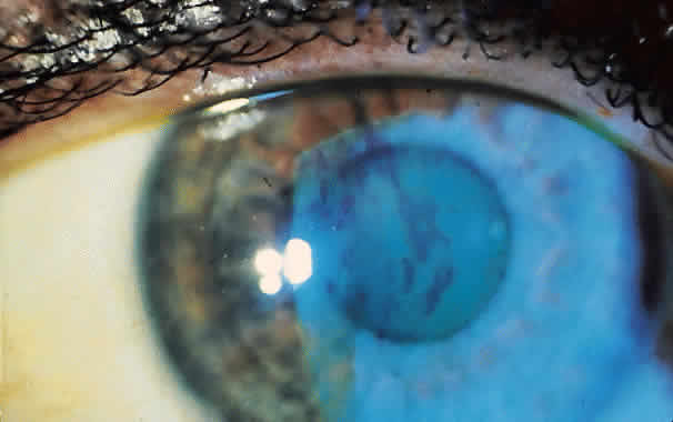
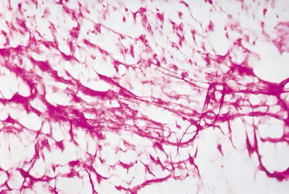
The movement of tears over the ocular surface serves to flush the surface of the eye, removing exfoliated cells, debris, and foreign bodies. This material is further entrapped in a mucin network on the ocular surface7 (Fig. 1). There has been general agreement that the tear film consists of a three-layer structure, with two layers resting on a semisolid mucin layer. The tear film thickness is approximately 7 to 10 μm. The outer layer is composed of lipid and originates primarily from the meibomian glands of the lids (Fig. 2); the glands of Zeis and Moll also contribute. The outer lipid layer of the precorneal tear film can be noted by microscopic observation of the interference patterns seen on the surface of the tear film. The thickness of this lipid layer varies considerably, depending on the interpalpebral area. As the space between the upper and lower lid is narrowed, the lipid layer is compressed. It has been reported to vary from 800 to more than 2000 nm in thickness. The lipid layer retards evaporation from the tear film, prevents contamination of the tear film by more polar liquids secreted by the sebaceous glands of the eyelids, and serves to thicken the tear film by drawing water across the ocular surface. The aqueous layer is 6 to 10 μm thick and constitutes some 90% of the preocular tear film. It is thought that the bulk of aqueous tears arise from the orbital and palpebral portions of the main lacrimal gland. The accessory lacrimal glands, varying considerably in number and weight, are scattered near the upper fornix and open on the conjunctival surface. Their contribution to tear volume varies greatly from one person to another. In addition, there is transconjunctival fluid transport; the contribution of transconjunctival fluid secretion to tear volume is unknown, but some researchers believe it approximates 10% of total volume.

Fig. 1. Mucin network on conjunctival surface stained with periodic acid-Schiff. (Courtesy of A. Adams, MD, Edinburgh.)

Fig. 2. Posterior lid view of structure of meibomian glands of the eyelids. (Snell RS, Lemp MA: Clinical Anatomy of the Eye. 2nd ed. Boston: Blackwell, 1998.)

The aqueous portion of the tears is secreted as an isotonic or slightly hypotonic solution. Aqueous tears flow out of the ductule openings of the main accessory lacrimal glands in the superior fornix. This fluid flows into the forniceal spaces and from there into the lacrimal rivers along the lid margins and over the exposed portions of the cornea and conjunctival surface. The direction of flow is toward the medial canthus. Tear flow is driven by the action of the orbicularis oculus muscle. Some aqueous fluid is lost through evaporation and reabsorption through the conjunctival surface, but most of the fluid flows out through the punctal openings into the superior and inferior canaliculi and then into the common canaliculus and down through the nasolacrimal duct. There is considerable reabsorption of fluid across the mucosa of the nasolacrimal duct during its passage. Total tear volume under relatively nonstimulated conditions is approximately 6 to 8 μl. The flow of aqueous tears has been reported to be about 1.2 μl/min. The previous distinction between basal and reflex tear secretion has been called into question.8 It is probable that all aqueous tear secretion is stimulus-driven. The main and accessory lacrimal glands have a similar histologic structure and are capable of rapidly increasing tear production in response to emotional and physical stimuli.

The aqueous layer contains the water-soluble contents of the tear film. These include inorganic salts, glucose, urea, proteins, and trace elements, all of which are secreted by the main and accessory lacrimal glands. Proteins secreted by the lacrimal glands include lysozyme, lactoferrin, free albumin, lactoperoxidase, transferrin, lipocalin, phospholipase, free albumin, and secretory IgA. Other compounds normally secreted by the lacrimal gland include epidermal growth factor, endothelin-1, basic fibroblastic growth factor, transforming growth factor α, transforming growth factor β, hepatocyte growth factor, thyroid hormone, and retinols.9,10

Lacrimal gland tissue has parasympathetic, sympathetic, and peptidergic innervation. Various substances act as neurostimulators and modulators. These include norepinephrine, acetylcholine, α-melanocyte-stimulating hormone, adrenocorticotropin, and corticotropin-like intermediate lobe peptide. Recently receptors for androgens have been identified in human lacrimal gland tissue in addition to meibomian gland tissue. This and other evidence suggests an important role for locally available bioandrogens in maintaining the normal glandular activity of both the lacrimal and meibomian glands.11

The inner loose mucin layer rests on the underlying surface of the epithelium. Mucin consists of hydrated glycoproteins. The thickness of this layer has been the subject of controversy, but it is probable that this loose mucin blanket extends well into the aqueous layer, contributing to the stability of the tear film. In addition, mucin may serve to affect the shear forces of the upper lid on the ocular surface.

The structure of the tear film is inherently unstable, and its dynamics are the result of a complex interaction between the ocular surface, the tear film itself, and the lids. The bulk of the tear volume is located in the upper and lower tear film menisci. On blinking, the superficial lipid layer is compressed to a thickness of approximately 0.1 mm adjacent to the junction of the closed lids. The underlying aqueous layer remains continuous under the closed eyelids and acts as a lubricant between the lids and the ocular surface. On opening the lids after a blink, the compressed lipid layer is spread in a monomolecular layer over the aqueous tear surface. Most of this spread occurs within 1 second of opening the eye. Between blinks, the tear film is constantly tending toward break-up secondary to evaporation and fluid retraction into the fornices. Periodic blinking resurfaces new tear film and prevents the tendency toward tear film break-up.

The maintenance of normal tear film and, indeed, the ocular surface is dependent on a critical servomechanism involving innervation of the ocular surface, lacrimal glands, and lids. These nervous innervations process signals from the ocular surface that modulate the secretion of hormones, growth factors, retinoids, and cytokines produced by both the lacrimal glands and the ocular surface. These substances in turn direct cellular turnover in the maintenance of the normal ocular surface and repair of injury to the ocular surface. In addition to the critical role of androgens in maintaining the normal glandular function of the lacrimal and meibomian glands, recent evidence points to an important role of apoptosis in maintaining normal lymphocyte populations in the lacrimal glands and in preventing inflammatory disease of the lacrimal glands. Further, dysfunction of normal apoptotic activity in the lacrimal acinar cells and the lacrimal lymphocytes has been suggested as a possible mechanism in the pathogenesis in lacrimal secretion deficiency.12

Back to Top
DEFINITION AND CLASSIFICATION OF DRY EYE STATES
In 1995, the National Eye Institute/Industry Workshop published the definition of dry eye states and a new classification (Fig. 3).13 This definition of dry eye states, “Dry eye is a disorder of the tear film due to tear deficiency or excessive tear evaporation which causes damage to the interpalpebral ocular surface and is associated with symptoms of ocular discomfort.” This definition recognizes multiple pathogenicities and the commonality of ocular surface disease underlying all of them. The classification system distinguishes two main categories of dry eye states, an aqueous tear deficiency state and an evaporative state. The aqueous tear deficiency state is further subdivided into Sjögren's syndrome-associated keratoconjunctivitis sicca (SS-KCS) and non- Sjögren's KCS (non-SS-KCS). There is controversy as to whether these two conditions represent two distinct pathogenetic processes or rather different loci on a spectrum of severity. In SS-KCS, there is characteristically intense, clinically discernible inflammatory activity on the ocular surface, in addition to that in the lacrimal glands. In non-SS-KCS (primary lacrimal deficiency), there may be a subclinical inflammatory state reflecting similar autoimmune-mediated processes resulting in inflammation in the lacrimal glands and on the ocular surface. Although the clinical presentation of these two conditions may differ, the possibility of a unified, pathogenetic mechanism offers hope for the development of a common treatment strategy.12

Fig. 3. Classification of dry eye. (Report of the National Eye Institute/Industry Workshops on Clinical Trials in Dry Eyes. CLAO J 1995;21:223.)

There are other less common causes of aqueous deficiency tear production, including the congenital abnormality of alacrima, hypoplasia of the lacrimal glands, or congenital paresis of cranial nerve VII. Another relatively rare cause is familial dysautonomia (Riley-Day syndrome). This condition occurs in Jews of eastern European origin. Both sexes are equally affected. In addition to decreased tearing, decreased corneal sensation and corneal ulceration are common. The condition is associated with a generalized dysfunction of the autonomic nervous system resulting in increased sweating, dermal discoloration, motor incoordination, cyclic vomiting, blood pressure lability, emotional difficulties, and frequent respiratory infections. Most patients with this condition have a shortened life span, frequently dying of infection.

Aqueous tear production decreases with age, but this decrease is usually insufficient to cause symptoms. It has been estimated that only 10% of normal aqueous tear production is necessary to maintain the normal ocular surface. In some people, particularly women, the deficiency can be extensive enough to lead to the development of ocular surface disease. Clinical symptoms include a scanty tear film meniscus over the inferior lid margin, increased debris in the tear film (the consequence of the decreased flushing action of tears), and frequently an increase in mucous threads in the inferior fornix. Another clinical sign associated with this condition is conjunctival pleating or conjunctivochalasis. Symptoms in primary lacrimal deficiency range from foreign body sensation irritation and grittiness, redness, and burning, to a more severe debilitating type of surface pain. Symptoms typically worsen throughout the day as evaporation from the tear film increases. Corneal surface findings include superficial punctate keratopathy. This is best seen on the cornea with the use of the water-soluble fluorescein dye and on the conjunctival surface with either a 1% rose bengal solution (Fig. 4) or lissamine green. The staining of the surface cells by rose bengal has been shown to be due to discontinuity of the mucin covering over the conjunctival surface in patients with dry eye, leading to absorption of the dye by the naked surface cells.

Fig. 4. Rose bengal staining of interpalpebral fissure in a patient with Sjögren's syndrome-associated keratoconjunctivitis sicca.

Although KCS is usually bilateral, there can be considerable asymmetry in its presentation. Unusual cases of unilateral KCS occur. These include seventh nerve paresis, viral dacryoadenitis, mechanical trauma to the upper fornix, or secondary to the surgical removal of the lacrimal glands and local irradiation. Systemic medications can diminish aqueous tear secretion. These include antihistamines, antimuscarinics, phenothiazines, mebendazole, diuretics, and beta-blockers.

Sjögren's syndrome classically consists of the triad of dry eyes, dry mouth, and arthralgia. The syndrome is thought to involve an autoimmune-mediated inflammation of the various glandular structures in the body, including the lacrimal glands, meibomian glands, and salivary glands.

When this condition occurs and there is no other discernible disease entity discovered, it is referred to as primary Sjögren's syndrome. Secondary Sjögren's syndrome is the term applied to this disease process when it occurs in association with another recognized disease, most commonly rheumatoid arthritis. Patients with Sjögren's syndrome can have multiple organ involvement including the liver, kidneys, spleen, gastrointestinal tract, lungs, thyroid, and adrenal glands. Because of the frequent involvement of the labial glands in the mouth, biopsy specimens of these glands have been used as a diagnostic test for Sjögren's syndrome.

There are various theories about the pathogenesis of Sjögren's syndrome. There is a 9:1 ratio of female to male patients, and it is thought that sex hormones, particularly estrogens, act as upregulators of immune activity in the body and probably have an important role in this gender difference.

The histologic differences in both SS-KCS and non-SS-KCS are those of generalized atrophy of the acinar and interstitial tissue of the lacrimal glands, along with varying degrees of lymphocytic and plasma cell infiltration of the lacrimal glands. In SS-KCS, the inflammatory infiltrates are particularly intense. These changes resemble those seen in other autoimmune diseases, reinforcing the impression that KCS has an autoimmune basis. There is a high prevalence of certain human leukocyte antigen (HLA) types in patients with Sjögren's syndrome, further buttressing this association. It has been suggested that certain viral infections, including Epstein-Barr, cytomegalovirus, and HIV, may play a role in the initiation of autoimmune inflammation of the lacrimal glands.14

Patients with SS-KCS represent a distinct subset of patients with more severe aqueous tear deficiencies. These patients are more prone to complications of the disease process, including severe inflammation of the ocular surface. These manifestations include scleritis (including necrotizing scleritis) (Fig. 5), rheumatoid nodules of the sclera, and corneal ulcerations, sometimes leading to perforation.15

Fig. 5. Necrotizing scleritis in a patient with secondary Sjögren's syndrome (with rheumatoid arthritis).

In some conditions, morphologic changes in the conjunctiva give rise to a decrease in the mucin secretion of the goblet cells. These include vitamin A deficiency, ocular pemphigoid, and Stevens-Johnson syndrome, as well as trachoma, chemical burns, and irradiation. In these chronic inflammatory conditions, there is cicatrization of the conjunctival surface that destroys not only the goblet cells but also the ductule openings of the main and accessory lacrimal glands and can therefore result in a decrease in the mucin component of the tear film and the aqueous component. This leads to severe tear film instability and secondary ocular surface changes.

Evaporative loss of aqueous tears is associated with dysfunction of the lipid layer. Although there are rare conditions in which there is an absence of meibomian gland secretions (e.g., congenital anhidrotic ectodermal dysplasia and an extensive injuries to the lid margins), much more common are qualitative and quantitative changes in lipid secretions associated with meibomian gland dysfunction.13,16 The lipids produced by the meibomian glands are important in the stabilization of the tear film. Abnormalities in its production have an adverse effect on tear film stability. Meibomian gland dysfunction is associated with abnormalities of excreta (meibum) thickness and volume. A qualitative shift probably occurs in the constituents of the meibum, with a change in melting point, leading to obstruction of the meibomian orifices. Alterations in lipid viscosity range from turbidity of the meibum to a more coagulated type of expression in meibomian gland dysfunction. Studies have demonstrated an association of increased evaporation leading to an increase in tear film osmolarity in obstructive meibomian gland dysfunction.17 This evaporative type of dry eye may occur in isolation or in combination with a deficiency of aqueous tear secretion. Indeed, recent studies have implicated deficiencies in bioavailable androgen as an important factor in dysfunction of both the lacrimal and meibomian glands. A recent study of Sjögren's syndrome patients found that 60% show evidence of meibomian gland dysfunction.18

Because the tear film is inherently unstable, periodic blinking is necessary to prevent disruption of the tear film, which is the ultimate consequence of the thinning that occurs between blinks. The shear force produced by the upper lids drives the removal of lipid-contaminated mucin strands and thus has a vital role in the rejuvenation of the mucous layer of the tear film. The action of the lid is also important in maintaining the normal corneal surface because it drives a preferential exfoliation of surface epithelial cells from the central area of the cornea, contributing to the centripetal movement of corneal epithelial cells and the renewal of the corneal surface.19

When normal lid movement is limited, that area of the cornea not surfaced by the lids develops a well-defined area of nonwetting, resulting in localized desiccation. Secondary changes (keratinization) eventually occur in the desiccated epithelium, making the area even less wettable. About 5% of sufferers have a poorly developed Bell's phenomenon. This reflex normally results in the upward rotation of the globe, protecting the cornea when the eyelid is closed. This portion of the population is therefore presumably at risk for the development of exposure keratitis if the lids are not completely closed. Examples of this type of exposure keratitis occur with cranial nerve VII paresis. Lid movement can also be restricted by the development of symblephara, as in ocular pemphigoid, Stevens-Johnson disease, and chemical burns. The successful maintenance of the tear film also requires reasonable congruity between the lids and the ocular surface to produce a sufficient and uniform shear with malapposition of the lid and the ocular surface. The shear diminishes the tear layers not resurfaced adequately. Localized nonwetting can also occur in contact lens wearers with incomplete blinking, giving rise to the so-called 3 and 9 o'clock peripheral corneal staining.

Because of the intimate relation between the ocular surface and the tear film, any abnormality in the surface can affect the stability of the overlying tear film. Corneal surface irregularities occur in corneal dystrophies, scars, elevations, and erosions and can produce localized drying. The normal innervation of the cornea and conjunctiva is essential in maintaining epithelial integrity. This innervation, which drives the servomechanism (see above), is important in the exquisite regulation of turnover of epithelial cells mediated by the various peptides (e.g., growth factors, hormones, cytokines) secreted by the lacrimal glands, which in turn affect the corneal surface cells by means of cell surface receptors.12 Decreases in corneal sensation and nervous innervation, as occur in lesions of the cranial nerve V and after zoster ophthalmicus, can lead to epithelial abnormalities ranging from superficial punctate erosions to coarse mucous plaques to frank corneal ulceration to corneal melting and even perforation. Abnormalities of the overlying tear film are common in these conditions.

Back to Top
DIAGNOSTIC METHODS
There is no single, readily available diagnostic test that has a high degree of sensitivity and specificity for the diagnosis of dry eye or its classification. Diagnosis, therefore, depends on the clinical history, clinical examination, and a combination of diagnostic studies, which will provide sufficient information to produce the correct clinical diagnosis. Individual methodologies will be reviewed, along with a simple algorithm for diagnosis of dry eye that allows not only accurate diagnosis but also classification.

SLIT-LAMP EXAMINATION

A careful slit-lamp examination is essential to the diagnosis of dry eye. Using the broad beam of the slit lamp, scan the ocular surface and adnexa, paying particular attention to the tear film meniscus (looking for a decrease to less than 0.1 mm in thickness), the tear film itself (looking for debris, which is a constant finding in dry eyes), the conjunctival surface (looking for increased mucous strands or conjunctivochalasis), and the corneal surface (looking for punctate erosions, coarse mucous plaques, or filamentary keratitis). It is also important to inspect carefully the inferior and superior fornices, looking for early symblephara, which may be present in diseases such as cicatrizing ocular pemphigoid or Stevens-Johnson disease (Fig. 6). Also look for coexisting meibomian gland dysfunction. Pay attention to the action of the lids, including the completeness of the blink and the presence or absence of Bell's phenomenon, looking for the presence of lagophthalmos.

Fig. 6. Cicatrizing conjunctivitis in a patient with ocular pemphigoid.

SCHIRMER'S TEST

Schirmer's test is a measure of tear secretion over a specified time. The test involves the wetting of a standardized strip of filter paper that rests over the lower lid and extends into the forniceal space. This test has been performed over the years in various ways. I prefer to perform the test without the use of topical anesthetics: anesthetics decrease some of the initial reflex tearing associated with stimulation of the conjunctival surface but do not limit the stimulation arising from touching the lid margins and lashes. The standardized strips are folded at the notch, placed in the outer third of the lower lid margins, and allowed to stay in place for 5 minutes. Patients are instructed to close their eyes to minimize eye movement.

This test has been criticized for its lack of reproducibility. Because the degree of stimulation associated with this test is quite variable, there is considerable variability in results. The test is quite useful when the results are serially consistent over several visits. Wetting of less than 5 mm of the strip after 5 minutes is normally considered to suggest an aqueous tear deficiency. If topical anesthetics are applied, a lower value of 3 mm of wetting should be used.

ROSE BENGAL STAIN

Rose bengal, a water-soluble dye, is useful in discerning desiccation damage to the ocular surface. In vitro studies show that rose bengal enters naked epithelial cells. In the clinical situation, in normal eyes there is a thick mucin blanket overlying the conjunctival surface, which protects the epithelial cells from staining with rose bengal. In dry eye, with desiccation of the ocular surface, the mucin blanket is discontinuous, allowing dye uptake. Punctate or confluent rose bengal staining, usually involving the interpalpebral area of the cornea and conjunctiva, is diagnostic of ocular surface disease. A 1% solution of rose bengal can be applied to the bulbar surface of the conjunctiva by placing a very small drop on the wooden end of an applicator stick and then touching this gently to the bulbar surface. This provides sufficient stain for diagnostic purposes without producing unpleasant irritation or overflow staining of facial skin. Rose bengal-impregnated strips, moistened with saline, can provide sufficient stain for diagnostic purposes. Alternatively, lissamine green is a stain with similar properties to rose bengal.

BREAK-UP TIME

Tear film break-up time is defined as the interval between a complete blink and the development of the first randomly distributed dry spot on the cornea.20 A time of less than 10 seconds is generally considered abnormal and suggests an unstable tear film. Mucin-deficient states especially cause a rapid break-up time, but surface abnormalities also can cause instability of the tear film and can result in a rapid break-up time (Fig. 7). Artificially induced rapid break-up times are associated with mechanically holding the lids open widely, the use of ointments that destabilize the tear film, or the use of topical anesthetics. Several devices that project grids onto the tear surface allow the measurement of tear break-up without the use of fluorescein dye.21

Fig. 7. Fluorescein tear film break-up.

TEAR FILM OSMOLARITY

Researchers have demonstrated a correlation between the increase of tear film osmolarity and the presence of KCS.22 The test for tear film osmolarity, as originally reported, involved the use of a micropipet to collect a very small sample of tears and the use of a freezing point osmometer. A commercially available device that makes this much simpler to perform has recently been marketed, but these devices are not in widespread clinical use.

Studies have demonstrated an increase in tear film osmolarity in association with aqueous deficiency tear secretion, thyroid disease, contact lens wear, and meibomian gland dysfunction (evaporative KCS). With increased evaporative loss of tears, there is a concentration of the constituents of aqueous tears. At present, the clinical use of this test is limited by the expense of the instrumentation.

LYSOZYME AND LACTOFERRIN ASSAYS

Lysozyme and lactoferrin are proteins secreted by the lacrimal glands. Both display antibacterial activity and are thought to be decreased in KCS. It is thought that this decrease parallels the secretion of aqueous tears by the lacrimal glands. Several clinical kits have been marketed for the measurement of these tear proteins, and more recently a spectrophotometric assay has been described.23–25

IMPRESSION CYTOLOGY

Conjunctival impression cytology is a relatively noninvasive diagnostic test that is useful in the diagnosis of ocular surface disease.26 Cellulose acetate filter discs are pressed onto the conjunctival surface and then removed. These discs pull off loosely attached conjunctival surface cells. After fixation and staining, the specimens are examined for morphologic abnormalities, such as a determination of goblet cell densities, squamous metaplasia, and keratinization. Studies using this technology have reported the presence of inflammatory cells on the ocular surface of patients with Sjögren's syndrome.27 This technology has been useful in delineating some of the mechanisms operative in the pathogenesis of ocular surface disease.

CONJUNCTIVAL BIOPSY

A conjunctival biopsy is sometimes useful to diagnose certain tear deficiency states in patients who have clinical evidence of symblephara formation. A biopsy may be helpful to distinguish chronic cicatricial pemphigoid from other inflammatory conditions. An ocular pemphigoid biopsy specimen can show characteristic immunohistologic and ultrastructural changes, although repeat biopsies may be necessary to establish the diagnosis.

FLUORESCEIN DILUTION TESTS

The instillation of fluorescein into the conjunctival sac and the measurement of the speed with which it is diluted constitutes an indirect measure of tear production. In the laboratory, this can be monitored precisely with a fluorophotometer. In the clinic, however, a simpler version of this test involves instilling 2% fluorescein into the conjunctival sac and observing, at the slit lamp, the rapidity with which its initial fluorescence diminishes; this provides a semiquantitative judgment of tear turnover.27

Back to Top
MANAGEMENT OF DRY EYE
Dry eye states are chronic disease states and as such challenge the management skills of the ophthalmologist and the patience of patients. The following treatment strategies have proved useful; the main objectives are to provide increased comfort and to maintain the ocular surface.

TEAR REPLACEMENT

Supplementation of tears with the use of tear substitutes is the mainstay of therapy for dry eyes. A variety of tear substitutes are available. Tear substitutes are useful in helping to hydrate the ocular surface and to increase lubrication between the lids and the ocular surface, but their utility is mainly limited by their short retention time in the conjunctival sac.28 Because the preservatives found in artificial tears can be toxic to the ocular surface, and an already compromised epithelium, as often encountered in KCS, the frequent use of preservative-containing drops can induce a significant degree of iatrogenic ocular surface disease.29 The introduction of preservative-free artificial tears has provided a major improvement in treatment. These products contain electrolytes and buffering agents and are usually either isotonic or slightly hypotonic. If the patient uses artificial tears more than four times per day, preservative-free artificial tears should be used. Recently, several formulations have been introduced that have “transiently preserved” agents. These preservatives are inactivated on contact with the tear film. Several preparations have been introduced with different electrolyte formulations and buffers; animal studies have indicated superior efficacy, but no large-scale human clinical trials have been performed on these agents.

Because aqueous artificial tears are limited by their short duration in the conjunctival sac, ointments or gels as a lubricating agent can be very useful, particularly when applied at night. These preparations increase the lubricity between the upper lid and the surface of the eye, reducing the abrasive action of the upper lid.

Sustained-release inserts and gels have been formulated in an attempt to reduce the need for frequent instillation of artificial tears. These inserts can be useful in a selected subset of patients with moderate to severe KCS. However, they tend to blur vision several hours after insertion, are difficult for some patients to handle, and are not readily available.

TEAR PRESERVATION

If frequent instillation of artificial tears proves insufficient for management and if there is objective evidence of significantly decreased tear production, occlusion of the punctum represents a logical extension of therapy.30 Both the superior and inferior puncta are important in tear drainage. Several temporary occlusive devices are available to assess the efficacy of punctal occlusion. Silicone plugs are also commercially available and can be used to occlude the puncta reversibly. Another temporary means of punctal occlusion is the application of a hot platinum spatula to the openings of the puncta, resulting in a temporary occlusion that usually lasts several days.

If it is decided that permanent punctal occlusion is desirable, the following recommendations are useful. Electrodesiccation is a simple method to effect occlusion. The entire circumference in the punctum must be desiccated to form a permanent scar. Alternatively, the argon laser has been used to effect punctal closure. It is possible with light application of the laser simply to shrink the punctal ring, thereby effecting a partial closure. Although the use of cyanoacrylate adhesives has been proposed for punctal occlusion, the adhesive does not adhere well to an intact epithelial surface and usually is extruded. The most unwanted complication of punctal occlusion is the production of a state of epiphora. To protect against this, punctal occlusion should be limited to patients who have a demonstrated Schirmer's test result of 2 mm or less on several occasions and also have evidence of ocular surface disease. Temporary closure of the inferior puncta initially to assess efficacy is recommended as a precautionary measure.

Patients with KCS are particularly sensitive to environmental changes such as low humidity and windy conditions. It is possible to fashion several types of moist chambers around the eye to decrease evaporation from the ocular surface and preserve a humid environment around the eye. Side shields for spectacles, including the use of attached moist pads, can also be quite useful. The use of humidifiers in the home can also provide significant relief from symptoms.

The use of secretagogues to increase lacrimal gland secretion is another therapeutic option. Several of these compounds have been used both topically and systemically, with some reported success. Bromhexine chloride31 and Eloisin are two such products. Tachyphylaxis has been reported to occur in some patients. Another secretagogue, 3-iso-butyl-1-methylxanthine (IBMX), has been reported to increase tear production, lower tear osmolarity, and decrease ocular surface rose bengal staining in an open-label clinical trial.32 Such a product, however, has not been marketed. Recently, improvement with a new M3 muscarinic stimulant has been reported. Salagen, a pilocarpine product taken orally, is used primarily for symptoms of dry mouth, but some patients with dry eyes have reported improvement with this agent.

CONSERVATION OF A MOIST SURFACE

The judicious but limited use of bandage contact lenses to treat ocular surface problems may be indicated in a small group of patients with KCS. Their use should be reserved for patients who have intractable filamentary keratitis and severe ocular surface disease that does not respond to alternative forms of treatment. The use of these lenses in KCS is fraught with serious risks, including infections, corneal ulcers, and annoying contact lens deposits. Bandage lenses must be kept moist with the addition of contact lens-compatible tear substitutes, and patients must be monitored closely to remove the bandage lens at the first sign of infection.

ANTI-INFLAMMATORY THERAPY

In general, the use of corticosteroids and anti-inflammatory agents has been discouraged for the treatment of dry eye conditions. In certain severe conditions, such as cicatrizing ocular pemphigoid and Stevens-Johnson disease, antimetabolites such as cyclophosphamide (Cytoxan) and azathioprine (Imuran) have been effective in halting the progress of these inflammatory processes. Studies in animal models and limited clinical experience with topical cyclosporin A have reported good results with this agent in the management of moderate to severe KCS.33,34 At this writing, a commercial product has not yet been introduced but is anticipated. Short-term corticosteroids can be useful in the treatment of patients with KCS displaying a significant degree of ocular surface inflammation.

SURFACE TREATMENT

Topical tretinoin has been used in a strategy to replace the squamous metaplasia noted in some dry eye conditions. Tretinoin is a natural retinoid and is a derivative of vitamin A. Although initial success with this product was reported in treating KCS and other dry eye conditions,35 subsequent controlled clinical studies have not confirmed this effect.36 It is possible, however, that topical tretinoin may have some place in the management of severe cicatrizing conditions such as ocular pemphigoid and erythema multiforme. Fibronectin is a component of basement membrane and may also have a role in managing KCS attended by severe ocular surface disease.

As mentioned earlier, patients with KCS have compromised ocular surface defense mechanisms. They have an increased prevalence of meibomian gland dysfunction, chronic blepharitis, or both. When these conditions lead to obstructive meibomian gland dysfunction, the induction of dry eye secondary to excessive evaporation from the tear film and increased tear film osmolarity may occur. To manage this component of the problem, it is usually necessary to resort to treatment with systemic tetracycline (250 mg twice daily) or doxycycline (50 mg twice daily). The exact mechanisms by which these systemic antibiotics are effective are incompletely understood. Both are highly lipid-soluble, however, and reach the meibomian glands in significant concentrations. Treatment for 4 to 12 weeks may be necessary to achieve maximum benefit. Patients with acne rosacea are particularly prone to meibomian gland dysfunction. These patients represent a challenge in the management of a chronic disease state, and periodic treatment with systemic antibiotics and attention to lid hygiene may be necessary.

DECREASING TEAR VISCOSITY

A subset of patients with dry eye have clinically observable, stagnant mucin in the tear film or mucous plaques and filaments. The use of a 10% to 20% solution of acetylcysteine four or five times a day can decrease the viscosity of surface mucin and can be quite effective. The usefulness of this drug is limited only in that it can be costly, irritating on application, and malodorous.

SURGERY

Older, unwieldy forms of surgical intervention in severe KCS such as parotid duct implantation have largely been abandoned. Lateral tarsorrhaphy sometimes is essential in maintaining the integrity of the ocular surface. In severe ocular surface disease, tarsorrhaphy decreases the exposed ocular surface, thereby decreasing evaporation. It immobilizes the lid, decreasing the abrasive shearing forces from the upper lid over the ocular surface. This is particularly useful in the treatment of lagophthalmos, persistent epithelial defects, and noninfectious ulcers. A partial tarsorrhaphy is preferable to a total tarsorrhaphy in most patients because it allows for greater oxygenation of the tear film from the atmosphere, providing the ocular surface with more oxygen.

FUTURE DIRECTIONS IN TREATMENT

During the next few years, we will probably see a variety of new alternative forms of treatment for dry eye or ocular surface disease states.37 There is considerable evidence to suggest that bioavailable androgens are necessary for the proper glandular activity of both the lacrimal and meibomian glands. Decreases in bioavailable androgens have been implicated in the pathogenesis of these dry eye states. The development of topical preparations to treat both aqueous tear deficiency dry eye states and evaporative dry eye states is ongoing.

Downregulation of the inflammatory process in the lacrimal glands, the ocular surface, and the meibomian glands with potent immunosuppressants such as cyclosporin offers another attractive strategy for reversing important inflammatory components of these disease processes.

Specific deficiencies of tear components have been identified in dry eyes, including tear proteins, growth factors, tear hormones, and retinols. The formulation of tear substitutes containing one or more of these components may well represent an important therapeutic innovation. The identification of two components of meibomian gland secretion that are decreased in obstructive meibomian gland disease—phosphatidylethanolamine and sphingomyelin—suggests that formulations containing these two components may be useful in preventing evaporative tear loss.

In addition to the secretagogues previously mentioned, a specific agonist for the P2Y2 purinergic receptors found on the conjunctiva has been identified. Initial in vitro work has suggested that these agents may be useful in increasing chloride and water transport across the conjunctival surface, augmenting tear volume. Cytokines that downregulate inflammation and are normally present in tears in low concentrations may represent another approach to the control of inflammation.

In this ever-increasing array of choices for the treatment of dry eye/ocular surface disease disorders, it is likely that more than one of these approaches would prove useful in managing these vexing and frequently debilitating chronic disease states.

Back to Top
REFERENCES

1. Homma M, Sugai S, Tojo T et al (eds): Sjögren's Syndrome: State of the Art. Amsterdam: Kugler Press, 1994

2. Mathers WP, Lane JA, Sutphin JE, Zimmerman MB: Model for ocular tear film function. Cornea 15:110, 1996

3. American Academy of Ophthalmology: Blepharitis and the Dry Eye in the Adult: Preferred Practice Pattern. San Francisco: AAO, 1991

4. Lemp MA: Recent developments in dry eye management. Ophthalmology 94:1299, 1982

5. Lemp MA: Mucin-deficient dry eye. Int Ophthalmol Clin 13:185, 1973

6. Gipson IK, Iantomi T: Cellular origin of mucins of the ocular surface and tear film in lacrimal gland: Tear film and dry eye syndromes. Adv Exp Med Biol 438:221, 1998

7. Holly FJ, Lemp MA: Tear physiology and dry eyes. Surv Ophthalmol 22:69, 1977

8. Jordan A, Baum JL: Basic tear flow: Does it exist? Ophthalmology 87:819, 1976

9. Wilson SE, Schultz GS, Chegini N et al: Epidermal growth factor, transforming growth factor alpha, transforming growth factor beta, fibroblast growth factor, basic growth factor and interleukin-1 proteins in the cornea. Exp Eye Res 59:63, 1994

10. Barton K, Nava A, Monroy DC, Pflugfelder SC: Cytokines and tear function in ocular surface disease in lacrimal gland, tear film and dry eye syndromes 2. Adv Exp Med Biol 438:461, 1998

11. Sullivan DH, Wickham A, Rocha EM et al: Influence of gender, sex steroid hormones and the hypothalamic-pituitary axis on the structure of the lacrimal gland in lacrimal gland, tear film and dry eye syndromes 2. Adv Exp Med Biol 438:11, 1998

12. Stern ME, Beuerman RW, Fox RI et al: A unified theory of the role of the ocular surface in dry eye in lacrimal gland, tear film and dry eye 2. Adv Med Biol 438:643, 1998

13. Lemp MA: Report of the National Eye Institute/Industry Workshop on Clinical Trials in Dry Eye. CLAO J 21:221, 1995

14. Pflugfelder SC, Crouse CA, Atherton SS: Epstein-Barr virus and the lacrimal gland pathology of Sjögren's syndrome. Adv Exp Med Biol 350:64, 1994

15. Pflugfelder SC, Wilhelmos KR, Osato MS et al: The autoimmune nature of aqueous tear deficiency. Ophthalmol 93:1513, 1986

16. Driver PJ, Lemp MA: Meibomian gland dysfunction. Surv Ophthalmol 40:343, 1996

17. Mathers WP, Shields WJ, Sachdev MS et al: Meibomian gland dysfunction and chronic blepharitis. Cornea 10:277, 1991

18. Shimazaki J, Goto E, Ono M et al: Meibomian gland dysfunction in patients with Sjögren's syndrome. Ophthalmology 105:1485, 1998

19. Lemp MA, Mathers WP: Corneal epithelial cell movement in humans. Eye 3:438, 1991

20. Lemp MA: The mucin-deficient dry eye. Int Ophthalmol Clin 13:185, 1973

21. Mengher LS, Bron AJ, Tonge SR, Gilbert DS: A non-invasive instrument for clinical assessment of the precorneal tear film stability. Exp Eye Res 4:1, 1985

22. Gilbard JP, Farris RL, Santamaria J II: Osmolarity of tear microvolumes in keratoconjunctivitis sicca. Arch Ophthalmol 96:677, 1978

23. McEwen W, Kimura S: Filter paper electrophoresis of tears I. Lysozyme and its correlation with keratoconjunctivitis sicca. Am J Ophthalmol 39:200, 1995

24. McCollum CJ, Foulks GN, Bodner B et al: Rapid assay of lactoferrin in keratoconjunctivitis sicca. Cornea 13:505, 1994

25. Tseng SCG: Staging of conjunctival squamous metaplasia by impression cytology. Ophthalmology 92:728, 1985

26. Pflugfelder SC, Huang AJ, Feuer W et al: Conjunctival cytologic features of primary Sjögren's syndrome. Ophthalmology 97:985, 1990

27. Pflugfelder SC, Tseng SC, Pepose JS et al: Chronic Epstein-Barr viral infection and immunologic dysfunction in patients with aqueous tear deficiency. Ophthalmology 97:313, 1990

28. Lemp MA: Artificial tear solutions. Int Ophthalmol Clin 13:221, 1973

29. Adams J, Wilcox MJ, Trousdale MD et al: Morphologic and physiologic effects of artificial tear formulations on corneal epithelial derived cells. Cornea 11:234, 1992

30. Dohlman CH: Punctal occlusion in keratoconjunctivitis sicca. Ophthalmology 85:1277, 1978

31. Frost-Larsen K, Isager H, Manthorpe R: Sjögren's syndrome treated with bromhexine: A randomized clinical study. Br J Med 1:1579, 1978

32. Gilbard JP, Rossi SR, Heyda KG, Dartt DA: Stimulation of tear secretion and treatment of dry eye disease with 3-isobutyl-1-methylxanthine. Arch Ophthalmol 109:972, 1991

33. Kaswan RL, Salsbury MA, Ward DA: Spontaneous canine keratoconjunctivitis sicca: A useful model for human keratoconjunctivitis sicca: Treatment with cyclosporin eye drops. Arch Ophthalmol 106:484, 1988

34. Laibovitz RA, Solch S, Andriano K et al: Pilot trial of cyclosporin 1% ophthalmic ointment in the treatment of keratoconjunctivitis sicca. Cornea 12:215, 1993

35. Tseng SCG, Maumanee AE, Stark WS et al: Topical retinoid treatment for various dry eye disorders. Ophthalmology 92:717, 1985

36. Soong HK, Martin NF, Wagoner MD et al: Topical retinoid therapy for squamous metaplasia of various ocular surface disorders. Ophthalmology 95:1442, 1988

37. Lemp MA: New strategies in the treatment of dry eye states. Cornea 18:625, 1999

Back to Top