Chapter 12
Bullous Diseases of the Skin and Mucous Membranes
BARTLY J. MONDINO, JOHN A. HOVANESIAN and UWE PLEYER
Main Menu   Table Of Contents

Search

CICATRICIAL PEMPHIGOID
ERYTHEMA MULTIFORME
OTHER BULLOUS DISEASES
REFERENCES

Bullous diseases of the skin and mucous membranes may be associated with severe ocular involvement. Of all the bullous diseases, cicatricial pemphigoid (CP) and erythema multiforme (EM) involve the conjunctiva most frequently and also cause the most severe conjunctival disease. Severe ocular involvement associated with other bullous diseases is rarely encountered in clinical practice.

There is no unifying pathogenic mechanism underlying the bullous diseases. Many bullous diseases (e.g., CP, pemphigus, bullous pemphigoid) are thought to have an autoimmune basis; epidermolysis bullosa, except for the acquired form, has a genetic basis.

Back to Top
CICATRICIAL PEMPHIGOID
The striking feature of CP is recurrent blisters or bullae of the mucous membranes and skin, with a tendency for scar formation.1 Ocular involvement is characterized by progressive conjunctival shrinkage, symblepharon, entropion with trichiasis, dry eye, and reduced vision from corneal opacification.2 The disease has no geographic or racial predilection and has been shown to be discordant in monozygotic twins.3 In a series of 108 patients, the mean age at initial examination was 70 years, with a range of 43 to 88 years (Table 1). CP affects more women than men, with ratios from 1.6:1 to 1.8:1.1,2,4


TABLE 1. Clinical Data on Patients with Cicatricial Pemphigoid


No. of patients108
Mean age (range)70 years (43–88)
Ratio of women to men1.8:1
No. of patients (%) with skin lesions17 (16%)
No. of patients (%) with lesions of oral mucosa45 (42%)


CUTANEOUS INVOLVEMENT

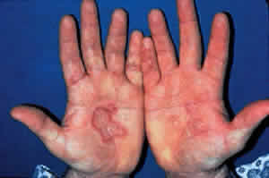
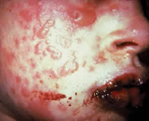
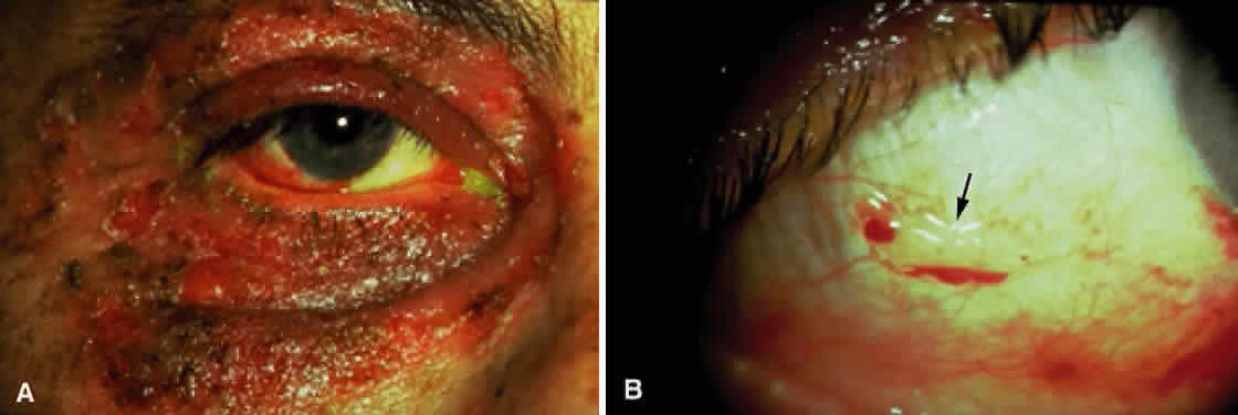
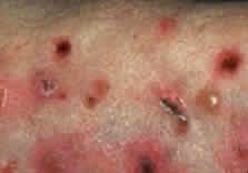
Skin involvement is found less frequently than mucuos membrane involvement (see Table 1) and is reported in 9% to 24% of cases.1–5 The skin lesions of CP may be divided into two types: (1) a recurrent, vesiculobullous, nonscarring eruption that may involve the inguinal area and extremities and occasionally becomes generalized (Fig. 1); and (2) localized, erythematous plaques with overlying vesicles and bullae that appear on the scalp and face near the affected mucous membranes and heal, leaving smooth, atrophic scars (Fig. 2).6

Fig. 1. Intact and ruptured blisters of forearm in a patient with cicatricial pemphigoid.

Fig. 2. Localized erythematous plaque, with intact and ruptured blisters on it, in a patient with cicatricial pemphigoid.

MUCOUS MEMBRANE INVOLVEMENT

Cicatricial pemphigoid may involve the following mucous membranes: conjunctiva, nose, mouth, pharynx, larynx, esophagus, anus, vagina, and urethra. In a dermatologic study, oral lesions were found in 91% and conjunctival lesions in 66% of patients with CP.5 In ophthalmic studies, 15% to 50% of patients showed involvement of the oral mucosa, whereas 100% had ocular involvement.1–4

CP is associated with two types of oral lesions.6 The first is a desquamative gingivitis that may be patchy or diffuse, heals slowly, and may persist for years. The second type is characterized by vesicles and bullae of the oral mucosa that develop rapidly, remain intact for a few days, and then rupture. Mucous membrane erosions may heal with scarring and even strictures. Stenosis of the nasopharynx or larynx may cause obstructive sleep apnea; esophageal strictures may result in asphyxiation and death when food is swallowed.7

OCULAR INVOLVEMENT

Ocular CP (OCP) is a bilateral disease. The initial symptoms are similar to those of any chronic conjunctivitis and include irritation, burning, and tearing.8 A mucopurulent discharge suggests a complicating bacterial blepharoconjunctivitis. Breakdown of the corneal epithelium leads to foreign-body sensation, photophobia, and reduced visual acuity.

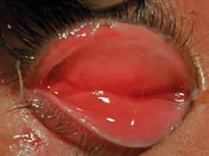
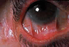
Fibrosis beneath the conjunctival epithelium is the hallmark of CP.9,10 Symblepharons, fibrotic bands that pass between the palpebral and bulbar conjunctiva, involve the inferior fornix first and are best demonstrated early in the disease by drawing the lower eyelid down and having the patient look up (Fig. 3). CP may be associated with a dry eye. Fibrosis beneath the conjunctival epithelium may cause occlusion of the ducts of the lacrimal and accessory lacrimal glands, leading to decreased aqueous tear secretion. The reduced numbers of mucus-producing goblet cells may contribute to an unstable tear film.11 Conjunctival scarring causes lagophthalmos with abnormal blinking and exposure and entropion with trichiasis and distichiasis. All these factors may cause breakdown of the ocular-surface epithelium.

Fig. 3. Symblepharon formation demonstrated by drawing lower lid down and having the patient look up.

Conjunctival or corneal bullae have been described10 but are rarely observed, perhaps because they rupture readily because of the blinking action of the eyelids. Breakdown of the corneal epithelium most commonly results from entropion with trichiasis; lagophthalmos with abnormal blinking and exposure; and a diminished, unstable tear film. These erosions may become complicated by secondary bacterial infiltrates and ulcers. Predisposing factors for the development of microbial keratitis include topical corticosteroids, bandage contact lenses, trichiasis, corneal surgery, lagophthalmos, and meibomianitis.12 Corneal neovascularization may develop either as a pannus or pseudopterygium.

Smears of conjunctival scrapings from patients with CP reveal neutrophils, keratinized squamous cells, and eosinophils.13 In a study of the bacterial flora of patients with CP, potential pathogens—mainly mannitol-positive staphylococci—were recovered from the eyelids and conjunctiva of 81% of patients with this disease.2 Glaucoma may be more prevalent in patients with CP.14

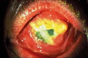
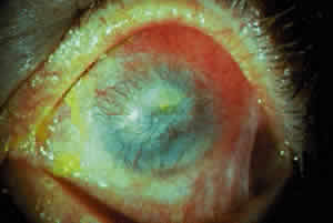
COURSE OF OCULAR DISEASE

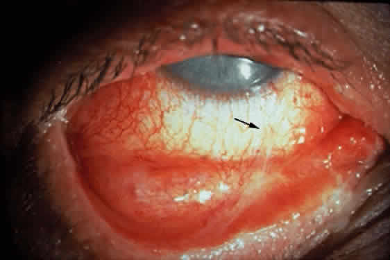
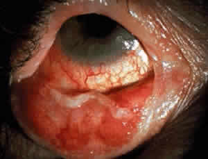
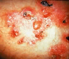
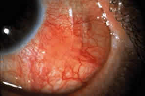
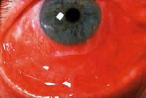
Cicatricial pemphigoid generally runs a chronic course, characterized by progressive shrinkage of the conjunctiva. When the end stage of this disease is reached, the eye lacks tears and has obliterated conjunctival fornices, ankyloblepharon, and a keratinized ocular surface epithelium. Episodes of acute disease activity may interrupt this chronic progressive course and result in rapid shrinkage of the conjunctiva.15 Acute disease activity may be precipitated by surgical procedures, including conjunctival biopsy, lysis of symblepharon, oculoplastic procedures on the eyelids, and cataract extraction. The acute manifestations consist of localized, ulcerated conjunctival mounds (Fig. 4) or diffuse, severe conjunctival hyperemia and edema (Fig. 5). Before concluding that acute inflammatory activity is caused by the disease process, it is necessary to eliminate other confounding factors such as trichiasis, exposure, or bacterial blepharoconjunctivitis.

Fig. 4. Localized ulcerated conjunctival mound indicated by slit-beam.

Fig. 5. Diffuse conjunctival hyperemia and edema as an acute manifestation of ocular cicatricial pemphigoid.

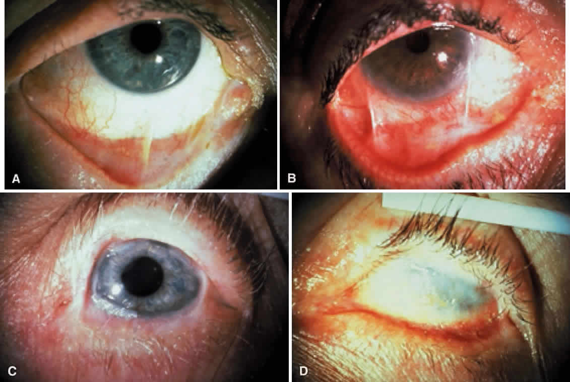
When evaluating a patient with CP, the extent of conjunctival shrinkage that has taken place should5 be determined. The most useful positions for examination and photography include upward gaze with the lower eyelid retracted and downward gaze with the upper eyelid retracted. The staging system of Mondino and Brown16 is based on the percentage of conjunctival shrinkage (Fig. 6).2 The staging system of Tauber and coworkers17 describes conjunctival destruction, as follows:

Fig. 6. Staging system of cicatricial pemphigoid. A. Stage I of cicatricial pemphigoid, showing 25% or less shrinkage of the conjunctival fornices. B. Stage II of cicatricial pemphigoid, showing 25% to 50% conjunctival shrinkage. C. Stage III of cicatricial pemphigoid showing conjunctival shrinkage of about 75%. Inferior fornix is nearly obliterated. Shallow superior fornix is still present. D. Stage IV or end stage of cicatricial pemphigoid, showing obliteration of conjunctival fornices.

  Stage I—chronic conjunctivitis and subepithelial fibrosis
  Stage II—early fornix foreshortening
  Stage III—symblephara
  Stage IV—ankyloblepharon and a frozen globe.

A patient with CP is more likely to have a dry eye, corneal vessels, trichiasis, and positive cultures of the lids and conjunctiva for potential pathogens as the disease advances in stage (Table 2).1


TABLE 2. Correlation of Clinical Data With Stage in Patients With Cicatricial Pemphigoid


StageNo. of EyesNo. With Dry Eye (%)*No. With Corneal Vessels (%)No. With Trichiasis (%)No. of Positive Cultures of Lid and Conjunctiva for Pathogens†No. Untreated Showing Progression of Conjunctival Shrinkage‡ (Mean Follow-up 2 Years)
I (25% or less shrinkage of conjunctival fornices)6519/65 (29%)20/65 (31%)38/65 (58%)22/54 (41%)13/32 (41%)
II (25% to 50% conjunctival shrinkage)6220/62 (32%)49/62 (79%)52/62 (84%)26/55 (47%)16/26 (62%)
III (approximately 75% conjunctival shrinkage)2817/28 (61%)28/28 (100%)24/28 (86%)17/25 (68%)8/11 (73%)
IV (end stage)77/7 (100%)7/7 (100%)7/7 (100%)5/5 (100%)-
All stages16263/162 (39%)104/162 (64%)121/162 (75%)70/139 (50%)37/69 (54%)

*Less than 5 mm of wetting on Schirmer filter strips after 5 minutes without topical anesthesia.
†Mannitol-positive or coagulase-positive Staphylococcus was the most prevalent potential pathogen recovered.
‡Progression defined as any increase in conjunctival shrinkage, including loss of fornix, new symblepharon formation, or extension of symblepharon.


In addition to staging the extent of conjunctival shrinkage that has taken place, it is important to assess the degree of conjunctival inflammatory activity. Stage and inflammatory activity are important parameters to assess but are not necessarily correlated. For example, an eye at stage IV may show no conjunctival inflammatory activity, whereas an eye at stage II may show severe conjunctival inflammatory activity.

Disease progression should be determined by clinical examination and comparison with previous photographs of the external eye.8,16 Progression is defined as increased conjunctival shrinkage—which may involve loss of fornix—new symblepharon formation, or enlargement of an existing symblepharon.

One study suggested that CP may be asymmetric in severity (i.e., both eyes may not be at the same stage) and progression (i.e., both eyes may not necessarily progress or fail to progress).16 Although most eyes progressed, the untreated disease had a variable course because there were eyes in all stages that did not progress. Progression was more likely to occur over a given period in the later stages (i.e., an eye at stage III was more likely to progress than an eye at stage I; see Table 2).

DRUG-INDUCED OCULAR CICATRICIAL PEMPHIGOID

Topical drugs associated with conjunctival scarring include idoxuridine,18 echothiophate iodide,19 pilocarpine,20 demecarium,20 epinephrine,21 and timolol.22 A study of drug-induced conjunctival shrinkage concluded that the spectrum of disease ranges from a self-limited toxic form to a progressive, immunologic form indistinguishable from CP.23 Patients in four studies did not have extraocular involvement of mucous membranes or skin.18–20,23 Patients with drug-induced conjunctival shrinkage may show immunoglobulins bound to conjunctival basement membrane.18,19,24 In a histopathologic study of drug-induced CP affecting the conjunctiva, light and electron microscopy of the conjunctiva revealed findings identical to those previously reported for idiopathic CP.22

There are five possible explanations offered for the relation between drugs and conjunctival scarring:

  1. The drug may cause conjunctival scarring that mimics CP but is not CP because the scarring is not progressive.
  2. The taking of a drug and the development of CP may be coincidental.
  3. The drug may promote or accelerate the development of CP that may have developed later anyway.
  4. The drug may be a cause of CP.
  5. CP may cause scarring that obstructs collecting vessels of the outflow pathway and results in glaucoma requiring drug therapy.

A practical approach to the problem of drug-induced conjunctival scarring is to discontinue the topical agents, if this can be done, and follow the patient for progressive conjunctival shrinkage.25 If the drugs are absolutely essential to control glaucoma, they should be continued. If progressive conjunctival shrinkage occurs, the patient probably has CP and immunosuppressant therapy may be warranted. The determination of whether these drugs simply cause scarring that mimics CP or whether they cause or promote CP in a particular patient can be made only by long-term follow-up after drug discontinuation for the development of chronic, progressive conjunctival shrinkage or the development of extraocular lesions.

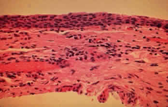
HISTOPATHOLOGIC FINDINGS

Cicatricial pemphigoid is characterized by subepithelial blisters or bullae. Electron-microscopic studies show that the separation at the margin of a blister is located within the lamina lucida between the plasma membrane of the basal cells and the electron-dense lamina densa.5

The conjunctival epithelium in CP shows squamous metaplasia, with parakeratosis and keratinization (Fig. 7).26 Mucus-producing goblet cells are scarce or lacking.11,27 Because the mitotic rate of conjunctival epithelium is higher in patients with CP, it has been suggested that CP is associated with hyperproliferation of the conjunctival epithelium, with a failure of conjunctival differentiation because of reduced goblet cells.28

Fig. 7. Conjunctival biopsy from patient with cicatricial pemphigoid showing squamous metaplasia, absent goblet cells, and mononuclear cells beneath conjunctival epithelium (hematoxylin and eosin, original magnification × 300).

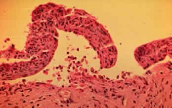
In the early stages of conjunctival disease, granulation tissue is found beneath the conjunctival epithelium, with an infiltration predominantly of lymphocytes and plasma cells, with occasional eosinophils and relatively few neutrophils.13,26 Later, pronounced fibrosis takes place in the conjunctival stroma and is responsible for the conjunctival shrinkage that characterizes the disease.9,10 Hyperproliferation of conjunctival fibroblasts from patients with CP has been demonstrated in tissue culture.29 One study described perivascular inflammatory cell infiltration in 20% of specimens and substantial mast cell participation and degranulation.4 In addition to the chronic inflammatory cells typical of CP, conjunctival biopsy specimens from patients with acute manifestations of CP show numerous neutrophils within and beneath the conjunctival epithelium (Fig. 8).15

Fig. 8. Subepithelial bulla with neutrophils within and beneath the conjunctival epithelium (hematoxylin and eosin, original magnification × 400).

An electron-microscopic study of conjunctival epithelium from patients with CP showed increased desmosomes and prominent tonofilaments and tonofibrils throughout the epithelial cytoplasm. The basal lamina showed areas of discontinuity, duplication, and focal thickening. Collagen fibrils were highly disorganized, and the vascular space was reduced.30 A scanning electron-microscopic study of conjunctival surfaces demonstrated a homogeneous granular sheet of amorphous, mucin-like material covering extensive areas of the conjunctiva in patients with CP that was absent from normal subjects.31

IMMUNOPATHOLOGY

CP is characterized by the binding of immunoglobulins (most commonly IgG) and components of both the classic and alternative complement pathways to the basement membrane zone of skin and oral mucosa.32,33 Circulating antibodies to the basement membrane zone can be demonstrated occasionally, and decreased serum levels of interleukin-6 and increased serum levels of tumor necrosis factor-alpha may be present.33,34 OCP and bullous pemphigoid antigens are distinct but have a common localization within the lamina lucida of the dermal-epidermal junction.35 Patients with systemic features of CP may also have antigens in the lamina densa.36 The bullous pemphigoid antigen is present on the blister roof in close association with basal cells; CP antigen is present on the blister floor.

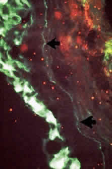
Conjunctival biopsies taken from patients with CP demonstrate immunoglobulins and complement bound to the basement membrane (Fig. 9) 24,37–43 One targeted basement membrane component may be laminin.44 The destruction of laminin, which normally binds basal epithelium to its basement membrane, leads to blister formation.45 An animal model of subepithelial blistering diseases has been developed using antilaminin antibodies in neonatal mice.46 Further damage causes fragmentation of the basement membrane and subsequent repair with aberrant forms of basement membrane collagen.47 Immunoglobulin deposition on the conjunctival basement membrane also may be found in other diseases.24,38,43,48

Fig. 9. Direct immunofluorescence shows linear deposition of IgA along basement membrane zone of conjunctiva (arrows). IgA deposition is also seen in overlying epithelium.

The percentage of patients who demonstrate circulating antibodies to conjunctival basement membrane varies from 0% to 50%.4,37–39,49 In one study, the detection of these antibodies correlated with the degree of clinical activity.49

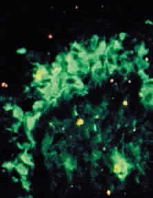
In CP, antibodies may be directed against specific T-cell antigens expressed in the conjunctival epithelium and its basement membrane (Fig. 10).38,39,43,50 In addition, circulating antibodies that bind to the conjunctival and corneal epithelium have been demonstrated.38,39 Studies showing the presence of transforming growth factor-beta 1 and 3, IL-2, and b-fibroblast growth factor suggest that these cytokines play a role in fibrosis.51,52

Fig. 10. Direct immunofluorescence shows IgM deposition in conjunctival epithelium (arrow).

The conjunctiva of patients with CP showed increased numbers of T lymphocytes within the epithelium and substantia propria.53 More helper than suppressor T cells were found. Macrophages were the next most common cells found in the substantia propria, followed by B cells and plasma cells.52 About half of patients with CP have elevated serum IgA levels.38,39 Patients with CP have reduced numbers of circulating T lymphocytes, as determined by E rosette formation.54 The presence of costimulatory molecules may upregulate T-cell activity.55 In one study, CP was associated with HLA-B12, suggesting an immunogenetic susceptibility to the development of this disease.56 Other studies have shown CP to be associated with HLA-DP, HLA-DR4, HLA-DR5, HLA-DQw3, HLA-A2, HLA-B8, HLA-B35, and HLA-B49,57,58 or HLA-DQB1.59,60

DIFFERENTIAL DIAGNOSIS

The clinical diagnosis of CP depends on the documentation of progressive conjunctival shrinkage. The presence of skin and extraocular mucosal lesions suggests the diagnosis. The finding of immunoglobulins and complement bound to the conjunctival basement membrane also support CP as the cause of cicatrization. Immunofluorescence studies on cultured skin keratinocytes may become a corroborative test for circulating antibodies associated with CP.61 Other causes of conjunctival shrinkage and symblepharon must be excluded.

Conjunctival scarring may result from irradiation and severe chemical burns, especially alkali. Symblepharons have also been reported with Sjögrens syndrome,62 atopic keratoconjunctivitis, and sarcoidosis63 but not to the extent and not with the relentless progression of CP. Conjunctival scarring may develop in association with scleral buckles and conjunctival carcinoma but these conditions are usually unilateral, unlike CP. Progressive systemic sclerosis (scleroderma) may be associated with a dry eye and progressive conjunctival shrinkage.64 Trachoma causes conjunctival scarring but this usually begins and predominates in the superior fornix and the upper palpebral conjunctiva.

A membranous conjunctivitis that results in conjunctival scarring may be caused by adenovirus types 8 and 19, a primary infection of herpes simplex virus, diphtheria, or β-hemolytic streptococcus.41,65 The acute self-limited nature of these infections contrasts with the chronic, progressive conjunctival shrinkage found with CP.

Conjunctival shrinkage may be associated with systemic practolol66 or penicillamine67 and topical epinephrine, echothiophate iodide, idoxuridine, pilocarpine, timolol, and dipivefrin.18–23 Whether drug-induced conjunctival shrinkage is self-limited or is actually CP is discussed in the section on “Drug-Induced Ocular Cicatricial Pemphigoid.”

Other bullous diseases usually do not cause much diagnostic confusion with CP (Table 3). Bullous pemphigoid and pemphigus do not usually cause conjunctival scarring.43,68 An acute episode of EM major may cause conjunctival shrinkage but the shrinkage is not chronically progressive, as it is with CP.


TABLE 3. Differential Diagnosis of Bullous Diseases


 Cicatricial PemphigoidPemphigus VulgarisBullous PemphigoidErythema MultiformeDermatitis Herpetiformis
CourseChronicChronicChronicAcute, self-limitedChronic
Age at onsetOlder than 6040–60Older than 600–30, but may occur at any age20–50
Sex preferenceWomenNoneNoneMenMen
Cutaneous involvement+++++
Mucous membrane involvement++Occasional+Rare
      
Ocular involvement++Occasional+Rare
PathologySubepithelial bullaAcantholysis, intraepithelial bullaSubepithelial bullaSubepithelial bullaSubepidermal vesicle and bulla
Immunofluorescence+ Basement membrane+ Intracellular+ Basement membrane+ Blood vessels+ Basement membrane
      

+ , present


TREATMENT

Artificial tears should be used to treat the dry eye associated with CP. Artificial tears without preservatives may be necessary if the preservatives in commercial tear preparations irritate the eyes or if allergies develop to them. Punctal occlusion can be performed if the puncta have not already been occluded by scarring. CP may be complicated by secondary bacterial blepharoconjunctivitis. As mentioned, potential pathogens—especially mannitolpositive staphylococci—were recovered from the eyelids or conjunctiva of 81% of patients with CP.2 Cultures of the eyelids and conjunctiva should be performed at initial examination and at regular intervals. Topical antibiotics should be administered, based on specific antibiotic-sensitivity testing. The staphylococcal blepharitis that frequently accompanies this condition may be treated with eyelid scrubs followed by an antibiotic ointment such as erythromycin or bacitracin. Oral tetracycline or doxycycline may be useful in the treatment of meibomian dysfunction.

In the presence of sufficiently deep fornices, therapeutic soft contact lenses may be used in selected patients to protect the corneal epithelium from trichiasis and drying. Some patients with CP develop large, recurrent corneal epithelial defects that are painful and require almost continuous use of a pressure patch or tarsorrhaphy. In these cases, therapeutic soft contact lenses may keep the corneal epithelium intact and enhance patient comfort. Artificial tears may have to be used frequently to prevent the lens from drying out. Patients who have dry eyes and wear contact lenses are at increased risk of infection and must be followed-up at regular intervals.

Cryotherapy or electrocautery using a hyfrecator may be performed to eliminate trichiasis. Entropion with trichiasis may be corrected early in the disease by oculoplastic surgical techniques. In the advanced stages of the disease, the benefits of oculoplastic procedures, including mucous membrane grafts, may be nullified by the disease. Surgery on the conjunctiva may entail a risk of triggering acute inflammatory activity that may lead to rapid and alarming shrinkage of the conjunctiva.15 Use of intraoperative mitomycin C may prevent this complication.69

If patients with CP show conjunctival inflammation or evidence of progressive conjunctival shrinkage, cataract surgery or oculoplastic surgery on the lids and conjunctiva should not be attempted until the disease is controlled by systemic immunosuppressant therapy, including corticosteroids. Once disease activity is controlled, mucous membrane grafts may be used to reconstruct or expand the fornices. Favorable results were reported for patients on systemic immunosuppression during cataract surgery.70 Conversely, CP patients who are stable and do not show conjunctival inflammation may not require systemic immunosuppression before surgery but may require postoperative systemic corticosteroids if acute inflammatory activity develops. Systemic corticosteroids are useful in the treatment of the acute manifestations of CP (Figs. 11 and 12).15

Fig. 11. Severe conjunctival hyperemia and edema as acute manifestation of cicatricial pemphigoid before oral prednisone.

Fig. 12. Resolution of conjunctival hyperemia and edema after treatment with oral prednisone.

Foster and associates37 and Mondino and Brown16 found that long-term systemic immunosuppressant therapy suppressed conjunctival inflammatory activity and inhibited progression of conjunctival shrinkage. In another report, the results of progression in patients treated with various systemic immunosuppressant agents were compared with those in a control group, with a mean follow-up of about 2 years for all groups (Table 4).1 An eye at stage I or II was less likely to progress and more likely to respond to immunosuppressant therapy than an eye at stage III. Not all patients responded to immunosuppressant therapy, however, and some developed complications that required tapering or even discontinuation. Therefore, older patients should not be subjected to the risk of immunosuppressant therapy unless their eyes demonstrate ongoing progression of conjunctival shrinkage. In addition to being followed-up by an ophthalmologist, patients taking immunosuppressant agents should be monitored by an internist, oncologist, or rheumatologist who is familiar with the toxicity and side-effects of these potent drugs. If immunosuppressant therapy is discontinued, the patient should be monitored for recurrence.71


TABLE 4. Number of Eyes Showing Progression of Conjunctival Shrinkage* in Control and Treatment Groups (Mean Follow-up, 2 Years)


GroupNo. of PatientsNo. of EyesStages  
   IIIIII
Azathioprine10201/3 >(3%)5/9 (56%)4/8 (50%)
Cyclophosphamide13261/4 (25%)1/10 (10%)9/12 (75%)
Prednisone11220/9 (0%)1/7 (14%)3/6 (50%)
Cyclophosphamide and Prednisone17†>331/6 (17%)4/19 (21%)2/8 (25%)
Treatment Groups Combined51†1013/22 (14%)11/45 (24%)18/34 (53%)
Control35†6913/32 (41%)16/26 (62%)8/11 (73%)

*Progression defined as any increase in conjunctival shrinkage including loss of fornix or new symblepharon formation.
†One eye from one patient was excluded from analysis because it was already at stage 4.


Dapsone has been used to treat CP.72 It should be avoided in patients who have a history of sulfa allergy or who are glucose-6-phosphate dehydrogenase-deficient. Tauber and associates73 believe that dapsone may be the best initial therapy for patients with CP that shows mild to modest inflammatory activity and is not rapidly progressive, whereas cyclophosphamide may be the best initial choice for highly active cases. Cyclophosphamide may also be combined with systemic prednisone in difficult cases.74 Sulphapyride may also be useful for treating CP.75

In the final stages of CP, when the conjunctival fornices are obliterated and the ocular surface epithelium is keratinized, a keratoprosthesis may be inserted to restore some sight to these patients.76

Back to Top
ERYTHEMA MULTIFORME
Erythema multiforme is an acute, generally self-limited inflammatory disorder of the skin and mucous membranes. For practical purposes, EM may be divided into minor or major forms. EM minor primarily involves the skin, with mucosal involvement either limited to one surface or absent. EM major, also known as Stevens-Johnson syndrome (SJS), is characterized by skin lesions, erosive involvement of two or more mucous membranes (frequently the conjunctiva), and systemic toxicity, usually including malaise, fever, headache, and prostration.77 Toxic epidermal necrolysis (TEN), which is characterized by massive denudation of the epidermis, may represent a severe variant of EM major.78–81 EM minor may last 1 to 4 weeks; EM major may last up to 6 weeks. EM has no racial or geographic predilection. It may be found in patients of almost any age but occurs primarily in young healthy patients in the first three decades and only rarely in infancy and old age. Men are affected more often than women.79,82

PRECIPITATING FACTORS

The most frequent and important precipitating factors are drugs and infections.79,83–85 The microbial agents associated with EM include viruses, bacteria, fungi, and Mycoplasma pneumoniae. Of these infectious agents, herpes simplex and M. pneumoniae have the strongest association with EM.82 Herpes simplex is more likely to be associated with EM minor; drugs and M. pneumoniae are more likely to be associated with EM major.79

Drugs are probably the most frequent and important precipitating factor in the development of EM.77,79,82,83 Drugs often associated with its development include sulfonamides, penicillin, barbiturates, salicylates, mercurials, arsenic, phenylbutazone, and phenytoin sodium. Of these drugs, sulfonamides have the best-documented association with EM. Recurrences of EM may develop on rechallenge with sulfonamides or other suspected drugs. In a study of 54 cases of EM, drugs were not associated with the development of this syndrome in 31% of patients.77 In 20% of patients, it was difficult to determine whether drugs caused EM or whether they were administered after the prodromal symptoms of EM had already begun. Drugs were strongly implicated as the cause of EM in 48% of patients because they were administered for symptoms unrelated to the prodromata of EM.

Herpes simplex is frequently associated with recurrent episodes of EM minor.86 Although a direct causal relation has not been proved, two studies have demonstrated viral particles within bullae.87,88 Other viral diseases associated with EM are hepatitis C, varicella,89 Epstein-Barr,90 and parvovirus 19.91

EM has been reported after the use of topical ophthalmic scopolamine, tropicamide,92 sulfonamides,93 proparacaine,94 timolol maleate, and chloramphenicol.95

EM has also been related to neoplasms, irradiation therapy, collagen vascular diseases, vaccinations, and administration of digestive enzymes.79,83,96

CLINICAL MANIFESTATIONS

The prodromal symptoms include malaise, fever, symptoms of upper respiratory tract infection, prostration, and headache.79 These symptoms are more prominent in the major form of EM.

The cutaneous lesions are distributed symmetrically. They are found most frequently on the extremities79,97 and spare the trunk, except for the more severe cases. Crops of skin lesions may develop every 2 weeks for about 6 weeks. Usually, the primary lesion of EM is a round erythematous macule that rapidly develops into a papule. Vesicles, bullae, and epidermal necrosis may develop. The characteristic target lesion consists of a red center surrounded by a pale zone, with another red ring peripheral to the pale zone (Fig. 13). If extensive necrosis and denudation of the epidermis result, the condition may be labeled TEN.80 The skin lesions may be associated with burning or itching and resolve leaving residual hyperpigmentation.

Fig. 13. Target lesions of the palms in patient with erythema multiforme (EM). EM major may involve any mucous membrane.

The mouth and eyes are affected most frequently and severely. In one study of predominant clinical features of EM major over a 10-year period, 100% of patients showed stomatitis, 63% showed conjunctivitis, and 61% showed balanitis, vaginitis, or urethritis.74 The lips are swollen and crusted (Fig. 14). Oral lesions, which occur in crops, begin as small erythematous macules that rapidly develop into clear or hemorrhagic bullae.79

Fig. 14. Swollen, crusted lips and facial blisters in patient with erythema multiforme.

Rupture of the bullae leaves an inflamed, painful hemorrhagic base with a white pseudomembrane. Over the course of 1 week, epithelialization occurs. The process may involve the external hares, pharynx, larynx, trachea, bronchi, and esophagus. Except for the conjunctiva, mucosal and cutaneous lesions usually disappear without scarring. Esophagitis with stricture formation has been reported, however.98

Complications of EM major include pneumonitis, septicemia, myocarditis, myositis, and glomerulonephritis. The reported mortality is less than 1% for EM minor and 2% to 25% for EM major.99 About 20% of EM patients develop recurrences.99 Recurrences are more common with the minor form of EM.

OCULAR INVOLVEMENT

The acute phase of ocular disease lasts 2 to 3 weeks.100 The eyelids are swollen, ulcerated, and crusted. Conjunctival vesicles have been reported101,102 but are rarely noted. Conjunctival involvement ranges from a mild catarrhal conjunctivitis, which terminates without sequela, to pseudomembranous or membranous conjunctivitis, which may involve both the palpebral and bulbar conjunctiva.103 Purulent conjunctivitis may develop secondary to bacterial infections.100–102

The severity of late ophthalmic complications depends more on the severity of systemic disease and conjunctival involvement than on the local treatment.104 In other words, patients with pseudomembranous or membranous conjunctivitis tend to develop late ophthalmic complications. The conjunctival surfaces of patients with membranous or pseudomembranous conjunctivitis may heal, leaving scarring, symblepharon, and even ankyloblepharon (Figs. 15 and 16). Late complications of conjunctival scarring include entropion with trichiasis and lagophthalmos with exposure.

Fig. 15. Pseudomembranes of upper and lower palpebral conjunctiva during acute attack of erythema multiforme.

Fig. 16. Symblepharon of nasal aspect of right inferior fornix (arrow) after resolution of acute attack of erythema multiforme.

Obliteration of the lacrimal puncta and canaliculi by fibrosis may cause epiphora in some patients. Destruction of conjunctival goblet cells and fibrotic obstruction of the ducts of the lacrimal and accessory lacrimal glands may result in a dry eye condition similar to CP. In severe cases, keratinization of the conjunctival and corneal epithelium may be found. The dry eye condition, entropion with trichiasis, and lagophthalmos with exposure may result in corneal complications that include punctate erosions, pannus, ulcers, opacification, and even perforation (Fig. 17).100–102

Fig. 17. Dry eye after resolution of acute attack of erythema multiforme. Note mucus in inferior fornix and vascularization of inferior cornea.

Recurrences of EM major rarely involve the conjunctiva.103 Thus, EM leaves conjunctival shrinkage and symblepharon in its wake but progressive scarring does not usually occur once the acute stage has subsided, unlike the chronic progressive course of CP. Further destruction of the eye usually depends on complications resulting from the acute event, such as entropion with trichiasis, lagophthalmos with exposure, and the dry eye condition with its propensity for secondary bacterial infections. All these complications may result in a chronically hyperemic eye with a tendency for breakdown of the corneal epithelium that may eventuate in corneal ulcers and even perforations.

Foster and associates105 reported a small subset of SJS patients with recurrent episodes of conjunctival inflammation. This inflammation was not associated with external factors such as lid margin keratinization, dry eye, trichiasis, or entropion. The histopathologic, ultrastructural, and immunopathologic characteristics of conjunctival biopsies from these patients suggested an underlying vasculitis or perivasculitis. Recurrent inflammation has been described in the skin and oral mucosa.79

Microbial keratitis in patients with SJS is most commonly caused by gram-positive cocci and is associated with the use of bandage contact lenses and topical corticosteroids.12

HISTOPATHOLOGY

A consistent finding in the skin lesions of EM is a mononuclear cell infiltrate in the dermis, predominantly around the upper dermal blood vessels.106 The perivascular infiltrate contains mainly lymphocytes and histiocytes.82 This perivascular infiltrate may be the only finding in the macules and papules of EM. A necrotizing vasculitis is not found.79 Damage to the overlying epidermis is a characteristic finding. The bullae of EM are subepithelial, with adjacent lymphocytes, histiocytes, and a few neutrophils or eosinophils.107

Histopathologic findings in mucosal lesions are similar to those in skin lesions.108 Unlike the skin, blisters are transient and rupture rapidly, leaving erosions. Neutrophils are found at an earlier stage and are more prominent in mucosal than cutaneous lesions.

A chronic, nonspecific inflammatory reaction of the conjunctiva is found with a perivascular infiltration of lymphocytes.101 Pseudomembranes may form from the fibrinous exudate made up of inflammatory cells that are predominantly neutrophils and necrotic epithelial cells. In some patients with severe necrotizing reactions, true membranes result. After the acute episode, the conjunctival epithelium from patients with SJS may show squamous metaplasia with surface keratinization. Conjunctival goblet cell densities are reduced in SJS.109,110 SJS may not represent a mucin-deficient condition because substantial amounts of ocular mucus glycoproteins are present in the eyes of these patients.111 The mucus from these patients was more viscous than normal, however, suggesting that qualitative differences may exist.

Conjunctival biopsies from SJS patients with recurrent conjunctival inflammatory episodes showed vascular involvement.105 In some patients, a classic vasculitis was found with inflammatory cell infiltration into the vessel wall and fibrinoid necrosis of the wall. Others showed prominent perivasculitis with lymphocytes, macrophages, and plasma cells surrounding the vessels. Substantial numbers of degranulating mast cells were noted.

IMMUNOPATHOLOGY

Circulating immune complexes have been demonstrated in the sera of patients with EM.106,108,112–114 Additionally, direct immunofluorescent studies of involved skin show deposition of C3, IgM, fibrin, and occasionally IgG in the blood vessel walls of the dermis.106,108,112–114 The normal unaffected skin from patients with EM does not have immunoglobulin and complement deposition in blood vessel walls. Injections of histamine can cause them to deposit there, however, suggesting that the immune complexes of EM circulate rather than form locally in the tissue.106 Immunoglobulin and complement may deposit at the dermal-epidermal junction, and may be directed against desmosomes.112,113,115,116 A study of the cellular infiltrate of skin lesions from patients with EM showed that T lymphocytes were the major infiltrating cells, with more cytotoxic or suppressor cells than helper or inducer cells in the epidermis but the reverse in the dermis.117

Foster and colleagues105 demonstrated a vasculitis or perivasculitis with immunoglobulin and complement deposition in vessel walls of conjunctival biopsies from a small subset of patients with SJS and recurrent conjunctival inflammation. SJS with ocular involvement has been associated with HLA-Bw44 (the dominant sub-group of B 12) and HLA-DQB1.118,119 These results suggested an immunogenetic susceptibility to the development of SJS with ocular manifestations. This association with HLA-B12 was supported in two other studies of patients with SJS and TEN (a severe variant of EM).78,120 Malo and associates121 found an association between herpes simplex virus-induced SJS and HLA-DQB1. These results suggest that there may be an immunogenetic susceptibility not only to the development of SJS with ocular involvement but to all the severe variants of EM.

TREATMENT

There is no specific treatment for EM. Eliminating any suspected etiologic factors and treating related infection are important.122 In patients taking multiple medications, patch testing has been used to identify the causative agent.123 Nonessential drugs should be discontinued.

Wet dressings may be used to debride crusted skin lesions, and baths may minimize discomfort.82 Hydration may be necessary to maintain fluid balance in patients with severe and extensive cutaneous and mucosal involvement. Analgesics may be needed to relieve pain. Treatment of oral lesions includes saline mouthwashes, topical anesthetics, and topical corticosteroids.82

Systemic corticosteroids have been recommended for the general manifestations of EM.82,124 An initial dose of 60 to 80 mg of prednisone daily has been recommended until improvement is noted, after which the prednisone is gradually tapered over 3 to 4 weeks. The value of systemic corticosteroids has not been established by a well-controlled, prospective, randomized clinical trial. Indeed, several studies have questioned and challenged the efficacy of systemic prednisone in EM.125–128 Retrospective reviews of hospitalized patients with EM suggest that patients treated with systemic corticosteroids had more complications and a prolonged recovery with a longer average length of hospital stay, compared with patients receiving only supportive care. Local treatment appears to have little influence on the severity of ophthalmic complications, which are related to the severity of systemic disease and conjunctival involvement.104 Early lysis of symblepharons should be attempted but may not be effective. Conjunctival cultures should be obtained and appropriate topical antibiotics should be administered for suspected bacterial infections.

In recurrent herpes simplex associated with EM, oral acyclovir may suppress recurrence of both viral disease and its accompanying EM rash.129

After the acute inflammation has subsided, patients with SJS may require artificial tears for the dry eye condition, destruction of aberrant lashes by electrocautery using a Hyfrecator or by cryotherapy, soft contact lenses to protect the cornea from drying and trichiasis, and lid scrubs followed by antibiotic ointment for chronic blepharitis.103 The lacrimal puncta are generally scarred and closed by the disease process. If not, they should be closed if the patient has a dry eye.130 Topical retinoids such as tretinoin (all-transretinoic acid, a vitamin A analog) may reverse conjunctival keratinization.131,132 Chronic corneal ulceration may require tarsorrhaphy. Mucous membrane grafts may be used to reconstruct the fornices. Patients with localized conjunctival epidermalization may be treated by tarsal polishing followed by grafting with full-thickness buccal mucous membrane.133 In patients with severe symblepharon or ankyloblepharon and keratinization of the ocular surface epithelium, a keratoprosthesis may be attempted.134

Back to Top
OTHER BULLOUS DISEASES

BULLOUS PEMPHIGOID

Bullous pemphigoid is a relatively benign disease that occurs predominantly in patients older than 60 years of age and primarily involves the skin, which develops large, tense, tough, subepidermal bullae.6 Oral mucosal lesions are occasionally found but are less frequent and severe than those in CP or pemphigus. Antibodies are directed against protein components of hemidesmosome, which are crucial to the integrity of the dermal-epidermal junction.135 Circulating antibody levels do not correlate consistently to disease severity.136 Bullous pemphigoid may involve the conjunctiva and cause shrinkage43 but this is not common.

PEMPHIGUS VULGARIS

Pemphigus vulgaris (Fig. 18) occurs primarily in middle-aged people and is characterized by small, flaccid, easily broken blisters of the skin in an intraepidermal location.6 Like CP, pemphigus may be associated with severe oral lesions. The cutaneous and mucosal lesions show little tendency to heal spontaneously but heal without scarring. Immunofluorescent studies show immunoglobulins and complement bound to the intercellular space of the epidermis, in addition to circulating antibodies to this location. Severity of disease correlates with circulating antibody levels.68 Immunity is directed against the desmosome portion of intercellular junctional complexes.137 Although pemphigus may be associated with conjunctivitis,9 conjunctival shrinkage is rare, although it has been reported.68

Fig. 18. A. Ruptured blisters of skin of upper and lower eyelids of patient with pemphigus. B. Conjunctival bulla in the same patient. Note layered hemorrhage at bottom of bulla. (Courtesy of Steven Berger, MD, and J. P. Dunn, MD)

EPIDERMOLYSIS BULLOSA

Epidermolysis bullosa refers to a group of skin diseases characterized by a tendency to form blisters after minor trauma138 and can be divided into hereditary and acquired forms.139 The hereditary forms of epidermolysis bullosa may be divided into three types. The simple form (autosomal dominant) is associated with blister formation in the basal cell layer of the epidermis, and mutations have been found in keratins localized to basal keratinocytes. The junctional form of epidermolysis bullosa (autosomal recessive) is thought to be caused by a defect in production of basement membrane laminins or integrins, causing a separation within the dermal-epidermal basement membrane at the lamina lucida. In the dystrophic form (autosomal dominant or recessive), separation occurs beneath the basement membrane in the papillary dermis, and mutations have been identified in the collagen VII gene.140

Ocular problems have been described in all types of epidermolysis bullosa but are most common in dystrophic epidermolysis bullosa.141 Patients with the more common dystrophic form have fewer corneal problems but more frequent and marked conjunctival scarring, including symblepharon formation. Patients with junctional epidermolysis bullosa, one of the rarest types, have more corneal problems (e.g., recurrent corneal erosions) but relatively little conjunctival involvement.141 In the acquired autoimmune form, the immune response is believed to be directed against the basement membrane proteins uncein and collagen VII.142 It may be associated with small subepithelial vesicles in the cornea, symblepharon, and scarring of the lacrimal puncta.143

DERMATITIS HERPETIFORMIS

Dermatitis herpetiformis is a rare, chronic, recurrent eruption characterized by successive crops of papules, vesicles, and blisters on an erythematous base and in a symmetric distribution.6 Men are affected more frequently than women, and the disease usually occurs between the second and fifth decades. The eruption is responsible for intense pruritus and burning. Superficial scars rarely develop. Mucous membrane lesions are rare and heal rapidly. Direct immunofluorescence of skin shows deposits of IgA and complement at the basement membrane zone of the epidermis. Circulating antibodies to the basement membrane zone of the skin are not found in the sera. Microabscesses of neutrophils and eosinophils form at the tips of dermal papillae and result in subepidermal vesicles and occasionally bullae.

Ocular changes are not common but cases have been reported with conjunctival involvement.9 Conjunctival vesicles, erosions, pseudomembranes, symblepharon, shrinkage, and keratinization have been described, resulting in a clinical picture that resembles CP. The conjunctival scarring found in dermatitis herpetiformis is not as extensive as that found in CP, however.

Back to Top
REFERENCES

1. Mondino BJ: Cicatricial pemphigoid and erythema multi-forme. Ophthalmology 97:939, 1990

2. Mondino BJ, Brown SI: Ocular cicatricial pemphigoid. Ophthalmology 88:95, 1981

3. Bhol K, Udell I, Foster CS et al: Ocular cicatricial pemphigoid. A case report of monozygotic twins discordant for the disease. Arch Ophthalmol 113: 202, 1995

4. Foster CS: Cicatricial pemphigoid. Trans Am Ophthalmol Soc 84:527, 1986

5. Lever WF: Pemphigus and pemphigoid. J Am Acad Dermatol 1:2, 1979

6. Moschella SL, Pillsbury DM, Hurley H J: Dermatology, vol 1, pp 460–476. Philadelphia, WB Saunders, 1975

7. Hanson RD, Olsen KD, Rogers RS: Upper aerodigestive tract manifestations of cicatricial pemphigoid. Ann Otol Rhinol Laryngol 97:493, 1988

8. Mondino BJ, Manthey R: Dermatological diseases and the peripheral cornea. Int Ophthalmol Clin 26(4):121, 1986

9. Duke-Elder S (ed): System of Ophthalmology, vol 8, part 1, Diseases of the Outer Eye: Conjunctiva, pp 498–516. St Louis, CV Mosby, 1965

10. Gazala JR: Ocular pemphigus. Am J Ophthalmol 48:355, 1959

11. Ralph RA: Conjunctival goblet cell density in normal subjects and dry eye syndromes. Invest Ophthalmol 14:299, 1975

12. Ormerod LD, Fong LP, Foster CS: Corneal infection in mucosal scarring disorders and Sjögren's syndrome. Am J Ophthalmol 105:512, 1988

13. Norn MS, Kristensen EB: Benign mucous membrane pemphigoid. II. Cytology. Acta Ophthalmol 52:282, 1974

14. Tauber J, Melamed S, Foster CS: Glaucoma in patients with ocular cicatricial pemphigoid. Ophthalmology 96:33, 1989

15. Mondino BJ, Brown SI, Lempert S et al: The acute manifestations of ocular cicatricial pemphigoid: diagnosis and treatment. Ophthalmology 86:543, 1979

16. Mondino BJ, Brown SI: Immunosuppressive therapy in ocular cicatricial pemphigoid. Am J Ophthalmol 96:453, 1983

17. Tauber J, Jabbur N, Foster CS: Improved detection of disease progression in ocular cicatricial pemphigoid. Cornea 5:446, 1992

18. Lass JH, Thoft RA, Dohlman CH: Idoxuridine-induced conjunctival cicatrization. Arch Ophthalmol 101:747, 1983

19. Patten JT, Cavanagh HD, Allansmith MR: Induced ocular pseudopemphigoid. Am J Ophthalmol 82:272, 1976

20. Hirst LW, Werblin T, Novak M et al: Drug-induced cicatrizing conjunctivitis simulating ocular pemphigoid. Cornea 1:121, 1982

21. Kristensen EB, Norn MS: Benign mucous membrane pemphigoid. I. Secretion of mucus and tears. Acta Ophthalmol 52:266, 1974

22. Pouliquen Y, Patey A, Foster CS et al: Drug-induced cicatricial pemphigoid affecting the conjunctiva. Ophthalmology 93:775, 1986

23. Fiore PM, Jacobs IH, Goldberg DB: Drug-induced pemphigoid. Arch Ophthalmol 105:1660, 1987

24. Leonard JN, Hobday CM, Haffenden GP et al: Immunofluorescent studies in ocular cicatricial pemphigoid. Br J Dermatol 118:209, 1988

25. Mondino BJ: Discussion of drug-induced cicatricial pemphigoid affecting the conjunctiva. Ophthalmology 93:782, 1986

26. Andersen SR, Jensen OA, Kristensen EB et al: Benign mucous membrane pemphigoid. III. Biopsy. Acta Ophthalmol 52:455, 1974

27. Kinoshita S, Kiorpes TC, Friend J, Thoft RA: Goblet cell density in ocular surface disease. Arch Ophthalmol 101: 1284, 1983

28. Thoft RA, Friend J, Kinoshita S et al: Ocular cicatricial pemphigoid associated with hyperproliferation of the conjunctival epithelium. Am J Ophthalmol 98:37, 1984

29. Roat MI, Sossi G, Lo C-Y, Thoft RA: Hyperproliferation of conjunctival fibroblasts from patients with cicatricial pemphigoid. Arch Ophthalmol 107:1064, 1989

30. Galbavy EJ, Foster CS: Ultrastructural characteristics of conjunctiva in cicatricial pemphigoid. Cornea 4:127, 1985

31. Foster CS, Shaw CD, Wells PA: Scanning electron microscopy of conjunctival surfaces in patients with ocular cicatricial pemphigoid. Am J Ophthalmol 102:584, 1986

32. Griffith MR, Fukuyama K, Tuffanelli D et al: Immunofluorescent studies in mucous membrane pemphigoid. Arch Dermatol 109:195, 1974

33. Rogers RS, Perry HO, Bean SF et al: Immunopathology of cicatricial pemphigoid: Studies of complement deposition. J Invest Dermatol 68:39, 1977

34. Lee SJ, Li Z, Sherman B, Foster CS: Serum levels of tumor necrosis factor-alpha and interleukin-6 in ocular cicatricial pemphigoid. Invest Ophthalmol Vis Sci 34:3522, 1993

35. Fine JD: Epidermolysis bullosa: variability of expression of cicatricial pemphigoid, bullous pemphigoid, and epidermolysis bullosa acquisita antigens in clinically uninvolved skin. J Invest Dermatol 85:47, 1985

36. Robin H, Prost C, Hoang-Xuan T: Pure ocular cicatricial pemphigoid is a unique entity: a direct immunoelectron microscopy study. Invest Ophthalmol Vis Sci 37:4701, 1996

37. Foster CS, Wilson LA, Ekins MB: Immunosuppressive therapy for progressive ocular cicatricial pemphigoid. Ophthalmology 89:340, 1982

38. Mondino BJ, Brown SI, Rabin BS: Autoimmune phenomena of the external eye. Ophthalmology 85:801, 1978

39. Mondino BJ, Ross AN, Rabin BS et al: Autoimmune phenomena in ocular cicatricial pemphigoid. Am J Ophthalmol 83:443, 1977

40. Bean SF, Furey N, West CE et al: Ocular cicatricial pemphigoid (immunologic studies). Trans Am Acad Ophthalmol Otolaryngol 81:806, 1976

41. Furey N, West C, Andrews T et al: Immunofluorescent studies of ocular cicatricial pemphigoid. Am J Ophthalmol 80:825, 1975

42. Proia AD, Foulks GN, Sanfilippo FP: Ocular cicatricial pemphigoid with granular IgA and complement deposition. Arch Ophthalmol 103:1669, 1985

43. Frith PA, Venning VA, Wojnarowska F et al: Conjunctival involvement in cicatricial and bullous pemphigoid: a clinical and immunopathological study. Br J Ophthalmol 73:52, 1989

44. Hsu R, Lazarova Z, Yee C et al: Noncomplement fixing, IgG4 autoantibodies predominate in patients with anti-epiligrin cicatricial pemphigoid. J Invest Dermatol 109:557, 1997

45. Kirtschig G, Marinkovich MP, Burgeson RE et al: Anti-basement membrane autoantibodies in patients with anti-epiligrin cicatricial pemphigoid bind the alpha subunit of laminin 5. J Invest Dermatol 105:543, 1995

46. Lazarova Z, Yee C, Darling T, Briggaman RA, Yancey KB: Passive transfer of anti-laminin 5 antibodies induces subepidermal blisters in neonatal mice. J Clin Invest 98: 1509, 1996

47. Dutt JE, Ledoux D, Foster CS: Collagen abnormalities in conjunctiva of patients with cicatricial pemphigoid. Cornea 15:606, 1996

48. Aultbrinker EA, Starr MB, Donnenfeld ED: Linear IgA disease—the ocular manifestations. Ophthalmology 95: 340, 1988

49. Franklin RM, Fitzmorris CT: Antibodies against conjunctival basement membrane zone. Arch Ophthalmol 101: 1611, 1983

50. Bialasiewicz AA, Ventura A, Schaudig U et al: Alpha/beta and gamma/delta T-cell receptors—analysis of conjunctival tissue of patients with ocular pemphigoid. Ophthalmologe 94:197, 1997

51. Sacks EH, Jakobiec FA, Wieczorek R et al: Immunophenotypic analysis of the inflammatory infiltrate in ocular cicatricial pemphigoid: further evidence for a T cell-mediated disease. Ophthalmology 96:236, 1989

52. Bernauer W: Chronic progressive forms of cicatrising conjunctivitis. In Pleyer U, Hartmann C, Sterry W (eds): Oculodermal Diseases, pp 193–221. Buren, The Netherlands, Aeolus Press, 1997

53. Elder MJ, Dart JK, Lightman S: Conjunctival fibrosis in ocular cicatricial pemphigoid—the role of cytokines. Exp Eye Res 65:165, 1997

54. Mondino B J, Rao H, Brown SI: T and B lymphocyte enumerations in ocular cicatricial pemphigoid. Am J Ophthalmol 92:536, 1981

55. Tesavibul N, Dorfman D, Foster CS et al: Costimulatory molecules in ocular cicatricial pemphigoid. Invest Ophthalmol Vis Sci 39:982, 1998

56. Mondino BJ, Brown SI, Rabin BS: HLA antigens in ocular cicatricial pemphigoid. Arch Ophthalmol 97:479, 1979

57. Lee SJ, Rice BA, Foster CS: HLA class II expression in normal and inflamed conjunctiva. Reg Immunol 3:177, 1990

58. Zaltas MM, Ahmed R, Foster CS: Association of HLA-DR4 with ocular cicatricial pemphigoid. Curr Eye Res 8(2):189, 1989

59. Haider N, Neuman R, Foster CS, Ahmed AR: Report on the sequence of DQB1*0301 gene in ocular cicatricial pemphigoid patients. Curr Eye Res 11:1233, 1992

60. Chan LS, Wang T, Wang XS et al: High frequency of HLA-DQB1*0301 allele in patients with pure ocular cicatricial pemphigoid. Dermatology 189(suppl):99, 1994

61. Batteux F, Franck N, Jaffray P et al: An extract from cultured human keratinocytes that contains the major autoantigens related to autoimmune bullous skin diseases. J Clin Immunol 3:228, 1997

62. Jones BR: The ocular diagnosis of benign mucous membrane pemphigoid. Proc R Soc Meal 54:109, 1961

63. Flach A: Symblepharon in sarcoidosis. Am J Ophthalmol 85:210, 1978

64. Foster CS: Ocular manifestations of the nonrheumatic acquired collagen vascular diseases. In Smolin G, Thoft RA (eds): The Cornea: Scientific Foundations and Clinical Practice, 1st ed, pp 279–281. Boston, Little, Brown & Co, 1983

65. Darougar S, Quinlan MP, Gibson JA et al: Epidemic keratoconjunctivitis and chronic papillary conjunctivitis in London due to adenovirus type 19. Br J Ophthalmol 61:76, 1977

66. Jones DB: Prospects in the management of tear deficiency states. Trans Am Acad Ophthalmol Otolaryngol 83:693, 1977

67. Bialy-Golan A, Brenner S: Penicillamine-induced bullous dermatoses. J Am Acad Dermatol 35:732, 1996

68. Bean SF, Holubar K, Gillett RB: Pemphigus involving the eyes. Arch Dermatol 111:1484, 1975

69. Secchi AG, Tognon MS: Intraoperative mitomycin C in the treatment of cicatricial obliterations of conjunctival fornices. Am J Ophthalmol 122:728, 1996

70. Sainz de la Maza M, Tauber J, Foster CS: Cataract surgery in ocular cicatricial pemphigoid. Ophthalmology 95:481, 1988

71. Neumann R, Tauber J, Foster CS: Remission and recurrence after withdrawal of therapy for ocular cicatricial pemphigoid. Ophthalmology 98:858, 1991

72. Rogers RS, Seehafer JR, Perry HO: Treatment of cicatricial (benign mucous membrane) pemphigoid with dapsone. J Am Acad Dermatol 6:215, 1982

73. Tauber J, Brookline MA, Sainz de la Maza M, Foster CS: Systemic chemotherapy for ocular cicatricial pemphigoid. Ophthalmology 95(suppl):146, 1988

74. Elder MJ, Leonard J, Dart JK: Role of cyclophosphamide and high dose steroid in ocular cicatricial pemphigoid. Br J Ophthal 79:264, 1995

75. Elder MJ, Leonard J, Dart JK: Sulphapyridine—a new agent for the treatment of ocular cicatricial pemphigoid. Br J Ophthal 80:549, 1996

76. Rao GN, Blatt HL, Aquavella JV: Results of keratoprosthesis. Am J Ophthalmol 88:190, 1979

77. Yetiv JZ, Bianchine JR, Owen JA: Etiologic factors of the Stevens-Johnson syndrome. South Med J 73:599, 1980

78. Roujeau JC, Huyn TN, Bracq C et al: Genetic susceptibility to toxic epidermal necrolysis. Arch Dermatol 123:1171, 1987

79. Huff JC, Weston WL, Tonnesen MG: Erythema multi-forme: a critical review of characteristics, diagnostic criteria, and causes. J Am Acad Dermatol 8:763, 1983

80. Halebian PH, Corder VJ, Madden MR et al: Improved burn center survival of patients with toxic epidermal necrolysis managed without corticosteroids. Ann Surg 204:503, 1986

81. Rzany B, Hering O, Mockenhaupt M et al: Histopathological and epidemiological characteristics of patients with erythema exudativum multiforme major Stevens-Johnson syndrome and toxic epidermal necrolysis. Br J Dermatol 135:6, 1996

82. Tonnesen MG, Soter NA: Erythema multiforme. J Am Acad Dermatol 1:357, 1979

83. Shelley WB: Herpes simplex virus as a cause of erythema multiforme. JAMA 201:153, 1967

84. Major PP, Morisset R, Kurstak C et al: Isolation of herpes simplex virus type I from lesions of erythema multiforme. Can Med Assoc J 118:821, 1978

85. Shelley WB: Bacterial endotoxin (lipopolysaccharide) as a cause of erythema multiforme. JAMA 243:58, 1980

86. Malo A, Keampgen E, Wank R: Recurrent herpes simplex virus-induced erythema multiforme: different HLA-DQB1 alleles associate with severe mucous membrane versus skin attacks. Scand J Immunol 47:408, 1998

87. Brice SL, Leahy MA, Ong L et al: Examination of non-involved skin previously involved skin and peripheral blood for herpes simplex virus DNA in patients with recurrent herpes-associated erythema multiforme. J Cutan Pathol 21:408, 1994

88. Imafuku S, Kokuba H, Aurelian L, Burnett J: Expression of herpes simplex virus DNA fragments located in epidermal keratinocytes and germinative cells is associated with the development of erythema multiforme lesions. J Invest Dermatol 109:550, 1997

89. Choy AC, Yarnald PR, Brown JE et al: Virus induced erythema multiforme and Stevens-Johnson syndrome. Allergy Proceed 16:157, 1995

90. Mitterer M, Pescosta N, Fend F et al: Chronic active Epstein-Barr virus disease in a case of persistent polyclonal B-cell lymphocytosis. Br J Haematol 90:526, 1995

91. Frank R, Glander HJ, Haustein UF: Dermatologic symptoms of parovirus B19 infections. Hautarzt 47:365, 1996

92. Guill MA, Goette DK, Knight CG et al: Erythema multi-forme and urticaria. Arch Dermatol 115: 742, 1979

93. Genvert GI, Cohen EJ, Donnenfeld ED et al: Erythema multiforme after use of topical sulfacetamide. Am J Ophthalmol 99:465, 1985

94. Ward B, McCulley JP, Segal RJ: Dermatologic reaction in Stevens-Johnson syndrome after ophthalmic anesthesia with proparacaine hydrochloride. Am J Ophthalmol 86: 133, 1978

95. Lazarov A, Amichai B: Skin reactions due to eye drops: report of two cases. Cutis 58:363, 1996

96. Miyoshi H, Kanzaki T: Drug eruption erythema multiforme type due to a digestive enzyme drug. J Dermatol 25:28, 1998

97. Huff JC: Erythema multiforme. Dermatol Clin 3:141, 1985

98. Boe J, Dalgaard JB, Scott D: Mucocutaneous-ocular syndrome with intestinal involvement. Am J Med 25:857, 1958

99. Chanda JJ: Erythema multiforme Perspect Ophthalmol 3:183, 1979

100. Dohlman CH, Doughman DJ: The Stevens-Johnson syndrome. In: Symposium on the Cornea: Transactions of the New Orleans Academy of Ophthalmology, pp 236–252. St Louis, CV Mosby, 1965

101. Howard GM: The Stevens-Johnson syndrome: ocular prognosis and treatment. Am J Ophthalmol 55:893, 1963

102. Patz A: Ocular involvement in erythema multiforme. Arch Ophthalmol 43:244, 1950

103. Mondino BJ: Cicatricial pemphigoid and erythema multiforme. Int Ophthalmol Clin 23:63, 1983

104. Arstikaitis MJ: Ocular aftermath of Stevens-Johnson syndrome. Arch Ophthalmol 90:376, 1973

105. Foster CS, Fong LP, Azar D et al: Episodic conjunctival inflammation after Stevens-Johnson syndrome. Ophthalmology 95:453, 1988

106. Wuepper KD, Watson PA, Kazmierowski JA: Immune complexes in erythema multiforme and the Stevens-Johnson syndrome. J Invest Dermatol 74:368, 1980

107. Korting GW, Denk R: Differential Diagnosis in Dermatology, pp 421–422. Philadelphia, WB Saunders, 1976

108. Buchner A, Lozada F, Silverman S: Histopathologic spectrum of oral erythema multiforme. Oral Surg 49:221, 1980

109. Nelson JD, Wright JC: Conjunctival goblet cell densities in ocular surface disease. Arch Ophthalmol 102:1049, 1984

110. Ohji M, Ohmi G, Kiritoshi A et al: Goblet cell density in thermal and chemical injuries. Arch Ophthalmol 105: 1686, 1987

111. Wells PA, Ashur ML, Foster CS: SDS-gradient poly-acrylamide gel electrophoresis of individual ocular mucus samples from patients with normal and diseased conjunctiva. Curr Eye Res 5:823, 1986

112. Bushkell LL, Mackel SE, Jordon RE: Erythema multi-forme: direct immunofluorescence studies and detection of circulating immune complexes. J Invest Dermatol 74:372, 1980

113. Huff JC, Weston WL, Carr RI: Mixed cryoglobulinemia, 125I Clq binding and skin immunofluorescence in erythema multiforme. J Invest Dermatol 74:375, 1980

114. Imamura S, Yanase K, Taniguchi S et al: Erythema multi-forme: demonstration of immune complexes in the sera and skin lesions. Br J Dermatol 102:161, 1980

115. Foedinger D, Sterniczky B, Elbe A et al: Autoantibodies against desmoplakin I and II define a subset of patients with erythema multiforme major. J Invest Dermatol 106: 1012, 1996

116. Foedinger D, Anhalt GJ, Boecskoer B: Autoantibodies to desmoplakin I and II in patients with erythema multiforme. J Exp Med 181:169, 1995

117. Margolis RJ, Tonnesen MG, Harrist TJ et al: Lymphocyte subsets and Langerhans cells/indeterminate cells in erythema multiforme. Invest Dermatol 81:403, 1983

118. Mondino BJ, Brown SI, Biglan AW: HLA antigens in Stevens-Johnson syndrome with ocular involvement. Arch Ophthalmol 100:1453, 1982

119. Power WJ, Saidman SL, Foster CS et al: HLA typing in patients with ocular manifestations of Stevens-Johnson syndrome. Ophthalmology 103:1406, 1996

120. Roujeau JC, Bracq C, Huyn NT et al: HLA phenotypes and bullous cutaneous reactions to drugs. Tissue Antigens 28:251, 1986

121. Malo A, Keampgen E, Wank R: Recurrent herpes simplex virus-induced erythema multiforme: Different HLA-DQB1 alleles associated with severe mucous membrane versus skin attacks. Scand J Immunol 47:408, 1998

122. Weston WL, Morelli JG: Herpes simplex virus-associated erythema multiforme in prepubertal children. Arch Pediatr Adolesc Med 151:1014, 1997

123. Gebhardt M, Wollina U: Allergy testing I serious cutaneous drug reactions—harmful or beneficial? Contact Dermatitis 37:282, 1997

124. Kakourou T, Klontza D, Soteropoulou F et al: Corticosteroid treatment of erythema multiforme major Stevens-Johnson syndrome in children. Eur J Pediatr 156:90, 1997

125. Rasmussen JE: Erythema multiforme in children: response to treatment with systemic corticosteroids. Br J Dermatol 95:181, 1976

126. Ginsburg CM: Stevens-Johnson syndrome in children. Pediatr Infect Dis 1:155, 1982

127. Nethercolt JR, Choi BCK: Erythema multiforme (Stevens-Johnson syndrome)—chart review of 123 hospitalized patients. Dermatologica 171:383, 1985

128. Power WJ, Ghoraishi M, Foster CS et al: Analysis of the acute ophthalmic manifestations of the erythema multiforme/Stevens-Johnson syndrome/toxic epidermal necrolysis disease spectrum. Ophthalmology 102:1669, 1995

129. Tatnall FM, Schofield JK, Leigh IM: A double-blind placebo-controlled trial of continuous acyclovir therapy in recurrent erythema multiforme. Br J Dermatol 132:267, 1995

130. Tuberville AW, Frederick WR, Wood TO: Punctal occlusion in tear deficiency syndromes. Ophthalmology 89: 1170, 1982

131. Wright P: Topical retinoic acid therapy for disorders of the outer eye. Trans Ophthalmol Soc UK 104:869, 1985

132. Soong HK, Martin NF, Wagoner MD et al: Topical retinoid therapy for squamous metaplasia of various ocular surface disorders. Ophthalmology 95:1442, 1988

133. McCord CD, Chen WP: Tarsal polishing and mucous membrane grafting for cicatricial entropion, trichiasis and epidermalization. Ophthalmic Surg 14:1021, 1983

134. Kozarsky AM, Knight SH, Waring GO: Clinical results with a ceramic keratoprosthesis placed through the eyelid. Ophthalmology 94:904, 1987

135. Lin MS, Mascaró JM Jr, Liu Z et al: The desmosome and hemidesmosome in cutaneous autoimmunity. Clin Exp Immunol 107(suppl):9, 1997

136. Lever WF, Shaumburg-Lever G: Histopathology of the Skin, pp 108–117. Philadelphia, JB Lippincott, 1975

137. Moll R, Moll I: Epidermal adhesion molecules and basement membrane components as target structures of autoimmunity. Virchows Arch 432:487, 1998.

138. Fine JD: Epidermolysis bullosa: clinical aspects, pathology, and recent advances in research. Int J Dermatol 25:43, 1986

139. Boothe WA, Mondino BJ, Donzis PB: Epidermolysis bullosa. In Gold DH, Weingeist TA (eds): The Eye in Systemic Disease, pp 634–636. Philadelphia, JB Lippincott, 1990

140. Korge BP, Krieg T: The molecular basis for inherited bullous diseases. J Mol Med 74:59, 1996

141. McDonnell PJ, Schofield OMV, Spalton DJ, Endy RAJ: Eye involvement in junctional epidermolysis bullosa. Arch Ophthalmol 107:1635, 1989

142. Dabelsteen E: Molecular biological aspects of acquired bullous diseases. Crit Rev Oral Biol Med 9:162, 1998

143. Zierhut M, Thiel H-J, Weidle EG et al: Ocular involvement in epidermolysis bullosa acquisita. Arch Ophthalmol 107: 398, 1989

Back to Top