Chapter 11
Therapeutic Hydrogel Lenses
ALAN M. KOZARSKY
Main Menu   Table Of Contents

Search

EXPECTED BENEFITS
CLINICAL APPLICATIONS
PHYSIOLOGY
DESIRED FITTING CHARACTERISTICS
CHOICE OF LENS
NEWER THERAPEUTIC LENSES
LENS MAINTENANCE
ADJUNCTIVE MEASURES
COMPLICATIONS
REFERENCES

In 1970, Gasset and Kaufman1 reported success in the therapeutic use of soft contact lenses in corneal disease. They described the use of two different types of lenses in patients with bullous keratopathy, advanced keratoconjunctival scarring, and neuroparalytic keratopathy. Since that report, therapeutic soft contact lenses have become firmly entrenched as a standard treatment modality.

There have been evolutionary changes in the indications for therapeutic soft contact lenses since 1970 (Table 1). Hydrogel lenses have largely replaced conjunctival flaps in relieving corneal pain and promoting healing. The success of modern keratoplasty has dramatically reduced the need for bandage contact lenses in painful bullous keratopathy. Nonetheless, the availability of affordable lenses for therapeutic use has expanded their usefulness and practicality for many clinicians.2


TABLE 1. Indications and Contraindications for the Therapeutic Use of Hydrogel Lenses

  1. Indications
    1. Persistent epithelial defect
      1. Shield ulcer—vernal conjunctivitis
      2. Chemical injuries

    2. Recurrent corneal erosion
    3. Filamentary keratitis
    4. Painful bullous keratopathy
    5. Small corneal lacerations
    6. After surgical procedures
      1. Application of cyanoacrylate adhesive
      2. Persistent poor surfacing (keratoplasty, cataract)
      3. Conjunctival autografts (Thoft procedure)
      4. Exposed sutures (eyelid repair, corneal laceration)
      5. Microperforation (radial keratotomy)

    7. As reservoir for topical medication

  2. Contraindications
    1. Unreliable patient
    2. Severe keratoconjunctivitis sicca
    3. Markedly diminished corneal sensation


Back to Top
EXPECTED BENEFITS
Therapeutic hydrogel lenses provide multiple benefits for a diseased ocular surface. First, the lens serves as a protective barrier separating the eyelids and the corneal surface, allowing the protected migration, replication, and secure attachment of corneal epithelial cells as they attempt to fill surface defects and remain attached during basement membrane repair. Second, the bandage lens acts to disperse an unstable tear film over the corneal surface. Third, in the presence of an epithelial defect, by preventing contact between the exposed corneal nerves and the overlying eyelids, foreign-body-type pain is relieved. Finally, in the presence of an unstable precorneal tear film or an irregular corneal surface, a therapeutic lens provides a smooth anterior optical surface, thereby improving vision.
Back to Top
CLINICAL APPLICATIONS
An excellent and frequent indication for the use of a therapeutic hydrogel lens is a persistently irregular corneal surface with punctate epitheliopathy, often to the extent of decreasing visual acuity. Such a clinical picture may follow prolonged topical antiviral or antibacterial treatment, as well as anterior segment surgery. When an irregular surface fails to respond quickly to decreasing topical drops and increased use of lubricants, insertion of a hydrogel lens instantly improves visual acuity and comfort while affording the corneal epithelium a protected chance to heal. The bandage lens is uniquely valuable in these eyes because they are generally healthy, with adequate tear film and complete eyelid closure, thus ensuring that the need for the lens will be brief and self-limited.

Bandage contact lenses can be quite valuable in the management of chemical injuries. Particularly in alkali injuries, when a large corneal (and conjunctival) epithelial defect is present and viable epithelium exists within the area of lens coverage, a therapeutic lens can promote the migration and adhesion of epithelial cells in damaged areas.

Recurrent corneal erosion is an indication for the use of a therapeutic lens. However, with the advent of anterior stromal puncture and related procedures, many clinicians choose to definitively treat typical post-traumatic recurrent corneal erosions, thereby minimizing the number of such patients requiring therapeutic lenses. Bandage lens treatment, if used for this indication, must be continued for up to 8 weeks to facilitate repair of the corneal epithelial basement membrane.

Painful bullous keratopathy responds quickly and dramatically to a therapeutic lens. Nonetheless, the long-term benefits and risks of chronic contact lens use must be weighed against the option of penetrating keratoplasty or a conjunctival flap.

The typical patient suffering from bullous keratopathy is elderly and burdened with the need for prolonged contact lens use and concomitant office visits. Furthermore, corneal vascularization caused by the contact lens may worsen the prognosis for future keratoplasty. Finally, the risk of infectious keratitis as a complication of therapeutic lens wear in patients with bullous keratopathy is quite real.3,4

Small corneal lacerations with aqueous leakage and well-apposed edges can be treated advantageously with a therapeutic lens. The lens causes swelling of the corneal stroma and decreases mechanical abrasion of the corneal surface by the eyelids, thereby providing an optimal setting for spontaneous closure of aqueous leaks without sutures, especially in conjunction with pharmacologic aqueous suppression. This application is quite valuable for surgical corneal incisions such as those used in radial keratotomy, in which inadvertent penetration may occur.

For sutured corneal incisions and lacerations, especially those with exposed knots and irregular, poorly apposed edges, a therapeutic lens can markedly increase patient comfort during the healing period.

Descemetocele formation may be improved with the use of a therapeutic lens. The lens can facilitate closure of an epithelial defect and promote stromal thickening and, eventually, desirable vascularization and cicatrization, especially in peripheral corneal lesions. In cases of perforation requiring the use of cyanoacrylate adhesive, a bandage contact lens is essential for separating the rough glue surface from the eyelids for comfort and stability.

A therapeutic hydrogel lens may be used as a reservoir for delivery of topical ocular medication. One study showed sustained therapeutic levels of gentamicin in the tear film for up to 3 days after lens immersion in 5% commercially available unpreserved gentamicin.5 Of clinical concern is the possibility that toxic levels of medication can be achieved with a saturated bandage lens. Nonetheless, I occasionally use a hydrogel lens previously immersed in a quinoline topical antibiotic in an eye with severe microbial keratitis (after diagnostic cultures) if I anticipate a prolonged period before therapy can be initiated, such as awaiting admission to a hospital or filling of the prescription by a pharmacy.

Back to Top
PHYSIOLOGY
Oxygen delivery to the cornea in the presence of a hydrogel contact lens occurs from oxygen dissolved in the tear film, which is exchanged around the lens when contact lens movement occurs with eye blinking. In addition, direct diffusion of oxygen occurs through the water-filled matrix of the hydrogel lens. Oxygen delivery to the cornea with a hydrogel lens in place is maximized by a high-water-content lens, reduced lens thickness, and adequate movement of the lens during blinking.

To allow extended wear, early bandage contact lenses, such as the Bausch and Lomb Plano T, were designed with relatively low water content but were very thin. In current practice, most therapeutic hydrogel lenses have a much higher water content than earlier lenses but are somewhat thicker than the original Plano T.

Back to Top
DESIRED FITTING CHARACTERISTICS
The fitting process for a therapeutic soft contact lens differs from that used for other soft contact lenses. Central keratometry is not an important parameter in choosing a therapeutic lens; the more important fitting parameters are lens diameter, centration, and movement. The following are desired characteristics of a therapeutic hydrogel lens:

  High water content (usually greater than 70%)
  Sufficient diameter to cover cornea and 1 mm of limbus (14–15 mm)
  Minimal but definite movement during blink
  No conjunctival molding
  No fluting of lens edge

The centration and movement of a hydrogel lens are related to the lens diameter and base curve; larger lenses with steeper base curves will have better centration and less movement. In contrast to fitting aphakic and myopic extended-wear soft contact lenses, in which 1 to 2 mm of vertical lens movement in upgaze during eye blink is desired, a therapeutic fit is satisfactory with only 0.5 to 1 mm of movement. Large lens excursions with each blink are usually undesirable in the presence of a diseased, unstable corneal epithelial surface. Poor centration, excessive movement with blinking, or fluting of the contact lens edge indicates the need for a lens with tighter fitting characteristics. On the other hand, the finding of conjunctival molding by the contact lens edge or the lack of any movement with blinking is a reliable sign of an overly tight fit, suggesting that a smaller or flatter lens design is required (Fig. 1).

Fig. 1. Insertion of a therapeutic lens through a small eyelid fissure. A. The “taco test” is performed to ensure that the lens is not inside out (the edges of the lens should curve inward when squeezed in the palm crease). B. After a drop of topical anesthetic and cleaning of the eyelid margins with alcohol or the equivalent, the lens is placed on the lower eyelid and a cellulose-tipped applicator is used to push the top of the lens under the upper eyelid and toward the superior conjunctival fornix. C. The bandage lens is pushed further into the superior fornix. D. Appearance of a well-centered bandage lens.END

Back to Top
CHOICE OF LENS
When therapeutic lenses were first described, there were few choices in terms of the specific lenses available. Today there are a number of specially designed hydrogel lenses for therapeutic use. These lenses generally have a very large diameter, a relatively flat base curve, a high water content, and no optical power.

Although specifically designed therapeutic hydrogel lenses perform very well, they are generally much more expensive than hydrogel lenses designed and marketed for optical correction. Many clinicians have successfully used disposable extended-wear cosmetic contact lenses for therapeutic use. Such lenses are available in suitable diameters, base curves, and oxygen permeability for therapeutic use. In addition, their relatively low cost in comparison with dedicated therapeutic lenses affords the clinician the option of discarding and replacing lenses rather than cleaning and disinfecting, thereby eliminating a potential source of microbial contamination.

In practice, I usually choose a disposable extended-wear cosmetic lens with minimal optical power for a therapeutic use. This fits successfully in over 90% of my patients. For the remaining few patients in whom such lenses are too loose, I generally choose a nondisposable myopic extended-wear lens of minimal power; such lenses are available with base curves that are steeper (tighter fitting) than those of disposable lenses. A patient with a large cornea or unusual fitting characteristics may be best served by a dedicated therapeutic lens with a diameter of approximately 15 mm (Table 2).


TABLE 2. Representative Lenses For Therapeutic Use*


  Diameter (mm)/Base Curves  
IndicationLens/Manufacturer(mm)PowerCost†
DisposableAcuvue/Johnson & Johnson14/8.4, 8.8±0.50–9.00$4.50
  14.4/9.1+0.50–+6.00 
SteeperPermalens/Cooper Vision13.5/7.7±0.25–20.0$28
Dedicated therapeutic lensPermalens/Cooper Vision15/9Plano$80

* These lenses are merely representative; many comparable lenses are available.
† Cost is approximate as of October 1994.


Back to Top
NEWER THERAPEUTIC LENSES

LARGE HYDROGEL LENSES

The Megasoft 20.5-mm-diameter lens has been successfully used after glaucoma filtration surgery and has been reported to deepen shallow anterior chambers and stop conjunctival wound leaks. Such a large lens may also be helpful in minimizing symblepharon formation after chemical injury.6

PORCINE COLLAGEN LENSES

Although an excellent concept, slowly dissolving lenses made from porcine collagen are not widely used for therapeutic use. A therapeutic lens that dissolves after 1 to 3 days is, at first glance, appealing, but most clinical indications for a therapeutic lens require the presence of the lens for far more than 3 days. An exception might be an uncomplicated corneal abrasion in which a collagen lens could be an alternative to a pressure dressing. Unfortunately, one study found that collagen lenses for common corneal abrasions resulted in unexpected discomfort rather than decreased symptoms.6 In most applications, collagen lenses have failed to find acceptance because of their expense, induced discomfort, difficulty in handling, and lack of optical clarity, and because of the need for constant replacement for applications in which more than 3 days of wear is required. Another study concluded that collagen lenses were not helpful in healing persistent epithelial defects after penetrating keratoplasty.7

Back to Top
LENS MAINTENANCE
The availability of inexpensive disposable and nondisposable extended-wear lenses suitable for therapeutic use has simplified lens maintenance. Rather than periodically cleaning and disinfecting expensive dedicated therapeutic lenses, the clinician and patient are often better served by frequent replacement of inexpensive disposable lenses. Advantages of replacement over cleaning, disinfection, and reinsertion include a decreased chance of microbial contamination and a decreased demand on clinician resources.

Although prolonged therapeutic lens use occurs infrequently, instruction in lens removal and new lens insertion can be given, decreasing the number of office visits required. It is advantageous for either the patient or a family member to be capable of removing the therapeutic lens when pain or increased inflammation occurs. In this regard, it is a frequent problem for the family member, in the setting of increased symptoms, to be unable to accurately report to the clinician whether or not the contact lens is present, dislocated, or lost from the eye. Providing the patient and family with fluorescein drops greatly facilitates visualization of the therapeutic lens and accurate reporting to the clinician.

Back to Top
ADJUNCTIVE MEASURES

FLUORESCEIN

Generations of clinicians have been taught about the clinical taboo of instilling fluorescein in the presence of a hydrogel lens. Indeed, it is a considerable disservice to discolor an aphakic or cosmetic hydrogel lens with fluorescein. However, with therapeutic lens use, and especially with disposable lenses, the need for fluorescein staining of the 
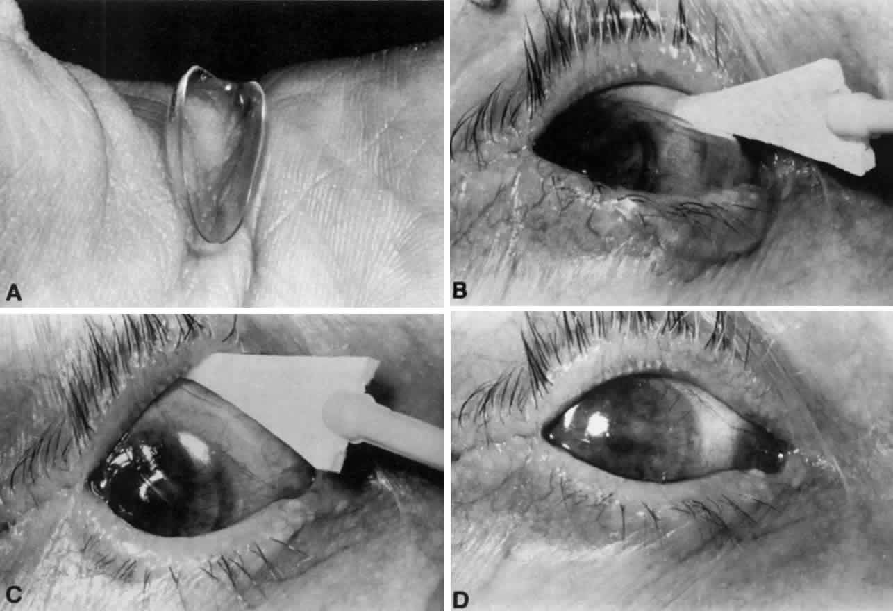
cornea outweighs any concern for lens spoilage and temporary discoloration.

In evaluating the corneal epithelium, it is often helpful to instill a fluorescein-containing topical anesthetic solution, place the patient at the slit lamp, slide the therapeutic lens temporally with a sterile cotton-tipped applicator, and ask the patient to blink once or twice. The corneal surface can then be evaluated with cobalt blue illumination before the contact lens is blinked or moved back into position with a cotton-tipped applicator.

TONOMETRY

Intraocular pressure must be monitored periodically during the use of a therapeutic lens. This can be accomplished with minimal ocular trauma by sliding the lens as described above before using an applanation tonometer. Alternatively, and with somewhat less trauma to the cornea, less difficulty for the examiner, and reasonable accuracy, the intraocular pressure can be measured with a Tono-Pen or Pneumatonometer with the contact lens in situ.8,9

TOPICAL MEDICATIONS

Topical medications administered for infectious keratitis, inflammation, or glaucoma may often accompany the use of a therapeutic lens. There is considerable uncertainty as to the precise amount of drug delivered to the cornea and anterior segment with a hydrogel lens in place. This issue has considerable clinical importance, which McCarey addressed by using a mathematical model of gentamicin delivery. This model demonstrated that although diffusion through the lens was increased by the presence of a higher water content, the more important pathway for drug delivery was around the edge of the lens.10

PRESERVATIVES

There is concern about the safety of using topical medications containing preservatives in the presence of a therapeutic lens. Lemp11 found that keratoconjunctivitis sicca patients treated with bandage lenses and frequent applications of preserved artificial tear solutions showed no accumulation of the preservative benzalkonium chloride in the contact lens after several weeks of treatment, nor any evidence of corneal epithelial toxicity. This is consistent with clinical experience using topical antibiotics and corticosteroids in the presence of therapeutic lenses without notable lens deterioration or epithelial toxicity. As always, especially with an already compromised ocular surface, preserved topical medications should be kept at the minimum level compatible with the desired therapeutic response.

PROPHYLACTIC TOPICAL ANTIBIOTIC

There is no definite consensus on the precise indications for topical antibiotic prophylaxis when using a therapeutic hydrogel lens, or on the antibiotic of choice. It would seem reasonable to use a topical antibiotic in the presence of measurable corneal epithelial defects or penetrating corneal lesions, but antibiotic prophylaxis may not be necessary when dealing with an intact epithelial surface or minimal punctate epitheliopathy. Indeed, potential unwanted consequences of topical antibiotics include toxicity, colonization by nonsusceptible organisms, and the theoretic emergence of resistant species.

No specific agent exists that uniquely qualifies for topical antibacterial prophylaxis. The aminoglycoside agents are lacking in the gram-positive spectrum. The newer quinoline medications have a somewhat broader spectrum than the aminoglycosides but are more effective against the virulent gram-negative rods than against streptococci and other gram-positive cocci. The present ophthalmic formulation of ciprofloxacin is not compatible with hydrogel lenses and leads to dense deposits on the lens surface. The newer quinoline formulations are more compatible with therapeutic lenses and may be the agents of choice when the risk-to-benefit ratio favors antibiotic use in the presence of a bandage contact lens. An alternative agent for broad-spectrum antibacterial prophylaxis might be a polymyxin-sulfa combination drop.

AVOIDING BANDAGE LENSES

As beneficial as therapeutic lenses may be, their use does entail additional patient expense, as well as potentially serious complications demanding greater physician responsibility. If at all possible, the use of therapeutic hydrogel lenses should be avoided in patients who are unreliable, have severe keratoconjunctivitis sicca, or have markedly diminished corneal sensation. In a significant number of cases, the use of bandage lenses may be avoided by substituting other more appropriate forms of treatment.

Many persistent epithelial defects may be persuaded to heal by merely decreasing or discontinuing unnecessary topical medications. A common example is the need to decrease rather than increase topical antiviral medication after prolonged treatment of herpes simplex keratitis. Systemic medications may be substituted for topical medications when surface toxicity is suspected (e.g., oral acyclovir may be substituted for topical trifluridine when antiviral coverage is needed but surface toxicity is present). Ophthalmic ointments, frequently formulated without preservatives, may be substituted for equivalent preserved aqueous solutions.

It is desirable, if possible, to identify and treat the responsible pathophysiologic factor rather than obtain instant and temporary improvement from the continued use of the bandage lens. Thus, punctal occlusion, often using removable plugs, may minimize the effect of dry eyes on an ocular surface problem. Defective eyelid closure and defective eyelid position are frequent causes of corneal epitheliopathy for which oculoplastic repair is more appropriate than prolonged bandage contact lens wear.

Therapeutic hydrogel lenses are more helpful in the short to medium term than in the long term. This relates to both the patient and clinician resources necessary for continued use, as well as the cumulative increased probability of lens-associated complications over time. Thus, self-limited conditions such as a postoperative “rough surface” are much more appropriate for bandage lens therapy than a chronic ocular surface condition without a definite end point.

ADDITIONAL MEASURES

I have found that the loss of therapeutic lenses occurs frequently at night during sleep. For that reason, we instruct our patients to wear an eye shield at bedtime, which seems to increase lens retention.

It is not uncommon for therapeutic lens wear to be indicated in the presence of marked blepharophimosis or lateral tarsorrhaphy. In such cases, insertion may be difficult or impossible through the narrowed interpalpebral fissure with the use of customary insertion techniques. Sliding the contact lens superiorly with a sterile applicator in an anesthetized eye will usually overcome this problem (see Fig. 1).

Back to Top
COMPLICATIONS

MICROBIAL KERATITIS

Kent and colleagues3 described 22 cases of microbial keratitis associated with therapeutic lenses, 15 of which were culture proven. Although some uncommon organisms were cultured, streptococci and other gram-positive organisms were common in this series. The prevalence of suppurative keratitis in bullous keratopathy patients was 13% in another series.4 Furthermore, culture-proven bacterial keratitis approached 10% prevalence in a large series of keratoplasty patients fitted with therapeutic lenses.12 This, however, must be balanced against the selection of patients fitted with bandage lenses; it is likely that these patients had a higher rate of keratoconjunctivitis sicca, blepharitis, and eyelid abnormalities, placing them at higher risk for ocular surface infection.

TIGHT LENS SYNDROME

Another complication, the tight lens syndrome, presents as conjunctival injection and discomfort in the presence of an essentially immobile hydrogel lens. The conjunctiva surrounding the lens may appear to be molded by the edge of the lens. Iritis of variable intensity, occasionally with hypopyon formation, may be present. Corneal edema usually accompanies the above signs.

McCarey and Wilson13 describe a mechanism by which contact lens dehydration and lowering of the surface pH cause a tighter fitting lens. This may explain why a previously well-fitting hydrogel lens may change its properties and cause a tight lens syndrome. A lens with a smaller diameter or a flatter base curve may be needed to prevent recurrences.

SURFACE DEPOSITS

Deposition of protein and calcium salts on the anterior lens surface is a soft contact lens complication that may occur with therapeutic use. Recently, however, the use of disposable lenses with frequent elective replacement has decreased the prevalence of lens deposits. The presence of deposits is an absolute indication for the replacement of the lens.

CORNEAL NEOVASCULARIZATION AND EDEMA

Other complications include corneal neovascularization and corneal edema. Contact lens-induced corneal neovascularization begins superficially and peripherally but can progress to threaten a penetrating graft or change the prognosis for subsequent penetrating keratoplasty. Mild corneal edema with subtle folds in Descemet's membrane is often observed after insertion of a bandage contact lens. More prominent corneal edema may occur, particularly in conjunction with an abnormal corneal endothelium or a tight-fitting lens.

GIANT PAPILLARY CONJUNCTIVITIS

Giant papillary conjunctivitis is characterized by mild conjunctival injection and mucoid discharge associated with progressive intolerance to the contact lens. Slit lamp examination demonstrates diffuse papillae of the superior tarsal conjunctiva (best detected with fluorescein staining and cobalt blue illumination), as well as much larger, typical papillary excrescences. This syndrome may occur with long-term use of a therapeutic lens. If a therapeutic lens must be worn despite the presence of giant papillary conjunctivitis, a smaller, thinner lens (or a disposable lens), frequent lens replacements, and the use of topical sodium cromolyn may be helpful.

Back to Top
REFERENCES

1. Gasset AR, Kaufman HE: Therapeutic uses of hydrophilic contact lenses. Am J Ophthalmol 69:252, 1970

2. Smiddy WE, Hamburg TR, Kracher GP et al: Therapeutic contact lenses. Ophthalmology 97:291, 1990

3. Kent HD, Cohen EJ, Laibson PR et al: Microbial keratitis and corneal ulceration associated with therapeutic soft contact lenses. CLAO J 15:49, 1990

4. Andrew NC, Woodward EG: The bandage lens in bullous keratopathy. Ophthalmic Physiol Opt 9(1):66, 1989

5. Busin M, Spitznas M: Sustained gentamicin release by presoaked medicated bandage contact lenses. Ophthalmology 95(6):796, 1988

6. Blok MD, Kok JH, van Mil C et al: Use of the Megasoft Bandage Lens for treatment of complications after trabeculectomy. Am J Ophthalmol 110(3):264, 1990

7. Groden LR, White W: Porcine collagen corneal shield treatment of persistent epithelial defects following penetrating keratoplasty. CLAO J 16(2):95, 1990

8. Khan JA, Lagreca BA: Tono-Pen estimation of intraocular pressure through bandage contact lenses. Am J Ophthalmol 108(4):422, 1989

9. Rubenstein JB, Deutsch TA: Pneumatonometry through bandage contact lenses. Arch Ophthalmol 103(11):1660, 1985

10. McCarey BE, Schmidt FH, Wilkinson KD et al: Gentamicin diffusion across hydrogel bandage lenses and its kinetic distribution on the eye. Curr Eye Res 3:977, 1984

11. Lemp MA: Bandage lenses and the use of topical solutions containing preservatives. Ann Ophthalmol 10:1319, 1978

12. Saini JS, Rao GN, Aquavella JV: Post-keratoplasty corneal ulcers and bandage lenses. Acta Ophthalmol (Copenh) 66(1):99, 1988

13. McCarey BE, Wilson CA: pH, osmolarity and temperature effects on the water content of hydrogel contact lenses. Contact Intraocular Lens Med J 8:158, 1982

Back to Top