Chapter 10
Tumors of the Conjunctiva and Cornea
CAROL L. SHIELDS and JERRY A. SHIELDS
Main Menu   Table Of Contents

Search

ANATOMY
DIAGNOSTIC APPROACHES
MANAGEMENT
CONGENITAL LESIONS
BENIGN TUMORS OF SURFACE EPITHELIUM
MALIGNANT LESIONS OF SURFACE EPITHELIUM
MELANOCYTIC TUMORS
VASCULAR TUMORS
FIBROUS TUMORS
NEURAL TUMORS
HISTIOCYTIC TUMORS
MYXOID TUMORS
LIPOMATOUS TUMORS
LYMPHOID TUMORS
LEUKEMIA
METASTATIC TUMORS
SECONDARY CONJUNCTIVAL INVOLVEMENT FROM ADJACENT TUMORS
CARUNCULAR TUMORS AND CYSTS
MISCELLANEOUS LESIONS THAT CAN SIMULATE CONJUNCTIVAL NEOPLASMS
REFERENCES

Tumors of the conjunctiva and cornea occupy a large spectrum of conditions ranging from benign inflammatory lesions, such as pinguecula or episcleritis, to aggressive, life-threatening malignancies, such as melanoma or Kaposi's sarcoma.1–3 The clinical differentiation of these tumors is based on the patient's medical background as well as certain typical clinical features of the tumor. The recognition and proper management of such tumors requires an understanding of the anatomy of the conjunctiva and cornea and knowledge of general principles of tumor management, both of which are described below. The specific clinical and histopathologic features, as well as the management of each tumor, are discussed, based on the authors' personal experience with more than 1600 patients with conjunctival tumors during a 30-year period.3
Back to Top
ANATOMY
The conjunctiva is a continuous mucous membrane that covers the anterior portion of the globe. It extends from the eyelid margin onto the back surface of the eyelid (palpebral portion), into the fornix (forniceal portion), onto the surface of the globe (bulbar portion), and up to the corneoscleral limbus (limbal portion). The conjunctiva is composed of epithelium and stroma. The epithelium consists of both stratified squamous and columnar epithelium. The squamous pattern is found near the limbus, and the columnar pattern is found near the fornix. The stroma is comprised of fibrovascular connective tissue that thickens in the fornix and thins at the limbus.

Special regions of the conjunctiva include the plica semilunaris and caruncle. The plica semilunaris is a vertically oriented fold of conjunctiva located in the medial portion of the bulbar conjunctiva. It is speculated that the plica semilunaris represents a remnant of the nictitating membrane found in certain animals. The caruncle is located in the medial canthus between the upper and lower puncta. It contains both conjunctival and cutaneous structures, such as nonkeratinized stratified squamous epithelium overlying the stroma of fibroblasts, melanocytes, sebaceous glands, hair follicles, and striated muscle fibers.

The conjunctiva can spawn neoplasms from both its epithelial and stromal structures. These are similar clinically and histopathologically to tumors that arise from other mucous membranes in the body. Similarly, the cornea can develop epithelial tumors, but stromal tumors are extremely rare from this structure. The caruncle, with its unique composition of both mucous membrane and cutaneous structures, can generate tumors found both in mucosa and skin.

Back to Top
DIAGNOSTIC APPROACHES
Unlike many other mucous membranes in the body, the conjunctiva is readily visible. Thus, tumors and related lesions that occur in the conjunctiva are generally recognized at a relatively early stage. Because many of these tumors have typical clinical features, an accurate diagnosis can often be made by external ocular examination and slit lamp biomicroscopy, provided that the clinician is familiar with their clinical characteristics. A diagnostic biopsy is not usually necessary in cases of smaller tumors that appear benign. If a smaller tumor does require a biopsy, it is often better to completely remove the lesion in one operation (excisional biopsy). In cases of larger lesions, however, it may be appropriate to remove a portion of the tumor (incisional biopsy) to obtain a histopathologic diagnosis before embarking on more extensive therapy. Because conjunctival tumors are readily accessible to incisional biopsy, it is rarely necessary to do exfoliative cytology or fine-needle aspiration biopsy, both of which provide less material than incisional biopsy.

In addition to evaluation of the conjunctival lesion, meticulous slit lamp examination of the cornea is essential in patients with suspected conjunctival tumors. Invasion of squamous cell carcinoma and melanoma into the peripheral cornea may appear as a subtle gray surface opacity. It is important to completely outline such corneal involvement before surgery because it is often less visible through the operating microscope than it is with slit lamp biomicroscopy in the office.

Back to Top
MANAGEMENT
Depending on the presumptive diagnosis and the size and extent of the lesion, management of a conjunctival tumor may consist of serial observation, incisional biopsy, excisional biopsy, cryotherapy, chemotherapy, radiotherapy, modified enucleation, orbital exenteration, or various combinations of these methods.4–7 If large areas of conjunctiva are removed, mucous membrane grafts from the conjunctiva of the opposite eye, buccal mucosa, or amniotic membrane may be necessary.8,9

OBSERVATION

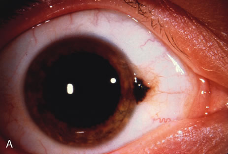
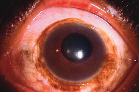
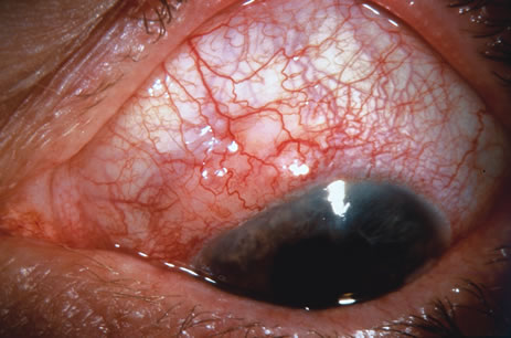
Observation is generally the management of choice for most benign, asymptomatic tumors of the conjunctiva. Selected examples of lesions that can be observed without interventional treatment include pinguecula, dermolipoma, and nevus. External or slit lamp photographs are advisable to document all lesions and are critical in following up suspicious lesions. Most patients are examined every 6 to 12 months for evidence of growth, malignant change, or secondary effects on normal surrounding tissues.

INCISIONAL BIOPSY

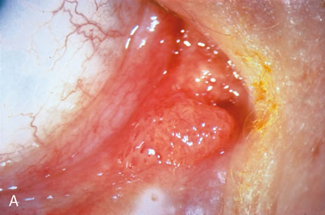
Incisional biopsy is reserved for extensive suspicious tumors that are symptomatic or suspected to be malignant. Examples include large squamous cell carcinoma, primary acquired melanosis (PAM), melanoma, and conjunctival invasion by sebaceous gland carcinoma. It should be understood that if such tumors occupy 4 clock hours or less on the bulbar conjunctiva, excisional biopsy is generally preferable to incisional biopsy. However, larger lesions can be approached by incisional wedge biopsy or punch biopsy. Further definitive therapy would then be planned based on the results of biopsy. Incisional biopsy is also appropriate for conditions that are ideally treated with radiotherapy, chemotherapy, or other topical medications. Such lesions include lymphoid tumors, metastatic tumors, extensive papillomatosis, and some cases of squamous cell carcinoma and PAM.

EXCISIONAL BIOPSY

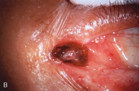
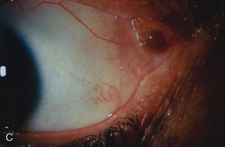
Primary excisional biopsy is appropriate for intermediate and small tumors that are symptomatic or suspected to be malignant. In such situations, excisional biopsy is preferred over incisional biopsy to avoid inadvertent tumor seeding. Examples of benign and malignant lesions that are ideally managed by excisional biopsy include symptomatic limbal dermoid, epibulbar osseous choristoma, steroid-resistant pyogenic granuloma, squamous cell carcinoma, and melanoma. When such lesions are located in the conjunctival fornix, they can be completely excised and the conjunctiva reconstructed primarily with absorbable sutures, sometimes with fornix-deepening sutures, or symblepharon ring to prevent adhesions. If the defect cannot be closed primarily, then a mucous membrane graft may be inserted.

Most primary malignant tumors of the conjunctiva, such as squamous cell carcinoma and melanoma, arise in the interpalpebral area near the limbus, and the surgical technique for limbal tumors is different than that for forniceal tumors.4–6 Limbal neoplasms have a propensity to invade through the corneal epithelium and sclera into the anterior chamber and also through the soft tissues into the orbit. Thus, it is often necessary to remove a thin lamella of sclera to achieve tumor-free margins and to decrease the chance for tumor recurrence. In this regard, we employ a partial lamellar sclerokeratoconjunctivectomy with primary closure for such tumors (Fig. 1). Because cells from these friable tumors can seed into adjacent tissues, a gentle technique without touching the tumor (“no–touch” technique) is mandatory. Additionally, the surgery should be performed using microscopic technique and the operative field should be left dry so that cells adhere to the resected tissue. It is wise to avoid wetting the field with balanced salt solution until after the tumor is completely removed, to minimize seeding of cells.

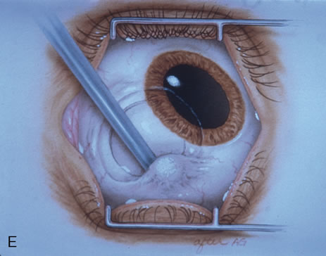
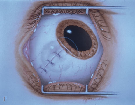
Fig. 1. Surgical excision of conjunctival malignancy using the “no-touch” technique. A. Absolute alcohol is applied by a cotton tip applicator to the involved cornea to allow for controlled corneal epitheliectomy. B. The corneal epithelium is scrolled off using a controlled sweeping motion with a Beaver blade. C. The conjunctival incision is made approximately 4 mm outside the tumor margin. A Beaver blade is used to create a thin lamella of tumor-free sclera underlying the limbal portion of the tumor. D. The conjunctival malignancy is removed, along with tumor-free margins, including underlying sclera and limbal corneal epithelium. E. Cryotherapy is applied to the conjunctiva at the site of resection. F. Closure of the conjunctiva with absorbable sutures is performed.

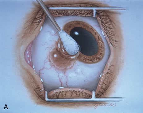
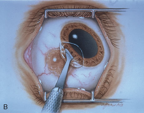
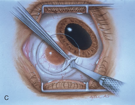
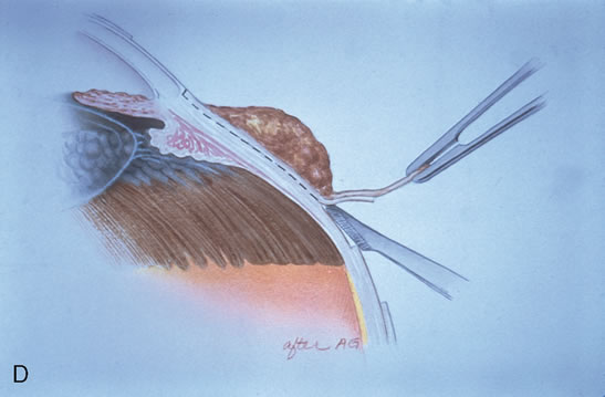
The technique for resection of limbal tumors is shown in Figure 1. Using retrobulbar anesthesia and the operating microscope, the corneal epithelial component is approached first and the conjunctival component is dissected second, with the goal of excising the entire specimen completely in one piece. Absolute alcohol soaked on an applicator is gently applied to the entire corneal component. This causes epithelial cellular devitalization and allows easier release of the tumor cells from Bowman's layer. A Beaver blade is used to microscopically outline the malignancy within the corneal epithelium using a delicate epithelial incision or epitheliorhexis technique 2 mm outside the corneal component. The Beaver blade is then used to sweep gently the affected corneal epithelium from the direction of the central cornea to the limbus, into a scroll that rests at the limbus. Next, a pentagonal or circular conjunctival incision based at the limbus is made 4 to 6 mm outside the tumor margin. The incision is carried through the underlying Tenon's fascia until the sclera is exposed so that full-thickness conjuctiva and Tenon's fascia is incorporated into the excisional biopsy. Cautery is applied to control bleeding. A second incision is then outlined by a superficial scleral groove approximately 0.2 mm in depth and 2.0 mm outside the base of the overlying adherent conjunctival mass. This groove is continued anteriorly to the limbus. The area outlined by the scleral groove is removed by flat dissection of 0.2 mm thickness within the sclera in an attempt to remove a superficial lamella of sclera, overlying Tenon's fascia and conjunctiva with tumor, and the scrolled corneal epithelium. In this way, the entire tumor with tumor-free margins is removed in one piece without touching the tumor itself. The removed specimen is then placed flatly on a piece of thin cardboard from the surgical tray and then placed in fixative and submitted for histopathologic studies. This step prevents the specimen from folding and allows better assessment of the tumor margins histopathologically. The used instruments are then replaced with fresh instruments for subsequent steps, to avoid contamination of healthy tissue with possible tumor cells.

After excision of the specimen, cryotherapy is applied to the margins of the remaining bulbar conjunctiva. This is performed by freezing the surrounding bulbar conjunctiva as it is lifted away from the sclera using the cryoprobe. When the ice ball reaches a size of 4 to 5 mm, it is allowed to thaw, and the cycle is repeated once. The cryoprobe is then moved to an adjacent area of the conjunctiva, and the cycle is repeated until all of the margins have been treated by this method. It is not necessary to treat the corneal margins with cryoapplication. The tumor bed is treated with absolute alcohol wash on cotton tip applicator and bipolar cautery, avoiding cryotherapy directly to the sclera.

Using clean instruments, the conjunctiva is mobilized for closure of the defect by loosening the intermuscular septum with Steven's scissor spreading and creating transpositional conjunctival flaps. Closure is completed with interrupted absorbable 6-0 or 7-0 sutures. If the surgeon prefers, an area of bare sclera may be left near the limbus; however, we prefer complete closure because it promotes better healing and allows for facility of further surgery if recurrence develops. The patient is treated with topical antibiotics and corticosteroids for 2 weeks, then followed at 3- to 6-month intervals.

CRYOTHERAPY

In the management of conjunctival tumors, cryotherapy may be used as a supplemental treatment to excisional biopsy as described above. In such cases, it can eliminate microscopic tumor cells and prevent recurrence of malignant tumors, such as squamous cell carcinoma and melanoma. It can also be used as a principal treatment for PAM and pagetoid invasion of sebaceous gland carcinoma. If cryotherapy can devitalize the malignant or potentially malignant cells in such instances, radical surgery, such as orbital exenteration, can often be delayed or avoided.

CHEMOTHERAPY

Recent evidence has revealed that topical eye drops containing mitomycin C, 5-fluorouracil, or interferon are effective in treating epithelial malignancies such as squamous cell carcinoma, PAM, and pagetoid invasion of sebaceous gland carcinoma.10–18 Mitomycin C or 5-fluorouracil is employed most successfully for squamous cell carcinoma, especially after tumor recurrence following previous surgery. This medication is prescribed in a topical dosage four times daily for a 1-week period, followed by a 1-week hiatus to allow the ocular surface to recover (Table 1). This cycle is repeated once again, so most patients receive a total of 2 weeks of the chemotherapy. Both mitomycin C and 5-fluorouracil are most effective for squamous cell carcinoma and less effective for PAM and pagetoid invasion of sebaceous gland carcinoma. Toxicities include most commonly dry eye findings, superficial punctate epitheliopathy, and punctal stenosis. Corneal melt, scleral melt, and cataract may develop if these agents are used in the presence of open conjunctival wounds or used excessively. Topical interferon can be effective for squamous epithelial malignancies and is less toxic to the surface epithelium, but this medication may require many months of use to effect a result.


Table 1. Protocol for Use of Mitomycin C for Conjunctival Squamous Cell Neoplasia and Primary Acquired Melanosis


WeekProtocol
1Slit lamp biomicroscopy
Place upper and lower punctal plugs
Cycle 1: mitomycin C 0.04% q.i.d. to the affected eye
2No medication
3Cycle 2: mitomycin C 0.04% q.i.d. to the affected eye
4No medication
Slit lamp biomicroscopy
Prescribe more cycles if residual tumor exists
Remove punctal plugs after all medication complete


RADIOTHERAPY

Two forms of radiotherapy are employed for conjunctival tumors, namely, external beam radiotherapy and custom-designed plaque radiotherapy. External beam radiotherapy to a total dose of 3000 to 4000 centigray (cGy) is used to treat conjunctival lymphoma and metastatic carcinoma when such lesions are too large or diffuse to excise locally. Side effects of dry eye, punctate epithelial abnormalities, and cataract should be anticipated.

Custom-designed plaque radiotherapy19 to a dose of 3000 to 4000 cGy can be used to treat conjunctival lymphoma or metastasis. A higher dose of 6000 to 8000 cGy can be employed to treat the more radiation-resistant melanoma and squamous cell carcinoma. In general, plaque radiotherapy is reserved for those patients who have diffuse tumors that are incompletely resected and for those who display multiple recurrences. There are two design choices for conjunctival custom plaque radiotherapy: a conformer plaque technique with six fractionated treatment sessions on an outpatient basis, or a reverse plaque technique with the device sutured onto the episcleral in an inpatient setting. In unique instances, plaque radiotherapy to a low dose of 2000 cGy is employed for benign conditions, such as steroid-resistant pyogenic granuloma, that show recurrence after surgical resection.20 Such treatment should be performed by experienced radiation oncologists and ocular oncologists.

MODIFIED ENUCLEATION

Modified enucleation is a treatment option for primary malignant tumors of the conjunctiva that have invaded through the limbal tissues into the globe, producing secondary glaucoma. Such an occurrence is quite rare but can occasionally occur with squamous cell carcinoma and melanoma. The uncommon mucoepidermoid variant of squamous cell carcinoma of the conjunctiva has a greater tendency for such invasion.21,22 At the time of enucleation, it is necessary to remove the involved conjunctiva intact with the globe to avoid spreading tumor cells. Thus, the initial peritomy should begin at the limbus, but when the tumor is approached, the incision should proceed posteriorly from the limbus to surround the tumor-affected tissue by at least 3 to 4 mm. The tumor will remain adherent to the globe at the limbus. Occasionally, a suture is employed through the surrounding conjunctiva into the episclera to secure the tumor to the globe so that it will not be displaced during subsequent manipulation. The remaining steps of enucleation are gently performed, and the globe is removed with tumor adherent after cutting the optic nerve from the nasal side. The margins of the remaining, presumed unaffected conjunctiva are treated with double freeze-thaw cryotherapy. Often this surgical technique leaves the patient with a limited amount of residual unaffected conjunctiva for closure. In these instances, a mucous membrane graft or amniotic membrane graft may be necessary for adequate closure and to provide fornices for a prosthesis. In some instances, a simple horizontal inferior forniceal conjunctival incision from canthus to canthus may suffice, as long as the conformer is constantly worn as a template, so the new conjunctival fornix grows deep and around this structure.

ORBITAL EXENTERATION

Orbital exenteration is probably the treatment of choice for primary malignant conjunctival tumors that have invaded the orbit or that exhibit complete involvement of the conjunctiva.7,23,24 Either an eyelid-removing or eyelid-sparing exenteration is employed, depending on the extent of eyelid involvement. The eyelid-sparing technique is preferred because the patients have better cosmetic appearance and heal within 2 or 3 weeks. Specifically, if the anterior lamella of the eyelid is uninvolved with tumor, an eyelid-sparing (eyelid-splitting) exenteration may be accomplished.4,7,24

MUCOUS MEMBRANE GRAFT

Mucous membrane grafts are occasionally necessary to replace vital conjunctival tissue after removal of extensive conjunctival tumors. The best donor sites include the forniceal conjunctiva of the ipsilateral or contralateral eye and buccal mucosa from the posterior aspect of the lower lip or lateral aspect of the mouth. Such grafts are usually removed by a freehand technique, fashioned to fit the defect, and secured into place with cardinal and running absorbable 6-0 or 7-0 sutures. Currently, in most instances, we employ a donor amniotic membrane graft to replace lost conjunctiva.8,9 The tissue is delivered frozen and must be defrosted for 20 minutes. The fine, transparent material is carefully peeled off its cardboard surface, laid basement membrane side up, and sutured into place with absorbable sutures. Topical antibiotic and steroid ointments are applied following all conjunctival grafting procedures.

It is important that the surgeon use a minimal manipulation technique for tumor resection. For graft harvest and placement, the surgeon should always use clean, sterile instruments at both the donor and the recipient sites. Free tumor cells can rest on instrument tips and later implant and grow in previously uninvolved areas if such precautions are not taken.

Back to Top
CONGENITAL LESIONS
A variety of tumors and related conditions may be present at birth or become clinically apparent shortly after birth.25,26 Most of the lesions to be considered here are choristomas, consisting of tissue elements that are not normally present at the involved site. Despite their presence at a young age, all of the conjunctival choristomas discussed herein are sporadic, without hereditary tendency.

DERMOID

Conjunctival dermoid is a congenital, well-circumscribed yellow-white solid mass that involves the bulbar conjunctiva at the corneoscleral limbus.25–29 It characteristically occurs near the limbus inferotemporally, and often this tumor has fine white hairs best seen with slit lamp biomicroscopy (Fig. 2). In rare cases, it can extend to the central cornea or be located in other quadrants on the bulbar surface. It may occur as an isolated lesion or may be associated with Goldenhar's syndrome. Hence, the patient should be evaluated for ipsilateral or bilateral preauricular skin appendages, hearing loss, eyelid coloboma, orbitoconjunctival dermolipoma, and cervical-vertebral anomalies that make up this nonheritable syndrome. Histopathologically, the conjunctival dermoid is a simple choristomatous malformation that consists of dense fibrous tissue lined by conjunctival epithelium with deeper dermal elements, such as hair follicles and sebaceous glands.

Fig. 2. Epibulbar dermoid. A. Limbal dermoid. B. Central corneal dermoid.

The management of an epibulbar dermoid includes simple observation if the lesion is small and visually asymptomatic. It is possible to excise the lesion for cosmetic reasons, but the remaining corneal scar is sometimes cosmetically unacceptable. Larger or symptomatic dermoids may produce visual loss from astigmatism. These can be approached by lamellar keratosclerectomy with primary closure of overlying tissue if the defect is superficial, or closure using corneal graft if the defect is deep or full thickness. It has been reported that the cosmetic appearance may improve, but the refractive and astigmatic error and visual acuity may not change.29 When the lesion involves the central cornea, a lamellar or penetrating keratoplasty may be necessary and long-term amblyopia may be a problem.28 Occasionally, extensive dermoids involve the lateral canthus, and carefully planned excision with lateral canthal repair is necessary.

DERMOLIPOMA

Dermolipoma is believed to be congenital and present at birth, but it typically remains asymptomatic for years and may not be detected until adulthood when it protrudes from the orbit through the conjunctival fornix superotemporally (Fig. 3). It appears as a pale yellow, soft, fluctuant, fusiform mass below the palpebral lobe of the lacrimal gland, best visualized with the eye in inferonasal gaze. It usually extends for a variable distance into the orbital fat and onto the bulbar conjunctiva, and occasionally it can extend anteriorly to reach the limbus. Unlike herniated orbital fat, dermolipoma may contain fine white hairs on its surface and cannot be reduced with digital pressure into the orbit.

Fig. 3. Dermolipoma in the superotemporal conjunctival fornix.

On computed tomography or magnetic resonance imaging, dermolipoma has features similar to orbital fat, from which it may be indistinguishable. Histopathologically, it is lined by conjunctival epithelium on its surface, and the subepithelial tissue has variable quantities of collagenous connective tissue. Pilosebaceous units and lacrimal gland tissue may occasionally be present. The majority of dermolipomas require no treatment, but larger symptomatic ones or those that are cosmetically unappealing can be managed by excision of the entire orbitoconjunctival lesion through a conjunctival forniceal approach or by simply removing the anterior portion of the lesion in a manner similar to that used to remove prolapsed orbital fat.

EPIBULBAR OSSEOUS CHORISTOMA

Epibulbar osseous choristoma is a rigid deposit of bone generally located in the bulbar conjunctiva superotemporally (Fig. 4).30 It is believed to be congenital, and typically remains undetected until palpated by the patient in the preteen years. It is clinically suspected because of its rock-hard consistency on palpation, although fibrous tissue tumors can feel similar. The diagnosis can be confirmed with ultrasonography or computed tomography to illustrate the calcium component. This tumor is generally best managed by periodic observation. Occasionally patients report a foreign-body sensation, and such symptomatic lesions can be excised with a circumtumoral conjunctival incision followed by dissection to bare sclera for full-thickness conjunctival resection. Tumors that might be adherent to the sclera may warrant a superficial sclerectomy.30

Fig. 4. Epibulbar osseous choristoma on the bulbar conjunctiva superotemporally, presenting as a firm, palpable mass.

LACRIMAL GLAND CHORISTOMA

Lacrimal gland choristoma is a congenital lesion often discovered in young children as an asymptomatic pink stromal mass, most often in the inferior bulbar or forniceal conjunctiva. It is speculated that this lesion presents in this location because of the pathway that the lacrimal gland takes during embryogenesis, from the inferior to the superotemporal region. The lacrimal gland choristoma can masquerade as a focus of inflammation because of its pink color. Rarely, a cystic appearance ensues from this secretory mass if there is no connection to the conjunctival surface. Excisional biopsy is usually performed to confirm the diagnosis.

RESPIRATORY CHORISTOMA

In unique instances, a cystic choristoma, appearing as congenital sclerocorneal ectasia, is found. In one report, such a case was found to manifest respiratory mucosa.31

COMPLEX CHORISTOMA

The conjunctival dermoid and epibulbar osseous choristoma are termed simple choristomas because they contain one tissue type, such as skin or bone. A complex choristoma contains a greater variety of tissue, such as dermal appendages, lacrimal gland tissue, cartilage, bone, and occasionally other elements. It is quite variable in its clinical appearance and may cover much of the epibulbar surface, or it may form a circumferential growth pattern around the limbus (Fig. 5). For example, a complex choristoma with extensive lacrimal tissue appears as a lobular pink mass, whereas one with dermal tissue appears yellow and thick and one with cartilage displays a smooth blue-gray hue. The complex choristoma has a peculiar association with the linear nevus sebaceus of Jadassohn.32–34 The nevus sebaceus of Jadassohn includes cutaneous features, such as sebaceous nevus in the facial region, and neurologic features, such as seizures, mental retardation, arachnoid cyst, and cerebral atrophy. The ophthalmic features of this syndrome include epibulbar complex choristoma and posterior scleral cartilage.32

Fig. 5. Epibulbar complex choristoma that was found histopathologically to have cartilage and ectopic lacrimal gland.

Management of the complex choristoma depends on the extent of the lesion. Observation or wide local excision with mucous membrane graft reconstruction are options. In the rare case of a very extensive lesion, where the lesion causes dense amblyopia with no hope for visual acuity, modified enucleation with ocular surface reconstruction may be necessary.

Back to Top
BENIGN TUMORS OF SURFACE EPITHELIUM
Several benign tumors and related conditions may arise from the squamous epithelium of the conjunctiva.

PAPILLOMA

Squamous papilloma is a benign tumor, documented to originate from human papillomavirus infection of the conjunctiva.35,36 This tumor can occur in both children and adults. It is speculated that the virus is acquired through transfer from the mother's vagina to the newborn's conjunctiva as the child passes through the mother's birth canal. Papilloma appears as a pink fibrovascular frond of tissue arranged in a sessile or pedunculated configuration. The numerous fine vascular channels ramify through the stroma beneath the epithelial surface of the lesion. In children, the lesion is usually small, multiple, and located in the inferior fornix (Fig. 6). In adults, it is usually solitary, more extensive, and can often extend to cover the entire corneal surface, simulating malignant squamous cell carcinoma. Histopathologically, the lesion shows numerous vascularized papillary fronds lined by acanthotic epithelium.

Fig. 6. Recurrent conjunctival papilloma in a child. A. The fibrovascular mass caused bloody tears. B. Following 3 months of oral cimetidine, the mass resolved.

In the case of a small sessile papilloma in a child, a trial of topical corticosteroids may bring about resolution of the lesion. Sometimes, periodic observation allows for slow spontaneous resolution of the viral-produced tumor. Larger or more pedunculated lesions are generally symptomatic, with foreign-body sensation, chronic mucus production, hemorrhagic tears, incomplete eyelid closure, and poor cosmetic appearance. Such lesions are unlikely to show a favorable response to observation or steroids and are best managed by surgical excision. Complete removal of the mass without direct manipulation of the tumor (no-touch technique) is generally advisable to avoid spreading the tumor-related virus. Double freeze-thaw cryotherapy is applied to the remaining conjunctiva around the excised lesion to help prevent tumor recurrence. In some instances, the pedunculated tumor is frozen alone and allowed to slough off the conjunctival surface later. For some large, unwieldy pedunculated tumors, complete cryotherapy of the mass down its stalk to its base is performed and excision while the mass is in the frozen state is achieved. This is especially important for large lesions, to allow for traction on the tumor without forceps manipulation. Closure is completed with absorbable sutures. For lesions that show recurrence, oral cimetidine for several months can resolve the papilloma virus–related tumor by boosting the patient's immune system and stimulating regression of the mass (Fig. 6B).36

KERATOACANTHOMA

The conjunctiva can give rise to benign reactive inflammatory lesions that simulate carcinoma, such as pseudocarcinomatous hyperplasia and its variant, keratoacanthoma.37 In some instances, a distinct nodule is found. This lesion appears gelatinous or leukoplakic, similar to squamous cell carcinoma of the conjunctiva, but its onset may be more rapid. Massive acanthosis, hyperkeratosis, and parakeratosis are found histopathologically.37

HEREDITARY BENIGN INTRAEPITHELIAL DYSKERATOSIS

Hereditary benign intraepithelial dyskeratosis (HBID) is a peculiar condition seen in an inbred isolate of Caucasian, African American, and American Indians (Haliwa Indians). This group resided initially in North Carolina. HBID has subsequently been detected in several other parts of the United States. It is an autosomal dominant disorder characterized by bilateral, elevated fleshy plaques on the nasal or temporal perilimbal conjunctiva (Fig. 7).38 Similar plaques can occur on the buccal mucosa. HBID may remain relatively asymptomatic, or it may cause severe redness and foreign-body sensation. In some instances, it can extend onto the cornea. It has no known malignant potential. It is characterized histopathologically by acanthosis, dyskeratosis, and prominent rete pegs.

Fig. 7. Hereditary benign intraepithelial dyskeratosis in a young woman who is a descendent of a Haliwa Indian. The opposite eye had a similar lesion.

HBID is a benign condition that does not usually require aggressive treatment. Smaller, less-symptomatic lesions can be treated with ocular lubricants and judicious use of topical corticosteroids. Larger, symptomatic lesions can be managed by local resection with mucous membrane grafting if necessary.

EPITHELIAL INCLUSION CYST

Conjunctival cysts may occur spontaneously or following inflammation, surgery, or nonsurgical trauma. Histopathologically, they are lined by conjunctival epithelium and are filled with clear fluid that often contains desquamated cellular debris (Fig. 8). Such cysts can simply be observed, or they can be excised completely, with primary closure of the conjunctiva.

Fig. 8. Epibulbar inclusion cyst with thick mucus from conjunctival glands.

DACRYOADENOMA

Dacryoadenoma is a rare conjunctival tumor, noted in children or young adults as a pink mass in the inferior bulbar or palpebral region.39 It is uncertain whether the lesion is congenital or acquired. This benign tumor appears to originate from the surface epithelium and proliferate into the stroma, forming glandular lobules similar to the lacrimal gland.

KERATOTIC PLAQUE

Keratotic plaque is a white limbal or bulbar conjunctival mass, usually in the interpalpebral region.1 It is composed of acanthosis and parakeratosis with keratinization of the epithelium. It appears similar to squamous cell carcinoma with leukoplakia.

ACTINIC KERATOSIS

Actinic keratosis is a frothy white lesion usually located over a chronically inflamed pinguecula or pterygium.1 Histopathologically, it is composed of a proliferation of surface epithelium with keratosis. Clinically, it resembles squamous cell carcinoma of the conjunctiva.

Back to Top
MALIGNANT LESIONS OF SURFACE EPITHELIUM
Squamous cell neoplasia can occur as a localized lesion confined to the surface epithelium (conjunctival intraepithelial neoplasia) or as a more-invasive squamous cell carcinoma that has broken through the basement membrane and invaded the underlying stroma.21,22,40–44 The former has no potential to metastasize, but the latter can gain access to the conjunctival lymphatics and occasionally metastasize to regional lymph nodes. It has been found that most squamous cell neoplasia is related to human papillomavirus infection of the conjunctival epithelium; this is most certain in patients with bilateral squamous cell neoplasia and immunosuppressed patients who develop this disease.45,46

The currently accepted term for the localized variety of squamous cell neoplasia is conjunctival intraepithelial neoplasia (CIN). When the abnormal cellular proliferation involves only partial thickness of the epithelium, it is classified as mild CIN, a condition also called dysplasia. When it affects full-thickness epithelium, it is classified as severe CIN, a condition also called carcinoma in situ. It is stressed that these are histopathologic terms and the differential between CIN mild and CIN severe cannot be made clinically.

CONJUNCTIVAL INTRAEPITHELIAL NEOPLASIA

Clinically, CIN appears as a fleshy, sessile or minimally elevated lesion usually at the limbus in the interpalpebral fissure and less commonly in the forniceal or palpebral conjunctiva (Fig. 9). The limbal lesion may extend for a variable distance into the superficial portion of the adjacent cornea. A white plaque (leukoplakia) may occur on the surface of the lesion as result of secondary hyperkeratosis.

Fig. 9. Conjunctival intraepithelial neoplasia (carcinoma in situ) with corneal involvement, displaying leukoplakia on both the conjunctiva and cornea.

Histopathologically, mild CIN (dysplasia) is characterized by a partial-thickness replacement of the surface epithelium by abnormal epithelial cells that lack normal maturation. Severe CIN (carcinoma in situ) is characterized by a full-thickness replacement of the epithelium by similar cells.

INVASIVE SQUAMOUS CELL CARCINOMA

Invasive squamous cell carcinoma occurs as a natural extension of CIN through the basement membrane to gain access to the conjunctival stroma. Clinically, invasive squamous cell carcinoma is generally larger and more elevated than CIN (Fig. 10). Leukoplakia may be variable. Uncommonly, lesions that are untreated or incompletely excised can invade through the corneoscleral lamella into the anterior chamber of the eye or they can transgress the orbital septum and invade the soft tissues of the orbit adjacent to the globe.43,44 A rather rare variant of squamous cell carcinoma of the conjunctiva is the mucoepidermoid carcinoma. Clinically, this variant occurs in older patients and has a yellow globular cystic component due to the presence of abundant mucus-secreting cells within cysts. It tends to be more aggressive than the standard squamous cell carcinoma and therefore deserves wider excision and closer follow-up.21,22

Fig. 10. Invasive squamous cell carcinoma of the conjunctiva. A. Gelatinous limbal squamous cell carcinoma. B. Nodular squamous cell carcinoma. C. Flat, diffuse squamous cell carcinoma.

Histopathologically, invasive squamous cell carcinoma is characterized by malignant squamous cells that have violated the basement membrane and have grown in sheets or cords into the stromal tissue. As mentioned above, the mucoepidermoid variant contains mucus-secreting cells that often produce mucus-containing cysts within the lesion. Even though the cells of invasive squamous cell carcinoma gain access to the blood vessels and lymphatic channels, regional and distant metastases are both rather uncommon. Patients who are medically immunosuppressed for organ transplantation or those with human immunodeficiency virus are at particular risk for developing conjunctival squamous cell carcinoma. In these cases, the risk for life-threatening metastatic disease is greater.46

Management of squamous cell carcinoma of the conjunctiva varies with the extent of the lesion. In general, the management of lesions in the limbal area involves alcohol epitheliectomy for the corneal component and partial lamellar scleroconjunctivectomy with wide margins for the conjunctival component, followed by freeze-thaw cryotherapy to the remaining adjacent bulbar conjunctiva, similar to the method used for limbal conjunctival melanoma.4–6 Lesions in the forniceal region are best managed by wide local resection and cryotherapy. In cases in which excessive conjunctiva is sacrificed, a mucous membrane graft or amniotic membrane graft may be employed for reconstruction. In all cases, the full conjunctival component along with the underlying Tenon's fascia should be excised using the no-touch technique, as mentioned previously. A thin lamella of underlying sclera should be removed with the tumor for lesions in the limbal region where the tumor is adherent to the globe. The surgical management of conjunctival squamous cell carcinoma is similar to the management of conjunctival melanoma and is discussed further in the subsequent section on melanoma.

For extensive tumors or tumors that are recurrent, especially those with an extensive corneal component, treatment with topical mitomycin C, 5-fluorouracil, or interferon is advised.10–16 We generally use mitomycin C for two cycles, with close monitoring of the patient (Table 1).12

Back to Top
MELANOCYTIC TUMORS
There are several tumors that arise from the melanocytes of the conjunctiva and episclera (Table 2). The most important ones include nevus, racial melanosis, PAM, and malignant melanoma. Ocular melanocytosis should also be included in this discussion because its scleral pigmentation can masquerade as conjunctival pigmentation.


Table 2. Differential Diagnosis of Pigmented Epibulbar Lesions*


ConditionAnatomic locationColorDepthMarginsLateralityOther featuresProgression
NevusInterpalpebral limbus usuallyBrown or yellowStromaWell definedUnilateralCysts <1% progress to conjunctival melanoma
Racial melanosisLimbus bulbar palpebral conjunctivaBrownEpitheliumIll definedBilateral Flat; no cystsVery rare progresses to conjunctival melanoma
Ocular melanocytosisBulbar conjunctivaGrayEpiscleraIll definedUnilateral more than bilateral Congenital; usually 2 mm from limbus; often with periocular skin pigmentation<1% progress to uveal melanoma
Primary acquired melanosisAnywhere but usually bulbar conjunctivaBrownEpitheliumIll definedUnilateral Flat no cystsProgresses to conjunctival melanoma in nearly 50% of cases that show cellular atypia
Malignant melanomaAnywhereBrown or pinkStromaWell definedUnilateral Vascular nodule; dilated feeder vessels; may be nonpigmented32% develop metastasis by 15 years

*See Figures 11 through 15 for clinical illustrations.


NEVUS

The circumscribed nevus is the most common melanocytic tumor of the conjunctiva. It generally becomes clinically apparent in the first or second decade of life as a discrete, variably pigmented, slightly elevated, sessile lesion that usually contains fine, clear cysts on slit lamp biomicroscopy (Fig. 11).47,48 It is typically located in the interpalpebral bulbar conjunctiva near the limbus and remains relatively stationary throughout life, with less than 1% risk for transformation into malignant melanoma.47,48 The interpalpebral location is so classic that one should doubt the diagnosis of nevus if a patient presents with a forniceal or palpebral pigment mass and should suspect PAM, racial melanosis, or malignant melanoma. During puberty, a nevus may become more pigmented and the previously inapparent nonpigmented portions may acquire pigment, simulating growth.

Fig. 11. Conjunctival nevus. A. Pigmented conjunctival nevus. B. Nonpigmented conjunctival nevus.

Histopathologically, a conjunctival nevus is composed of nests of benign melanocytes in the stroma near the basal layers of the epithelium.49 Like cutaneous nevus, it can be junctional, compound, or deep. The best management is usually periodic observation with photographic comparison; if growth is documented, local excision of the lesion should be considered. In some cases, excision for cosmetic reasons is desired. At the time of excision, the entire mass is removed using the no-touch technique; if it is adherent to the globe, a thin lamella of underlying sclera is removed intact with the tumor.4 Standard double freeze-thaw cryotherapy is applied to the remaining conjunctival margins. These precautions are employed to prevent recurrence of the nevus and also to prevent recurrence should the lesion prove to be a melanoma.

RACIAL MELANOSIS

Racial melanosis is a relatively common, bilateral condition of flat conjunctival pigmentation found in darkly pigmented individuals. This pigment is generally present at the limbus, often for 360 degrees, and a variable amount of this pigment can be noted on the limbal cornea and bulbar conjunctiva (Fig. 12). Uncommonly, this pigment involves the fornix and rarely the palpebral conjunctiva. This pigmentation may occasionally be inhomogeneous, with a patchy appearance. It is extremely rare for conjunctival melanoma to arise from racial melanosis. Histopathologically, the pigmented cells are benign melanocytes located in the basal layer of the epithelium. Recommended management is periodic observation.

Fig. 12. Racial melanosis found bilaterally in a patient with a dark complexion.

OCULAR MELANOCYTOSIS

Ocular melanocytosis is a congenital pigmentary condition of the periocular skin, sclera, orbit, meninges, and soft palate. Typically, there is no conjunctival pigment. However, this condition is commonly confused with PAM because of their similar appearance. In ocular melanocytosis, flat gray-brown pigment scattered posterior to the limbus on the sclera is visualized through the thin overlying conjunctival tissue (Fig. 13). The entire uvea is also generally affected by similarly increased pigment. This condition imparts a one in 400 risk for the development of uveal melanoma and not conjunctival melanoma.50 Such patients should be followed once or twice yearly for the development of uveal, orbital, or meningeal melanoma.

Fig. 13. Ocular melanocytosis with episcleral gray pigment, heavy uveal pigment, and little conjunctival pigment.

PRIMARY ACQUIRED MELANOSIS

Primary acquired melanosis is an important benign conjunctival pigmented condition that can give rise to conjunctival melanoma. In contrast to conjunctival nevus, it is acquired in middle age and appears diffuse, patchy, flat, and noncystic (Fig. 14). In contrast to ocular melanocytosis, the pigment is acquired, located within the conjunctiva, and appears brown, not gray, in color. The pigmentation can wax and wane over time.17,51,52 In contrast to racial melanosis, PAM generally is found in fair-skinned individuals as a unilateral, patchy condition.53

Fig. 14. Primary acquired melanosis of the conjunctiva, showing the characteristic irregular, patchy, flat pigmentation.

Histopathologically, PAM is characterized by the presence of abnormal melanocytes near the basal layer of the epithelium. Pathologists should attempt to classify the melanocytes as having atypia or no atypia, based on nuclear features.51,52 PAM with atypia carries a nearly 50% risk for ultimate evolution into malignant melanoma, whereas PAM without atypia carries a nearly 0% risk for melanoma development (Table 3).51,52


Table 3. Histopathologic Classification of Primary Acquired Melanosis (PAM) of the Conjunctiva and Risks for Evolution Into Conjunctival Melanoma


General ClassificationRisk for Development of Conjunctival Melanoma
PAM without atypia 0%
PAM with atypia46%
  If atypical melanocytes in the epithelium are located in
  other than the basal layer of the epithelium
90%
  If atypical melanocytes show epithelioid cellular features
  (abundant cytoplasm)
75%

Folberg R. McLean IW, Zimmerman LE: Primary acquired melanosis of the conjunctiva. Hum Pathol 16:136. 1985.


Management of PAM depends on the extent of involvement and the association with melanoma. If there is only a small region of PAM—occupying less than 3 clock hours of the conjunctiva—periodic observation or complete excisional biopsy and cryotherapy are options.4 If the PAM occupies more than 3 clock hours, incisional map biopsy of all four quadrants is warranted, followed by double freeze-thaw cryotherapy to all affected pigmented sites. If the patient has a history of previous or current conjunctival or cutaneous melanoma or if there are areas of nodularity or vascularity within the presumed PAM that are suspicious for melanoma, a more aggressive approach is warranted, with complete excisional biopsy of the suspicious areas using the no-touch technique, as described previously. Additional small incisional map biopsies should be performed in the regions of flat PAM and even in the apparently uninvolved quadrants of the bulbar conjunctiva to determine if there are melanocytes with atypia. Cryotherapy should be applied to all remaining pigmented areas. We manage patients who have PAM associated with melanoma more aggressively than those with PAM alone. If there is recurrent PAM on follow-up, prompt excisional biopsy and cryotherapy in the operating room or in the outpatient clinic are provided. Topical mitomycin C can also be beneficial, especially if there is recurrent corneal PAM.

MALIGNANT MELANOMA

Malignant melanoma of the conjunctiva most commonly arises from PAM, but it can also arise from a pre-existing nevus or de novo.23,54 Conjunctival melanoma shows considerable clinical variability. It is generally a pigmented or tan elevated conjunctival lesion that can be located on the limbal, bulbar, forniceal, or palpebral conjunctiva (Fig. 15). Often prominent feeder vessels and surrounding flat PAM are present. Conjunctival melanoma can show both local tumor recurrence and distant metastasis (Tables 4, 5, and 6).23,54–61 Multiple recurrences, especially those that occur within the orbit, frequently necessitate orbital exenteration.23,24,56 Ipsilateral facial lymph nodes, brain, lung, and liver are the most common sites of metastasis.23,59


Table 4. Risks for Local Tumor Recurrence, Exenteration, Metastasis, and Death in Follow-Up of Patients With Conjunctival Melanoma*


OutcomeAt 5 Years, %At 10 Years, %At 15 Years, %
Recurrence265165
Exenteration 81632
Metastasis162632
Death 713NA

*Kaplan-Meier life table analysis.
Shields CL, Shields JA, Gunduz K. et al: Conjunctival melanoma: risk factors for recurrence, exenteration, metastasis, and death in 150 consecutive patients. Arch Opthalmol 118:1497, 2000.


Table 5. Clinical Factors Predictive of Local Tumor Recurrence Following Resection of Conjunctival Melanoma


Factor p ValueRelative Risk
Tumor location extralimbal0.012.3
Tumor extends to surgical margin (histopathologically)0.022.9

Shields CL, Shields JA, Gunduz K, et al: Conjunctival melanoma: risk factors for recurrence, exenteration, metastasis, and death in 150 consecutive patients. Arch Ophthalmol 118:1497, 2000.


Table 6. Clinical Factors Predictive of Tumor Metastasis From Conjunctival Melanoma


Factor p ValueRelative Risk
Tumor extends to surgical margin (histopathologically)0.0055.7
Tumor location extralimbal0.03 3.1

Shields CL, Shields JA, Gunduz K, et al: Conjunctival melanoma: risk factors for recurrence, exenteration, metastasis, and death in 150 consecutive patients. Arch Ophthalmol 118:1497, 2000.


Fig. 15. Conjunctival melanoma. A. Pigmented melanoma that arose de novo. B. Pigmented melanoma that arose from primary acquired melanosis. Note the flat extension of the melanoma into the cornea. C. Nonpigmented melanoma, recurrent following previous excisions.

Histopathologically, conjunctival melanoma is composed of variably pigmented malignant melanocytes within the conjunctival stroma. There may be microscopic evidence of PAM or a nevus.

Management of conjunctival melanoma varies with the extent of the lesion.60 This malignancy is particularly difficult to treat. Despite excellent microscopic excision of the mass, further disease may develop from associated PAM in 26% of patients by 5 years and 65% of patients by 15 years (Table 4).23 Classic limbal tumors are removed by absolute alcohol epitheliectomy for the flat corneal component, and wide no-touch partial lamellar scleroconjunctivectomy with 4 mm margins followed by double freeze-thaw cryotherapy for the conjunctival portion. Larger lesions that extend into the forniceal region or orbit may require more extensive excision, always with tumor-free margins encapsulating the tumor and with no-touch, dry technique (see Fig. 1). Closure is achieved by primary apposition of conjunctiva or with conjunctival rotational flaps, mucous membrane graft from the opposite eye or buccal mucosa, or amniotic membrane transplantation.8 Often, fornix-deepening sutures or a symblepharon ring is required to reform the fornix. Lesions that extend into the globe may require a modified enucleation, and those that extend into the orbit may require orbital exenteration as described above.23,24,56

CONDITIONS THAT CAN SIMULATE CONJUNCTIVAL MELANOCYTIC TUMORS

There are several benign, non-neoplastic conditions that can resemble conjunctival PAM or melanoma, including pinguecula, pterygium, Axenfeld's nerve loops at the site of a scleral emissarial canal, mascara deposition in the inferior fornix, silver deposition on the entire conjunctival surface in patients who have used Argyrol eye drops, gunpowder deposition in patients exposed to gunpowder explosions, adrenochrome pigment in the inferior fornix in patients using epinephrine eye drops, hemorrhagic conjunctival cyst following previous surgery, pigmented cells trapped within a nonmelanocytic tumor (fellow travelers),63 ochronosis pigmentation at the site of muscle insertion and in pinguecula in patients with alkaptonuria, and calcified Cogan's scleral plaque at the horizontal rectus muscle insertions in older adults.1 Understanding and recognition of these pseudomelanomas should be achieved by clinicians managing patients with conjunctival malignancies.

Back to Top
VASCULAR TUMORS

PYOGENIC GRANULOMA

Pyogenic granuloma is a proliferative fibrovascular response to prior tissue insult by inflammation, surgery, or nonsurgical trauma. It is sometimes classified as a polypoid form of acquired capillary hemangioma.64 It appears clinically as an elevated red mass, often with a florid blood supply. Microscopically, it is composed of granulation tissue with chronic inflammatory cells and numerous small-caliber blood vessels (Fig. 16). Because the lesion is neither pyogenic nor granulomatous, the term pyogenic granuloma is a misnomer. Pyogenic granuloma sometimes responds to topical corticosteroids, but many cases ultimately require surgical excision. In bothersome, recurrent cases, low-dose plaque radiotherapy can be applied.20

Fig. 16. Pyogenic granuloma.

CAPILLARY HEMANGIOMA

Capillary hemangioma of the conjunctiva generally presents in infancy, several weeks following birth, as a red stromal mass sometimes associated with cutaneous or orbital capillary hemangioma (Fig. 17). Similar to its cutaneous counterpart, the conjunctival mass might enlarge over several months, then spontaneously involute. Management includes observation most commonly, but surgical resection or local or systemic prednisone can be employed.

Fig. 17. Capillary hemangioma of the conjunctiva in a newborn infant.

CAVERNOUS HEMANGIOMA

Cavernous hemangioma of the conjunctiva is rare.65 This benign tumor appears as a red or blue lesion usually in the deep stroma in young children (Fig. 18). It may be similar to the orbital cavernous hemangioma that is generally diagnosed in young adults. It can be managed by local resection.

Fig. 18. Cavernous hemangioma of the conjunctiva in a young child.

RACEMOSE HEMANGIOMA

Occasionally, dilated arteriovenous communication without intervening capillary bed (racemose hemangioma) is found in the conjunctiva. This appears as a loop or neatly wound monolayer of a dilated, noncrossing vessel in the stroma, with no evident stimulus or planned direction. It can remain stable for years and is generally monitored conservatively. It is important to rule out Wyburn-Mason's syndrome in such cases.

LYMPHANGIOMA

Conjunctival lymphangioma may occur as an isolated conjunctival lesion or, more often, may represent a superficial component of a deeper diffuse orbital lymphangioma.66 It usually becomes clinically apparent in the first decade of life and appears as a multiloculated mass containing variable-sized, clear, dilated cystic channels (Fig. 19). In most instances, blood is observed in many of the cystic spaces, which gives the appearance of “chocolate cysts.” The treatment of conjunctival lymphangioma is often extremely difficult because surgical resection or radiotherapy cannot completely eradicate the mass.

Fig. 19. Lymphangioma of the conjunctiva.

VARIX

Varix is a venous malformation that can be found in the orbit and rarely the conjunctiva. It is a mass of dilated venous channels that can enlarge with Valsalva maneuver. Some authorities believe that this condition is in the spectrum of lymphangioma. Treatment involves cautious observation. If clots and pain are present, cold compresses and aspirin may be useful. Surgical resection should be done cautiously because of the risk for prolonged bleeding at surgery.67

HEMANGIOPERICYTOMA

Hemangiopericytoma is a tumor composed of the pericytes that surround blood vessels. It can show both benign and malignant cytologic features. It appears as a red conjunctival mass originating from the stroma. Wide surgical resection with tumor-free margins is advised.

KAPOSI'S SARCOMA

Kaposi's sarcoma is best known as a cutaneous malignancy that occurs in elderly immunosuppressed patients. With the advent of acquired immune deficiency syndrome (AIDS), this tumor has become more common and often affects mucous membranes, such as conjunctiva. Clinically it appears as one or more reddish vascular masses that may resemble a hemorrhagic conjunctivitis (Fig. 20). It is moderately responsive to chemotherapy and markedly responsive to low-dose radiotherapy.68

Fig. 20. Kaposi's sarcoma of the conjunctiva with typical surrounding hemorrhage.

Back to Top
FIBROUS TUMORS

FIBROMA

Fibroma is a rare conjunctival tumor that appears as a white stromal mass, either unifocal or multifocal. Surgical resection is advised.

FIBROUS HISTIOCYTOMA

Fibrous histiocytoma is a rare mass of the conjunctiva and is composed of fibroblasts and histiocytes. Clinically and histopathologically, it resembles many other amelanotic stromal tumors. In the conjunctiva, it can be benign, locally invasive, or malignant. Wide excision with tumor-free margins is advised.

NODULAR FASCIITIS

Nodular fasciitis is a benign proliferation of connective tissue that most commonly occurs in the skin and less commonly in the eyelid, orbit, and conjunctiva. Clinically and histopathologically, it may resemble fibrosarcoma. The lesion appears as a solitary white mass in Tenon's fascia. Complete excision is advised as the lesion can recur.

Back to Top
NEURAL TUMORS
Neural tumors of the conjunctiva are rare. They tend to manifest a more yellow appearance than the fibrous tumors.

NEUROFIBROMA

Neurofibroma can occur in the conjunctiva as a solitary mass or as a diffuse or plexiform variety. The former is not usually associated with systemic conditions; the latter is generally a part of von Recklinghausen's neurofibromatosis.33,34 The solitary tumor is a slowly enlarging elevated stromal mass that is best managed by complete surgical resection. The plexiform type is more difficult to surgically excise, and debulking procedures are often necessary.

NEURILEMOMA

Neurilemoma, also known as schwannoma, is a benign proliferation of Schwann cells that surround the peripheral nerves. This tumor more commonly arises in the orbit, but there are reports of such rare tumor in the conjunctiva.69 Clinically, this lesion is a yellowish pink, nodular mass in the stroma. Complete excision is warranted, to minimize recurrence.

GRANULAR CELL TUMOR

Granular cell tumor is a rare tumor and of disputed origin, but currently, most authorities speculate that it is of Schwann cell origin.1 This benign tumor clinically appears smooth, vascular, and pink, and is located in the stroma or within Tenon's fascia. Histopathologically, it is comprised of large round cells with pronounced granularity to the cytoplasm. Complete excision is advised.

Back to Top
HISTIOCYTIC TUMORS

XANTHOMA

Xanthoma most often occurs within the cutaneous dermis near extensor surfaces, and its location on the conjunctiva is exceptionally rare. Conjunctival xanthoma appears as a yellow subepithelial smooth mass affecting one or both epibulbar surfaces. Bilateral conjunctival involvement has been found in a condition termed xanthoma disseminatum. Histopathologically, subepithelial infiltrate of lipidized histiocytes, eosinophils, and Touton giant cells are seen.

JUVENILE XANTHOGRANULOMA

Juvenile xanthogranuloma is a relatively common cutaneous condition that presents as painless, pink skin papules with spontaneous resolution, generally in children under the age of 2 years. Rarely, conjunctival, orbital, and intraocular involvement is noted. In the conjunctiva, the mass appears as an orange-pink stromal mass, typically in children (Fig. 21). If the classic skin lesions are noted, the diagnosis is established clinically and treatment with observation or topical steroid ointment is provided. Otherwise, biopsy is suggested and recognition of the typical histopathologic features of histiocytes admixed with Touton giant cells confirms the diagnosis.

Fig. 21. Juvenile xanthogranuloma of the conjunctiva in a child.

RETICULOHISTIOCYTOMA

Reticulohistiocytoma is a rare tumor, often found as part of a systemic multicentric reticulohistiocytosis. Clinically, the tumor appears as a pink, vascular limbal mass in an adult. Histopathologically, it is composed of large histiocytes with granular cytoplasm.70

Back to Top
MYXOID TUMORS

MYXOMA

Myxoma is a rare conjunctival tumor that appears as an orange-pink mass within the stroma. This tumor can be associated with Carney's complex, a syndrome of cardiac myxomas, systemic myxomas, cutaneous lentigines, and Sertoli cell tumor of the testicle.71,72 Histopathology reveals slender stellate and spindle cells interspersed in a loose stroma.

Back to Top
LIPOMATOUS TUMORS

LIPOMA

Conjunctival lipoma is quite rare and generally is found in adults as a yellowish-pink stromal mass.71,72 Most lipomas are of the pleomorphic type, with large lipid vacuoles surrounded by stellate cells.

HERNIATED ORBITAL FAT

Occasionally, orbital fat presents in the conjunctiva as a herniation from the superotemporal orbit. The condition is often bilateral and represents deficiency in the orbital connective tissue to maintain the proper location of the normal orbital fat. Clinically, the mass is deep to Tenon's fascia and is most prominent on inferonasal gaze (Fig. 22). Digital reposition of the fat into the orbit can be performed but is only temporary. Management is observation, unless the condition causes symptoms of dry eye from eyelid malposition. In such cases, resection of the herniated fat and resuspension of the orbit position of the fat are advised. Histopathologically, the tissue is composed of large lipid cells.

Fig. 22. Herniated orbital fat.

LIPOSARCOMA

Liposarcoma of the conjunctiva has been rarely recognized and shows clinical features similar to lipoma. Histopathologically, neoplastic stellate lipid cells and signet-ring cells have been observed.72

Back to Top
LYMPHOID TUMORS
Lymphoid tumors can occur in the conjunctiva as isolated lesions, or they can be a manifestation of systemic lymphoma.73–76 Clinically, the lesion appears as a diffuse, slightly elevated pink mass located in the stroma or deep to Tenon's fascia, most commonly in the forniceal region (Fig. 23). This appearance is similar to that of smoked salmon; hence, it is termed the salmon patch.75 It is not usually possible to differentiate clinically between a benign and malignant lymphoid tumor. Therefore, biopsy is necessary to establish the diagnosis, and a systemic evaluation should be done in all affected patients to exclude the presence of systemic lymphoma (Table 7). Histopathologically, sheets of lymphocytes are found and classified as reactive lymphoid hyperplasia or malignant lymphoma. Most are B cell lymphoma (non-Hodgkin's type). Rarely, T cell lymphoma is noted.77 Treatment of the conjunctival lesion should include chemotherapy if the patient has systemic lymphoma or external beam irradiation (2000 to 4000 cGy) if the lesion is localized to the conjunctiva. Other options include excisional biopsy and cryotherapy,78 local interferon injections, or observation.


Table 7. Risks for the Development of Systemic Lymphoma in Patients Who Present With Conjunctival Lymphoid Infiltrate and No Sign of Systemic Lymphoma


 Development of Systemic Lymphoma, %
At 1 YearAt 5 YearsAt 10 Years
Generally, if conjunctival lymphoid tumor 71528
Specifically, if conjunctival lymphoma123879

Shields CL, Shields JA. Carvalho C, et al: Conjunctival lymphoid tumors: clinical analysis of 117 cases and relationship to systemic lymphoma. Ophthalmology 108:979, 2001.


Fig. 23. Conjunctival lymphoma. A. Limbal tumor. B. Forniceal tumor.

Back to Top
LEUKEMIA
Leukemia generally manifests in the ocular region as hemorrhages from associated anemia and thrombocytopenia rather than leukemic infiltration. However, leukemic infiltration can be found with chronic lymphocytic leukemia. In such cases, the tumor appears as a pink smooth mass within the conjunctival stroma either at the limbus or the fornix, similar to a lymphoid tumor. Biopsy reveals sheets of large leukemic cells. Treatment of the systemic condition is advised, with secondary resolution of the conjunctival infiltration.
Back to Top
METASTATIC TUMORS
Metastatic tumors rarely occur in the conjunctiva, but conjunctival metastasis can occur from breast carcinoma, cutaneous melanoma, and other primary tumors.79 Metastatic carcinoma appears as one or more fleshy pink vascularized conjunctival stromal tumors (Fig. 24). Melanoma metastatic to the conjunctiva usually is pigmented.79

Fig. 24. Breast carcinoma metastatic to the conjunctiva.

Back to Top
SECONDARY CONJUNCTIVAL INVOLVEMENT FROM ADJACENT TUMORS
The conjunctiva can be secondarily involved by tumors of adjacent structures, particularly by direct extension from tumors of the eyelids. The most important tumor to exhibit such behavior is sebaceous gland carcinoma of the eyelid.80,81 This tumor can exhibit pagetoid invasion and extend directly into the conjunctival epithelium. This can result in a clinical picture compatible with chronic unilateral blepharoconjunctivitis. Uveal melanoma in the ciliary body region can extend extrasclerally into the subconjunctival tissues, simulating a primary conjunctival tumor. Rhabdomyosarcoma of the orbit, a tumor typically found in children, occasionally presents first with its conjunctival component before the mass is discovered in the orbit.82,83
Back to Top
CARUNCULAR TUMORS AND CYSTS
The caruncle is a unique anatomic structure that contains elements of both conjunctiva and skin. The tumors and related lesions that develop in the caruncle are similar to those that occur in mucous membranes and cutaneous structures. By histopathologic analysis, 95% of caruncular tumors are benign and 5% are malignant.84 The most common lesions include papilloma and nevus (Table 8; Fig. 25).84,85 Other caruncular lesions include pyogenic granuloma, inclusion cyst, sebaceous hyperplasia, sebaceous adenoma, and oncocytoma.86 Malignant tumors, such as squamous cell carcinoma, melanoma, lymphoma, and sebaceous carcinoma, are relatively rare in the caruncle. The oncocytoma is a benign tumor that occurs more commonly in the lacrimal or salivary glands. In the caruncle, it probably arises from accessory lacrimal gland tissue and often has a blue cystic appearance (Fig. 25). The treatment of most caruncular masses is either observation or local resection, depending on the final diagnosis.


Table 8. Types and Frequency of Tumors of the Caruncle: Comparison of Two Major Surveys


Tumor TypeFrequency of Tumor %
Luthra et al84 (n = 112)Shields et al85 (n = 57)
Papilloma1332
Nevus4324
Pyogenic granuloma  3  9
Epithelial inclusion cyst  4  7
Chronic inflammation  4  7
Oncocytoma  4  4
Normal caruncle  0  4
Sebaceous gland hyperplasia  8  2
Sebaceous gland adenoma  0  2
Lipogranuloma  0  2
Seborrheic keratosis  1  2
Lymphangiectasia  0  2
Histiocytic lymphoma  0  2
Squamous cell carcinoma  0  2
Basal cell carcinoma  0  2
Reactive lymphoid hyperplasia  4  0
Foreign-body granuloma  3  0
Malignant melanoma  2  0
Capillary hemangioma  2  0
Senile keratosis  1  0
Freckle  1  0
Adrenochrome pigment  1  0
Cavemous hemangioma  1  0
Dermoid  1  0
Granular cell myeloblastoma  1  0
Plasmacytoma  1  0
Apocrine hydrocystoma  1  0
Pilar cyst  1  0
Sebaceous gland carcinoma  1  0
Ectopic lacrimal gland  1  0


Fig. 25. Caruncular tumors. A. Papilloma of the caruncle. B. Nevus of the caruncle. C. Oncocytoma of the caruncle.

Back to Top
MISCELLANEOUS LESIONS THAT CAN SIMULATE CONJUNCTIVAL NEOPLASMS
A number of non-neoplastic conditions can simulate neoplasms, including pinguecula, pterygium, foreign body, inflammatory granuloma, and amyloidosis.1 In most instances, the history and clinical findings should make the diagnosis obvious. In some cases, however, excision of the mass may be necessary in order to exclude a neoplasm.
Back to Top
REFERENCES

1. Shields JA, Shields CL (eds): Tumors and pseudotumors of the conjunctiva. In: Atlas of Eyelid and Conjunctival Tumors, pp 199–334. Philadelphia, Lippincott Williams & Wilkins, 1999

2. Grossniklaus HE, Green WR, Luckenbach M, Chan CC: Conjunctival lesions in adults. A clinical and histopathologic review. Cornea 6:78, 1987

3. Shields CL, Demirci H, Karatza KC, Shields JA: Clinical survey of 1643 tumors and simulating lesions of the conjunctiva. Ophthalmology (in press)

4. Shields JA, Shields CL (eds): Management of conjunctival tumors. In: Atlas of Eyelid and Conjunctival Tumors, pp 332–334. Philadelphia, Lippincott Williams & Wilkins, 1999

5. Shields JA, Shields CL, De Potter P: Surgical management of circumscribed conjunctival melanomas. Ophthal Plast Reconstr Surg 14:208, 1998

6. Shields JA, Shields CL, De Potter P: Surgical management of conjunctival tumors. The 1994 Lynn B. McMahan Lecture. Arch Ophthalmol 115:808, 1997

7. Shields JA, Shields CL, Suvarnamani C, et al: Orbital exenteration with eyelid sparing: indications, technique and results. Ophthalmic Surg 22:292, 1991

8. Shields CL, Shields JA, Armstrong T: Management of conjunctival and corneal melanoma with combined surgical excision, amniotic membrane allograft, and topical chemotherapy. Am J Ophthalmol 132:576, 2001

9. Paridaens D, Beekhuis H, van Den Bosch W, et al: Amniotic membrane transplantation in the management of conjunctival malignant melanoma and primary acquired melanosis with atypia. Br J Ophthalmol 85:658, 2001

10. Frucht-Pery J, Rozenman Y: Mitomycin C therapy for corneal intraepithelial neoplasia. Am J Ophthalmol 117:164, 1994

11. Frucht-Pery J, Sugar J, Baum J, et al: Mitomycin C treatment for conjunctival-corneal intraepithelial neoplasia: a multicenter experience. Ophthalmology 104:2085, 1997

12. Shields CL, Naseripour M, Shields JA: Topical mitomycin C for extensive, recurrent conjunctival-corneal squamous cell carcinoma. Am J Ophthalmol 133:601, 2002

13. Yeatts RP, Engelbrecht NE, Curry CD, et al: 5-Fluorouracil for the treatment of intraepithelial neoplasia of the conjunctiva and cornea. Ophthalmology 107:2190, 2000

14. Midena E, Angeli CD, Valenti M, et al: Treatment of conjunctival squamous cell carcinoma with topical 5-fluorouracil. Br J Ophthalmol 84:268, 2000

15. Yamamoto N, Ohmura T, Suzuki H, Shirasawa H: Successful treatment with 5-fluorouracil of conjunctival intraepithelial neoplasia refractive to mitomycin-C. Ophthalmology 109:249, 2002

16. Karp CL, Moore JK, Rosa RH Jr: Treatment of conjunctival and corneal intraepithelial neoplasia with topical interferon alpha-2b. Ophthalmology 108:1093, 2001

17. Shields CL, Demirci H, Shields JA, Spanich C: Dramatic regression of conjunctival and corneal acquired melanosis with topical mitomycin C. Br J Ophthalmol 86:244, 2002

18. Shields CL, Naseripour M, Shields JA, Eagle RC Jr: Topical mitomycin C for pagetoid invasion of the conjunctiva by eyelid sebaceous gland carcinoma. Ophthalmology 109:2129, 2002

19. Shields CL, Shields JA, Gunduz K, et al: Radiation therapy for uveal malignant melanoma. Ophthalmic Surg Lasers 29:397, 1998

20. Gunduz K, Shields CL, Shields JA, Zhao D: Plaque radiotherapy for recurrent conjunctival pyogenic granuloma. Arch Ophthalmol 116:538, 1998

21. Brownstein S: Mucoepidermoid carcinoma of the conjunctiva with intraocular invasion. Ophthalmology 88:1126, 1981

22. Gunduz K, Shields CL, Shields JA, et al: Intraocular neoplastic cyst from mucoepidermoid carcinoma of the conjunctiva. Arch Ophthalmol 116:1521, 1998

23. Shields CL, Shields JA, Gunduz K, et al: Conjunctival melanoma: risk factors for recurrence, exenteration, metastasis, and death in 150 consecutive patients. Arch Ophthalmol 118:1497, 2000

24. Shields JA, Shields CL, Gunduz K, Cater J: Clinical features predictive of orbital exenteration for conjunctival melanoma. Ophthal Plast Reconstr Surg 16:173, 2000

25. Cunha RP, Cunha MC, Shields JA: Epibulbar tumors in childhood. A survey of 283 biopsies. J Ped Ophthalmol 24:249, 1987

26. Shields JA, Shields CL: Pediatric ocular and periocular tumors. Pediatr Ann 30:491, 2001

27. Dailey EB, Lubowitz RM: Dermoids of the limbus and cornea. Am J Ophthalmol 53:661, 1962

28. Shields JA, Laibson PR, Augsburger JJ, Michon CA: Central corneal dermoid. A clinicopathologic correlation and review of the literature. Can J Ophthalmol 21:23, 1986

29. Scott JA, Tan DT: Therapeutic lamellar keratoplasty for limbal dermoids. Ophthalmology 108:1858, 2001

30. Shields JA, Eagle RC, Shields CL, et al: Epibulbar osseous choristoma. Computed tomography and clinicopathologic correlation. Ophthalmic Practice 15:110, 1997

31. Young TL, Buchi ER, Kaufman LM, et al: Respiratory epithelium in a cystic choristoma of the limbus. Arch Ophthalmol 108:1736, 1990

32. Shields JA, Shields CL, Eagle RC Jr, et al: Ophthalmic features of the organoid nevus syndrome. Ophthalmology 104:549, 1997

33. Shields JA, Shields CL (eds): Systemic hamartomatoses (“phakomatoses”) In: Intraocular Tumors. A Text and Atlas, pp 513–539. Philadelphia, WB Saunders, 1992

34. Shields CL, Shields JA: Phakomatoses. In Regillo CD, Brown GC, Flynn HW Jr (eds): Vitreoretinal Disease. The Essentials, pp 377–390. New York, Thieme Medical Publishers, 1999

35. Sjo NC, Heegaard S, Prause JU, et al: Human papillomavirus in conjunctival papilloma. Br J Ophthalmol 85:785, 2001

36. Shields CL, Lally MR, Singh AD, et al: Oral cimetidine (Tagamet) for recalcitrant, diffuse conjunctival papillomatosis. Am J Ophthalmol 128:362, 1999

37. Munro S, Brownstein S, Liddy B: Conjunctival keratoacanthoma. Am J Ophthalmol 116:654, 1993

38. Shields CL, Shields JA, Eagle RC: Hereditary benign intraepithelial dyskeratosis. Arch Ophthalmol 105:422, 1987

39. Jakobiec FA, Perry HD, Harrison W, Krebs W: Dacryoadenoma. A unique tumor of the conjunctival epithelium. Ophthalmology 96:1014, 1989

40. Tunc M, Char DH, Crawford B, Miller T: Intraepithelial and invasive squamous cell carcinoma of the conjunctiva: Analysis of 60 cases. Br J Ophthalmol 83:98, 1999

41. Cha SB, Shields CL, Shields JA, et al: Massive precorneal extension of squamous cell carcinoma of the conjunctiva. Cornea 12:537, 1993

42. McKelvie PA, Daniell M, McNab A, et al: Squamous cell carcinoma of the conjunctiva: A series of 26 cases. Br J Ophthalmol 86:168, 2002

43. Iliff WJ, Marback R, Green WR: Invasive squamous cell carcinoma of the conjunctiva. Arch Ophthalmol 93:119, 1975

44. Shields JA, Shields CL, Gunduz K, Eagle RC Jr: The 1998 Pan American Lecture. Intraocular invasion of conjunctival squamous cell carcinoma in five patients. Ophthal Plast Reconstr Surg 15:153, 1999

45. Scott IU, Karp CL, Nuovo GJ: Human papillomavirus 16 and 18 expression in conjunctival intraepithelial neoplasia. Ophthalmology 109:542, 2002

46. Shelil AE, Shields CL, Shields JA, Eagle RC: Aggressive conjunctival squamous cell carcinoma in a patient following liver transplantation. Arch Ophthalmol 121:280, 2003

47. Gerner N, Norregaard JC, Jensen OA, Prause JU: Conjunctival naevi in Denmark 1960-1980. A 21-year follow-up study. A 21-year follow-up study. Acta Ophthalmol Scand 74:334, 1996

48. Shields CL, Fasiuddin A, Mashayekhi A, et al: Clinical features and natural course of 417 conjunctival nevi. Arch Ophthalmol (submitted)

49. Crawford JB, Howes EL Jr, Char DH: Combined nevi of the conjunctiva. Arch Ophthalmol 117:1121, 1999

50. Singh AD, DePotter P, Fijal BA, et al: Lifetime prevalence of uveal melanoma in white patients with oculo(dermal) melanocytosis. Ophthalmology 105:195, 1998

51. Folberg R, McLean IW, Zimmerman LE: Primary acquired melanosis of the conjunctiva. Hum Pathol 16:136, 1985

52. Folberg R, McLean IW: Primary acquired melanosis and melanoma of the conjunctiva: Terminology, classification, and biologic behavior. Hum Pathol 17:652, 1986

53. Gloor P, Alexandrakis G: Clinical characterization of primary acquired melanosis. Invest Ophthalmol Vis Sci 36:1721, 1995

54. Seregard S: Conjunctival melanoma. Surv Ophthalmol 42:321, 1998

55. Paridaens AD, Minassian DC, McCartney AC, Hungerford JL: Prognostic factors in primary malignant melanoma of the conjunctiva: A clinicopathological study of 256 cases. Br J Ophthalmol 78:252, 1994

56. Paridaens AD, McCartney AC, Minassian DC, Hungerford JL: Orbital exenteration in 95 cases of primary conjunctival malignant melanoma. Br J Ophthalmol 78:520, 1994

57. Strempel I, Kroll P: Conjunctival malignant melanoma in children. Ophthalmologica 213:129, 1999

58. Tuomaala S, Aine E, Saari KM, Kivela T: Corneally displaced malignant conjunctival melanomas. Ophthalmology 109:914, 2002

59. Esmaeli B, Eicher S, Popp J, et al: Sentinel lymph node biopsy for conjunctival melanoma. Ophthal Plast Reconstr Surg 17:436, 2001

60. Shields CL: Conjunctival melanoma [editorial]. Br J Ophthalmol 86:127, 2002

61. Anastassiou G, Heiligenhaus A, Bechrakis N, et al: Prognostic value of clinical and histopathological parameters in conjunctival melanomas: A retrospective study. Br J Ophthalmol 86:163, 2002

62. Shields JA, Shields CL, Demirci H, et al: Experience with eyelid-sparing orbital exenteration. The 2000 Tullos O. Coston Lecture. Ophthal Plast Reconstr Surg 17:355, 2001

63. Shields JA, Shields CL, Eagle RC Jr, et al: Pigmented conjunctival squamous cell carcinoma simulating a conjunctival melanoma. Am J Ophthalmol 132:104, 2001

64. Ferry AP: Pyogenic granulomas of the eye and ocular adnexa: A study of 100 cases. Trans Am Ophthalmol Soc 87:327, 1989

65. Ullman SS, Nelson LB, Shields JA, et al: Cavernous hemangioma of the conjunctiva. Orbit 6:261, 1988

66. Shields JA, Shields CL, Scartozzi R: Survey of 1264 orbital tumors and pseudotumors. The 2002 Montgomery Lecture. Ophthalmology (in press)

67. Shields JA, Eagle RC Jr, Shields CL, et al: Orbital varix presenting as a subconjunctival mass. Ophthal Plast Reconstr Surg 11:37, 1995

68. Shields JA, De Potter P, Shields CL, Komarnicky LT: Kaposi's sarcoma of the eyelids: Response to radiotherapy. Arch Ophthalmol 110:1689, 1992

69. Perry HD: Isolated episcleral neurofibroma. Ophthalmology 89:1095, 1982

70. Eagle RC Jr, Penne RA, Hneleski IS Jr: Eyelid involvement in multicentric reticulohistiocytosis. Ophthalmology. 102:426, 1995

71. Shields JA (ed): Lipomatous and myxomatous tumors. In: Diagnosis and Management of Orbital Tumors, pp 236–238. Philadelphia, WB Saunders, 1989

72. Shields JA, Shields CL (eds): Lipomatous and myxomatous tumors. In: Atlas of Orbital Tumors, pp 143–152. Philadelphia, Lippincott Williams & Wilkins, 1999

73. Knowles DM II, Jakobiec FA: Ocular adnexal lymphoid neoplasms: Clinical, histopathologic,electron microscopoic, and immunologic characteristics. Hum Pathol 123:148, 1982

74. Cockerham GC, Jakobiec FA: Lymphoproliferative disorders of the ocular adnexa. Int Ophthalmol Clin 37:39, 1997

75. Shields CL, Shields JA, Carvalho C, et al: Conjunctival lymphoid tumors: Clinical analysis of 117 cases and relationship to systemic lymphoma. Ophthalmology. 108:979, 2001

76. McKelvie PA, McNab A, Francis IC, et al: Ocular adnexal lymphoproliferative disease: A series of 73 cases. Clin Experiment Ophthalmol 29:387, 2001

77. Shields CL, Shields JA, Eagle RC: Rapidly progressive T-cell lymphoma of the conjunctiva. Arch Ophthalmol 120:508, 2002

78. Eichler MD, Fraunfelder FT: Cryotherapy for conjunctival lymphoid tumors. Am J Ophthalmol 118:463, 1994

79. Kiratli H, Shields CL, Shields JA, DePotter P: Metastatic tumours to the conjunctiva: Report of 10 cases. Br J Ophthalmol 80:5, 1996

80. Chao AN, Shields CL, Krema H, Shields JA: Outcome of patients with periocular sebaceous gland carcinoma with and without conjunctival intraepithelial invasion. Ophthalmology 108:1877, 2001

81. Honavar SG, Shields CL, Maus M, et al: Primary intraepithelial sebaceous gland carcinoma of the palpebral conjunctiva. Arch Ophthalmol 119:764, 2001

82. Shields CL, Shields JA, Honavar SG, Demirci H: Clinical spectrum of primary ophthalmic rhabdomyosarcoma. Ophthalmology 108:2284, 2001

83. Shields JA, Shields CL: Rhabdomyosarcoma: Review for the ophthalmologist [review]. Surv Ophthalmol 48:39, 2003

84. Luthra CL, Doxanas[fi1] MT, Green WR: Lesions of the caruncle. A clinicopathologic study. Surv Ophthalmol 23:183, 1978

85. Shields CL, Shields JA, White D, Augsburger JJ: Types and frequency of lesions of the caruncle. Am J Ophthalmol 102:771, 1986

86. Shields CL, Shields JA, Arbizo V: Oncocytoma of the caruncle. Am J Ophthalmol 102:315, 1986

Back to Top