Chapter 5
Bacterial Conjunctivitis
MARK J. MANNIS and RONALD D. PLOTNIK
Main Menu   Table Of Contents

Search

NATURAL DEFENSES
RISK FACTORS
NORMAL OCULAR SURFACE FLORA
EPIDEMIOLOGY
CLINICAL CONSIDERATIONS
CLINICAL CLASSIFICATION
PRINCIPLES OF TREATMENT
REFERENCES

Bacterial conjunctivitis occurs the world over and is among the most common forms of ocular infection. Associated with a wide variety of microorganisms, the clinical characteristics and course of bacterial conjunctivitis are related primarily to the disease-producing capabilities of the infecting microbe, including its invasiveness and its ability to elaborate toxins.
Back to Top
NATURAL DEFENSES
The external eye possesses an indigenous microbial flora that is acquired early in life.1–4 The balance between host, normal flora, and pathogenic organisms at the ocular surface is maintained by both native and acquired defense mechanisms of the external eye.5,6

The native (nonspecific) defense mechanisms include the anatomic features of the eye and its adnexa. The mechanical action of the lids provides a continuous mechanism to sweep and flush potential bacterial pathogens from the eye. Tears are removed from the eye with each blink, decreasing the number of organisms in the tear film. This, along with tear film immune components, helps prevent mucosal adhesion of microorganisms. Additionally, the integrity of the surface epithelium, with its tight junctions, represents a protective barrier to microbial invasion. Certain bacteria can penetrate an intact corneal epithelium, but most cannot.

Acquired (specific) defense mechanisms are provided by both the cellular and humoral components of the immune system that are present in abundance in the highly vascular conjunctiva and the tear film. Natural antimicrobial components of the tear film include secretory immunoglobulin A (IgA), other immunoglobulins, complement system components, lysozyme, lactoferrin, and beta-lysin.6–8 Further, the normal bacterial flora itself may help resist infection because of production of metabolic products toxic to other bacteria and other factors that adversely affect the survival of more pathogenic species.9

Back to Top
RISK FACTORS
Both native and acquired defense mechanisms can be disrupted. Abnormalities of the lid margins, incomplete lid closure, inadequate blinking, and abnormalities of lid apposition to the globe (such as ectropion, horizontal lid laxity, or entropion) may compromise corneal surfacing and promote epithelial erosion.

Dry eye or obstruction of the lacrimal drainage system may decrease tear film turnover. Tear film abnormalities, including defects of the water, lipid, or mucus layers, may compromise the ocular surface. This may lead to altered immune function of the tears, poor wetting of the ocular surface, or surface epithelial cell damage. Surface trauma may promote infection by causing a break in the epithelial barrier and by introducing foreign, potentially virulent bacteria to the eye.

Specific ocular defense mechanisms may be depressed by systemic or local immunosuppression secondary to aging, disease, alcohol abuse, human immunodeficiency virus (HIV), or immunosuppressive therapy. Under these conditions, bacteria not normally pathogenic may become opportunists and sources of conjunctival infection. Further, some bacteria have evolved to be able to penetrate host mechanisms of defense. These include glycocalyx adherence (by such organisms as Pseudomonas) and the production of IgA proteases (Streptococcus pneumoniae, Neisseria species, and Haemophilus influenzae).10,11

Back to Top
NORMAL OCULAR SURFACE FLORA
The indigenous microbial flora of the ocular surface primarily are gram-positive organisms, composed of Staphylococcus species (primarily coagulase negative) and diphtheroids. Studies have shown that Staphylococcus epidermidis has developed strategies that have allowed it to overcome tear defenses and become part of the normal ocular flora. Isolates have demonstrated resistance to the action of lysozyme, lactoferrin, and other constitutive tear proteins.12,13 Other organisms have been isolated from the conjunctival surface, but they usually are transient. These include S. pneumoniae, Haemophilus species, Moraxella, and Staphylococcus aureus. Occasionally, gram-negative coliform rods can be isolated from the lids or conjunctiva. Anaerobic skin and mucous membrane flora, including Propionibacterium acnes, Lactobacillus species, Eubacterium species, and Peptostreptococcus species, have been isolated from the outer eye. There is little evidence that there is any significant indigenous fungal flora.14 In infants, there is evidence that bacterial normal flora are acquired after birth and not from the birth canal.14
Back to Top
EPIDEMIOLOGY
The incidence of bacterial conjunctivitis is difficult to establish because many patients are treated empirically in the offices of family, pediatric, and internal medicine physicians. Bacterial conjunctivitis is ubiquitous in humans. Its frequency, cause, distribution, and course are influenced by age, climate, social and hygienic conditions, and coexisting epidemic disease. Epidemiologic distribution is, to some extent, peculiar to bacterial species. Age is an important factor, with children, adults, and the elderly more likely to be infected, respectively, with specific organisms. In children, the most commonly isolated organisms are S. pneumoniae and Haemophilus species. In one childhood-based study, Staphylococci, Corynebacteria, and alpha-hemolytic Streptococci were the predominant organisms recovered from the lids of control subjects, whereas H. influenzae, S. pneumoniae, and Moraxella catarrhalis were the major pathogens cultured from the conjunctival specimens from patients with bacterial conjunctivitis.15 In adults and the elderly, Staphylococcus species often predominate. Some organisms result in more seasonal infections, and others are more common in certain climates. Certain bacteria are responsible for an increased incidence of conjunctivitis during seasons when there is a higher incidence of upper respiratory infection.
Back to Top
CLINICAL CONSIDERATIONS
Specific organisms may produce a characteristic clinical picture that is helpful in diagnosis and in specifying a course of treatment. Very often, however, the clinical picture is nonspecific, and cultures provide the criteria for defining a specific bacteria.

Although the clinical expression and severity of conjunctivitis is the result of the pathogenicity, virulence, invasiveness, and toxigenicity of an organism, the basic mechanism underlying each clinical expression is similar. The pathogen elicits a hyperemic response, vascular stasis, cellular exudate, and vascular leakage, producing edema. The intensity of these common responses varies with the organism and the host. Signs and symptoms include a red eye, mucopurulent or purulent discharge, conjunctival thickening and chemosis, and a papillary reaction. Certain virulent organisms may produce marked lid edema, conjunctival hyperemia, and a copious, purulent, exudative response. Infections by other organisms are characterized by a membranous or pseudomembranous conjunctivitis. The membrane or pseudomembrane consists of condensed fibrin, inflammatory cells, and other exudates. A true membrane results from penetration of the fibrinous exudate into the epithelium, producing bleeding when peeled; pseudomembranes do not bleed when peeled. Specific organisms may produce a follicular response, but this is unusual and more characteristic of chlamydial or viral infections. The follicular response represents a lymphoproliferative reaction to bacterial antigen in the inferior conjunctival cul-de-sac. Enlarged tender preauricular lymph nodes typically are absent but may be present with certain bacteria.

Back to Top
CLINICAL CLASSIFICATION
Bacterial conjunctivitis can be characterized according to the mode of onset, the severity of the clinical response, and the time course of the disease. Descriptions of the three clinical types follow.

ACUTE BACTERIAL CONJUNCTIVITIS

Acute bacterial conjunctivitis commonly begins unilaterally with irritation and tearing accompanied by the development of mucopurulent or purulent discharge and mattering of the lids. The second eye often is involved 24 to 48 hours later. Symptoms are accompanied by epibulbar and tarsal conjunctival hyperemia, and there often is punctate epithelial keratitis. There generally is minimal or no lymphadenopathy. The pathogens most commonly associated with acute bacterial conjunctivitis include Staphylococcus aureus, coagulase-negative Staphylococcus, S. pneumoniae, and H. influenzae. In children, the most common pathogens are similar, with H. influenzae and S. pneumoniae the more frequent.

S. aureus is the most common cause of bacterial conjunctivitis worldwide. This ubiquitous aerobic gram-positive organism, which colonizes humans within a few days after birth, normally is not part of the ocular flora. It is seen both singly and in pairs, and, less commonly, in clusters in conjunctival smears. It is 1 μm in diameter, and it produces cream-colored or golden colonies and complete hemolysis on blood agar. Conjunctival infection probably is caused by spread from the adjacent facial skin or nares.16 The tissue-cytologic response is polymorphonuclear. Although the clinical manifestations of infection induced by S. aureus are numerous, the organism can induce a self-limited inflammatory episode with an acute onset associated with a mucopurulent discharge. There is no seasonal incidence of infection.

S. epidermidis, a type of coagulase-negative Staphylococcus species, also may produce conjunctivitis, despite its appearance as normal flora in many individuals. Strains causing infections may elaborate toxins similar to S. aureus.17 In one study, of those with conjunctivitis and blepharitis versus controls, S. epidermidis was the most frequent species isolated from the conjunctiva and lids of both groups, whereas S. aureus was isolated only from infected patients.18

S. pneumoniae is an aerobic, encapsulated, gram-positive diplococcus carried most commonly in the upper respiratory tract of healthy children of preschool or grammar school age. It is more commonly seen in more temperate climates during the colder months of the year. The serotypes causing eye disease are those that are found in the healthy carrier. On gram-stained smears of conjunctival scrapings, the organism occurs in pairs, is lancet shaped, and elicits a substantial polymorphonuclear response. Some appear encapsulated (polysaccharide). Colonies grow on blood or chocolate agar. S. pneumoniae produces a self-limited (7 to 11 days), mucopurulent conjunctivitis that generally reaches its peak in 2 to 3 days and commonly resolves without damage to the conjunctiva. It is more common in children and may be associated with institutional epidemics. Other Streptococcal species, such as the Streptococcus viridans group also may act as pathogens. Streptococcus species usually cause a red eye and a sticky mucopurulent discharge. Subconjunctival hemorrhages and chemosis may occur. In some subjects, beta-hemolytic Streptococci may produce a severe purulent conjunctivitis that can progress to pseudomembrane or membrane formation.

H. influenzae biotype III (formerly Haemophilus aegyptius) is a fastidious, aerobic, gram-negative, coccobacillary organism that, like other bacterial causes of conjunctivitis, may be isolated from the upper respiratory tract of healthy carriers. It is found more often in children than in adults, appears with greater frequency in warmer climates, and is one of the most common cause of bilateral conjunctivitis in children. Occasionally, a few colonies of the organism may be found on the surface of the healthy eye. Because Haemophilus species require an iron protoporphyrin (X-factor) and nicotinamide adenine dinucleotide (V-factor) for growth, chocolate agar, composed of heat-disrupted erythrocytes, provides optimal growth conditions. H. influenzae produces an acute conjunctivitis that sometimes is more severe and lengthy (10 to 15 days) than conjunctivitis produced by gram-positive organisms. It reaches a peak on the third or fourth day after inoculation and is characterized by a mucopurulent discharge and fine, petechial conjunctival hemorrhages. In children aged 6 months to 3 years, the conjunctivitis may be accompanied by a bluish periorbital discoloration and swelling suggestive of preseptal cellulitis.8,19 It often is associated with fever, upper respiratory tract infection, and leukocytosis. Further, the “conjunctivitis-otitis” syndrome may result, requiring systemic treatment for the otitis media. Systemic treatment for younger children to prevent the otitis and upper respiratory tract infection has been recommended.19 The organism associated with this symptom complex is H. influenzae, type B, and may be accompanied by bacteremia and metastatic meningitis, septic arthritis, or endophthalmitis. Other strains causing acute Haemophilus conjunctivitis generally are not as virulent. In areas of endemic trachoma, Haemophilus organisms may be associated with seasonal waves of epidemic conjunctivitis.

With the exception of Moraxella lacunata, which may produce chronic follicular conjunctivitis, and S. aureus, which can progress to chronic blepharoconjunctivitis, most cases of acute bacterial conjunctivitis resolve either spontaneously or promptly with antibiotic therapy. Of note, these two organisms also may cause enlarged preauricular nodes, more characteristic of infection of other etiology.

HYPERACUTE BACTERIAL CONJUNCTIVITIS

Hyperacute conjunctivitis is so named because of its abrupt onset and copious suppurative discharge, which characteristically reaccumulates immediately after it is wiped away. The patient experiences redness and irritation with rapid progression of symptoms, including lid swelling, aching, tenderness to palpation, and purulent discharge. There is marked conjunctival hyperemia and chemosis, and an inflammatory membrane may form. Preauricular adenopathy commonly is seen. The severity of the symptoms generally prompts the patient to seek medical help early in the course of the disease. The two pathogens most commonly associated with hyperacute conjunctivitis are the Neisseria species Neisseria gonorrhoeae and Neisseria meningitidis. Other members of the Neisseria family (Neisseria catarrhalis and Neisseria sicca) frequently are isolated from the normal oropharynx, but they may be a cause of chronic conjunctivitis.

Pathogenic Neisseria organisms are coffee bean shaped, gram-negative aerobic diplococci seen within the cytoplasm of polymorphonuclear leukocytes. They are fastidious and are best grown on Thayer-Martin agar in 2% to 10% CO2 at 36°C. The pathogenicity of N. gonorrhoeae is related to its ability to attach to the epithelial surface. It is a true epithelial parasite, and by invading epithelial cells as well as polymorphonuclear leukocytes, it escapes phagocytosis. Other organisms that are capable of epithelial parasitism include Listeria monocytogenes, Corynebacterium diptheriae, and Haemophilus species.

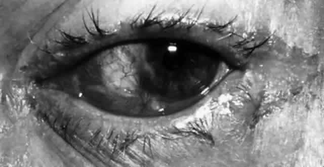
N. gonorrhoeae is by far the more common of the two Neisseria species infections and is considered to be an oculogenital disease. It is seen primarily in neonates and in sexually active adolescents and young adults. Transmission generally is from genitalia to hand to eye. A careful history of sexual activity should be taken, including contact with infected individuals and symptoms of urethritis, vaginitis, or proctitis. Initially, ocular symptoms are similar to those of acute conjunctivitis but progress quickly to include swelling of the lids, profuse tenacious yellow-green purulent discharge, hyperemia and chemosis of the conjunctiva, pain, tenderness to touch, and prominent, tender preauricular nodes (Fig. 1). In cases that go untreated, corneal involvement is a consistent feature, and for this reason, gonococcal conjunctivitis represents a clinical condition of considerable importance. Corneal involvement begins with a loss of corneal luster, or a corneal haze. This is followed by a breakdown of the corneal epithelium, most often in the periphery. An ulcerative gutter surrounded by stromal infiltrate then forms; this may progress circumferentially to form a ring abscess or may progress centrally. Such ulcers may burrow and perforate rapidly, resulting in endophthalmitis. In addition to keratoconjunctivitis, iritis, lid abscesses, dacryoadenitis, and septicemia may occur. If left untreated, conjunctival inflammation caused by these organisms may progress to involve the cornea, with peripheral ulceration, abscess formation, and ultimately, perforation.

Fig. 1. A. Hyperacute purulent conjunctivitis produced by Neisseria gonorrhoeae. There is copious discharge, conjunctival hyperemia, and chemosis. B. Massive chemosis in gonococcal conjunctivitis with associated corneal involvement, including loss of corneal luster, epithelial defect, and early ring infiltrate. (Courtesy of I.R. Schwab)

Meningococci are carried in the oropharynx of up to 38% of healthy persons and may be the cause of epidemic meningitis. They are not commonly the cause of conjunctivitis but may cause serious conjunctival infection in children and young adults with resultant meningococcemia. The purulent conjunctivitis caused by N. meningitidis is clinically identical to that caused by gonococcal infection, but it usually is seen in younger patients and frequently is bilateral.20,21 The corneal findings generally are milder than in gonococcal infection. In addition, N. meningitidis infection may be complicated by meningococcemia, metastatic meningitis, or endogenous endophthalmitis.

CHRONIC BACTERIAL CONJUNCTIVITIS

Symptoms of chronic bacterial conjunctivitis are present for 4 weeks or more and usually consist of redness, irritation, lid excoriation, daily discharge, morning crusting, or mild mattering of the lids. Clinical signs often are nonspecific and include diffuse conjunctival hyperemia, papillae or follicles, minimal mucoid or mucopurulent discharge, and thickening of the conjunctiva. Treatment failures and recurrences are common. In some patients, specific organisms typically cause a more drawn-out course. In others, the organism simply is resistant to the antibiotic initially chosen. Masquerade syndromes are common and include viral conjunctivitis (molluscum contagiosum), lid disease (meibomian gland dysfunction, blepharoconjunctivitis, acne rosacea), allergic/atopic conjunctivitis, toxic conjunctivitis, and chronic dacryocystitis. The search for adnexal sources of infection is especially important because they may influence culture results, often may not be treated effectively by topical drops, may be caused by resistant organisms, or may require more long-term, intensive, or systemic treatment.

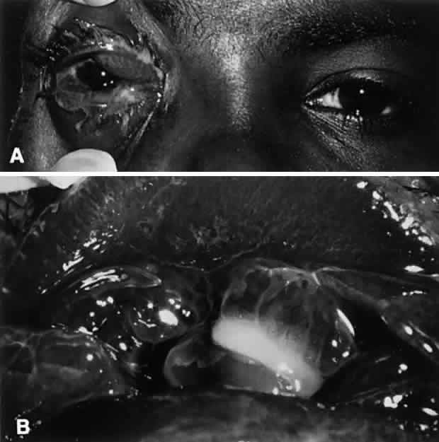
Chronic bacterial conjunctivitis is differentiated from other causes of chronic conjunctivitis on the basis of history, physical findings, and laboratory work-up. History taking should include questions regarding infection exposure, genitourinary symptoms, application of cosmetics, and instillation of topical medicines. Pressure over the lacrimal drainage system may produce a discharge indicative of dacryocystitis. The lids and canthal angles should be inspected for excoriation, ulceration, and erythema. The lids should be inspected closely for signs of anatomic abnormalities or signs of meibomianitis and blepharitis (Fig. 2). The involvement of the lids with eczematoid dermatitis, lash loss, trichiasis, collarettes, redness or telangiectasis of the lid margin, or recurrent hordeolum strongly suggest meibomian gland dysfunction. The exudate in such cases is crusted and yellowish and often encases the base of the cilia. Laboratory work-up includes culturing of the lid margin and conjunctiva to look for heavy growth of a predominant pathogen. Impression cytology with of the ocular surface is less helpful because of lack of specificity.22 Microscopic cytologic evaluation of conjunctival scrapings can add valuable information that correlates well with microbiology. Neutrophils are correlated with positive bacterial cultures, lymphocytosis with viral etiologies, characteristic basophilic inclusions with chlamydial infection, and eosinophils with allergic conjunctivitis.22

Fig. 2. Chronic staphylococcal blepharoconjunctivitis. A. Chronic erythema and induration of the lid margin due to colonization of the lid margin with pathogenic staphylococci. B. Characteristic features of the lid margin with staphylococcal disease are seen, including collarettes and hordeolum.

S. aureus is the organism isolated most commonly in cases of chronic bacterial conjunctivitis.23 Coagulase-negative Staphylococcus species, including S. epidermidis, may cause a similar clinical picture. Staphylococci may infect the conjunctiva primarily or may colonize the lid margin. Conjunctival inflammation occurs either by direct infection or through elaboration of exotoxins.24,25 The latter are believed to be responsible for a nonspecific conjunctivitis, punctate epithelial keratitis, phlyctenular keratoconjunctivitis, marginal corneal ulceration, and angular conjunctivitis. These entities are responsive to steroid-antibiotic combinations, which serve to decrease inflammation as well as bacterial counts. Such preparations, however, must be employed with caution.

Staphylococcus species conjunctivitis may result in inferior cornea fine punctate epithelial keratitis, which may be the result of bacterial toxigenicity. This may be responsible for a foreign-body sensation and is most prominent during the morning hours. Phlyctenular keratitis may complicate chronic Staphylococcus blepharoconjunctivitis. The gelatinous nodular lesions occur most commonly at the limbus, but they may be wholly corneal or conjunctival in location and acutely painful.

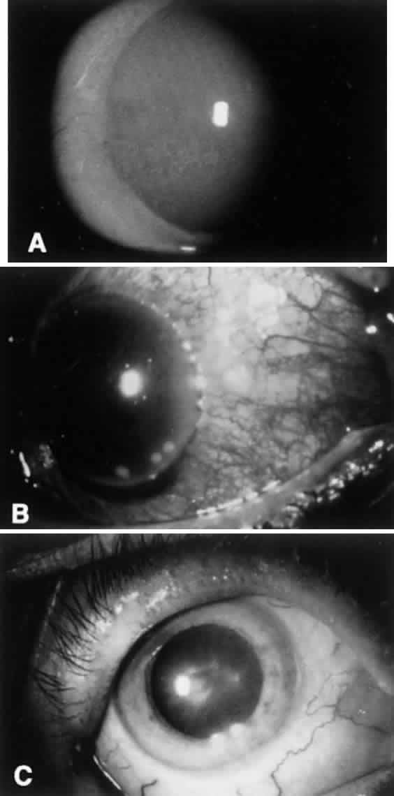
Marginal corneal ulceration due to hypersensitivity to staphylococcal exotoxin also may occur. Findings include the formation of single or multiple gray-white infiltrates with or without subsequent overlying epithelial breakdown. These are most often seen along the inferior limbus between the 4- and 8-o'clock positions. The ulcers frequently are associated with paralimbal conjunctival vascular engorgement and generally are culture negative (Fig. 3).

Fig. 3. The corneal manifestations of chronic staphylococcal blepharoconjunctivitis. A. Fine, inferior punctate epithelial keratitis. B. Marginal staphylococcal hypersensitivity ulcers located near the inferior limbus. C. Corneal phlyctenules resulting from chronic staphylococcal infection.

Maceration, excoriation, and crusting of the lateral canthal angle, along with localized, lateral epibulbar injection indicate the presence of angular blepharitis, a form of chronic conjunctivitis most commonly caused by Staphylococcus or Moraxella. Bacterial elaboration of dermonecrotoxins produce the characteristic findings.

M. lacunata organisms are large, symmetric, gram-negative diplobacilli that are cultured readily on blood or chocolate agar. The Moraxella organisms may retain gentian violet in the Gram staining process and thus may appear gram positive. In the conjunctiva, Moraxella organisms do not evoke a significant polymorphonuclear response. Infection may result in a chronic follicular conjunctivitis, angular conjunctivitis, or an enlarged preauricular lymph node (Fig. 4). Other organisms, especially in the elderly or immunocompromised, also may be implicated. These organisms are largely enteric bacteria and include Proteus mirabilis, Escherichia coli, Branhamella catarrhalis, Klebsiella pneumoniae, Serratia marcescens, and Pseudomonas. Serratia marcescens organisms are gram-negative rods with parallel sides and rounded ends. N. catarrhalis, which normally is found in the upper respiratory tract, also may be a cause of chronic conjunctivitis. Pseudomonas organisms appear as single rods that are straight or slightly curved. Although all of these organisms occasionally can be isolated from the ocular surface, they are located in much larger numbers in the presence of an infection.

Fig. 4. Angular blepharoconjunctivitis associated with Moraxella infection.

Back to Top
PRINCIPLES OF TREATMENT
Microbiologic culture and sensitivity testing can be helpful, but most forms of conjunctivitis respond well to empiric topical antibiotic treatment. Many believe that cultures should be reserved for nonresponding acute conjunctivitis, for chronic conjunctivitis not responsive to a course of antibiotic treatment, and for any suspicion of cornea infection. To culture for cases of treatment failures, antibiotics can be discontinued for 2 or 3 days to allow effective culturing at that time. This may be an effective strategy in an era of cost-consciousness and a reliance on outcome evaluations to help make therapeutic decisions.

Arguments against the use of empiric broad-spectrum antibiotics include that nonspecific antimicrobial use can infrequently lead to complications, such as drug toxicity, sensitization, and treatment failures. Further, although a given antibiotic may be ineffective in resolving a conjunctivitis, it may lead to sterilization of future culture and sensitivity evaluations while the patient is taking the medication and for several days after. This can delay effective treatment and result in further morbidity.

As an adjunctive measure, Gram and Giemsa staining of conjunctival scrapings has been shown to an effective modality and correlates well with microbiologic testing in terms of etiology.15 In a study looking at childhood conjunctivitis, Gram stain of conjunctival scrapings provided a means of predicting the pathogen in 51 of 55 cases of bacterial conjunctivitis and Giemsa stains provided etiologic information in 81 of 84 cases, showing neutrophilia in bacterial infections, lymphocytosis in viral infections, and eosinophilia in allergic disease.15 Nasal swabbing may be of additional help, especially in children, in the diagnosis of chlamydia, adenovirus, and H. influenzae.26

An additional consideration is that in vitro susceptibility of bacterial isolates recovered from patients with conjunctivitis may not correlate with clinical response. Further, studies show that no antibiotic provides 100% broad-spectrum coverage in vitro. Comparing newer to more established drugs, one study found the following susceptibilities of recovered isolates to antibiotics (in decreasing order): chloramphenicol, bacitracin/polymyxin B, ofloxacin, sulfa, ciprofloxacin, trimethoprim/polymyxin B, norfloxacin, gentamicin, bacitracin, trimethoprim, tobramycin, neomycin, erythromycin, and polymyxin B.27

The treatment of acute bacterial conjunctivitis consists of topical antimicrobial drops or ointment. The choice of the antimicrobial agent should be based on cultures, if available. If treatment is based on clinical characteristics alone, however, a broad-spectrum antibacterial should be chosen and antibiotic treatment should be discontinued when the inflammatory process has resolved. Streptococcal conjunctivitis should be treated with penicillin or topical erythromycin. If C. diphtheriae is identified as the pathogen, diphtheria antitoxin (10,000 to 100,000 units) should be given parenterally along with systemic penicillin or erythromycin. H. influenzae conjunctivitis should be treated systemically if associated with conjunctivitis-otitis syndrome. In any type of acute bacterial conjunctivitis, lack of response to antimicrobial therapy should prompt discontinuation of treatment and repeat laboratory studies.

Resistance of certain organisms to the fluoroquinolones and to vancomycin is emerging. In general, these antibiotics (especially vancomycin) should be reserved for those with resistant forms of conjunctivitis with cultures and sensitivities indicating potential effectiveness. The treatment with these medications for chronic blepharoconjunctivitis, on an ongoing basis, should be avoided. Vancomycin, penicillin, and other nonstandard antibiotic drops can be made up readily by many hospital pharmacies.

Based on organism counts, antibiotic levels, and rates of mutation, some predicted that organisms resistant to the fluoroquinolones would be uncommon. In actuality, reasonably high rates of Staphylococcus species have shown in vitro resistance. Nonetheless, drugs such as ofloxacin and ciprofloxacin have proven to be highly effective in the treatment of bacterial conjunctivitis and corneal ulcers.28,29 In a study evaluating ciprofloxacin for the treatment of bacterial conjunctivitis and blepharitis, the infecting organism was eradicated in 81%, and clinical cure or improvement was seen in 95%.30 In another study, ciprofloxacin eradicated or reduced organisms in 96%, with clinical cure or improvement also in 96%.31 A study evaluating the treatment of methicillin-resistant S. aureus in a long-term care facility showed the efficacy of oral ciprofloxacin and topical vancomycin in clinical resolution of conjunctivitis.32

In certain types of chronic conjunctivitis and angular conjunctivitis, antibiotic-steroid combinations may be indicated and effective. There use should be limited, however, and there probably is no place for topical steroids in the treatment of acute or hyperacute conjunctivitis. There often is the temptation to treat chronic conjunctivitis with long-term topical corticosteroids. In addition to the potential complications from long-term steroid use, antibiotic-steroid combinations have shown to be more effective than steroids alone.33 Even so, the risks of chronic steroid use remain a hazard. In general, antibiotic therapy should be as specific as possible, limited to the duration of the inflammatory process, and augmented by appropriate lid and periocular hygiene measures. The importance of lid hygiene often is underestimated by both patients and physicians. The use of antibiotics should be limited to a short-term course and terminated when inflammation has resolved, usually within 5 to 7 days.

Treatment of hyperacute conjunctivitis must be specific to the causative pathogen. Gonococcal conjunctivitis should be treated both with topical and full doses of systemic antibiotics.34 In the early stages of the disease, frequent lavage also is helpful in removing contaminated exudate from the ocular surface. Treatment recommendations for N. gonorrhoeae change periodically, and current recommendations should be consulted. Parenteral therapy includes ceftriaxone sodium, 1 g given intramuscularly (IM) daily for 5 consecutive days or penicillin G, 10 million units given intravenously (IV) for 5 consecutive days. Alternatives to penicillin include spectinomycin 4 g (10 ml) IM in two divided doses or tetracycline hydrochloride 1.5 g as a loading dose followed b, 0.5 g four times a day for 4 days. Ampicillin 3.5 g administered orally, with 1 g of probenecid given simultaneously, is another alternative. Single-dose treatments have been advocated, including ceftriaxone 1 g IM. Additional treatment to cover Chlamydia trachomatis is recommended and includes doxycycline and azithromycin. Treatment regimens for N. meningitidis include penicillin IV or IM.

Back to Top
REFERENCES

1. Okumoto M: Normal flora in the defense of the conjunctive against infection. In Oconnor GR (ed): Immunologic Diseases of the Mucous Membranes: Pathology, Diagnosis and Treatment, p 33. New York: Masson, 1980

2. Perkins RE, Kundsin RB, Pratt MV et al: Bacteriology of normal and infected conjunctiva. J Clin Microbil 1:147, 1975

3. Locatcher-Khorazo D, Seegal BC: The bacterial flora of the healthy eye. In Locatcher-Khorazo D, Seegal B (eds): Microbiology of the Eye, p l3. St Louis: CV Mosby, 1972

4. Torrey JC, Reese MK: Initial aerobic flora of newborn infants: Selective tolerance of the upper respiratory tract for bacteria. Am J Dis Child 69:208, 1945

5. Allansmith MR: The Eye and Immunology. St Louis: CV Mosby, 1982

6. Friedlaender MH: Immunology of ocular infections. In Friedlaender MH (ed): Allergy and Immunology of the Eye, p 107. Philadelphia: Harper & Row, 1979

7. Rotkis WM: Lysozyme: Its significance in external ocular disease. In Oconnor GR (ed): Immunologic Diseases of the Mucous Membranes: Pathology, Diagnosis, and Treatment, p. 27. New York: Masson, 1980

8. Londer L, Nelson DL: Orbital cellulitis due to Haemophilus influenzae. Arch Ophthalmol 91:89, 1974

9. McClellan KA: Mucosal defense of the outer eye. Surv Ophthalmol 42:233, 1997

10. Poulsen K, Reinholdt J, Kilian M: Characterization of the Streptococcus pneumoniae immunoglobulin A1 protease gene (iga) and its translation product. Infect Immunol 64:3957, 1996

11. Lomholt H, Poulsen K, Kilian M: Comparative characterization of the iga gene encoding IgA1 protease in Neisseria meningitidis, Neisseria gonorrhoeae and Haemophilus influenzae. Mol Microbiol 15:495, 1995

12. Leitch EC, Willcox MD: Interactions between the constitutive host defences of tears and Staphylococcus epidermidis. Aust NZ J Ophthalmol 25:S20, 1997

13. Krohn MA, Hillier SL, Bell TA et al: The bacterial etiology of conjunctivitis in early infancy: Eye Prophylaxis Study Group. Am J Epidemiol 138:326, 1993

14. Wilson L, Ahearn D, Jones D, Sexton R: Fungi from the normal outer eye. Am J Ophthalmol 67:52, 1969

15. Weiss A, Brinser JH, Nazar-Stewart V: Acute conjunctivitis in childhood. J Pediatr 122:10, 1993

16. Locathcer-Khorazo D, Sullivan N, Gutierrez E: Staphylococcus aureus isolated from normal and infected eyes. Arch Ophthalmol 77:370, 1967

17. Valenton MJ, Okumoto M: Toxin-producing strains of Staphylococcus epidermidis (albus): Isolates from patients with staphylococcal blepharoconjunctivitis. Arch Ophthalmol 89:186, 1973

18. Au YK, Jensen HG, Rowsey J et al: Coagulase-negative staphylococci in conjunctivitis and blepharitis. Yen Ko Hsueh Pao 9:129, 1993

19. Feingold M, Gellis SS: Cellulitis due to Haemophilus in fluenzae type B. N Engl J Med 272:788, 1965

20. Mangiaracine AB, Pollen A: Meningococcic conjunctivitis. Arch Ophthalmol 91:89, 1974

21. Wald ER: Conjunctivitis in infants and children. Pediatr Infect Dis J 16:S17, 1997

22. Cvenkel B, Globocnik M: Conjunctival scrapings and impression cytology in chronic conjunctivitis: Correlation with microbiology. Eur J Ophthalmol 7:19, 1997

23. Thygeson P, Kimura S: Chronic conjunctivitis. Trans Am Acad Ophthalmol Otolaryagol 67:494, 1963

24. Okumoto M: Microbiology. In Smolin G, Thoft RA (eds): The Cornea: Scientific Foundations and Clinical Practice, p 105. Boston: Little Brown & Co, 1983

25. Thygeson P: Bacterial factors in chronic catarrhal conjunctivitis. Arch Ophthalmol 18:373, 1937

26. Morton CE, Mallinson H, Clearkin LG et al: Per-nasal swabbing as an aid to the diagnosis of chlamydial and adenovirus conjunctivitis. Eye 4:510, 1990

27. Everett SL, Kowalski RP, Karenchak LM et al: An in vitro comparison of the susceptibilities of bacterial isolates from patients with conjunctivitis and blepharitis to newer and established topical antibiotics. Cornea 14:382, 1995

28. Ofloxacin monotherapy for the primary treatment of microbial keratitis: A double-masked, randomized, controlled trial with conventional dual therapy: The Ofloxacin Study Group. Ophthalmology 104: 1902, 1997

29. Hyndiuk RA, Eiferman RA, Caldwell DR et al: Comparison of ciprofloxacin ophthalmic solution 0. 3% to fortified tobramycin-cefazolin in treating bacterial corneal ulcers: Ciprofloxacin Bacterial Keratitis Study Group. Ophthalmology 103:1854, 1996

30. Adenis JP, Colin J, Verin P et al: Ciprofloxacin ophthalmic solution in the treatment of conjunctivitis and blepharitis: A comparison with fusidic acid. Eur J Ophthalmol 6:368, 1996

31. Adenis JP, Brasseur G, Demailly P et al: Comparative evaluation of efficacy and safety of ciprofloxacin and norfloxacin ophthalmic solutions. Eur J Ophthalmol 6:287, 1996

32. Brennen C, Muder RR: Conjunctivitis associated with methicillin-resistant Staphylococcus aureus in a long-term-care facility. Am J Med 88:14N, 1990

33. Shulman DG, Sargent JB, Stewart RH et al: Comparative evaluation of the short-term bactericidal potential of a steroid-antibiotic combination versus steroid in the treatment of chronic bacterial blepharitis and conjunctivitis. Eur J Ophthalmol 6:361, 1996

34. Ullman S, Roussel TJ, Forster RK: Gonococcal keratoconjunctivitis. Surv Ophthalmol 32:199, 1987

Back to Top