Chapter 4
Parinaud's Oculoglandular Syndrome
Elmer Y. Tu
Main Menu   Table Of Contents

Search

CAT-SCRATCH DISEASE
PATHOLOGY
LABORATORY DIAGNOSIS
SKIN TEST (HANGER-ROSE TEST)
DIAGNOSIS
TREATMENT
OCULOGLANDULAR TULAREMIA
CONJUNCTIVAL SPOROTRICHOSIS
CONJUNCTIVAL TUBERCULOSIS
CONJUNCTIVAL SYPHILIS
CONJUNCTIVAL COCCIDIOIDOMYCOSIS
RARE CAUSES OF PARINAUD'S OCULOGLANDULAR SYNDROME
REFERENCES

Parinaud's oculoglandular conjunctivitis is a form of follicular, granulomatous conjunctivitis (with or without ulceration), often associated with a visible swollen preauricular and/or submandibular lymph node on the same side1 (Fig. 1). The conjunctival granuloma may occur in bulbar or palpebral conjunctiva, upper or lower. Fever and other systemic signs may or may not be present. The conjunctival granulomas and the regional lymph node swelling usually diminish in 4 to 5 weeks, although in some cases the enlarged lymph nodes may progress to suppuration.

Fig. 1 Parinaud's oculoglandular syndrome demonstrating markedly swollen eyelids, mucopurulent discharge, and enlarged parotid lymph node on left side in a patient with tularemia. (Courtesy of George N. Chin, MD; Seattle, WA)

Parinaud's oculoglandular syndrome (POS) consists of the characteristic granulomatous conjunctival inflammation, usually unilateral, with extension to or involvement of local, ipsilateral lymph nodes. Although cat-scratch disease (CSD) is by far the most common etiology, as a syndrome it does not constitute a disease with a specific origin or pathology but is a common endpoint for multiple infectious and noninfectious processes. The clinical findings in CSD are most representative of the originally described Parinaud's conjunctivitis. Other known causative agents are listed in Table 1. Although cat-scratch disease is usually a benign, self-limiting disease, laboratory examinations should be obtained to determine the exact causal agent and to rule out other more serious infections. Diagnostic considerations for Parinaud's oculoglandular conjunctivitis are listed in Table 2.


TABLE 1. Causes and Treatment of Parinaud's Oculoglandular Conjunctivitis


Frequency Disease Agents Drugs/Treatment of Choice
Most commonCat-scratch diseaseBartonella henselaeSymptomatic, Azithromycin, Ciprofloxacin, Rifampin, Doxycycline and others
CommonTularemia*Francisella tularensisStreptomycin or Gentamicin
 Sporotrichosis*Sporotrichum schenckiiItraconazole or Fluconazole
OccasionalTuberculosis*Mycobacterium tuberculosisIsoniazid, streptomycin
   Ethambutol or rifampin
 Syphilis*Treponema pallidumPenicillin G
 Coccidioidomycosis*Coccidioides immitisTriazoles (i.e., Itraconazole or ketoconazole)
RareParacoccidioidomycosisParacoccidioidomycosis brasiliensisItraconazole
 Actinomycosis*Actinomyces israelii,Penicillin G, drain abscess
  A. propionicus 
 BlastomycosisBlastomyces dermatitidisItraconazole
 Infectious mononucleosisEpstein-Barr virusSymptomatic
 MumpsParamyxovirusSymptomatic
 PasteurellosisPasteurella multocida (septica)Penicillin G or tetracycline
   Amoxicillin/ clavulanate
 Yersinia infectionYersinia pseudotuberculosisGentamicin or tetracycline
  Yersinia enterocoliticaTetracycline
 Glanders*Burkholderia malleiImipenum, Azithromycin, and Doxycycline
 Chancroid*Haemophilus ducreyiCiprofloxacin or Ceftriaxone
 Lymphogranuloma venereum (LGV)*Chlamydial LGV agent L, L2, L3Tetracycline, doxycycline, or erythromycin
 Rickettsiosis (Mediterranean spotted fever)Rickettsia conoriiTetracycline
 ListerellosisListeria monocytogenesAmpicillin or TMP/SMZ
 Ophthalmia nodosa (noninfectious)Lepidoptera (caterpillars) Tarantula HairsSymptomatic, corticosteroids or surgical excision

*May cause conjunctival ulcers


TABLE 2. Laboratory Diagnosis of Parinaud's Oculoglandular Conjunctivitis


Immediate Considerations
Culture conjunctival secretions or scrapings of blood agar, broth (thioglycolate, brain-heart infusion), Sabouraud medium, and L#auowenstein-Jensen medium.
Stains: Gram, Giemsa, acid-fast, and fungal stains of secretions or scrapings
Blood studies: Serology—based on history.
B. henselae—IFA or ELISA, F. tularensis enzyme-linked immunoabsorbent assay, infectious mononucleosis—Monospot, Mumps—IgM/ IgG titers, Syphilis—VDRL and FTA-ABS or MHA-TP Blood cultures if patient is febrile, complete blood cell count
PPD
Later Considerations (if warranted)
Biopsy or aspirate lesions for culture/ special media
Histopathology (Warthin-Starry silver-impregnation stain, GMS, DFA for sporotrichosis)
PCR—B. henselae, F. tularensis
Convalescent serum sample for serologic tests (e.g., tularemia, mumps)


Back to Top
CAT-SCRATCH DISEASE
Cat-scratch disease is caused by Bartonella henselae (formerly Rochalimaea) and rarely by Afipia felis.2,3 Eye involvement (benign inoculation lymphoreticulosis)4,5 develops in those persons who have close contact with cats, especially children younger than 10 years of age. Over 90% of patients report a history of being scratched by a healthy cat (see Fig. 2), often a kitten.6 Kittens are the most susceptible to infection with B. henselae and may manifest an asymptomatic bacteremia for several weeks, whereas some adult cats have been shown to be chronic carriers. Transmission from cat to cat is mediated by the cat flea, whereas cat-to-human transmission may occur from trauma (scratch), inhaling materials infected by cats, being licked by infected animals, or rubbing one's eye after contact. Human-to-human transmission has not been reported, and patient isolation is, therefore, not required. It is also not considered necessary to remove the cat from the home because of the low overall rate of human transmission.7 However, multiple infections within the same family have been reported.8 A primary inoculation lesion in the conjunctiva, skin, or mucous membrane occurs in about 76% of patients.5

Fig. 2 Cat scratch marks with nonsuppurative preauricular lymphadenopathy. (Courtesy of Dr. Andrew M. Margileth. Margileth, A: A new look at old cat scratch. Contemp Pediatr 7:27, 1990)

The annual incidence of CSD is approximately 24,000, with children or adolescents making up 80% of cases.6,9 Parinaud's is classified as an atypical manifestation of CSD and occurs in approximately 3% to 7% of all infections.4,10 Seven to 14 days after exposure, redness, irritation, watery conjunctival discharge, and the sensation of a foreign body develop in the eye. The typical lesion consists of a unilateral granulomatous nodule located in the upper or the lower palpebral conjunctiva, surrounded by follicles (Fig. 3), intense chemosis, and injection. The fornices and bulbar conjunctiva may be involved (Fig. 4). The granuloma may be single, large, and flat, or there may be multiple elevated vegetations (Fig. 5). Corneal involvement usually consists of superficial punctate keratitis, but rarely may cause significant keratitis.11

Fig. 3 Bartonella henselae (cat-scratch disease) conjunctivitis. Multiple tarsal (conjunctive) follicles are present. (Courtesy of Daniel A. Johnson, MD; San Antonio, TX)

Fig. 4 Bartonella henselae conjunctivitis demonstrating multiple bulbar (arrows) conjunctival follicles. (Courtesy of Daniel A. Johnson, MD; San Antonio, TX)

Fig. 5 B. henselae conjunctivitis multiple nonulcerative granulomas (arrows) are seen in the bulbar conjunctiva. (Courtesy of Daniel A. Johnson, MD; San Antonio, TX)

Finding the inoculation site is often helpful in the diagnosis of cat-scratch disease. If the skin is involved first, a primary, nonpruritic papule or pustule appears 3 to 7 days after the animal scratch or bite. The skin papules persist for 7 to 21 days and disappear without a scar. A variety of other skin lesions including erythema nodosum, erythema multiforme, and nonspecific morbilliform rashes may occur.

Regional granulomatous lymphadenopathy is present in all patients with cat-scratch disease and may involve the head, neck, and axilla (Fig. 6). One to 3 weeks after inoculation, preauricular, cervical, and submaxillary nodes are visible on the affected side. The nodes are firm, are usually nontender, and may be enlarged to the size of a golf ball. Approximately 10% of these lymph nodes progress to suppuration. Systemic symptoms and signs are common but are usually mild. Low-grade fever, chills, headaches, anorexia, and general malaise may also be present. Without treatment, the course is benign, and complete recovery within 2 to 3 months is the rule.

Fig. 6 Cat-scratch disease with golf-ball-sized axillary lymph node. (Courtesy of J. Brian Reed, MD; Wilford Hall, TX)

Immunocompromised hosts are more likely to develop the disseminated form of the disease as bacillary angiomatosis, a reactive vascular skin tumor mimicking Kaposi's sarcoma, or bacterial peliosis, an infiltration of systemic organs.12,13 B. henselae is an emerging human pathogen and was noted in 10% of a group of HIV patients in South Africa screened by PCR.14,15 Although immunocompetent individuals are not advised to avoid infected cats, studies in immunocompromised patients have produced conflicting recommendations.16

Back to Top
PATHOLOGY
Cat-scratch disease has been recognized as a clinical entity for more than 50 years. B. henselae is a small, pleomorphic, gram-negative bacillus that has been identified in conjunctival granuloma, skin, and lymph node specimens. The organism is best demonstrated using the Warthin-Starry silver impregnation stain. With a modified Brown-Hopp tissue Gram stain, the organism can be detected as a pleomorphic, gram-negative, nonacid-fast bacterium measuring 0.3 to 1 μm by 0.6 to 3 μm. The bacteria are found in the vessel walls of capillaries and in macrophages in the germinal centers, appearing as a single organism or in chains. Focal areas of necrosis are noted with activated histiocytes and polymorphonuclear in the surrounding cells, forming a granuloma.

B. henselae is very difficult to isolate using standard laboratory techniques. First described in 1990 by Relman et al., polymerase chain reaction (PCR) amplifying the 16S rRNA of B. henselae is both a highly sensitive and specific method for identifying the organism.17 Its successful use has been described on conjunctival specimens as well as lymph node biopsies and aspirates.18–21 There is no cross reactivity with other Bartonella species.

Back to Top
LABORATORY DIAGNOSIS
Two serologic tests are available for the confirmation of B. henselae exposure—an indirect immunofluorescence test (IFA) produced by the CDC and various enzyme-linked immunoassays (ELISA). These tests have varying sensitivities and specificities, but CDC-processed tests yield above 90% for both.22 Predictability at other centers may be different depending on the laboratory. Because these tests rely on the robust host immune response seen in CSD, accuracy in immunocompromised patients is significantly less.9 Studies of various ELISAs show a greater predictability with IgM titers than IgG, but to achieve high specificity and sensitivity, the results need to be combined with the following diagnostic criteria. Cross reactivity with other Bartonella species is possible resulting in false positives.23–25
Back to Top
SKIN TEST (HANGER-ROSE TEST)
A skin test for cat-scratch disease was one of the original diagnostic criteria for CSD. The antigen was obtained from a suppurative lymph node and used as a skin-test antigen. A dose of 0.1 ml of the antigen is inoculated intradermally with a positive reaction creating a central papule or erythema measuring 0.5 to 1 cm in diameter after 48 to 72 hours. The size of the skin reaction did not correlate with the severity of the illness. With the advent of other serology and PCR, the risk of using foreign human lymphoid tissue outweighs its benefits.
Back to Top
DIAGNOSIS
Diagnosis of cat-scratch disease includes the following criteria.
  1. History of contact with a cat or kitten with presence of a scratch
  2. Primary inoculation site either cutaneous or ocular
  3. Serology (as listed earlier)
  4. Biopsy of conjunctival granuloma or lymph node—demonstrating pleomorphic gram-negative bacilli with the Warthin-Starry silver impregnation stain or by PCR

Clinical characteristics combined with either serologic or pathologic confirmation make the diagnosis of CSD-related Parinaud's oculoglandular syndrome. Other, more serious, infections should also be considered at the time of presentation because of the morbidity related to those diseases. In addition, noninfectious causes, primarily malignancies such as lymphoma or metastatic disease, should be considered and a biopsy taken if tests for infectious agents are inconclusive.

Back to Top
TREATMENT
The prognosis of cat-scratch disease is excellent, with few reports of permanent ocular damage. Even without treatment, CSD will resolve in 2 to 4 months in the majority of cases. Complications in immunocompetent individuals are rare, although encephalopathy, thrombocytopenic purpura, osteomyelitis, lymph node enlargement simulating malignant lymphoma, and parotid gland tumor have been reported.26 Because of the affinity of B. henselae for vascular endothelium, immunocompromised individuals may develop disseminated vasoproliferative disease, such as bacillary angiomatosis and bacterial peliosis.

Topical antibiotics are of questionable benefit because a cure is achieved primarily through systemic medication. Supportive management may include analgesics and antipyretic agents. For systemic and posterior ocular manifestations, oral corticosteroids have been effective in reduction of inflammation and improvement in function, but its need and utility in isolated POS has not been established.27,28

Aspiration of the lymph node, if suppuration occurs, can be done to relieve painful adenopathy and to provide material for serologic testing and histopathology. A 16-gauge needle and a 20-ml syringe may be used after anesthetizing and prepping the skin. Incision and drainage is, however, not recommended because a fistulous tract may develop, and discharge may persist for several months resulting in permanent scarring.

The self-limited nature of CSD both makes the need for systemic antibiotics unclear and antibiotic efficacy difficult to evaluate. Success has been reported with erythromycin and doxycycline. Prior to the identification of B. henselae as the causative agent in CSD, Margileth reviewed available studies on antibiotic treatment of CSD and concluded that four agents had the most reasonable probability of efficacy based on a varied set of outcome criteria: Rifampin, Ciprofloxacin, Gentamicin, and Trimethoprim/Sulfamethoxazole. Rifampin 10–20 mg/kg/day q 12–24 hours being the most effective in the group.5 In the only prospective, randomized study of therapy of CSD, Bass et al. found that a single 500-mg dose of azithromycin followed by four daily doses of 250 mg resulted in more rapid resolution of lymphadenopathy than placebo at 1 month.29 CSD in immunocompromised individuals predisposes to more disseminated disease, and therefore, the threshold for systemic antibiotics use is lower. Azithromycin (as in dosage listed earlier) or doxycycline (100 mg b.i.d) has been shown to be efficacious in these individuals.9,30 Antibiotic treatment and observation should continue for several months because of the possibility of recurrence.

Back to Top
OCULOGLANDULAR TULAREMIA
Tularemia is caused by Francisella tularensis (formerly Pasteurella tularensis). It is primarily an infectious disease of small wild animals such as rabbits, hares, ground squirrels, rats, mice, and foxes. The infection usually occurs from direct contact with infected animals, especially rabbits, and is typically seen in hunters or meat cutters (Fig. 7). The disease can also be transmitted indirectly by insect vectors, including deerflies, mosquitoes, lice, and especially ticks. Incubation time is 2 to 10 days.31

Fig. 7 Oculoglandular tularemia. Bulbar conjunctival granuloma with ipsilateral regional adenopathy with loss of jaw angularity (arrow). (Courtesy of Joel S. Sugar, MD; Chicago, IL)

Oculoglandular tularemia is an acute infectious conjunctivitis characterized by unilateral, necrotizing, granulomatous conjunctival inflammation with subsequent ulceration.32–34 The granulomas may occur in the upper or lower tarsal or bulbar conjunctiva. The ocular findings are associated with tender regional lymphadenitis on the same side and severe systemic manifestations, including fever, malaise, headaches, chills, vomiting, and occasionally pneumonia and severe prostration. Corneal ulcers with perforation, panophthalmitis, optic neuritis, and dacryocystitis may also occur but are rare.35,36

Conjunctival tularemia should be suspected when a patient has killed, skinned, or cleaned rabbits or has been bitten by ticks. Findings include multiple yellow tarsal conjunctival nodules with necrotic, shallow ulcers; tender regional lymphadenopathy; and moderate to severe systemic signs with an acute onset. These features distinguish this entity from other more benign causes of Parinaud's conjunctivitis. Smears or cultures showing pleomorphic gram-negative coccobacilli can confirm the disease. Serology titers of 1:160 or higher within 2 to 3 weeks after onset of clinical disease are considered diagnostic of the disease.35 Because of its highly contagious nature, F. tularensis is a potential bioterrorism organism, and clinical samples should be handled carefully.

Administration of the drug of choice, streptomycin, 7.5–10 mg/kg intramuscularly b.i.d. for 7 days, results in rapid recovery and prevents relapse. Gentamicin at 1.7 mg/kg IM or IV every 8 hours is an acceptable alternative.31 In addition, for the conjunctivitis, topical antibiotics such as gentamicin ophthalmic drops should be instilled every 2 hours for 1 week, then five times daily until resolution. Anecdotal reports of success with systemic fluoroquinolones have also been noted. Subconjunctival antibiotics may be used for corneal or uveal complications. Corticosteroids should be avoided. It is best to manage these patients with the help of an internist, because patients with systemic complications, such as septicemia or endocarditis, and immunologically compromised persons need close medical supervision. Mortality is noted to be between 8% and 30%.35

Back to Top
CONJUNCTIVAL SPOROTRICHOSIS
Ocular sporotrichosis is a chronic infection caused by Sporothrix schenckii, a fungus found in soil and vegetative material.37 Sporotrichosis may be localized to the conjunctiva as a primary infection or may be secondary to involvement of the lid and face. The first evidence of infection in the skin of the eyelid is the appearance of a hard, spherical, elastic movable, nontender nodule that may later become attached to the skin. The nodule is initially pink, then purple, and finally black and necrotic, described as a sporotrichotic chancre. Subcutaneous nodules appear along the course of the lymphatics draining extending from the area.38 Numerous soft, yellow, granulomatous nodules, which may ulcerate, develop in the palpebral or bulbar conjunctiva (Fig. 8) of the involved eye. The conjunctival ulcers usually discharge a small amount of purulent material. The preauricular and submandibular lymph nodes are grossly enlarged and may suppurate. The patient does not usually appear systemically ill.

Fig. 8 Oculoglandular sporotrichosis. Nonulcerative bulbar conjunctival granuloma refractive to topical therapy. (Courtesy of James C. Chodosh, MD; Oklahoma City, OK)

The organism grows in 3 to 5 days on Sabouraud agar. Biopsy of the nodules may reveal the cigar-shaped organism on PAS or Gomori's methenamine silver (GMS) staining. Direct fluorescent antibody staining is also available for rapid diagnosis. The infection responds well to systemic itraconazole 200 mg daily. Ketoconazole is not effective.39 Topical fluconazole therapy may be added for conjunctival disease.37 Sporotrichosis may also respond to orally administered iodides (SSKI), which increase the host immune response to the organism. Therapy begins with a saturated solution of potassium iodide, 1 ml in a glass of water three times daily. The dose is increased by 0.5 to 1 ml/day until a daily dose of 9 to 12 ml is reached. To avoid recurrence, iodides should be continued for 4 to 6 weeks after clinical resolution.39 With all treatments, therapy should be continued past the appearance of clinical resolution.

Back to Top
CONJUNCTIVAL TUBERCULOSIS
The acid-fast bacillus Mycobacterium tuberculosis causes conjunctival tuberculosis. The incidence in the United States is low, but continues to be important internationally. Other mycobacteria are less-common causes. Eye involvement may occur with ocular trauma, direct inoculation, or extension from contiguous tuberculous sites, such as the skin lesions in cutaneous tuberculosis (lupus vulgaris).40,41 The conjunctival infection consists of a small, unilateral, painless, and indolent ulcer on the tarsal conjunctiva and fornix; this is surrounded by a small nodule. Rarely, there are larger nodules and hypertrophic lesions. The regional lymph nodes are often enlarged.

A chest roentgenogram and a tuberculin skin test should be performed. Scrapings from the conjunctival lesion usually show acid-fast bacilli. Histologically, the conjunctiva nodules consist of typical tubercles with epithelioid cells, Langhans' giant cells, and caseous necrosis. Although smears and biopsy findings suggest the diagnosis, it should be confirmed by culture because many diseases may mimic tuberculosis.

Systemic therapy with two effective antituberculous medications, usually isoniazid plus ethambutol, rifampin, or streptomycin, is required.42

Back to Top
CONJUNCTIVAL SYPHILIS
Conjunctival syphilis is caused by Treponema pallidum and may occur during the primary, secondary, or tertiary stages of the disease.43 In primary syphilis the conjunctival lesion is an indolent ulcer with an indurated margin and a gray base. The lid may be involved and is markedly swollen. A dense scar follows healing of the ulcer. Secondary syphilis of the conjunctiva is characterized by multiple granulomatous nodules or a diffuse, rose red, jelly-like thickening of the tarsal conjunctiva. An acute granulomatous iridocyclitis is usually present in the early secondary stage of dissemination accompanied by other secondary manifestations in the skin and mucous membranes. In tertiary syphilis (gumma), the conjunctiva may be involved as a result of syphilitic tarsitis. Regional lymph nodes are often enlarged in all three stages of syphilis. Complications such as uveitis, pannus, perforation, and panophthalmitis may occur. Diagnosis is confirmed by dark-field examination of scrapings or exudates and serologic tests (VDRL, fluorescent treponemal antibody-absorption testing [FTA-ABS], and microhemagglutination [MHA-TP]). Treatment is the systemic administration of penicillin; the dose depends on the stage of the disease. Local ocular disease may be treated with supportive care.
Back to Top
CONJUNCTIVAL COCCIDIOIDOMYCOSIS
Some pathogenic fungi that are responsible for facial and lid skin infections may secondarily invade the conjunctiva with an associated regional lymphadenopathy. These include Coccidioides immitis (coccidioidomycosis)44,45 and Blastomyces dermatitidis (blastomycosis).46 C. immitis is endemic in the southwestern United States, parts of Mexico, and Central and South America and is the etiologic agent for coccidioidomycosis.47

Ocular involvement may occur with primary pulmonary infection and is often associated with erythema nodosum. Palpebral conjunctival granulomas are often a manifestation of the disseminated disease. Phlyctenular conjunctivitis, episcleritis, scleritis, keratoconjunctivitis, or iridocyclitis may be present. These are considered hypersensitivity responses to the coccidioidal antigen and usually subside with time. In the posterior eye, manifestations may range from an asymptomatic focal chorioretinitis to a fulminating granulomatous process involving the entire eye. C. immitis is a dimorphic fungi that produces characteristic barrel-shaped arthroconidia, but will exhibit multinucleated spherules in pathologic specimens.48 The diagnosis of coccidioidomycosis is confirmed by culture and serologic tests. The treatment of choice for extrapulmonary disease is the triazole antifungals 400 mg/day (ketoconazole, fluconazole) or itraconazole 200 mg b.i.d.39 Amphotericin B is reserved for worsening or severe disease. All these mycoses produce a regional nonspecific lymphadenopathy.

Back to Top
RARE CAUSES OF PARINAUD'S OCULOGLANDULAR SYNDROME
Parinaud's oculoglandular syndrome is an uncommon disorder with the vast majority of cases due to the agents noted previously. Other infectious etiologies, listed following, rarely occur, but should be considered when the history or evaluation for more common diseases is negative. Certain noninfectious disorders resulting in conjunctival granulomas and regional lymphadenopathy can normally be elucidated by a careful history. Finally, lymphoma and metastatic disease may mimic the findings in POS.

The actinomycete Actinomyces israelii, a higher order anaerobic bacterium, is often introduced by trauma with a high incidence among farmers, laborers, and horticulturists. Initially the skin of the face and eyelids is involved, and then the infection extends to the conjunctiva. Large fungating verrucous plaques and ulcerating granulomas appear, and the active borders show numerous minute abscesses.

Infectious mononucleosis is an acute self-limited lymphoproliferative disease caused by the Epstein-Barr virus and characterized by fever, pharyngitis, and cervical lymphadenopathy.49,50 A unilateral tarsal conjunctival granuloma may occur. The presence of atypical lymphocytes and heterophil antibodies (Monospot test) in the blood establishes the diagnosis. Tests for antibodies to the Epstein-Barr virus are also available, which can help determine stage of infection.

Mumps is an airborne viral infection that causes localized swelling of the parotid glands. Ocular involvement may occur in the form of granulomatous conjunctivitis, with dacryoadenitis, optic neuritis, tenonitis, scleritis, and extraocular muscle palsies. Mumps may be accompanied by complications such as orchitis, pancreatitis, and meningoencephalitis.

Several organisms found primarily in animals may cause Parinaud's conjunctivitis in humans. These organisms include Pasteurella multocida,51 a gram-negative bacterium found in the oral cavity of cats and dogs; Yersinia pseudotuberculosis; Yersinia enterocolitica;52 Burkholderia mallei (glanders),53 an organism studied for bioterrorism purposes; Listeria monocytogenes, a common food contaminant; and Rickettsia conorii, an organism conveyed by ticks from dogs and responsible for Mediterranean spotted fever. Humans acquire these infections from animal sources, and airborne infection may occur in laboratory workers.

Granulomatous conjunctivitis caused by Haemophilus ducreyi (chancroid)54 or lymphogranuloma venereum is rare and is usually due to direct primary inoculation by venereal contact through the fingers or by accidental contamination of the eye in laboratory workers.55 Corneal ulcers with perforation have been reported.

Caterpillar hairs (ophthalmia nodosa)56 accidentally lodged in the palpebral conjunctiva may also cause a granulomatous conjunctivitis and regional lymphadenopathy. Tarantula hairs are barbed in similar fashion to encourage progressive penetration and have been reported to cause granulomatous inflammation in every layer of the eye including the cornea (Fig. 9) and conjunctiva.57–59 Implantation of synthetic fibers may result in a “Teddy Bear Granuloma” of the palpebral conjunctiva.60 Simple removal is curative without need for other medications.61

Fig. 9 Tarantula hair keratitis. Granulomatous keratitis surrounding each penetrating hair. Inset: High magnification photo of inflammation surrounding a single tarantula hair (arrow). (Courtesy of Dr. Ann C. Guidera; Sellersville, PA)

The author and editors wish to express the appreciation to George N. Chin, MD, author of this chapter in the previous edition. Some of that material has been used in this revision.

Back to Top
REFERENCES

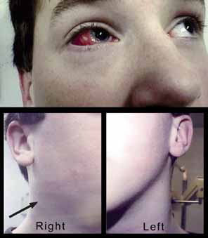
1. Parinaud H: Conjonctivité infectieuse transmisé par les animaux. Ann Ocul 101:252, 1889

2. Giladi M, Avidor B, Kletter Y, Abulafia S, Slater LN, Welch DF, et al: Cat scratch disease: The rare role of Afipia felis. Journal of Clinical Microbiology 36:2499–502, 1998

3. Bergmans AM, Groothedde JW, Schellekens JF, van Embden JD, Ossewaarde JM, Schouls LM: Etiology of cat scratch disease: Comparison of polymerase chain reaction detection of Bartonella (formerly Rochalimaea) and Afipia felis DNA with serology and skin tests. Journal of Infectious Diseases 171:916–23, 1995

4. Carithers HA: Cat-scratch disease. An overview based on a study of 1,200 patients. American Journal of Diseases of Children 139:1124–33, 1985

5. Margileth AM: Cat scratch disease: No longer a diagnostic dilemma. Seminars in Veterinary Medicine & Surgery (Small Animal) 6:199–202, 1991

6. Ormerod LD, Dailey JP: Ocular manifestations of cat-scratch disease. Current Opinion in Ophthalmology 10:209–16, 1999

7. Kordick DL, Wilson KH, Sexton DJ, Hadfield TL, Berkhoff HA, Breitschwerdt EB: Prolonged Bartonella bacteremia in cats associated with cat-scratch disease patients. Journal of Clinical Microbiology 33:3245–51, 1995

8. Murano I, Yoshii H, Kurashige H, Sugio Y, Uchida M, Shinohara T, et al: Three children with systemic cat scratch disease. Kansenshogaku Zasshi—Journal of the Japanese Association for Infectious Diseases 73:248–52, 1999

9. Cunningham ET, Koehler JE: Ocular bartonellosis. Am J Ophthalmol 130:340–9, 2000

10. Murakami K, Tsukahara M, Tsuneoka H, Iino H, Ishida C, Tsujino K, et al: Cat scratch disease: Analysis of 130 seropositive cases. Journal of Infection & Chemotherapy 8:349–52, 2002

11. Gabler B, Linde HJ, Reischl U, Lohmann CP: [Disciform keratatis caused by Bartonella henselae infection: detection of a rare ocular complication of cat-scratch disease with PCR]. Klinische Monatsblatter fur Augenheilkunde 217:299–302, 2000

12. Resto-Ruiz S, Burgess A, Anderson BE: The role of the host immune response in pathogenesis of Bartonella henselae. DNA & Cell Biology 22:431–40, 2003

13. Torok L, Viragh SZ, Borka I, Tapai M: Bacillary angiomatosis in a patient with lymphocytic leukaemia. British Journal of Dermatology 130:665–8, 1994

14. Koehler JE, Sanchez MA, Tye S, Garrido-Rowland CS, Chen FM, Maurer T, et al: Prevalence of Bartonella infection among human immunodeficiency virus-infected patients with fever. Clinical Infectious Diseases 37:559–66, 2003

15. Frean J, Arndt S, Spencer D: High rate of Bartonella henselae infection in HIV-positive outpatients in Johannesburg, South Africa. Transactions of the Royal Society of Tropical Medicine & Hygiene 96:549–50, 2002

16. Schwartzman WA, Patnaik M, Angulo FJ, Visscher BR, Miller EN, Peter JB: Bartonella (Rochalimaea) antibodies, dementia, and cat ownership among men infected with human immunodeficiency virus. Clinical Infectious Diseases 21:954–9, 1995

17. Relman DA, Loutit JS, Schmidt TM, Falkow S, Tompkins LS: The agent of bacillary angiomatosis. An approach to the identification of uncultured pathogens [comment]. New England Journal of Medicine 323:1573–80, 1990

18. Starck T, Madsen BW: Positive polymerase chain reaction and histology with borderline serology in Parinaud's oculoglandular syndrome. Cornea 21:625–7, 2002

19. Margolis B, Kuzu I, Herrmann M, Raible MD, Hsi E, Alkan S: Rapid polymerase chain reaction-based confirmation of cat scratch disease and Bartonella henselae infection. Archives of Pathology & Laboratory Medicine 127:706–10, 2003

20. Labalette P, Bermond D, Dedes V, Savage C: Cat-scratch disease neuroretinitis diagnosed by a polymerase chain reaction approach. American Journal of Ophthalmology 132:575–6, 2001

21. Hara H, Ito K, Akimoto M, Suzuki H, Asai S, Maruyama S: Detection of Bartonella henselae DNA using polymerase chain reaction assay in patient with cat scratch disease. Acta Dermato-Venereologica 83:67–8, 2003

22. Dalton MJ, Robinson LE, Cooper J, Regnery RL, Olson JG, Childs JE: Use of Bartonella antigens for serologic diagnosis of cat-scratch disease at a national referral center. Archives of Internal Medicine 155:1670–6, 1995

23. Bergmans AM, Peeters MF, Schellekens JF, Vos MC, Sabbe LJ, Ossewaarde JM, et al: Pitfalls and fallacies of cat scratch disease serology: Evaluation of Bartonella henselae-based indirect fluorescence assay and enzyme-linked immunoassay [comment]. Journal of Clinical Microbiology 35:1931–7, 1997

24. Giladi M, Kletter Y, Avidor B, Metzkor-Cotter E, Varon M, Golan Y, et al: Enzyme immunoassay for the diagnosis of cat-scratch disease defined by polymerase chain reaction. Clinical Infectious Diseases 33:1852–8, 2001

25. Sander A, Berner R, Ruess M: Serodiagnosis of cat scratch disease: Response to Bartonella henselae in children and a review of diagnostic methods. European Journal of Clinical Microbiology & Infectious Diseases 20:392–401, 2001

26. Fanous MM, Margo CE: Parinaud's oculoglandular syndrome simulating lymphoma. American Journal of Ophthalmology 112:344–5, 1991

27. Conrad DA: Treatment of cat-scratch disease. Current Opinion in Pediatrics 13:56–9, 2001

28. Lerdluedeeporn P, Krogstad P, Roberts RL, Stiehm ER: Oral corticosteroids in cat-scratch disease. Clinical Pediatrics 42:71–3, 2003

29. Bass JW, Freitas BC, Freitas AD, Sisler CL, Chan DS, Vincent JM, et al: Prospective randomized double blind placebo-controlled evaluation of azithromycin for treatment of cat-scratch disease [comment]. Pediatric Infectious Disease Journal 17:447–52, 1998

30. Mui BS, Mulligan ME, George WL: Response of HIV-associated disseminated cat scratch disease to treatment with doxycycline. American Journal of Medicine 89:229–31, 1990

31. Thompson S, Omphroy L, Oetting T: Parinaud's oculoglandular syndrome attributable to an encounter with a wild rabbit. American Journal of Ophthalmology 131:283–4, 2001

32. Chappell CW, Brainard J, Shock JP: Oculoglandular tularemia—a case report. Journal of the Arkansas Medical Society 78:128–30, 1981

33. Halperin SA, Gast T, Ferrieri P: Oculoglandular syndrome caused by Francisella tularensis. Clinical Pediatrics 24:520–2, 1985

34. Peter R, Banyai T: Erythema nodosum revealing oculoglandular tularemia. Dermatology 202:79–80, 2001

35. Steinemann TL, Sheikholeslami MR, Brown HH, Bradsher RW: Oculoglandular tularemia. Archives of Ophthalmology 117:132–3, 1999

36. Parssinen O, Rummukainen M: Acute glaucoma and acute corneal oedema in association with tularemia. Acta Ophthalmologica Scandinavica 75:732–4, 1997

37. Hampton DE, Adesina A, Chodosh J: Conjunctival sporotrichosis in the absence of antecedent trauma. Cornea 21:831–3, 2002

38. Trent JT, Kirsner RS: Identifying and treating mycotic skin infections. Advances in Skin & Wound Care 16:122–9; quiz 130–1, 2003

39. Sobel JD: Practice guidelines for the treatment of fungal infections. For the Mycoses Study Group. Infectious Diseases Society of America. Clinical Infectious Diseases 30:652, 2000

40. Cook CD, Hainsworth M: Tuberculosis of the conjunctiva occurring in association with a neighbouring lupus vulgaris lesion. British Journal of Ophthalmology 74:315–6, 1990

41. Whitford J, Hansman D: Primary tuberculosis of the conjunctiva. Medical Journal of Australia 1:486–7, 1977

42. Horsburgh CR Jr, Feldman S, Ridzon R: Infectious Diseases Society of A: Practice guidelines for the treatment of tuberculosis. Clinical Infectious Diseases 31:633–9, 2003

43. Spektor FE, Eagle RC Jr , Nichols CW: Granulomatous conjunctivitis secondary to Treponema pallidum. Ophthalmology 88:863–5, 1981

44. Maguire LJ, Campbell RJ, Edson RS: Coccidioidomycosis with necrotizing granulomatous conjunctivitis. Cornea 13:539–42, 1994

45. Wood TR: Ocular coccidioidomycosis. Report of a case presenting as Parinaud's oculoglandular syndrome. American Journal of Ophthalmology 64:Suppl:587–90, 1967

46. Slack JW, Hyndiuk RA, Harris GJ, Simons KB: Blastomycosis of the eyelid and conjunctiva. Ophthalmic Plastic & Reconstructive Surgery 8:143–9, 1992

47. Galgiani JN: Coccidioidomycosis: A regional disease of national importance. Rethinking approaches for control. Annals of Internal Medicine 130:293–300, 1999

48. Rodenbiker HT, Ganley JP: Ocular coccidioidomycosis. Survey of Ophthalmology 24:263–90, 1980

49. Meisler DM, Bosworth DE, Krachmer JH: Ocular infectious mononucleosis manifested as Parinaud's oculoglandular syndrome. American Journal of Ophthalmology 92:722–6, 1981

50. Wilhelmus KR: Ocular involvement in infectious mononucleosis. American Journal of Ophthalmology 91:117–8, 1981

51. Ho AC, Rapuano CJ: Pasteurella multocida keratitis and corneal laceration from a cat scratch. Ophthalmic Surgery 24:346–8, 1993

52. Chin GN, Noble RC: Ocular involvement in Yersinia enterocolitica infection presenting as Parinaud's oculoglandular syndrome. American Journal of Ophthalmology 83:19–23, 1977

53. Srinivasan A, Kraus CN, DeShazer D, Becker PM, Dick JD, Spacek L, et al: Glanders in a military research microbiologist [comment]. New England Journal of Medicine 345:256–8, 2001

54. Buus DR, Pflugfelder SC, Schachter J, Miller D, Forster RK: Lymphogranuloma venereum conjunctivitis with a marginal corneal perforation. Ophthalmology 95:799–802, 1988

55. Macnie J: Ocular lymphogranuloma venereum. Arch Ophthalmol 25:255, 1941

56. Martin X, Uffer S, Gailloud C: Ophthalmia nodosa and the oculoglandular syndrome of Parinaud. British Journal of Ophthalmology 70:536–42, 1986

57. Bernardino CR, Rapuano C: Ophthalmia nodosa caused by casual handling of a tarantula. CLAO Journal 26:111–2, 2000

58. Shrum KR, Robertson DM, Baratz KH, Casperson TJ, Rostvold JA: Keratitis and retinitis secondary to tarantula hair. Archives of Ophthalmology 117:1096–7, 1999

59. Watts P, McPherson R, Hawksworth NR: Tarantula keratouveitis. Cornea 19:393–4, 2000

60. Weinberg JC, Eagle RC Jr , Font RL, Streeten BW, Hidayat A, Morris DA: Conjunctival synthetic fiber granuloma. A lesion that resembles conjunctivitis nodosa. Ophthalmology 91:867–72, 1984

61. Lueder GT, Matsumoto B, Smith ME: Pathological case of the month. Synthetic fiber granuloma (“Teddy Bear Granuloma”). Archives of Pediatrics & Adolescent Medicine 150:327–8, 1996

Back to Top