Chapter 2
Immunology of the External Eye
C.D. SEAMONE and W.B. JACKSON
Main Menu   Table Of Contents

Search

BASICS OF THE IMMUNE SYSTEM
ALLERGIC OCULAR DISEASE
IMMUNOLOGIC CONSIDERATIONS IN SELECTED EXTERNAL OCULAR DISEASES
REFERENCES

BASICS OF THE IMMUNE SYSTEM
Immunity includes those active mechanisms that protect the host from foreign substances, microbial invasion, and neoplasia. Innate immunity is genetically determined, and resistance is not improved by repeated exposure. It is a first line of defense consisting of soluble factors such as acute-phase proteins (e.g., C-reactive protein), lysozyme, and complement, as well as cells such as phagocytes and natural killer cells. Inherent in this innate immune system is nonspecificity. On the other hand, acquired immunity is an adaptive type of immunity that develops after exposure and improves with repeated challenge. The hallmarks of this form of immunity are specificity and memory.1

All immune-related cells arise from a common pluripotential stem cell. This stem cell gives rise to committed stem cells for the lymphoid and myeloid series. Under the influence of particular colony-stimulating factors, the lymphoid and myeloid stem cells give rise to various cell types. The lymphoid cell line consists of lymphocytes such as T cells, B cells, and large granular lymphocytes (see Third Population Cells). The myeloid cell line consists of nonlymphoid cells such as erythrocytes, monocytes/macrophages, polymorphonuclear granulocytes (neutrophils, basophils, eosinophils), megakaryocytes/platelets, and mast cells. The bone marrow is the site of maturation of most of these cell types. A major exception is the T cell, which leaves the bone marrow and matures under thymic influence.

Before continuing with a discussion of immune cell types and function, two topics will be reviewed: the major histocompatibility gene complex (MHC) and cell markers.

MAJOR HISTOCOMPATIBILITY GENE COMPLEX

Alloantigens are antigens that differentiate between members of a species (i.e., intraspecies). The histocompatibility antigens are alloantigens that play a role in the rejection of allogeneic tissue grafts. These surface molecules are coded in the human genome in the major histocompatibility gene complex (MHC). This localized group of genes codes for cell surface antigens, which play a major role in immunity, self-recognition, and tissue graft rejection. Human leukocyte antigen (HLA) is the name for the human MHC. The HLA region is located on the 21 region of the short arm of chromosome 6 (Fig. 1). In addition, there are minor histocompatibility antigens coded throughout the genome that play a definite but less well-defined role in the immune response. There are three classes of gene products of the MHC.2

Fig. 1. Human major histocompatibility gene complex (HLA).

Class I Molecules

These antigens are expressed on virtually all nucleated cell surfaces, including B cells, T cells, and platelets, but not on mature red blood cells. They are products of the HLA-A, HLA-B, and HLA-C loci. Each individual has six serologically defined HLA-A, HLA-B, and HLA-C antigens, three from each parent.

Class II Molecules

These antigens are a product of the HLA-D region, which includes the subregions -DR, -DQ, and -DP. The genes of the HLA-D region that code for class II determinants are known as immune response (Ir) genes. Class II molecules, often called Ia (immune-associated) antigens, are expressed on antigen-presenting cells (monocytes/macrophages, Langerhans cells, dendritic cells), B lymphocytes, and activated T lymphocytes. Immune suppressor (Is) genes also exist in the HLA-DQ region.

Class III Molecules

These antigens include the complement components C4, C2, and factor B.

Although HLA antigens do not directly cause disease, certain HLA types are known to predispose individuals to particular autoimmune diseases (e.g., HLA-B27 and ankylosing spondylitis). The mechanism is not clearly defined but involves immunologic alteration.

SURFACE MARKERS

Cell surface molecules are identifiable by specific monoclonal antibodies. The CD system is presently the standard nomenclature (CD stands for cluster designation). Surface markers can be used to differentiate immune cell types, stages of maturation of a given immune cell type, and immune cell activation.

T-Cell Markers

Pan-T cell markers include CD2, CD5, and CD7 (Table 1). CD2 is responsible for the binding of T cells to sheep erythrocytes forming rosettes (B cells cannot do this). This was formerly the major means of identification of T cells.


TABLE 1. Important T-Cell Markers


CD GroupOther NamesCell Type/Function
CD1T6Immature cortical thymocytes, Langerhans cells
CD2T11, sheep erythrocyte receptorOriginal T-cell marker (rosette formation with sheep RBC); T-cell adhesion, activation
CD3 TCR-associated molecules
CD4T4T helper; MHC class II restriction
CD5 Pan-T cell, some B cells
CD7 Earliest marker of T-cell line; T-cell activation;pan-T cell, including all third population cells;?receptor for Fc portion of IgM
CD8T8Suppressor/cytotoxic T cells; class I restriction
TCR Recognizes antigen; T-cell activation induced by antigen
CD294B4, fibronectin receptorPresent on helper/inducer T cells
CD45R Present on suppressor-inducer T cells (i.e., CD4+ cells that induce suppressor/cytotoxic CD8+ cells)

RBC, red blood cells; TCR, T-cell antigen receptor; MHC, major histocompatibility gene complex


The T-cell antigen receptor (TCR) is the definitive T-cell marker.3 There are two types of TCR, TCR1 and TCR2, the latter being the most common (present on 95% of T cells). TCR2 is composed of two peptide chains, alpha and beta, linked to form a heterodimer. This is closely associated with CD3 molecules (CD3 subunit), forming the TCR2 receptor complex. TCR1 is similar in structure to TCR2 but contains gamma and delta polypeptides.

The TRC2 group can be divided into those cells that are CD4+ (helper T cells) and those that are CD8+ (suppressor/cytotoxic T cells). Helper T cells recognize antigen associated with class II molecules. The functions of CD4+ cells include promotion of T-cell and B-cell differentiation, regulation of suppressor/cytotoxic T-cell proliferation/function, and production of lymphokines (see below). They also play a role in regulation of erythropoiesis. Those TCR2 cells associated with CD8+ are suppressor/cytotoxic T cells. These cells recognize antigen associated with class I molecules and function as cytotoxic cells. Also, they act as suppressors of T- and B-cell function.

B-Cell Markers

The classic B-cell marker is surface immunoglobulin produced by the cell itself. These immunoglobulins act as B-cell receptors for antigen. On circulating B cells, the surface immunoglobulins are predominately IgM and IgD molecules, and, for a given cell, they share the same antigen specificity. CD19, CD20, and CD22 are presently the markers commonly used to identify B cells.3 Other markers found on B cells include MHC class II antigens, which are important in cooperation with T cells, complement receptors CR1 (for C3b) and CR2 (for C3d), CD5 (also expressed on all T cells), and surface receptors for the Fc portion of IgG.

CELLS OF THE IMMUNE SYSTEM

T Lymphocytes

T cells regulate the immune response, are the major players in cell-mediated immunity, and induce antibody production by B cells. T-cell precursors arise from stem cells in the yolk sac, the fetal liver, and, later in prenatal development, the bone marrow. These immature cells migrate to the thymus, where maturation follows. Mature lymphocytes migrate via the circulation to the spleen, lymph nodes, tonsils, and unencapsulated lymphoid tissue. The latter includes mucosa-associated lymphoid tissue (MALT), such as that in the gut (GALT), bronchi (BALT), and conjunctiva (CALT). Lymphocytes make up 20% of the total circulating leukocyte count. Mature T lymphocytes make up 70% to 80% of peripheral blood lymphocytes. In lymph nodes, T cells reside in deep paracortical areas around B-cell germinal centers (Fig. 2) and in periarteriolar areas of white pulp of spleen. The T-cell pool is maintained throughout the years by antigen-driven expansion of long-lived T cells that reside in the peripheral lymph organs. There are two major subsets of T lymphocytes--helper T cells and suppressor/cytotoxic T cells.

Fig. 2. Lymph node structure and lymphocyte distribution. Arrows indicate afferent and efferent lymph flow. (Adapted from Haynes BF: Enlargement of lymph nodes and spleen. In Wilson JD et al (eds): Harrison's Principles of Internal Medicine, p 353. New York, McGraw-Hill, 1991)LABELS

HELPER T CELLS.

Helper T cells are CD4+ lymphocytes. They are class II restricted (i.e., they respond to antigens presented in conjunction with class II molecules on the surface of antigen-presenting cells [APC], such as macrophages). The CD4 molecule has an adhesive function and binds to the class II (HLA-DR, -DQ, or -DP) molecule on the surface of the APC. This enables TCR to bind to the antigen in a stable fashion. Helper T cells function as regulator/modulator cells. Those CD4+ cells that display the helper function (i.e., positively influence the immune response of T cells and B cells) are CD29+ . These cells induce B cells to produce antibodies. A subset of CD4+ cells that are CD45R+ display the suppressor-inducer function (i.e., induce suppressor/cytotoxic function in CD8+ cells, which results in suppressed antibody production by B cells).4

SUPPRESSOR/CYTOTOXIC T CELLS.

Suppressor/cytotoxic T cells are CD8+ and are class I restricted (i.e., cytotoxic T cells recognize antigens presented in association with class I [HLA-A, -B, or -C] molecules). CD8+ binds to the class I molecule of the antigen-presenting cell. This stabilizes TCR binding to antigen on the surface of the antigen-presenting cell. When this condition is met, lysis of the target cell can occur (see below). These cells play an important role against intracellular parasites and viruses. Suppressor cells may act on either T cells or B cells and perform a down-regulating function. Suppressor/cytotoxic T cells may be activated by helper T cells that are CD45R+ (see suppressor-inducer function above). No surface markers are yet known that distinguish between CD8+ suppressor and cytotoxic cells.

Helper T cells constitute 45% of circulating lymphocytes and suppressor/cytotoxic T cells 25% of circulating lymphocytes, giving a normal helper T/suppressor T ratio of 1.8:1.

B Lymphocytes

B cells are responsible for humoral immunity (i.e., that arm of the immune response mediated by antibodies). B lymphocytes arise from precursor cells in the bone marrow (in birds, in the bursa of Fabricius, hence the name B cell). Mature B cells express surface immunoglobulins, which, as previously discussed, act as receptors for foreign antigen. Each B cell produces only one type of immunoglobulin heavy and light chain variable region; thus, each B cell has specificity for only one antigen.

B cells constitute 10% to 15% of circulating lymphocytes. In lymph nodes, B cells are located in cortical germinal centers and medullary cords, where they are the principal cell type (see Fig. 2). B cells are located in primary and secondary germinal centers of the white pulp of the spleen.

When mature unprimed B cells are stimulated by antigen, they either develop into plasma cells that produce antibody or become memory cells. This is known as the primary response. Memory cells are long lived and respond when re-exposed to the same antigen in the future, enabling an amplified secondary response. Memory cells have higher-affinity antigen receptors and are prone to make IgG earlier than unprimed B cells.5

Plasma cells are restricted in location to tissues and are not found to any significant extent in the circulation. These cells possess an eccentric round nucleus with chromatin arranged in a cartwheel pattern. The cytoplasm is basophilic; large amounts of RNA are present in the rough endoplasmic reticulum, which is actively involved in antibody synthesis. Each plasma cell produces only one class of immunoglobulin, specific for a particular antigen.

Third Population Cells

Third population cells make up 5% to 10% of peripheral blood lymphocytes. These cells typically possess granular morphology and are called large granular lymphocytes. They lack the usual lymphocyte antigen receptors of T and B lymphocytes (i.e., TCR and immunoglobulin, respectively) and thus are also known as non-T, non-B cells, or null cells.3 The definitive marker for this group of cells is CD16. Third population cells do share some common markers with T cells (e.g., CD2, CD7), and to a lesser extent cells of the myelomonocytic series, but are CD4-(30) and often CD8 -(31). Third population cells also express interleukin-2 (IL-2) receptor and thus can be activated by IL-2. Third population cells probably develop in bone marrow, but their sequence of differentiation is not well defined.

Large granular lymphocytes have the ability to kill certain tumor cells and virus-infected cells by NK (natural killer) activity, and target cells primed with IgG by antibody-dependent cell-mediated cytotoxicity (K cells). Also, these cells may release cytokines (e.g., interferon-γ [IFN-γ]), which are important in regulation of the immune response.

Natural Killer (NK) Cells

Natural killer activity describes the non-antibody-mediated, nonphagocytic killing of target cells when there has been no previous contact. Natural killer cells have the ability to recognize the altered surface of infected or neoplastic cells, bind to those cells, and cause lysis of the target cell (i.e., cytotoxicity). The term natural killer describes a function, not an exclusive cell type. Large granular lymphocytes (LGL) are the major NK cells (also note that LGL can function in antibody-dependent cell-mediated cytotoxicity). Other NK cell types include macrophages/monocytes.

Killer (K) Cells

K cells participate in antibody-dependent cell-mediated cytotoxicity. K cells possess surface Fc receptors, allowing binding to the Fc portion of IgG bound to the target cell surface. Binding results in cell lysis.

Monocytes/Macrophages

Monocytes arise from myeloid precursor cells in the bone marrow under the influence of monocyte colony-stimulating factor (M-CSF), granulocyte/macrophage colony-stimulating factor (GM-CSF), and IL-3 (multi-CSF). Mature monocytes circulate in the blood (3% to 8% of peripheral blood leukocytes) and migrate into peripheral tissues, where they are termed macrophages. Usual locations of tissue macrophages include skin, connective tissue, perivascular connective tissue, peritoneum, pleura, synovium, lung, spleen, and lymph nodes. In the liver, they are known as Kupffer cells, in bone as osteoclasts, and in the central nervous system (CNS) as microglia.

Macrophages play an important role in the immune response. Macrophages can function as phagocytes (i.e., can engulf particles and destroy them) by binding via Fc receptors to the Fc portion of antibody bound to the target cell, by binding to complement on the target cell via complement receptors, or by engulfing without opsonization. This is not cytotoxicity (i.e., NK activity) by definition. However, macrophages also do possess NK activity. Macrophages are important in antigen presentation to lymphocytes. Their role also includes immunoregulation by monokine production. The most important example is T-cell activation promoted by interleukin-1 (IL-1) secreted by macrophages. Tumor necrosis factor (TNF) is another important monokine.

Macrophages possess surface class II molecules, and with activation, the expression of class II molecules increases. In addition to Fc receptors and complement receptors (CR1 and CR3), macrophages possess surface receptors for migration inhibitory factor (MIF) and macrophage activation factor (MAF), IFN-γ, and other lymphokines, all of which play a role in macrophage activation. IL4 and transforming growth factor-ß (TGF-ß) antagonize macrophage activation.

Granulocytes

Granulocytes arise from precursors in the bone marrow and include neutrophils, eosinophils, and basophils. Granulocytes are found in the circulation, where they constitute 60% to 70% of total blood leukocytes. They are also present in extravascular tissues.

NEUTROPHILS.

Neutrophils make up 90% of the granulocyte population. Neutrophils spend a relatively short time in the circulation with a half-life of 6 to 7 hours; they survive longer in extravascular tissues, dying after 1 to 4 days. Neutrophils express receptors for C5a and other chemotactic factors.

These chemotactic factors stimulate migration of neutrophils from the blood into extravascular sites via margination and diapedesis. Neutrophils also express Fc receptors for IgG and receptors for C3b; these facilitate binding to microorganisms and subsequent phagocytosis. Engulfed microorganisms are contained in vacuoles called phagosomes, which fuse with lysosomes (see below) to form phagolysosomes, in which the foreign microorganisms are digested. Lymphocyte function-associated antigen type 1 (LFA-1) is found on neutrophils as it is on all leukocytes. LFA-1 mediates intercellular adhesion between leukocytes and other cells (non-antigen specific).

Neutrophils possess primary granules (lysosomes) that contain acid hydrolases, myeloperoxidase, elastase, lysozyme (muramidase), and other enzymes. Also, there are secondary granules that contain lactoferrin, lysozyme, collagenase, and others. Neutrophils are important in type III hypersensitivity reactions, binding to the Fc portion of immune complexes with subsequent degranulation. The extracellular release of enzymes is followed by generation of superoxide radicals that kill miocroorganisms but also lead to injury to surrounding tissue. Persons with abnormal neutrophil function are particularly prone to pyogenic infection.

EOSINOPHILS.

Eosinophils constitute 2% to 5% of blood leukocytes. These granulocytes possess a bilobed nucleus and numerous acidophilic granules. Eosinophils were classically believed to down-regulate the immune response because of anti-inflammatory enzymes such as histaminase (inactivates histamine), arylsulfatase (inactivates slow-reacting substance of anaphylaxis [SRS-A]), and phospholipase D. However, eosinophils are now recognized as playing a more active role in inflammatory and allergic processes.

Eosinophils possess receptors for complement fragments and the Fc portion of IgG, IgM, and IgE. Eosinophils are chemotactic to platelet activating factor (PAF) and eosinophilic chemotactic factor of anaphylaxis (ECF-A), both of which are produced by mast cells. Eosinophils are capable of phagocytosis but also possess cytotoxic abilities achieved through degranulation. Eosinophilic granules contain cytotoxic basically charged proteins, including eosinophilic major basic protein (EMBP), eosinophilic cationic protein, and eosinophil peroxidase. Each is toxic to schistosomes and other parasites. EMBP is particularly important in allergic diseases such as asthma and vernal keratoconjunctivitis because of its ability to stimulate mast cell degranulation and damage host tissue.6 Eosinophils generate lipid-derived mediators (e.g., leukotriene C4 and PAF).

BASOPHILS.

Basophils make up less than 0.2% of circulating leukocytes. Basophils are also found in tissue and like mast cells play a role in allergic mechanisms and cutaneous basophil hypersensitivity (Jones-Mote). IgE receptors on the basophil surface bind IgE. Antigen binding stimulates release of inflammatory mediators similar to those of mast cells (see below).

Mast Cells

Tissue mast cells play the major role in type I hypersensitivity reactions. The mast cell membrane has as many as 500,000 IgE receptors, 10% of which are occupied in vivo.7 The Fc portion of the IgE molecule binds to these receptors, leaving the Fab portion exposed to bind antigen. With antigen binding, calcium influx ensues and the mast cell immediately releases preformed chemical mediators located within granules (e.g., histamine, ECF-A, neutrophil chemotactic factor). In addition, the arachidonic pathway is stimulated, resulting in de novo synthesis of prostaglandins and leukotrienes. PAF, a potent eosinophilic chemotactic factor, is also produced. Mast cell degranulation is believed to be responsible for the immediate wheal and flare response in skin tests and arachidonic acid metabolites, the late-phase response.

A negative feedback mechanism exists to keep the reaction in check. Released histamine binds to histamine receptors on the surface of mast cells, leading to activation of adenyl cyclase, which converts ATP to cyclic AMP (cAMP). Increased cAMP turns off mast cell degranulation, resulting in a negative feedback control. Beta-adrenergic receptor activation and certain prostaglandins also activate adenyl cyclase and increase cAMP levels. The enzyme phosphodiesterase degrades cAMP; thus, phosphodiesterase inhibitors such as theophylline play a beneficial role in allergic disease by maintaining levels of cAMP. Alpha-adrenergic receptor stimulation decreases cAMP levels. Cyclic GMP (cGMP) also modulates the mast cell response, having the opposite effect of cAMP. Increased cGMP levels stimulate mast cell degranulation mediator release. Cholinergic stimulation increases the concentration of cGMP.

Antigen-Presenting Cells

T cells cannot recognize free antigen. Suppressor/cytotoxic T cells recognize antigen in association with MHC class I molecules on the surface of target cells (i.e., a class I-restricted phenomenon). Helper T cells recognize antigen presented in association with MHC class II molecules on the surface of antigen-presenting cells (class II-restricted) (Fig. 3).

Fig. 3. Antigen recognition and T-cell binding. TCR, T-cell receptor complex. (Adapted from Steward M: Molecules which recognize antigen. In Roitt I et al: Immunology, p 7.10. St. Louis, CV Mosby, 1989)

Antigen-presenting cells pick up antigen, which is then processed (i.e., large antigen complexes or cells are degraded into an optimal size of 8 to 24 amino acids to allow fitting into HLA surface molecules) and presented on the surface in conjunction with HLA class II molecules. This enables helper

T cell binding and activation. Phagocytes (monocytes/macrophages), B cells, and dendritic cells (including Langerhans cells) can all present antigen to class II-restricted helper T cells. In addition, cells that do not usually express class II MHC antigen, in particular vascular endothelial cells, can be induced by IFN-γ and TNF to express class II MHC on their surfaces and thus can present antigen to helper T cells. This phenomenon may play a role in autoimmune disease.

Langerhans cells are bone marrow-derived cells normally present in the epidermis, conjunctiva, and corneal limbus. They possess characteristic Birbeck granules in the cytoplasm. These cells travel via the afferent lymphatics to the local lymph nodes, where they settle in the paracortical areas and interdigitate (thus called interdigitating dendritic cells) with T cells, thereby presenting antigen to T cells in the draining nodes. Langerhans cells are rich in class II MHC molecules and thus are particularly involved with presenting antigen to CD4+ cells.

LYMPHOCYTE ACTIVATION

The key to activation of both T and B lymphocytes in vivo is antigen binding. Antigen is processed by antigen-presenting cells that present antigenic peptides (epitopes) in association with class II MHC molecules on their surface (Fig. 4). This is recognized by the helper T-cell receptor (first signal), resulting in helper T-cell activation and proliferation. This process is enhanced by IL-1 (second signal) produced by macrophages. The resulting increase in helper T-cell function induces cytotoxic T cells and other effector cells and B-cell differentiation. In addition, B cells themselves are able (in the absence of T cells) to recognize directly both free antigen and antigen presented on the surface of antigen-presenting cells by binding via surface immunoglobulin. However, T cells are required for full B-cell response. The result is B-cell stimulation, plasma cell differentiation and antibody production, and memory cell formation.

Fig. 4. Immune response to antigen stimulation. (Adapted from Goodman JW: The immune response. In Stites DP et al (eds): Basic and Clinical Immunology, p 36. East Norwalk, CT, Appleton and Lange, 1991)

T and B lymphocytes carrying specific antigen receptors exist in the body as a resting lymphoid pool. When stimulated by a specific antigen for the first time, these cells proliferate and differentiate into effector cells (plasma cells from B cells; helper and/or suppressor/cytotoxic cells from T cells) and memory cells (memory B cells, memory T cells). This initial response is called the primary response. Memory cells are long lived and respond when re-exposed to the same antigen in the future. When the memory cells are subsequently rechallenged by the same specific antigen, they are capable of generating a more efficient and magnified response, known as the secondary response (anamnestic response). Again, both effector and memory cells are produced, but in greater numbers. The antibodies produced in a secondary response appear more quickly, consist predominantly of IgG, attain a higher titer, and have a higher affinity for antigen than those produced in the primary response.

Activation causes changes in surface markers. IL-2 receptors and class II (HLA-DR) molecules are expressed with T-cell activation and are major markers for this activation. B-cell activation markers include receptors for IL-2 as well as IL-3, -4, -5, -6, and transferrin, as well as HLA-DR (class II) molecules.

CELL-MEDIATED IMMUNITY

The term cell-mediated immunity (CMI) includes those immune mechanisms in which cellular interactions predominate and antibody, although not totally excluded, plays a lesser role. The key player in cell-mediated immunity is the helper T cell, which, when activated by a specific antigen, modulates the activity of various other immune cells. B cells are stimulated to produce antibody. Cytotoxic T cells, NK cells, K cells, macrophages, and granulocytes are influenced by helper T cells and participate in the cell-mediated responses. Cytokines produced by these cells influence the immune processes. T-suppressor function dampens B-cell activity and the function of other T cells. The major components of CMI include antigen presentation and lymphocyte activation (described previously), cell-mediated cytotoxicity, macrophage involvement, and cytokine production.

Cell-Mediated Cytotoxicity

Lymphoid and some myeloid cells can bind to and lyse target cells (not phagocytosis). Cytotoxicity occurs by three main mechanisms (Fig. 5): cytotoxic T-cell activity, antibody-dependent cellmediated cytotoxicity, and natural killer (NK) activity.

Fig. 5. Types of cell-mediated cytotoxicity. TC, cytotoxic T cell; NK, natural killer cell; K, killer cell. (Adapted from Rook G: Cell-mediated immune responses. In Roitt I et al: Immunology, p 9.3. St. Louis, CV Mosby, 1989)

CYTOTOXIC T-CELL ACTIVITY.

Via their MHCrestricted T-cell receptors, cytotoxic T cells recognize antigen in conjunction with class I MHC molecules present on the surface of target cells. The result is lysis of the target cell. Although the majority of cytotoxic cells are CD8+ , 10% of MHC-restricted cytotoxic T cells are CD4+ and thus class II restricted. An important role of cytotoxic T cells is elimination of virus-infected cells.

ANTIBODY-DEPENDENT CELL-MEDIATED CYTOTOXICITY.

This form of cytotoxicity involves K cells that possess surface Fc receptors. The K cell Fc receptor binds to the exposed Fc portion of antibody (IgG) bound to antigen on the surface of the target cell. The result is cell lysis. Fc receptors are found on macrophages, neutrophils, and large granular lymphocytes (LGL).

NATURAL KILLER (NK) ACTIVITY.

Most NK cells are LGL. These cells are able to recognize nonspecific and MHC-unrestricted determinants on the surface of targets cells and thereby induce cell lysis. Lymphokine-activated killer cells (LAK) are cells activated by culturing in high levels of IL-2; these cells also exhibit NK activity. K and NK activity can be properties of the same cell.

Macrophage Functions in CMI

As mentioned previously, macrophages are involved in initiation of lymphocyte activation by presenting processed antigen and production of IL-1. Macrophages are also effector cells. Activated helper T cells release lymphokines such as macrophage activation factor IFN-γ and migration inhibition factor, resulting in macrophage stimulation. The resulting macrophage effector functions are numerous. Macrophages play a role in inflammation and fever, microbicidal and tumoricidal activity, and tissue damage and repair. These functions are achieved by various mechanisms, including phagocytosis, NK activity, monokines (e.g., IL-1, TNF), enzymes (e.g., acid hydrolases, proteases, lysozyme, elastase, hyaluronidase, collagenase), products of oxidative metabolism (H2O2), complement, and prostaglandins.

Cytokines

Cytokines8 include both lymphokines and monokines, produced by lymphocytes (primarily T cells but also B cells) and macrophages, respectively, as well as mediators secreted by other cells (NK, LGL). These soluble molecules are intercellular messengers and have in common the function of mediating the action and interaction of cells involved in the immune response. Cytokines may work alone or in cooperation with other cytokines. Some of the more important cytokines are discussed below (Table 2).


TABLE 2. Cytokines


CytokineOther NamesSourceFunction
Interleukin-1 (IL-1)Lymphocyte activating factor, leukocytic pyrogenMacrophages, LGL, B cellsMacrophage stimulation; lymphocytedactivation; increased NK activity;dchemoattractant for PMNs, lymphocytes; induces fever
IL-2 T cellsT- & B-cell proliferation
IL-3Multi-CSFT cellsStimulates hematopoietic stem cells of several lines (neutrophils, monocytes, eosinophils, basophils, erythrocytes, megakaryocytes)
IL-4 T cellsB-cell & T-cell proliferation; regulation of IgE synthesis
IL-5 T cellsB-cell & eosinophil differentiation
IL-6 T cells, B cells, macrophagesB-cell growth, differentiation; acute-phase proteins
IL-7 Bone marrow stromal cellsGrowth of B-cell & T-cell precursors
IL-8 Fibroblasts, monocytes, endotheliumChemotactic for PMNs, T lymphocytes; promotes adhesion of PMNs to vascular endothelium
Tumor necrosis factor (TNF) Lymphocytes, macrophagesMacrophage, granulocyte & cytotoxic cell activation; enhances MHC expression; tumoricidal activity; circulatory collapse in septicemia
Interferon-<mh3><piga Virus-infected cellsMHC class I induction, antiviral activity
Interferon-<mh3><pigb Virus-infected cellsMHC class I induction, antiviral activity
Interferon-<mh3><piggMacrophage activation factorT cells, LGLMHC class II induction; macrophage activation; augments NK activity; B-cell proliferation
Migration inhibition factor T cellsInhibition of migration of macrophages from site of inflammation
Colony-stimulating factors (CSF) Monocytes, macrophages, T cellsStimulate growth & differentiation of bone marrow progenitor cells fordneutrophils
G-CSF  (G-CSF); monocytesd(M-CSF); andneutrophils, monocytes, eosinophils,erythrocytes &dmegakaryocytes (GM-CSF)
M-CSF   
GM-CSF   


Interleukin-1 is produced primarily by macrophages but also by LGL and B-cells. IL-1 induces helper T-cell activation, lymphokine release, and IL-2 expression, as well as B-cell proliferation and differentiation (lymphocyte activating factor). IL-1 promotes macrophage chemotaxis and cytocidal activity, polymorphonuclear leukocyte (PMN) activation and chemotaxis, and increased NK activity. IL-1 induces fever and has been called leukocytic pyrogen. The tumor necrosis factors share many of the actions of IL-1.

Interleukin-2 is produced by T cells and plays a key role in T-cell and B-cell proliferation and maturation. It enhances cytotoxic T-cell activity as well as NK and LAK activity.

Interleukin-3 is produced by T cells and stimulates differentiation of hematopoietic precursors of multiple cell lineages; it is also called multi-colonystimulating factor (multi-CSF). IL-4 is produced by activated T cells and induces B- and T-cell proliferation. IL-4 increases IgE secretion by B cells. IL-5 promotes B-cell and eosinophil growth and differentiation. IL-3, -4, and -5 play a role in allergic and antiparasitic inflammatory responses.9 IL-6 is produced by T and B cells and macrophages and fosters B-cell growth and differentiation. It also induces production of acute-phase proteins by hepatocytes. IL-7 produced by bone marrow stromal cells promotes growth of B- and T-cell precursors. IL-8 is chemotactic and may be involved in adhesion of PMNs to vascular endothelium.

The tumor necrosis factors (TNF) are produced by lymphocytes and macrophages and result in activation of macrophages, granulocytes, and cytotoxic cells. TNF enhances MHC expression and antigen presentation. TNF promotes the tumoricidal activity of monocytes and protection against microorganisms. TNF may mediate the circulatory collapse and tissue necrosis seen in septicemia.

Interferon-α and -ß promote MHC class I induction and antiviral protection. IFN-α and IFN-ß are produced by virus-infected cells early in infection and induce resistance to virus in noninfected cells. IFN-γ (macrophage activation factor) is released by T cells and LGL. It has the ability to induce class II receptor expression on many cell types (e.g., vascular endothelial cells) that do not usually express HLA-DR molecules. This promotes the participation of these cells in the immune process. IFN-γ also stimulates macrophage activation, NK activity, and B-cell proliferation.

Migration inhibition factor is produced by T cells and has the effect of keeping macrophages at the site of inflammation. Colony-stimulating factors (e.g., M-CSF, G-CSF, and GM-CSF) stimulate stem cell division and differentiation.

HUMORAL IMMUNITY

Humoral immunity (i.e., antibody-mediated immunity) involves production of antibodies locally (e.g., IgA) or systemically, triggered by antigen stimulation (antigen = antibody generator). The basic function of antibody is to bind antigen. Antigenic targets include extracellular bacteria and viruses, allergens, toxins and other foreign proteins, viral antigens expressed on infected cells, and altered autologous cellular or soluble proteins or carbohydrates. Immunoglobulins play a role in opsonization, immune complex formation, complement activation, antibody-dependent cellmediated cytotoxicity, type I hypersensitivity, and autoimmunity.

The phenomenon of change from IgM to IgG while maintaining the same specificity for antigen that occurs in the course of an immune response is known as isotype switching, an event under T-cell control. Free serum antibodies provide short-term specific immunity with a half-life of about 25 days; they are essentially absent after 1 year.

Structure

Each immunoglobulin molecule consists of two identical heavy chains and two identical light chains joined by disulfide bonds (Fig. 6). The type of heavy chain (alpha, delta, gamma, mu, epsilon) determines the immunoglobulin isotype or class (i.e., IgA, IgD, IgG, IgM, IgE, respectively). Light chains are of two types, kappa and lambda. IgG can be divided into four subclasses, IgG1, IgG2, IgG3, and IgG4. IgA has two subclasses, IgA1, IgA2. In each case, the differentiation is on the basis of specific antigenic determinants on the heavy chains. Each differentiated B cell/plasma cell produces only one type of antibody with identical heavy and light chains.

Fig. 6. Basic structure of the immunoglobulin molecule. (Adapted from Haynes BF, Fauci AS: The immune system. In Wilson JD et al (eds): Harrison's Principles of Internal Medicine, p 81. New York, McGraw-Hill, 1991)

It should be noted that numerous molecules involved in the immune processes possess similarity in amino acid structure and sequence to immunoglobulin heavy and light chains (e.g., HLA, CD2, CD4, CD8, TCR), hence the concept of the immunoglobulin supergene family.

Each heavy and light chain is made up of constant (C) and variable (V) regions (domains). Light chains have one V and one C region, VL and CL, respectively. Heavy chains have one variable (VH) region and three or four constant (CH) regions, depending on the immunoglobulin class. Constant regions share the same structure for each immunoglobulin molecule of the same isotype and subclass.

Constant regions are involved in IgM and IgG binding to complement or IgG binding to Fc receptors of macrophages, LGL, B cells, neutrophils, and eosinophils. The Fc (Fc = crystallizable fragment) region of the immunoglobulin molecule is the CH region at the carboxyterminus end. Variable regions (both VL and VH) are found at the amino terminal end of the molecule. The variable regions are involved in binding to antigen, being known as the Fab (ab = antigen binding) region of the molecule (see Fc above). There are two antigen-binding sites per monomer. Within the VL and VH regions are hypervariable regions whose amino acid sequence is unique to that immunoglobulin molecule and specific for binding to a particular antigen. This constitutes the antigen-binding site.

Each antibody has unique antigenic determinants in its variable regions called idiotopes, the sum of which determines its idiotype. A second set of antibodies is generated that is directed at the idiotype; these are called anti-idiotype antibodies. These can regulate the response to the initial antigen by either suppressing or stimulating production of the initial antibody. Anti-anti-idiotype antibodies can also form.

Immunoglobulin Classes

IGG.

IgG is a monomer and makes up 75% of total serum immunoglobulin. It is distributed equally between the intravascular and extravascular pools. Four subclasses exist, IgG1, G2, G3, and G4, in decreasing order of frequency. IgG is the main immunoglobulin produced on second challenge to antigen. IgG binds to Fc receptors of macrophages, neutrophils, eosinophils, LGL, and B cells and can act as an opsonin. IgG (IgG1, IgG2, and IgG3, but not IgG4) is able to activate complement via its CH2 domain. Blocking antibodies are of the IgG type. It is the only immunoglobulin that can cross the placental barrier.

IGM.

IgM is primarily intravascular in location and makes up about 10% of the immunoglobulin pool. In the circulation, IgM antibodies are found as a star-shaped pentamer consisting of five IgM monomers joined by a molecule known as the J-chain (Fig. 7). As a monomer, IgM is the major antigen receptor expressed on the surface of the mature B cell. It is the main immunoglobulin produced in the primary response to antigen. IgM is an effective agglutinator of particulate antigens. It is a potent activator of complement (binds C1 via CH4). IgM antibodies are important in defense against bacterial infections. IgM is important in the etiology of autoimmune disease because of its role in immune complex formation (e.g., rheumatoid factor is an IgM against IgG). Its large size prevents diffusion or transport across membranes (e.g., placenta).

Fig. 7. Pentameric structure of IgM. (Theodore FH et al: Basic immune mechanisms. In Clinical Allergy and Immunology of the Eye, p 9. Baltimore, Williams & Wilkins, 1983)

IGA.

IgA constitutes 15% of total serum immunoglobulin. About 80% exists in a monomeric form, but a dimeric form exists. It is the major class of immunoglobulin in secretions such as tears, saliva, milk, and those of the respiratory and gastrointestinal (GI) tract (secretory IgA). Secretory IgA is a dimer consisting of two IgA monomers joined by a J-chain and associated with a glycoprotein called the secretory piece (Fig. 8). The secretory piece may facilitate the transport of IgA through mucosal surfaces and make the IgA molecule more resistant to degradation by proteolytic enzymes present in external secretions. There are two IgA subtypes; IgA1 is found mostly in serum, whereas IgA2 is found more in secretions. IgA fixes complement via the alternative pathway. IgA possesses potent antiviral activity in man by prevention of viral binding to respiratory and GI epithelial cells. Secretory IgA is the main immunoglobulin in the tear film, and IgA is found regularly in the superficial conjunctival epithelium in various types of conjunctivitis.10

Fig. 8. Secretory IgA. (Van Oss CJ: The immunoglobulins. In Rose NR et al (eds): Principles of Immunology, p 135. New York, Macmillan, 1973)

IGD.

IgD is found in minimal amounts in serum. Like IgM, it acts as a major receptor for antigen on the surface of B cells.

IGE.

IgE is present in low amounts in serum. It binds to the surface of basophils and mast cells via its Fc region and acts as an antigen-binding site. Cross-linking of surface IgE by antigen promotes mast cell mediator release (type I hypersensitivity reaction). IgE plays a major role in allergy and immunity to helminths.

COMPLEMENT

The complement system comprises a cascade of about 25 soluble serum proteins. Each acts as asubstrate for the preceding protein (enzyme) in the cascade, then acts as an enzyme on the next protein in the sequence. There are two major complement pathways, the classic and alternative pathways.

In the classic pathway, which is the more rapid and efficient of the two pathways, IgG- and IgM-containing antigen-antibody complexes activate C1, causing a cascade of activation in the following order: C4, C2, C3, C5, C6, C7, C8, and C9 (Fig. 9). The alternative pathway is a slower system in which C3 is activated by microbial products (e.g., endotoxin), properdin, and/or IgA- or IgD-containing immune complexes. With C3 activation, the cascade continues in a manner similar to that of the classic system.

Fig. 9. Pathways of complement activation. (Adapted from Foster CS: Basic ocular immunology. In Kaufman HE et al (eds): The Cornea, p 100. New York, Churchill-Livingstone, 1988)

The functions of the complement system include opsonization, chemotaxis, anaphylotoxin activity, and cytotoxicity. Opsonization refers to the process of binding by C3b (or antibody) to microorganisms, facilitating phagocytosis by macrophages and neutrophils with C3b receptors (or Fc receptors in the case of opsonization by antibody). Neutrophil chemotaxis is promoted by C5a. C3a, C4a, and C5a can bind to receptors on mast cells and basophils, promoting degranulation and release of histamine and other inflammatory mediators. The cytotoxic function of complement is performed by the membrane attack complex (C5b–C9). This complex forms transmembrane channels in target cells, leading to osmotic lysis.

CLASSIC HYPERSENSITIVITY REACTIONS

The Gell-Coombs classification of hypersensitivity reactions11 has provided an organized approach to understanding the major mechanisms of immune response (Fig. 10). However, seldom does any one of the reactions occur purely by itself. Most often, more than one mechanism is at work in any immune reaction, and there is considerable interaction and overlap.

Fig. 10. Classic hypersensitivity reactions. (Brostoff J et al: Hypersensitivity--type I. In Immunology, p 19.1. St. Louis, CV Mosby, 1989)

Type I: Immediate

Hypersensitivity (Anaphylaxis)

Type I hypersensitivity involves antigen binding to IgE bound to the surface of mast cells and subsequent cross-linking. This stimulates mast cell degranulation and release of preformed mediators (e.g., histamine). In addition, there is de novo synthesis of inflammatory mediators such as prostaglandins, leukotrienes, and PAF. The result is smooth muscle contraction, increased vascular permeability, and vasodilation. This is the predominant mechanism of allergic reactions and is discussed more fully elsewhere (see Mast Cells, and Allergic Conjunctivitis).

Type II: Antibody-Dependent Cytotoxicity

In type II reactions, complement-fixing antibody (e.g., IgM, IgG1, IgG2, IgG3) binds to the target cell. Complement then binds to this antibody, activating the classic pathway. The result is cell lysis. This is the pathogenic mechanism seen in Goodpasture's syndrome and in hemolysis in transfusion reactions. Pemphigoid, pemphigus, Mooren's ulcer, and thyroid-related orbitopathy are considered examples. Antibody-dependent cell-mediated cytotoxicity (i.e., K-cell activity) has also been categorized under type II reactions, although there is an obvious overlap with cell-mediated immunity.

Type III: Immune Complex-Mediated Hypersensitivity (Arthus Reaction)

Low levels of immune complexes are found in normal individuals as a result of antibody binding to antigen; these complexes are cleared by cells of the reticuloendothelial system. This process represents an effective mechanism of normal host defense. However, depending on certain factors (e.g., size of the immune complex, immunoglobulin class, antigen characteristics, hemodynamic turbulence), immune complexes can be deposited in tissues such as vascular walls and renal glomeruli. If these complexes contain IgM or IgG1–3, the complement cascade can be activated with release of chemotactic factors, neutrophil accumulation, and subsequent degranulation and enzyme release, causing local tissue damage. This mechanism is responsible for vasculitis and glomerulonephritis. Corneal immune ring formation is the result of immune complex deposition. Immune complex deposition was classically considered a major player in noninfectious uveitis; however, now its role is less clear. For example, Behçet's disease was thought to be the prototype of immune complex-mediated disease, yet T-cell-mediated mechanisms are also involved.12

Type IV: Cell-Mediated Hypersensitivity (Delayed Hypersensitivity)

Type IV hypersensitivity was initially termed delayed hypersensitivity to describe those reactions that took more than 12 hours to develop. These reactions were found to be transferable to nonimmunized hosts by lymphoid cells but not serum, indicating the cell-mediated nature of this type of immune response. Classically, four types of type IV delayed hypersensitivity reactions have been described, based on characteristic skin reactions to antigen. These types include the Jones-Mote reaction, contact hypersensitivity, tuberculin-type hypersensitivity, and granulomatous inflammation. However, in practical terms, there is considerable overlap.

To generalize, type IV cell-mediated hypersensitivity occurs as a result of class II-restricted antigen presentation by Langerhans cells or macrophages and IL-1 production, leading to helper T-cell stimulation and lymphokine release. This triggers a complex cellular response including recruitment of B cells, cytotoxic T cells, macrophages, and, to a lesser extent, neutrophils. Mechanisms of damage include cell-mediated cytotoxicity, phagocytosis, enzymatic processes, and, in the case of persistent antigen, epithelioid and giant cell formation (i.e., granulomatous inflammation).

Type IV reaction plays a major role in virus infection, tuberculosis, leprosy, and fungal infection. Sympathetic ophthalmia and Vogt-KoyanagiHarada syndrome, corneal graft rejection, phlyctenulosis, and contact allergy are all type IV reactions.

The Jones-Mote reaction is also known as cutaneous basophil hypersensitivity. It has been described as a unique form of type IV hypersensitivity in which there is a predominance of basophils. This form of hypersensitivity peaks at 24 hours, as opposed to contact and tuberculin reactions, which peak at 48 to 72 hours, and granulomatous reactions, which take 3 to 4 weeks to develop. It plays a role in vernal keratoconjunctivitis and contact lens-related giant papillary conjunctivitis (GPC).

Type V: Stimulating Antibody

Type V hypersensitivity is an antibody-mediated reaction in which autoantibody binds to cell receptors normally occupied by another molecule. This may lead to stimulation of the target cell (e.g., Graves' disease).

AUTOIMMUNITY

Tolerance

Through its inherent specificity and through certain protective mechanisms, the immune system is able to differentiate self from nonself. Tolerance to self may be attained by removal of autoreactive T-cell clones during development (clonal deletion). Another possible mechanism is clonal anergy by which B cells are rendered unresponsive to self-antigen by exposure at an early stage of development. Other possible mechanisms include sequestration of autoantigen, absence of processing and presentation of self-antigen, receptor blockade by antigen, anti-idiotype antibodies, and activity of suppressor T cells that down-regulate potential autoreactivity.13,14 However, the safeguards of immunologic self-tolerance can malfunction, resulting in immune reactions against self (i.e., autoimmunity). Several mechanisms have been implicated.

Mechanisms of Autoimmune Disease

In normal persons, autoreactive B cells and T-effector cells and autoantigens are present; however, these are not normally active because autoreactive helper T cells are functionally absent.13 Under certain circumstances, functional autoreactive helper T cells may emerge either by dysfunction of suppressor cells or by the action of T-contrasuppressor cells (which enable anti-self helper T cells to resist suppression). Also, it is possible that in some cases autoantigen may bypass helper T cells altogether and directly stimulate T-effector cells and B cells. Also, suppressor T cells that normally prevent antibody production to self-antigen could become inactivated.

Another possible autoimmune mechanism involves inappropriate expression of class II molecules on cells that normally do not possess this cell marker. This can occur in some diseases, especially in the presence of IFN-γ. This conveys to these cells the ability to act as antigen-presenting cells promoting inappropriate presentation of self-antigen. This may result in activation of helper T cells, leading to B-cell production of autoantibodies and/or activation of cytotoxic T cells.

Cross-reactivity may play a role. Alteration of self-antigen (e.g., by drugs or viruses) may enable helper T cells to function as self-reactive T-helpers and thus to generate an autoimmune response. Alternatively, the normal T- and B-cell response to foreign antigen that possesses a structure resembling self-antigen may produce antibodies that cross-react with those self-antigens of similar structure. This is known as molecular mimicry.

Nonspecific activation of the immune system by polyclonal activators (e.g., Epstein-Barr virus or bacterial lipopolysaccharides) may overwhelm normal immunoregulatory mechanisms and promote autoimmune disease. Anti-idiotype antibodies may normally play a role in controlling the immune response by binding the idiotype portion of surface immunoglobulin on B cells and suppressing B-cell antibody production. Dysfunction of anti-idiotype antibodies could lead to B-cell hyperactivity and could play a role in autoimmune disease.

Some autoantibodies play a role in autoimmune disease by their effect on surface receptors. Autoantibodies can bind to cell-surface receptors and act as agonists. An example is Graves' disease, in which an autoantibody (long-acting thyroid stimulator [LATS]) binds to the thyroid-stimulating hormone (TSH) receptor, delivering the same stimulus as TSH and driving thyroid hormone production. Other autoantibodies can block cell-surface receptors, preventing binding of hormones or other molecules to their appropriate receptors and resulting in dysfunction. An example is myasthenia gravis, in which autoantibodies bind to acetylcholine receptors on the motor endplate, resulting in defective neuromuscular transmission.

Genetic factors play a role in autoimmunity, as there are HLA associations with certain autoimmune diseases (e.g., Graves' disease and HLA-B8, -DRw3 in whites; ankylosing spondylitis and HLA-B27).

Tissue injury in autoimmune disease may occur by a number of mechanisms. Type II reactions can be involved. Circulating autoantibodies may react with self-antigens on the cell surface, triggering complement activation and cell lysis via the membrane attack complex. Also, autoantibody binding can lead to activation of cytotoxic cells, resulting in cell lysis. A second mechanism involves type III reactions. Autoantibodies may bind to free antigen, forming circulating immune complexes that deposit in tissue, followed by complement activation and subsequent inflammation. Type IV (cell-mediated) hypersensitivity mechanisms also play a role in autoreactivity.

IMMUNOSUPPRESSIVE AGENTS

A classification of immunosuppressive agents is presented in Table 3. The mechanism of action of selected agents more commonly used in the treatment of ocular inflammatory disease is discussed.


TABLE 3. Immunosuppressive Agents


GroupExamples
CorticosteroidsHydrocortisone, prednisone
Alkylating agentsNitrogen mustards, cyclophosphamide, chlorambucil, thiotepa
Antimetabolites 
 Purine analogues6-Mercaptopurine, azathioprine
 Pyrimidine analoguesCytosine arabinoside, bromodeoxyuridine, 5-fluorouracil
 Folic acid analogues<paMethotrexate
Natural products 
 AntibioticsCyclosporine, mitomycin, Adriamycin, actinomycin D
 Vinca alkaloidsVinblastine, vincristine
 OtherColchicine, L-asparaginase
Ionizing radiation 


Corticosteroids

Corticosteroids exert their effect by binding with receptor proteins in the cytoplasm, forming a steroid-receptor complex. This complex migrates to the nucleus, where it binds to chromatin and influences RNA synthesis and ultimately the synthesis of specific proteins.15 The result is modified cell function. The anti-inflammatory and immunosuppressive effects of corticosteroids include inhibition of macrophage and neutrophil migration, as well as induction of lymphocytopenia, eosinopenia, and monocytopenia. Glucocorticoids affect inflammatory chemical mediators by inhibiting degranulation of neutrophils, mast cells, and basophils; stabilizing lysosomes; and suppressing the action of lymphokines. Corticosteroids reduce capillary permeability and suppress vasodilation. Corticosteroids act as inhibitors of prostaglandin and leukotriene synthesis by inhibiting the enzyme (phospholipase A2) responsible for production of arachidonic acid from cell membrane phospholipid.16 Unlike cytotoxic agents, corticosteroids do not cause lysis of immune cells in humans.

Cytotoxic Agents

Cytotoxic agents include alkylating agents and antimetabolites such as purine analogues (6-mercaptopurine, azathioprine), pyrimidine analogues, and the folic acid analogue methotrexate. As a group, these compounds interfere with the synthesis of nucleic acids and proteins. Any rapidly proliferating cells (e.g., lymphocytes involved in an immune response) are extremely sensitive to such agents. Although interference with nucleic acid and protein synthesis has classically been considered the major mode of action of these agents, other mechanisms are undoubtedly involved.

ALKYLATING AGENTS.

Alkylating agents include nitrogen mustard and its derivatives cyclophosphamide and chlorambucil. Cyclophosphamide must be metabolized in the liver before becoming active, whereas chlorambucil can act directly. Alkylating agents cause covalent links (alkylation) with various nucleophilic substances, such as phosphate, amino, sulfhydryl, hydroxyl, carboxyl, and imidazole groups. In DNA, the key target is the 7 nitrogen of the purine base guanine. Alkylation of these guanine residues induces abnormal base pair formation between the altered guanine and thymine (rather than cytosine, as occurs normally). The result is DNA miscoding. Also, breaks in DNA strands can be induced and cross-linking of DNA strands can occur, resulting in failure of separation during division.17 Other mechanisms are also involved.

At clinical doses, these agents are cytotoxic for lymphocytes and affect B and T cells about equally. At higher doses, alkylating agents have a greater effect on B cells than on T cells.18,19 Alkylating agents are more potent immunosuppressive agents than the antimetabolites.18

ANTIMETABOLITES.

Purine Analogues. Purine analogues, including 6-mercaptopurine and azathioprine, interfere with purine synthesis and thus alter DNA, RNA, and protein synthesis. Azathioprine is a prodrug, being converted in the liver to 6-mercaptopurine. This is further converted to thioinosinic acid and other metabolites. These metabolites inhibit enzymes involved in DNA synthesis and are also incorporated into nucleic acids, producing abnormal base sequences. The effect of these antimetabolites probably involves other mechanisms as well.17

The major effect of purine analogues at usual doses appears to be on T cells, but at higher doses, B cells are affected.19,20

Methotrexate.

This antimetabolite is a potent inhibitor of dihydrofolate reductase, which catalyzes the reduction of dihydrofolate to tetrahydrofolate. Inhibition of this process by methotrexate limits the production of thymidylate, which is essential for DNA synthesis and cell division. Thus, methotrexate produces an antiproliferative effect, which explains, at least in part, its immunosuppressive effect.21 Both B- and T-cell functions are suppressed.20

Cyclosporine

This lipid-soluble cyclic peptide containing 11 amino acid residues is a metabolite of the fungus Tolypocladium inflatum Gams. The major effect of cyclosporine is inhibition of the early stage of T-cell activation by primarily reducing IL-2 production, although this is probably only one of several mechanisms. Cyclosporine diffuses through the plasma membrane of the T lymphocyte, where it binds to the cytoplasmic protein cyclophilin. This enzyme plays a role in a series of steps that lead to stimulation of specific gene transcription, including that for IL-2.22 By this method, cyclosporine causes a selective inhibitory effect on the synthesis of IL-2 by helper T cells and thus an inhibitory effect on T-helper lymphocyte activation. Production of lymphokines other than IL-2 (IL-1, IL-3, MAF, MIF, IFN-γ) is also affected. Cyclosporine may inhibit expression of IL-2 receptors on the surface of T lymphocytes and may interfere with T-cell surface receptors that recognize HLA-DR antigens on antigen-presenting cells.20 Cyclosporine also inhibits induction and proliferation of cytotoxic T lymphocytes.23 The overall result is suppression of cell-mediated immunity. Cyclosporine also may suppress humoral immunity through inhibition of T- and B-cell cooperation.24 Cyclosporine has also been reported to have an immunosuppressive effect by binding to prolactin receptors on the surface of lymphocytes; this prevents binding by prolactin, a known immunostimulator. Bromocriptine, a dopamine agonist that suppresses prolactin secretion, increases the effectiveness of cyclosporine.25 Cyclosporine has no effect on hematopoietic stem cells or nonlymphocytic leukocytes.

This section has been solely an overview of the mechanism of action of more commonly used immunosuppressive agents. Clinical indications, dosage, and adverse effects have not been discussed. As a group, these are potent agents, and the possibility of severe side effects (e.g., bone marrow suppression and secondary malignancies with cytotoxic agents) is great. Thus, immunosuppressives must be used selectively and cautiously.

ANATOMY/IMMUNOLOGY CORRELATION

Nonimmune Protective Mechanisms of the External Eye

The eye features several protective mechanisms that are nonimmune in nature. First of all, the eyelids and blink reflex provide a physical barrier to foreign materials reaching the eye. The tears themselves have a diluting and flushing effect. Also, the tear film plays a role in maintaining the integrity of the corneal and conjunctival epithelia, which provide a protective anatomic barrier. The normal bacterial flora of the eyelids also has a beneficial role in inhibiting growth of potential pathogens.

TEARS.

In addition to its nonimmune protective functions, the tear film provides an immunologic barrier to the environment. The tear film contains lactoferrin, lysozyme, and beta-lysin, which have antimicrobial effects. Immunoglobulins, complement, and cytokines are present. In addition, inflammatory mediators such as histamine and prostaglandins are found. Lysozyme is a cationic, low-molecular-weight enzyme that attacks the mucopeptides of the cell walls of susceptible bacteria. Lactoferrin has bacteriostatic properties by making certain metals unavailable for microbial agents.

IgA is the major immunoglobulin in tears and is found mostly in the secretory form. IgG is usually present with lesser amounts of IgM and IgE. Plasma cells found in the main and accessory lacrimal glands produce IgA in the dimeric form (two IgA monomers joined by a J-chain). This passes through the basement membrane of the acinar cells, which attach a secretory component. Secretory IgA is then released by the acinar cells into the lumen of the lacrimal ducts. IgM can bind secretory component and probably follows a similar route. It was once thought that IgG and IgE came solely from the serum, but there is evidence of local production as well. However, the exact route of entry of IgG and IgE into the tears is unknown at present.26 IgA is important in promoting microbial phagocytosis, inhibiting epithelial binding by microbials, interfering with bacterial exotoxins, and facilitating antibody-dependent cell-mediated cytotoxicity.27

CONJUNCTIVA.

The conjunctiva also provides an immunologic as well as a physical barrier to the environment. The conjunctiva contains blood vessels, lymphoid tissue, and immunoreactive cells, including lymphocytes, neutrophils, Langerhans cells, neutrophils, and mast cells. The lymphoid cell population of the conjunctiva-associated lymphoid tissue (CALT) is analogous to that of other mucosa-associated lymphoid tissues (MALT).28 In the conjunctival epithelium of normals, small numbers of lymphocytes are interspersed in the epithelial layer, mainly suppressor/cytotoxic T cells.10,29 In the substantia propria, T lymphocytes represent about one third of the cell population, with helper T cell and suppressor/cytotoxic T-cell populations in about equal proportions. In a quiet conjunctiva, most of the CD8+ cells would be expected to be suppressor cells. B cells are present in the substantia propria, but in smaller numbers, arranged in aggregates mostly in the fornices. Langerhans cells are the major antigen-presenting cells of the conjunctiva (as they are for cornea and skin) and are located in the epithelium.30 In addition, non-Langerhans dendritic cells are found in the substantia propria and basilar epithelium and also function as antigen-presenting cells.30

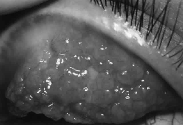
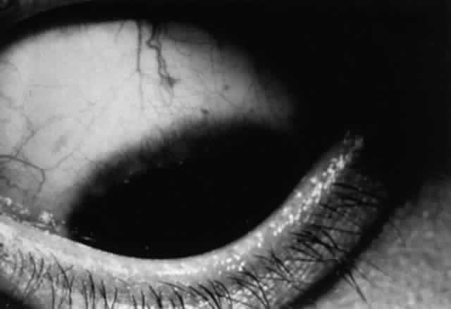
There are approximately 50 million mast cells in the ocular and adnexal tissues of the human eye.31 In normal conditions, mast cells are concentrated in the substantia propria of the conjunctiva but are not found in the conjunctival epithelium. However, in disease states such as vernal and contact lens- induced giant papillary conjunctivitis (GPC), mast cells infiltrate the conjunctival epithelium. Each mast cell has as many as 500,000 receptors for IgE; IgE is bound to 10% of these receptors, thus coating the mast cell surface.7 The Fab portion of the IgE molecule extends from the mast cell surface, poised to bind antigen and thus trigger mast cell degranulation, resulting in type I immune reaction. There are two main types of mast cells in the conjunctiva, defined by their protease (tryptase and chymase) content. The T mast cell is tryptase positive and chymase negative, whereas the TC mast cell is tryptase positive and chymase positive. In the normal conjunctival substantia propria, both T and TC mast cells are present, with the latter being predominant. In inflammation (e.g., vernal, GPC), the TC mast cells greatly increase in number and also are found in the epithelium.32

Scattered neutrophils are found in the epithelium and substantia propria. Eosinophils and basophils are not present in normal conjunctiva but are present in disease states.

Lymphoid cells sensitized to antigen exit via lymphatic channels to regional lymph nodes (submandibular and preauricular), then via efferent lymphatics to the thoracic duct and to the bloodstream. The sensitized lymphocytes return via the bloodstream to take up residence in conjunctival substantia propria (mostly T cells) and in lacrimal and accessory lacrimal glands (mostly B cells). Some cells probably migrate to other sites of MALT as well.

CORNEA.

The normal cornea is devoid of blood vessels, lymphatics, and inflammatory cells. Class II antigen-bearing Langerhans cells are found in corneal epithelium, with the greatest density at the periphery, decreasing toward the central cornea. Although complement components C3, C4, and C5 are found throughout the corneal stroma, there is more complement in the peripheral cornea than in the central cornea overall, especially with regard to C1. The major source of complement is the limbal vasculature, with complement components diffusing from the peripheral to the central cornea. Being the largest complement component, C1 is less likely to diffuse centrally. Immunoglobulins in the human cornea probably diffuse inward from the limbal vessels. IgG is the predominant immunoglobulin in the cornea and probably the most important in microbial defense. Both IgG and, to a lesser extent, IgA are found through the whole stroma, each distributed smoothly from peripheral to central cornea. However, IgM is found only in the peripheral cornea, probably because its large size restricts diffusion. Stromal edema associated with corneal inflammation facilitates diffusion of immunoglobulins and complement toward the central cornea from the limbal vessels, reducing differences in distribution.33

Thus, immunologic differences between the central and peripheral cornea exist. The peripheral cornea has more Langerhans cells, more IgM, and more C1 and is adjacent to conjunctival lymphatics and vasculature. This has implications in the generation of peripheral corneal disease. Antigens in the peripheral cornea are closer to the conjunctival vessels and lymphatics and thus should be able to elicit an immune response more easily; likewise, conjunctival immune responses should be more likely to affect the peripheral cornea. Because C1 is the recognition unit for the classic pathway of complement activation, the complement cascade is more likely to be triggered in the peripheral cornea. The increased amount of IgM in the peripheral cornea may play a role in peripheral corneal rheumatoid melts, which is consistent with the fact that rheumatoid factor is an IgM antibody directed against IgG.33

The immune privilege of the cornea is related to several factors, including the lack of blood vessels and lymphatics in the cornea and the fact that the central cornea is relatively free of C1, IgM, and Langerhans cells. Anterior chamber-associated immune deviation (ACAID) also plays a role.34 This term describes the phenomenon of down-regulation of cell-mediated immunity in the face of antigen exposure, which is unique to the anterior chamber. Production of transforming growth factor-ß (TGF-ß) by cells in the iris and ciliary body seems to be an integral component of this phenomenon.12 Also, classical antigen-presenting cells (MHC class II) are not normally found in the anterior chamber of the eye.

Back to Top
ALLERGIC OCULAR DISEASE
Approximately 20% of the general population have allergic symptoms of one form or another. Of these, approximately one third have ocular symptoms. Fortunately, most affected persons have only mild ocular involvement. The majority tolerate their symptoms or receive sufficient relief from over-the-counter medications and never seek medical attention. Nevertheless, some forms of ocular allergy can cause severe symptoms and result in significant ocular morbidity. A classification of ocular allergic disease is presented in Table 4.


TABLE 4. Classification of Allergic Ocular Disease

  1. Allergic conjunctivitis
    1. Allergic rhinoconjunctivitis
      1. Seasonal (hay fever)
      2. Perennial

    2. Acute anaphylaxis

  2. Giant papillary conjunctivitis
    1. Vernal keratoconjunctivitis
    2. Atopic keratoconjunctivitis
    3. Contact lens/induced giant papillary conjunctivitis
    4. Reaction to sutures, prostheses

  3. Contact dermatoconjutivitis/keratitis
  4. Microbioallergic disease


ALLERGIC RHINOCONJUNCTIVITIS (HAY FEVER)

Allergic rhinoconjunctivitis is the most common ocular allergic disease.35 This condition usually begins early in life. There is often a family history of allergy. There may be a personal history of other allergic disease (e.g., asthma).

Pathophysiology

In predisposed individuals, airborne environmental antigens (allergens) contact the conjunctival and/or nasal mucosa, stimulating a type I hypersensitivity reaction (a local anaphylaxis). Mast cells and basophils possess high-affinity surface receptors for the Fc portion of IgE; thus, IgE coats the surface of these cells. Allergen binds to this surface IgE on conjunctival basophils and mast cells. Bridging occurs, resulting in a change in membrane permeability and influx of calcium. Degranulation follows (i.e., release of preformed mediators from secretory granules such as histamine, proteoglycans [heparin], and proteases [tryptase]). Eosinophilic chemotactic factor of anaphylaxis (ECF-A), responsible for influx of eosinophils, is also released by mast cells. Also, de novo synthesis of inflammatory mediators occurs. Calcium influx activates phospholipase A2, which liberates arachidonic acid from membrane-bound phospholipids. This results in the formation of eicosanoids such as prostaglandins via the cyclo-oxygenase pathway and leukotrienes via the lipoxygenase pathway (Figs. 11 and 12). Platelet activating factor is produced from modified phospholipids.

Fig. 11. Cyclo-oxygenase pathway. Heavy arrows indicate sites of inhibition by corticosteroids and nonsteroidal anti-inflammatory drugs (NSAIDs). (Abelson MB, Schaefer K: Conjunctivitis of allergic origin: Immunologic mechanisms and current approaches to therapy. Surv Ophthalmol 38(suppl): 120, 1993)

Fig. 12. Lipoxygenase pathway. (Abelson MB, Schaefer K: Conjunctivitis of allergic origin: Immunologic mechanisms and current approaches to therapy. Surv Ophthalmol 38(suppl):121, 1993)

Histamine is the most abundant mediator, making up 10% of the weight of the mast cell granule.36 Histamine causes increased vasopermeability, vasodilation, and bronchoconstriction, mediated through histamine receptors. Both H1 and H2 receptors have been identified on the ocular surface. Slow-reacting substance of anaphylaxis (SRS-A) is actually a combination of leukotrienes C4, D4, and E4 and is believed to be 1000 times more potent than histamine at eliciting vasopermeability and bronchoconstriction.37 The activity of ECF-A has been attributed to lipoxygenase products, including leukotriene B4 (LTB4).38 Prostaglandin D2 is the major prostaglandin produced by the mast cell and increases conjunctival microvascular permeability. It is considered to be 10-fold more potent than histamine.37 PAF is chemotactic for eosinophils and is believed to be 100-fold more effective than ECF-A and LTB4. In addition, PAF causes vasodilation, increased vasopermeability, and bronchoconstriction. Prostacyclin and thromboxane may also play a role. Other autacoids such as the kinins are involved. Together these mediators are responsible for the redness, chemosis, mucus production, and pruritus seen in hay fever conjunctivitis and other ocular allergic disease.

More severe allergic reactions may demonstrate a late-phase reaction. These are either sustained reactions or more discrete second peaks of response 4 to 11 hours after antigen exposure. Prostaglandins are thought to play a role. The significance of late-phase reactions is not yet clearly defined but may contribute to the mechanism by which mild self-limited allergic disease evolves into a more chronic process such as vernal conjunctivitis.39

Why atopic individuals are affected by allergens while other exposed persons are spared symptoms is uncertain. Genetic factors play a role. Increased mucosal permeability or impaired local defense mechanisms such as opsonization and phagocytosis may allow increased penetration of allergens. The percentage of occupied receptors for IgE on the surface of mast cells and basophils is higher in allergic individuals and may be a factor.40

Clinical Features

Symptoms include itchy, watery eyes with mild redness. The most important symptom is itching; if there is no itching, an allergic component is not likely present. Eye symptoms may be accompanied by nasal symptoms such as rhinorrhea, sneezing, nasal obstruction, and itching. Ocular signs include mild to moderate eyelid edema, dilatation of conjunctival vessels, and minimal to moderate conjunctival chemosis. The conjunctival edema of the inferior fornix typically has a milky appearance. There may be a small amount of mucus. Small papillae may be present under the upper lid, but there are never large papillae or follicles.* Lid swelling may be accompanied by venous congestion, causing the lids to have a dark color called an “allergic shiner.” There may be a typical horizontal fold in the lower eyelid skin known as “Dennie's line.” The cornea is seldom involved, and vision is not significantly affected.


Papillae are moundlike elevations of the conjunctiva with a visible central vessel when viewed with the slit lamp. Histopathologically, there are inflammatory cells and edema surrounding the central vessel. Papillae may be small (less than 0.3 mm in diameter) or large (0.3 to 1 mm for macropapillae; more than 1 mm for giant papillae). Follicles are also moundlike but aresurrounded by a wreath of vessels and do not possess a central vessel. Histopathologically, they consist of a central collection oflymphocytes--a lymph follicle. They may vary in size from small to large. They are most commonly seen in viral or chlamydial conjunctivitis.

Seasonal Versus Perennial Allergic Rhinoconjunctivitis

Seasonal allergic conjunctivitis typically occurs in May and June (pollen from grasses and trees) and in late summer and fall (ragweed). Grass and ragweed are the chief offenders for the eye, as they are for the nose and lung. Molds and fungi are common offenders. House dust, animal dander, and industrial chemicals may be allergenic. Food allergy (e.g., chocolate, strawberries, seafood) may play a role in some cases.

Perennial (chronic) allergic rhinoconjunctivitis is a variant of seasonal allergic rhinoconjunctivitis. Symptoms are much the same as for the seasonal form but are generally milder and occur on a year-round basis; there may be superimposed exacerbations. Objective findings are often minimal and include conjunctival injection, more pronounced milky edema of the conjunctiva of the inferior fornix, and small papillae on the superior tarsal bulbar conjunctiva. Corneal involvement is unusual. Rhinitis may accompany the ocular manifestations. Conjunctival mast cells are in a continuous state of activation, releasing mediators throughout the year. Airborne allergens (mites and molds, house dust, feathers, animal dander) are the usual inciting agents (70%).41 These patients may be more symptomatic indoors and during the winter months.

Diagnosis

The diagnosis of allergic conjunctivitis is usually a clinical one made on the basis of the symptoms and signs. By far the most important clinical clue is the presence of itching. In most cases, the laboratory is not helpful. Nevertheless, lab studies are indicated in those who are nonresponsive to therapy and have chronic complaints. Conjunctival smears for cytology may show eosinophils in chronic cases but are seldom present acutely. Eosinophils are not present in the normal human conjunctiva; thus, the presence of even one eosinophil or eosinophilic granule may indicate that an allergic process is present. However, because eosinophils may be located deep in the conjunctiva in allergic disease, conjunctival scraping can be falsely negative; the absence of eosinophils on conjunctival scraping does not rule out an allergic process.42

Scratch or prick skin tests to determine sensitivity to suspected allergens can be helpful. A wheal and flare reaction within 15 to 20 minutes indicates sensitivity. Weak skin test reactions to airborne allergens are characteristic of localized ocular allergy; strong skin test responders often have nasal, respiratory, and cutaneous allergic disease, as well as ocular problems.41 In questionable or negative skin tests, intradermal injection of a more dilute preparation of allergen can be administered.40 Conjunctival provocation testing (ocular challenge testing) can be performed by the topical application of a minimal dose of a specific allergen, usually one that has given a positive skin test result. A positive ocular challenge test (subsequent ocular itching and redness) will verify a suspected allergen as the cause of a patient's ocular allergic symptoms. Also, ocular challenge tests may be positive when skin tests are negative.43 Presently, however, this test has limited office application and is used primarily experimentally. As with skin testing, an antianaphylaxis kit must be available.

Serum and tear IgE levels may be increased in allergic conjunctivitis but not consistently. IgE against specific allergens (dust, pollens) can be identified by use of the radioallergosorbent test (RAST). However, measurement of IgE levels is seldom performed clinically. The level of tryptase, a product of mast cells and an indicator of mast cell activity, is elevated in tears in seasonal allergic conjunctivitis and may prove helpful in the diagnosis of ocular allergic disorders.44 Bacterial cultures should be performed to rule out a chronic blepharitis or conjunctivitis. A Schirmer's test can help determine whether there is a dry eye problem either mimicking or accompanying allergic conjunctivitis.

Treatment

Exposure to allergens should be reduced as much as possible. For those with seasonal allergic conjunctivitis, this may require minimizing going outdoors during peak seasons. For both the seasonal and perennial sufferer, use of air conditioners and air filters can be helpful. Especially for those with perennial symptoms, immaculate household cleanliness is necessary. Household pets may need to be removed. Cigarette smoking should be eliminated. Patients with concurrent dry eyes benefit from artificial tears, especially without preservatives (e.g., Refresh), to maintain an adequate tear film to flush allergens out of the eye. Lid hygiene at bedtime consisting of careful scrubbing of the eyelid margins for a few minutes with commercial preparations (e.g., Eye-Scrub) or diluted baby shampoo can help those with a concurrent mild chronic blepharitis.

Treatment of flare-ups of allergic conjunctivitis includes application of cold compresses and nonpreserved artificial tears. Topical vasoconstrictors such as naphazoline (e.g., Vasocon, Naphcon, Albalon) or combination vasoconstrictor-antihistamines such as naphazoline and pheniramine or antazoline (e.g., Vasocon-A, Naphcon-A, Albalon-A) can be used three to four times a day as needed to lessen itching and redness. However, chronic use should be avoided because these drugs can result in rebound hyperemia and toxic follicular conjunctivitis. Oral antihistamines (H1-receptor blockers) can be given to lessen symptoms in the acute stage, but patients should be warned concerning possible sedation and systemic anticholinergic side effects. Some of the newer oral antihistamines such as astemizole (Hismanal) and terfenadine (Seldane) supposedly possess efficacy with lesser side effects. Topical antihistamine therapy is now available in the form of levocabastine (Livostin), an H1-receptor blocking agent with proven efficacy.45–47 There are no topical H2-receptor antagonists presently available for ocular use, but such agents have theoretic potential, especially in combination with an H1-receptor blocker.39

Topical cyclooxygenase inhibitors offer another treatment option. Topical flurbiprofen (Ocufen) has shown a beneficial effect in subjects with allergic conjunctivitis induced by topical antigen challenge.48 Topical ketorolac49,50 has been shown to be efficacious in seasonal allergic conjunctivitis. Both topical ketorolac (Acular) and diclofenac (Voltaren) have been approved for treatment of allergic conjunctivitis in the United States.

Topical cromolyn sodium 2% or 4% (Opticrom), a mast cell stabilizer that inhibits mast cell degranulation, is an effective preventive and maintenance therapy for allergic conjunctivitis. Unfortunately, sodium cromoglycate requires at least 2 weeks to produce a clinical effect. Topical cromolyn can be started as soon as a flare-up begins, but topical lubricants, decongestants, or nonsteroidal anti-inflammatory drugs must be relied upon temporarily until cromolyn takes effect. The seasonally allergic patient can start using topical cromolyn four times a day at the onset of his or her allergy season before signs and symptoms begin and can continue until the end of the season to prevent flare-ups. The perennially affected patient may require the drops two to four times a day all year round. Fortunately, there are few ocular side effects (mild stinging or burning immediately after application). A newer mast cell stabilizer, lodoxamide 0.1% (Alomide), is now available as an alternative to cromolyn sodium.51,52 Nedocromil sodium, a pyranoquinoline and chemically distinct from cromolyn, is a newer inhibitor of mast cell mediator release and has been shown to be effective in seasonal allergic conjunctivitis.53,54

Topical corticosteroids55 such as prednisolone acetate or phosphate 0.1% (Pred Mild, Inflamase Mild) and 1% (Pred Forte, Inflamase Forte) or fluorometholone 0.1% (FML Liquifilm) are very effective in alleviating symptoms. However, allergic conjunctivitis is a benign disease that does not cause significant ocular sequelae. On the other hand, corticosteroids can cause glaucoma, cataract, and exacerbation of herpetic ocular disease. Therefore, the use of corticosteroids must be limited to those cases with marked symptoms, and then for only short periods.

Immunotherapy in the form of desensitization can be helpful in some patients. However, desensitization is not as effective for ocular allergic conjunctivitis as it is for allergic rhinitis. The presumed mechanism of action is the production of blocking antibodies of the IgG class or stimulation of production of suppressor T cells that inhibit IgE synthesis.56 Desensitization involves injection of sequentially increasing doses of allergen extract until a maintenance dose and frequency that successfully prevent symptoms is attained. Improvement should occur in a few weeks. Maintenance injections may be required every 1 to 6 weeks and may be continued for 3 to 5 years. Some patients may relapse after injections are discontinued. The risk of anaphylaxis must be considered.57

Acute Anaphylactic Reaction

An explosive onset of severe eyelid edema and conjunctival redness and swelling can be stimulated by ocular exposure to allergen. Common agents include topical medication, animal dander, pollens, molds, insect bites, and stings. In addition, exposure may occur at a distant site yet be manifest predominately in the eye. Also, there may be associated allergic signs at other body sites (e.g., rhinitis, urticaria). There may be serious manifestations of anaphylaxis as well (angioedema, bronchospasm, hypotension).

Treatment of the ocular manifestations includes cold compresses, topical vasoconstrictor-antihistamines, topical corticosteroids, and oral or, if severe, parenteral antihistamines such as intramuscular or intravenous diphenhydramine (Benadryl) 50 mg. Those with serious concurrent systemic manifestations of anaphylaxis may require subcutaneous epinephrine (adult dose of 0.3 to 0.5 mL of 1:1000 solution), intravenous theophylline and inhaled bronchodilators, intravenous corticosteroids, intravenous fluids, oxygen, and even intubation to maintain an airway, depending on the extent and severity of systemic involvement.

GIANT PAPILLARY CONJUNCTIVITIS

This heading actually includes several entities (see Table 4) having in common the presence of giant papillae. These entities include vernal keratoconjunctivitis and atopic keratoconjunctivitis as well as reactions to ocular prostheses, exposed nylon suture, cyanoacrylate glue, exposed scleral buckles, and contact lenses. In addition, the term giant papillary conjunctivitis is frequently used to denote the specific entity that occurs as a response to contact lens wear (i.e., contact lens-induced giant papillary conjunctivitis).

VERNAL KERATOCONJUNCTIVITIS

Vernal keratoconjunctivitis typically begins in children before puberty and has a tendency to lessen in the second and third decades. In the majority, the onset is before age 10. Rarely, the diagnosis is made in adults. Before puberty, males are affected two to three times more frequently than females; however, the prevalence in females increases after puberty and equals that of males after age 20. Vernal is commonly found in individuals with a personal or family history of allergic disease. It is usually seasonal, occurring in the spring and summer; however, some persons are affected year-round. The disease is prevalent in hot, dry climates such as the Mediterranean area, Middle East, West Africa, the Indian subcontinent, Mexico, and the southwestern United States. Vernal does occur elsewhere in North America and Canada but less frequently. Despite its seasonal profile, vernal may occur throughout the year, especially in tropical countries.

The most important ocular symptom is itching, often severe. There can be burning, photophobia, and complaints of lacrimation and mucus discharge. Affected persons often rub their eyes vigorously and exhibit blepharospasm.

Vernal is a bilateral disease but may be asymmetric. It has two conjunctival forms, palpebral and limbal. The palpebral form is characterized early by a dull, pale, thickened tarsal conjunctiva. Eventually, papillae form that may be small and few in number or may evolve into multiple giant cobblestone (flat-topped) papillae on the superior tarsal conjunctiva (Fig. 13). There is often copious tenacious mucus draped over these papillae. There may be an associated mechanical ptosis. The lower tarsal conjunctiva shows at most a mild papillary reaction. Pseudomembrane formation when the upper lid is everted and exposed to heat is known as the Maxwell-Lyons sign.

Fig. 13. Giant papillae involving the superior tarsal conjunctiva in vernal keratoconjunctivitis. (Courtesy of the Francis I. Proctor Foundation)

In the limbal form, papillae are located on the bulbar conjunctiva at the corneal limbus, usually superiorly, but they can involve the limbus elsewhere (Fig. 14). These papillae often have a gelatinous appearance; they may coalesce and form a ridge along the limbus. There may be associated micropannus formation. Small, fine white dots may be seen on the limbal papillae; these are Horner-Trantas' dots and represent focal collections of eosinophils (Fig. 15). Horner-Trantas' dots are usually seen at the superior limbus but can be seen elsewhere on the limbus and on the bulbar and tarsal conjunctiva. The limbal and palpebral forms of vernal can coexist. The limbal form is more common in blacks.

Fig. 14. Limbal papillae in vernal keratoconjunctivitis.

Fig. 15. Horner-Trantas' dots at the superior limbus in vernal keratoconjunctivitis.

Either form of conjunctival vernal can be accompanied by changes in the upper one half of the cornea, including micropannus and a farinaceous epithelial keratopathy known as keratitis epithelialis vernalis of Tobgy, consisting of tiny gray-white intraepithelial opacities (“dusting of flour”). Punctate epithelial keratitis may evolve into epithelial macroerosions. Such macroerosions may evolve into a shield ulcer (Fig. 16). This is an indolent, oval, superficial ulceration with thickened, opaque edges and plaquelike deposition of mucus, fibrin, and inflammatory debris. This deposition prevents re-epithelialization. Shield ulcers usually affect the superior cornea but also can occur inferiorly. With resolution, oval or ring scarring can remain in the superficial stroma. Another corneal finding typical of vernal is pseudogerontoxon, an arclike or annular deposition of unknown nature in the corneal periphery. There is an association between vernal and keratoconus.58 Pellucid marginal degeneration, keratoglobus, and Terrien's marginal degeneration have also been described.59,60

Fig. 16. Shield ulcer in vernal keratoconjunctivitis.

Pathophysiology

Vernal keratoconjunctivitis involves type I and type IV (including cutaneous basophil hypersensitivity) mechanisms.61 Eosinophils and free eosinophilic granules on examination of conjunctival scrapings are characteristic. Elevated levels of tear IgE and/or IgG, histamine, tryptase, eosinophilic major basic protein, Charcot-Leyden crystal protein (found when eosinophils have undergone fragmentation),62 prostaglandins, and leukotrienes are present.

Histopathologically, the conjunctival epithelium is thickened; the epithelium shows mast cells, basophils, and eosinophils, none of which are seen in normal conjunctival epithelium. The substantia propria shows hyperplasia of connective tissue and infiltration by mast cells, basophils, eosinophils, plasma cells, lymphocytes, and macrophages. Eosinophils and basophils are not normally found in the substantia propria. Mast cells are found in higher numbers in the conjunctiva, they are located more superficially, and the percentage of degranulated mast cells is increased over normal controls.61,63 These findings support the concept that this is a mixed type I and IV reaction.

Although it was previously thought that eosinophils may dampen allergic inflammation, evidence now suggests that eosinophils play an active role and contribute to tissue damage in allergic disease. Eosinophil granule major basic protein is known to stimulate mast cell degranulation and may explain the extensive and sustained mast cell degranulation seen in vernal keratoconjunctivitis (and contact lens-induced GPC).64

The corneal epithelial manifestations of vernal may be in part related to mechanical friction caused by tarsal conjunctival papillae. More likely is that corneal epithelial damage occurs from chemical mediators released by mast cells and eosinophils, particularly eosinophil major basic protein. Eosinophil major basic protein possesses cytotoxic capabilities65,66 and prevents epithelial healing.67 Eosinophil major basic protein has been detected in the inflammatory debris of vernal ulcers.68 Unless the underlying inflammatory process is treated with success medically or, in some cases, the inflammatory debris of the ulcer is removed surgically, healing will not occur, implicating eosinophil major basic protein in the ulcerative process by inhibition of corneal epithelial healing.68

Treatment

General measures include “climatotherapy” (i.e., moving to a cooler and moister climate), air-conditioning, humidification, and air filtering to remove dust and other allergens. Cold compresses are helpful to relieve itching. Patching can be helpful.

The cornerstone of medical therapy is the use of mast cell stabilizers. Topical cromolyn sodium 2% or 4% (Opticrom) is given four times a day, and in severe cases up to six times a day. If a patient has a history of seasonal exacerbation, he or she can be instructed to start cromolyn a month or so prior to the expected onset of the symptoms; some require cromolyn all year round two to four times a day as maintenance therapy. As mentioned previously, this drug has few side effects and is well tolerated.69 A clinical response takes 1 to 2 weeks to become evident. In the interim, mild cases can be treated with artificial tears, topical vasoconstrictor-antihistamines, or topical cyclooxygenase inhibitors. Topical lodoxamide 0.1%, a newer mast cell stabilizer, has been shown to be effective in vernal, eliciting an earlier and greater response than cromolyn sodium.51 Also, oral antihistamines can be helpful. Topical levocabastine (H1-receptor blocker) offers promise as adjunctive therapy. Topical mucolytic agents (e.g., acetylcysteine) are seldom helpful.

More severe cases (i.e., marked symptoms or corneal involvement) will require topical corticosteroids four or more times a day for a few weeks until cromolyn takes effect. Thereafter, topical corticosteroids should be tapered over a week or so to prevent rebound, and then should be discontinued to minimize the chance of side effects. A few weeks of topical corticosteroids are usually sufficient, but some patients may require maintenance therapy with as low a dose as possible. Very severe cases, especially if unresponsive to treatment, will require a course of oral corticosteroids. Topical cyclosporine may be helpful also in refractory cases.70,71

A new direction in therapy is the use of cyclooxygenase inhibitors. Oral aspirin has been reported to aid remission of vernal72-74; however, one must be wary of acetylsalicylic acid (ASA) in those with asthma.75 Other nonsteroidal anti-inflammatory agents, such as indomethacin, tolmetin, and suprofen, applied topically, have been shown to be effective in the treatment of vernal keratoconjunctivitis.76–78

Desensitization is not commonly used; its efficacy is a subject of debate. Cryotherapy to the tarsal surface is effective in providing immediate short-term relief, probably related to the mechanical degranulation of large numbers of mast cells.79 As previously discussed, surgical removal of corneal plaque by superficial keratectomy is sometimes required to heal shield ulcers.80

Prognosis

Fortunately, vernal keratoconjunctivitis is a self-limited disease that resolves with age, usually after an interval of 2 to 10 years. Unlike atopic keratoconjunctivitis, vernal does not typically cause significant conjunctival cicatricial changes. However, shield ulcers can leave corneal scarring. In some patients, vernal does not resolve and instead transforms into adult atopic keratoconjunctivitis.79

CONTACT LENS-INDUCED GIANT PAPILLARY CONJUNCTIVITIS (GPC)

Giant papillary conjunctivitis affects approximately 1% to 5% of the 30 million contact lens wearers in the United States.81 GPC can occur in those wearing hard, semi-rigid, and soft hydrogel lenses; however, soft lens wearers are by far the most commonly affected.

Clinical Features

The earliest symptoms are itching on lens removal and mild mucus formation. Later, patients complain of contact lens intolerance with irritation, itching, redness, tearing, mucus discharge, and blurriness of vision while wearing their lenses. Symptoms usually precede signs.82 Although symptoms and signs usually progress together, some patients with minimal objective findings may have severe symptoms; on the other hand, occasionally a patient with severe objective changes may have minimal symptoms.

On examination, early there is bulbar and palpebral conjunctival hyperemia; occasional mucus strands may be present. With progression, conjunctival thickening with loss of translucency occurs. The upper tarsal conjunctiva will demonstrate macropapillae (0.3 to 1 mm in diameter) or giant papillae (more than 1 mm in diameter) (Fig. 17). The number of papillae can vary from a few to innumerable. More pronounced mucus strands can be found at the canthi, in the fornices, and among the papillae. The papillae often have a dome-shaped appearance with fibrotic whiteyellow tops. Horner-Trantas' dots may be seen. Fluorescein staining of the papillae, which indicates ulceration of the conjunctival surface, reflects severity. There may also be corneal staining, but this is uncommon. Pseudoptosis may occur in more severe cases. There is often significant deposit formation on the surface of the contact lenses.

Fig. 17. Giant papillary reaction in contact lens-induced giant papillary conjunctivitis. (Courtesy of the Francis I. Proctor Foundation)

Pathogenesis

Several factors may be involved in pathogenesis. Mechanical irritation by the contact lens probably plays a role, especially lenses with thick edges, as seen in myopes. Accumulated deposits of protein, lipid, and minerals on the contact lens surface act as a source of antigens that can sensitize the conjunctiva. Chronic microtrauma by the lens and its deposits disrupts tight junctions between conjunctival epithelial cells, allowing allergens to penetrate. Damage to conjunctival epithelial cells induced by contact lenses and their deposits stimulates the production of neutrophil chemotactic factors, which play a role in the inflammatory response.83–85 Certain lens polymers (e.g., hydrogels) are more prone to accumulation of deposits. Also, larger lenses provide a greater surface area for deposits to accumulate and a greater surface area for antigens to contact the conjunctiva. Long wearing times allow a longer period of conjunctival exposure to antigen. Meibomian gland dysfunction may play a role in deposit formation and contact lens intolerance.86–88

The allergic mechanism in GPC is probably a basophil-rich delayed hypersensitivity (type IV) reaction with a possible IgE (type I) humoral component.82,89 An abnormal distribution of inflammatory cells is seen. There are mast cells in the conjunctival epithelium (not seen in normals) and increased numbers of mast cells in the substantia propria. Basophils and/or eosinophils, not usually seen in normal conjunctiva, may be found in increased numbers in both the epithelium and substantia propria.82 Although tear histamine levels are normal, tear immunoglobulins are increased.90,91 As with vernal, eosinophil major basic protein, which stimulates mast cell degranulation and possesses cytotoxic effects that may enhance the conjunctival inflammatory reaction, is found in increased amounts in the conjunctiva (but not in tears as in vernal) and undoubtedly plays a role.64 Tear tryptase levels in patients with GPC, a measure of mast cell degranulation, may provide an early marker for this disease.92

Treatment

Asymptomatic patients with signs only or those with minimal symptoms and signs may not need to discontinue lens wear if good lens hygiene is maintained; these patients may take topical cromolyn 2% or 4% q.i.d. with their contact lenses in place to maintain control.

Initial treatment of the symptomatic patient is discontinuation of lens wear. Ocular lubricants are helpful during this time period. Symptoms may improve within 2 or 3 days, but lenses should not be resumed until about 1 week after all mucus, redness, and irritation are gone. Papillae may last longer, but contact lenses can be resumed if there is no fluorescein staining. Topical cromolyn sodium or corticosteroids are usually not required if lens wear is stopped but may be helpful in more severe cases. The nonsteroidal anti-inflammatory agent suprofen used topically has also been shown to be effective in GPC.93

When contact lenses are resumed, proper lens hygiene must be stressed. This is especially important because as a group, contact lens wearers with GPC are neglectful with lens care. Hydrogen peroxide cleaning systems (preferably nonpreserved) and nonpreserved saline are best for patients with GPC. Cold disinfection systems increase the possibility of exposure to sensitizing agents and should be avoided. Thermal disinfection bakes deposits on the lenses and thus is also less favorable for patients with GPC. Enzymatic cleaning, particularly papain, is superior to surfactant cleaning.82 Whereas the usual contact lens wearer uses enzymatic cleaners once a week and surfactant cleaners daily, the patient with GPC needs to use enzymatic cleaners several times a week, and in some cases daily. Maximum wearing time should be reduced. A change to a soft contact lens made of a different polymer may be required; in particular a nonionic, low-water-content polymer is ideal. Another alternative is to use disposable soft contact lenses (e.g., Acuvue) on a daily-wear basis, using regular cleaning, as noted above, and discarding the lens after 1 to 2 weeks.94

If a recurrence occurs after resumption of lens wear, there must be at least a 1-month period off lenses, preferably 3 months. Again, a soft contact lens of a different polymer can be tried, or the patient can switch to a gas-permeable semi-rigid lens. Some will benefit from topical cromolyn 2% or 4% four times a day with the contact lenses in place to prevent recurrences.

ATOPIC KERATOCONJUNCTIVITIS

This entity usually develops in patients with a personal or family history of atopic disease, such as atopic dermatitis, asthma, perennial rhinitis, urticaria, or food allergy. Asthma and atopic dermatitis are the most common atopic diseases in patients with atopic keratoconjunctivitis.95 Atopic keratoconjunctivitis is seen in 25% of all patients with atopic dermatitis.96 When there is concurrent atopic dermatitis, it can be isolated to the periocular area or can be found elsewhere. Eczema may have been present in infancy or childhood but has long since resolved. Atopic dermatitis and atopic keratoconjunctivitis may also be associated with the hypergammaglobulinemia E syndrome.97

Atopic keratoconjunctivitis is a bilateral, usually symmetric disease. It can occur in early childhood but more commonly begins in the late teens and early twenties. The peak incidence is age 30 to 50 years. Its onset is typically later than that of atopic dermatitis alone and is also later than that of vernal keratoconjunctivitis. Atopic keratoconjunctivitis usually will eventually settle but does so gradually and can leave serious ocular sequelae.

Seasonal exacerbations occur in about one third of patients, although there does not appear to be a particular season more prone to exacerbations.95,98 Symptoms may be perennial. Patients may be able to identify particular allergens that stimulate flare-ups, common ones being animals, dusts, and foods.

Usual symptoms include itching, burning, tearing, photophobia, and pain. Signs include eczematous changes of lids; there may be associated blepharitis and meibomianitis. In atopic keratoconjunctivitis, conjunctival involvement is predominately found inferiorly. The conjunctiva may be hyperemic and chemotic in exacerbations but often is pale and thickened. The palpebral conjunctiva displays papillae that may be large; however, they are not usually as striking as those in vernal keratoconjunctivitis and often are scarred in appearance. Mucus strands may be present. There may be limbal papillae. As in vernal and contact lens-induced GPC, Horner-Trantas' dots may be seen. Conjunctival scarring is typical of this disease, with subepithelial fibrosis, symblepharon formation, and foreshortening of the fornices. Punctal ectropion may cause epiphora. Corneal changes include punctate keratitis (especially involving the inferior one third), vascularization, shieldlike ulceration, and subsequent anterior stromal scarring. Associated ocular conditions include keratoconus and cataract (Fig. 18). These patients often have deficient immunoregulation with depressed T-cell function. As a result, they are prone to develop staphylococcal blepharitis and herpetic keratitis.

Fig. 18. Typical anterior subcapsular cataract in atopic keratoconjunctivitis.

Serum IgE levels are increased in patients with atopic dermatitis, and a high percentage have positive skin prick tests.95,98 Nevertheless, identification of a specific causative allergen is seldom made.

Histopathology/Immunopathology

Foster and colleagues99 have provided an excellent description of the histopathology and immunopathology of atopic keratoconjunctivitis. Histopathologic examination of the conjunctiva shows mast cell and eosinophil invasion of the epithelium, goblet cell proliferation, epithelial pseudotubular formation, and increased numbers of mast cells in the substantia propria in various states of degranulation, along with eosinophils and large numbers of mononuclear cells. Evidence of frank granuloma formation and perivasculitis can be seen.

The mononuclear cell population of the epithelium shows increased numbers of T cells, helper T cells, activated T cells (IL-2 receptor positive), macrophages, and Langerhans cells. The helper/suppressor T-cell ratio is increased. In addition, the conjunctival epithelium shows increased MHC class II expression. In the substantia propria, increased numbers of helper T cells and suppressor T cells are present, with an elevated CD4/CD8 ratio. About 50% of the T cells are activated. B cells and antigen-presenting cells (macrophages and Langerhans cells) are also increased in the substantia propria. There is increased expression of MHC class II antigens on fibroblasts and vascular endothelium.99 These findings are in keeping with a complex immunologic mechanism involving both type I and type IV mechanisms.

As mentioned previously, a reduction in delayed hypersensitivity exists in patients with atopic dermatitis,100 making these patients more prone to herpes simplex virus (HSV) and staphylococcal infection. HSV keratitis is often bilateral with extensive corneal epithelial ulceration.101–103 These patients are prone to eczema herpeticum.104

Treatment

Patients should avoid exposure to precipitants historically suspected by the individual or identified on skin tests. Hyposensitization may be helpful.

Emollients and nonfluorinated corticosteroid creams or ointments (e.g., 0.5% to 1% hydrocortisone, desonide 0.05%) are used to treat eyelid eczema. Lid hygiene, topical antibiotic application, and/or low-dose systemic tetracycline may be required to treat staphylococcal blepharitis and meibomianitis.

Cold compresses, topical vasoconstrictors, topical antihistamines (e.g., levocabastine), and oral antihistamines such as terfenadine, astemizole, or hydroxyzine are used to control itching. Topical lubricants (especially nonpreserved) may give symptomatic relief. Mast cell stabilizers (2% or 4% cromolyn sodium q.i.d.) are used for maintenance therapy. Topical lodoxamide and nedocromil sodium offer other options to cromolyn sodium. Topical steroids are reserved for flare-ups as much as possible; however, some patients require chronic maintenance topical corticosteroid therapy. Of course, any maintenance topical corticosteroid therapy should be kept to as small a dose as possible. Severe exacerbations require a course of systemic prednisone. In fact, many of these patients will be on maintenance systemic corticosteroids to control their asthma or eczema. Topical cyclosporine has been used.98 Plasmapheresis may be helpful in severe cases.97,98

CONTACT ALLERGY

Contact inflammation of the eye may affect the lids, conjunctiva or cornea, or a combination of these and may involve allergic or nonallergic (irritant/toxic) mechanisms.

Contact allergic reactions involve exposure to a sensitizing substance that is absorbed through the skin. These substances are generally haptens (i.e., partial antigens) of low molecular weight that bind to dermal proteins, forming complete antigens. Sensitization of the cellular immune system results. Upon re-exposure, a type IV hypersensitivity reaction ensues. Humoral mechanisms are not typically involved. Sensitization may take weeks to years to develop, depending on the ability of the hapten to act as a sensitizer, the amount applied and the duration, pre-existing lid or ocular disease, and individual susceptibility. In sensitized persons, contact allergic reactions can occur in 48 to 72 hours upon rechallenge, in keeping with type IV delayed-type hypersensitivity reactions.

Nonallergic mechanisms involve contact irritation (i.e., toxicity). Contact inflammation is produced on the basis of a direct chemical effect on tissue; the resulting inflammation is not allergic in origin. Frequent defatting of the skin caused by excessive moisture also plays a role.105 Toxic reactions are much more common than allergic ones, accounting for about 90% of all reactions to topical ophthalmic medications. Toxic reactions include papillary conjunctivitis, follicular conjunctivitis, keratitis, pseudopemphigoid, and pseudotrachoma. Allergic contact dermatoconjunctivitis is the second most common type of drug reaction, accounting for 10% of reactions to topical ophthalmic medications.106,107

In contact reactions of the eye and elsewhere, irritant and allergic mechanisms may coexist. Clinically, it is usually difficult to separate the two. Classically, a contact allergic reaction will begin in 48 to 72 hours, in keeping with type IV hypersensitivity reactions. An irritant reaction may begin within a few hours of contact or may occur only after prolonged use of a topical medication.

Although sometimes the offending agent may be obvious, often identification is difficult. Patch testing may provide the answer. A true allergic response will occur in 48 to 72 hours. An irritant can also cause a positive reaction, but this usually develops within a few hours and can be avoided by using lower doses of testing substances.

The acute lesions of contact allergic dermatitis/blepharitis resemble acute eczema, with erythema, vesicles, edema, oozing, and crusting. The chronic phase is characterized by dryness, crusting, fissuring, and thickening of the skin (Fig. 19). Contact allergic conjunctivitis involves conjunctival injection and chemosis; there may be a papillary response and serous or mucoid discharge. Initially, the lower conjunctiva and lid are usually more affected; later, the entire conjunctiva and upper lid are involved as well. Keratitis can occur, including superficial punctate changes, subepithelial opacities, marginal infiltration and ulceration, erosions, or stromal edema. Itching can be prominent.

Fig. 19. Contact dermatitis involving the face, eyelids, and conjunctiva.

Several topical ocular drugs are known to act as sensitizers, including aminoglycosides (gentamicin, tobramycin, and neomycin), sulfonamides, atropine and its derivatives, topical antiviral agents (idoxuridine, trifluridine), topical anesthetics, echothiophate, epinephrine and phenylephrine, and preservatives (thimerosal, benzalkonium chloride, EDTA). Other sensitizing agents include lanolin and parabens (cosmetics, skin creams, and lotions), nickel sulfate (jewelry), chromates (jewelry, leather products, fabrics, industrial chemicals), and p-phenylenediamine (hair sprays, clothing, shoes). Rubbing the eyes after handling soaps, detergents, or chemicals may explain a localized ocular or periocular reaction after a more general exposure.108 The thinness of the eyelid skin and accessibility of the conjunctiva to sensitizing agents undoubtedly play a role.

Treatment

Treatment includes removal of the offending agent (including discontinuing eye drops) or avoidance (use of protective wear such as goggles, worksuits). In the acute stage of periocular contact dermatitis, cool compresses and topical nonfluorinated corticosteroids (e.g., hydrocortisone, desonide) can be used. In some cases, especially if severe, unresponsive to therapy, or associated with cutaneous ulcerations, cultures should be performed to rule out secondary infectious eczematoid dermatitis (usually staphylococcal or streptococcal in origin). Chronic periocular dermatitis requires maintenance of skin hydration and topical corticosteroids. In some cases (either acutely severe or chronic and unresponsive cases), a course of oral prednisone may be indicated. Contact conjunctivitis/keratitis may be soothed by application of nonpreserved artificial tears. A short course of topical corticosteroids may speed recovery, but one always should consider the possibility of further sensitization by the vehicle or the steroid itself.

MICROBIOALLERGIC DISEASE

Catarrhal Infiltrates and Ulcers

Catarrhal infiltrates typically occur in association with chronic staphylococcal blepharoconjunctivitis and are located in the peripheral cornea, separated from the limbus by a lucid interval. They are most commonly found at the two, four, eight, and ten o'clock positions, where the lid margins cross the peripheral cornea. These infiltrates are gray-white in color, may be single or multiple, and spread circumferentially rather than centrally, sometimes making partial rings. Overlying superficial ulceration often develops. There may be associated conjunctival injection or mild anterior chamber reaction. Symptoms include irritation, tearing, pain, and photophobia. Catarrhal ulcers are more common in adults than in children (compare with phlyctenulosis).

Although catarrhal infiltrates usually occur in a background of staphylococcal blepharoconjunctivitis, cultures of catarrhal lesions are usually negative. Rather than direct infection, the mechanism may involve sensitization to staphylococcal cell wall antigens, in particular, ribitol teichoic acid. There seems to be an antigen-antibody reaction and immune complex deposition in the peripheral cornea, with complement activation and neutrophil infiltration.109 In fact, Ig and C3 have been demonstrated in catarrhal infiltrates.110

These lesions are not to be confused with infected corneal ulcers caused by direct staphylococcal infiltration of the corneal stroma. These usually follow trauma, are central, and yield positive bacterial cultures from scrapings of the lesion.

Catarrhal ulcers have also been described in acute beta-hemolytic streptococcal conjunctivitis associated with chronic dacryocystitis (lacrimal conjunctivitis of Morax), the mechanism perhaps being hypersensitivity to streptococcal antigens,111 although others have suggested direct infection.112 Other causes of lesions similar to catarrhal infiltrates include herpes simplex, Chlamydia, Haemophilus aegyptius, Moraxella lacunata, Neisseria gonorrhoeae, Escherichia coli, lymphogranuloma, actinomyces, and drug allergy. Sterile infiltrates related to contact lens wear can be confused with catarrhal infiltrates.

Management includes cultures of the lids and conjunctiva to identify the presence of staphylococcal blepharoconjunctivitis or, less commonly, other organisms. Treatment should stress proper hygiene of the eyelid margins using lid scrubs with diluted baby shampoo or commercially available preparations, warm compresses, and topical antibiotics (e.g., bacitracin, erythromycin). Topical corticosteroids elicit a rapid improvement but should be used with caution. They should not be used without topical antibiotics and lid hygiene to reduce the presence of staphylococci. Corticosteroids should not be used if there is any question of an ulcer caused by direct bacterial invasion (central location). Also, herpes simplex keratitis can sometimes mimic the picture of staphylococcal marginal keratitis. It is wise to check for a history suggestive of herpetic disease (cold sores, recurrent skin vesicles, or dendritic keratitis). Corneal sensation should be checked (reduced in herpetic disease, normal in staphylococcal disease) and/or HSV studies performed. If there is any doubt, topical corticosteroid use should be deferred.

PHLYCTENULAR KERATOCONJUNCTIVITIS

Phlyctenules are nodular lesions of the cornea and conjunctiva, more commonly seen in children than adults. The symptoms of conjunctival phlyctenulosis include mild to moderate itching, tearing, and irritation. Corneal phlyctenules are more severe with photophobia, foreign-body sensation, and tearing.

Phlyctenules usually appear at the corneal limbus as small, red, rounded elevations; there may be more than one present at a time. Lesions typically develop yellowish white centers representing infiltrate, followed by ulceration. Usually, spontaneous resolution occurs after about 2 weeks, but lesions can last longer. When ulcerated, limbal phlyctenules may be confused with marginal catarrhal ulcers, except that the long axis is perpendicular to the limbus and there is no intervening clear zone between the ulcer and limbus. Limbal phlyctenules leave triangular scars with the base at the limbus. Phlyctenules may recur at the edge of pannus left as a residual from previous episodes. Phlyctenules can “wander” onto the cornea, the advancing edge being active but the trailing edge healing, often leaving a vascular trail (i.e., fascicular keratitis). This may resemble HSV keratitis. Occasionally, corneal lesions can begin distant from the limbus and leave a nonvascularized scar. Corneal perforations have occurred, more so in tuberculous phlyctenulosis, but also in staphylococcal disease. Previous phlyctenulosis can lead to Salzmann's nodular degeneration.

Conjunctival phlyctenules can occur, although they are less common (Fig. 20). Conjunctival phlyctenules are typically seen as small, pinkish white nodules on the bulbar conjunctiva near the interpalpebral limbus and are surrounded by dilated vessels. Phlyctenules can also occur on the tarsal conjunctiva. In miliary phlyctenulosis, multiple small lesions are found at the limbus and on the cornea.

Fig. 20. Conjunctival phlyctenule.

Phlyctenules are raised subepithelial inflammatory nodules composed of leukocytes (macrophages, PMNs, lymphocytes, and plasma cells) and blood vessels. Helper T cells outnumber suppressor T cells; mature B cells are present.113 The immunopathogenesis has classically been described as a type IV hypersensitivity reaction.

Currently, the most common cause of phlyctenulosis is hypersensitivity to staphylococcal antigens. Mondino109 was able to induce phlyctenular lesions in rabbits with staphylococcal cell wall antigen (ribitol teichoic acid), suggesting that hypersensitivity to ribitol teichoic acid may be a key factor in the development of Staphylococcus-associated phlyctenules. Historically, hypersensitivity to tuberculoprotein was the most common cause. Other causes include Candida, Coccidioides immitis, nematodes, lymphogranuloma venereum, adenovirus, and HSV. Phlyctenulosis can also be associated with ocular rosacea, probably because of the high frequency of staphylococcal blepharitis seen in this entity.

Investigations should include lid and conjunctival cultures to identify the etiologic agent. Because tuberculosis is less common than in the past, investigation for systemic tuberculosis is usually not required; however, tuberculosis should be considered in endemic areas if another etiology is not identified. Treatment includes topical and/or systemic antibiotics, depending on the causative agent. Phlyctenules are usually very sensitive to topical corticosteroids. Recurrent disease may respond to systemic tetracycline.

Back to Top
IMMUNOLOGIC CONSIDERATIONS IN SELECTED EXTERNAL OCULAR DISEASES

VERSUS-HOST DISEASE

Bone marrow transplantation is widely used in the treatment of severe aplastic anemia, hematologic malignancies (e.g., leukemia), and genetic disease (e.g., immunodeficiency states). Recipients are immunologically competent to some degree and require preparation with immunosuppressive agents (e.g., cyclophosphamide) to prevent marrow graft rejection. In addition, hematologic malignancies (e.g., leukemia) require destruction of all malignant cells; this is accomplished with cyclophosphamide followed by total body irradiation. Transplantation itself is achieved by intravenous injection of marrow aspirate from an HLA-matched donor to restore the recipient's hematologic function. It takes about 2 to 4 weeks for the new marrow to produce peripheral blood elements.114

Immunocompetent donor T lymphocytes can recognize host tissue as nonself and thus mount an immunologic response. This is known as graft-versus-host disease (GVHD). Graft-versus-host disease is a cell-mediated/cytotoxic immune reaction involving T-lymphocyte participation. In addition, NK cells115 and cytokines, especially IL-1,116 are believed to play a critical role.

Acute GVHD occurs early in the post-transplant period and affects primarily the skin (erythema, bullae), liver (elevated bilirubin and enzymes), and GI tract (diarrhea). Chronic GVHD occurs later (by definition, more than 100 days) and is typified by skin disease, buccal mucosal involvement, esophageal strictures, small and large intestinal involvement, chronic hepatitis, pulmonary insufficiency, and generalized wasting. Recurrent bacterial infections occur. Bone marrow transplant recipients are treated prophylactically with immunosuppressive agents to minimize the development of GVHD.114

Ocular involvement in GVHD includes keratitis sicca (especially in chronic GVHD) and conjunctivitis, which may range from mild injection, chemosis, and serosanguineous exudate to severe pseudomembranous/membranous conjunctivitis (Fig. 21). Cicatricial lagophthalmos and uveitis can occur.117,118 About 11% to 12% of patients with either acute or chronic GVHD will have conjunctival involvement.119 Conjunctival biopsies in milder cases of conjunctival involvement show lymphocyte migration into the basal epithelium and dyskeratosis. Biopsies in patients with more severe (pseudomembranous) conjunctivitis show a mono nuclear cell infiltrate of the substantia propria, subepithelial microvesicle formation, and loss of conjunctival epithelium, in keeping with the fact that this is an immune reaction against epithelial cells. These findings are similar to those seen in skin biopsy specimens.120 In acute GVHD, the involved mononuclear cells are mostly T lymphocytes, with helper T cells predominant over suppressor/cytotoxic T cells.119

Fig. 21. Tarsal conjunctival hyperemia and membrane formation at the superior tarsal border in a patient with graft-versus-host disease.

Conjunctival involvement in acute and chronic GVHD seems to be a marker for severe systemic GVHD. Survival of patients with acute GVHD and conjunctival involvement is significantly lower than it is for those with acute GVHD without conjunctival involvement.119

ROSACEA

Rosacea is a chronic skin disease, usually occurring in middle age, that involves the face and neck. It is characterized by erythema, telangiectasia, papules and pustules without comedones, and rhinophyma. Ocular rosacea occurs in up to 58% of patients with rosacea. Typical features include chronic blepharitis and meibomianitis with telangiectasia of the lids and lid margins, styes, and chalazia. Also characteristic are chronic conjunctival hyperemia, episcleritis, and keratitis. Corneal melting can occur.121

The immunopathology of epibulbar conjunctival biopsy specimens in eight patients with ocular rosacea has been well described by Hoang-Xuan and associates.122 The epithelium demonstrated an increased T-cell population; both CD4+ and CD8+ cells were increased over normals, with the former being predominant, causing the CD4/CD8 ratio to reverse (1.6 versus 0.85 in normal conjunctival epithelium). The number of activated T cells was elevated. B cells were present in the epithelium in rosacea specimens but not in controls. Phagocytes (macrophages and neutrophils) and Langerhans cells were increased. Immunoglobulins, especially IgA, were detected on the epithelial surface.

The substantia propria showed large infiltrates of chronic inflammatory cells, mostly plasma cells and lymphocytes with some mast cells. T lymphocytes were the predominant cell type in the substantia propria, constituting 50% of all mononuclear cells in numbers 25 times greater than those in normals, with an increased CD4/CD8 ratio of 5.0 (versus 1.4 in normals). The substantia propria showed significant increases in activated T cells, B cells, NK cells, and monocytes/macrophages, and increased HLA-DR expression over controls. Immunofluorescence studies demonstrated cells with surface immunoglobulin and/or complement components. Perivasculitis and vasculitis were present. Granulomatous inflammation characterized by epithelioid cells but not giant cells was noted in one half of specimens. The granulomas showed a mixture of helper T cells, suppressor/cytotoxic T cells, B cells, NK cells, and macrophages. Conjunctival granulomas with epithelioid and multinucleated giant cells surrounded by lymphocytes and plasma cells have been reported in patients with rosacea.123 Granulomas of the eyelid skin have also been described.124

Hoang-Xuan and colleagues122 hypothesize that an unknown antigenic stimulus reaches the ocular surface via the tear film. Potential antigen sources include chronic staphylococcal lid infection, Demodex folliculorum, and meibomian gland secretions.121 Unknown local factors, such as a change in the tear film constituents or pH, may facilitate antigen penetration, resulting in an inflammatory response. The presence of increased numbers of macrophages and Langerhans cells promotes antigen presentation. Also, IL-1 secretion by macrophages and Langerhans cells stimulates increased numbers of CD4+ cells and increase of the CD4/CD8 ratio. Thus, the stage is set for cell-mediated immune mechanisms. The presence of granulomatous inflammation in the substantia propria supports a type IV hypersensitivity reaction as the predominant immunologic mechanism accounting for inflammation in ocular rosacea. Ocular lesions such as episcleritis, scleritis, peripheral keratitis, and corneal neovascularization are probably secondary to the toxic products and mediators released by the inflammatory cells.122

SCLERITIS

Noninfectious scleritis has been classically described as the result of granulomatous inflammation of the sclera and its vasculature.125 The presence of vasculitis has suggested the underlying immune mechanism to be a type III reaction with deposition of immune complexes and subsequent complement activation. Supporting evidence for an immune complex-related mechanism has included the association of scleritis with autoimmune diseases (e.g., rheumatoid arthritis) known to have autoantibodies and circulating immune complexes, the association of necrotizing scleritis with systemic vasculitis,126 and the favorable response to immunosuppressive therapy.

Rao and associates127 described the histopathologic features of necrotizing scleritis secondary to rheumatoid arthritis and other systemic autoimmune diseases versus idiopathic necrotizing scleritis. The former group showed granulomatous inflammation, ischemic necrosis, and vasculitis, whereas the idiopathic group showed predominantly a nongranulomatous chronic inflammation without vasculitis. Rao and colleagues127 concluded that the immunopathogenesis of necrotizing scleritis related to rheumatoid arthritis and other connective tissue diseases is an immune complex-mediated vasculitis, whereas that for idiopathic necrotizing scleritis is more consistent with a delayed-type hypersensitivity reaction.

More recently, Fong and co-workers128 have described immunopathologic studies that suggest that necrotizing scleritis represents an immune complex-mediated vasculitis combined with T-cell mechanisms. On histopathologic examination of conjunctival and scleral biopsies in patients with non-necrotizing and necrotizing scleritis, Fong and colleagues demonstrated granulomatous inflammation consisting of histiocytes, plasma cells, and giant cells in the sclera surrounded by chronic inflammatory cells in the overlying episcleral and conjunctival layers. Vasculitis with fibrinoid necrosis and cellular infiltration of the vascular wall was present in both sclera and conjunctiva. More important, by using immunohistochemical techniques, they demonstrated immunodeposits in scleral and conjunctival vessel walls in the majority of samples (IgM, IgD, IgA, C3, and C4 being most commonly identified). Conjunctival epithelium and substantia propria contained significantly increased numbers of T cells, macrophages, and B cells. The sclera itself contained large numbers of T cells (both suppressor/cytotoxic T cells and helper T cells) and macrophages. Both conjunctiva and sclera showed increased HLA-DR expression over controls. Circulating immune complexes were also demonstrated in about 80% of patients.

These findings support the concept of an immune complex-mediated (type III) vasculitis in the etiology of scleritis combined with concurrent significant involvement of T-cell mechanisms undoubtedly leading to a granulomatous response.128

CORNEAL GRAFT REJECTION

In the United States, about 40,000 persons per year undergo corneal transplantation.129 The majority of cases will do well, with only 10% or so of those recipients with avascular (low-risk) host corneas experiencing graft failure, whether HLA-matched or not.130 The main factor for this success is the “immune privilege” of the cornea, as previously discussed. However, in the subset of high-risk patients (i.e., those with previous graft rejection or vascularization in more than two quadrants), the risk of failure climbs to 35% to 65%.131–134 The major cause of corneal graft failure in these high-risk patients is immunologic allograft rejection, which occurs in about 50% of cases.

Mechanism of Corneal Graft Rejection

The mechanism of corneal graft rejection is classically believed to be a type IV cell-mediated immune reaction. Foreign class II antigens act as a strong stimulus and can be recognized by host helper T cells (CD4+ ). Also, host Langerhans cells process foreign class I antigen and present it in conjunction with self-class II molecules to host helper T cells.135 The result of either mechanism is helper T-cell activation.

Activated helper T cells release IL-2 and other lymphokines that stimulate the proliferation and activation of helper T cells, cytotoxic T cells, and B lymphocytes. Host cytotoxic T cells (CD8+ ) can recognize foreign class I cell-surface antigens on the surface of donor cells, resulting in lysis of donor cells.135 NK activity also plays a cytotoxic role. Antibody production by B cells enables opsonization, complement binding, and facilitation of antibody-dependent cell-mediated cytotoxicity (K cell activity). Suppressor T cells modulate the response. Release of IFN-γ induces class II antigen expression on donor cells. This increased class II antigen expression probably potentiates the cell-mediated allograft reaction.

Clinical Types of Corneal Graft Rejection

By definition, immunologic graft rejection occurs after the first 2 weeks of transplantation. Although most rejections occur within the first year, they may occur years later. One or all corneal layers can be involved.

EPITHELIAL REJECTION.

Epithelial rejection usually is seen as an elevated epithelial line, often starting at the graft-host junction adjacent to a blood vessel, that progresses across the corneal transplant over a few days to 2 weeks. This line is made up of PMNs, lymphocytes, and disorganized epithelial cells. Recipient epithelium fills in behind the advancing line. Epithelial graft rejection usually does not affect the stroma unless a persistent epithelial defect occurs. This phenomenon occurs in about 10% of grafts but is often missed because it is asymptomatic. Average time of onset is 3 months after surgery.

SUBEPITHELIAL INFILTRATES.

Subepithelial infiltrates have been described as a form of corneal graft rejection.136 Typically, there are white epidemic keratoconjunctivitis (EKC)-like infiltrates in the donor tissue located below Bowman's membrane. There may be an associated mild anterior chamber reaction. This phenomenon is estimated to occur in about 15% of corneal graft rejections, about 10 months after surgery on the average. Patients are usually asymptomatic.

STROMAL REJECTION.

Stromal rejection is seldom seen alone; it usually occurs in association with endothelial cell rejection. The clinical appearance is that of stromal edema and haze. There is stromal infiltration by monocytes, lymphocytes, plasma cells, and fibroblasts. There is loss of the normal lamellar structure of the stroma and destruction of the epithelial basement membrane.137

ENDOTHELIAL REJECTION.

Endothelial rejection (Fig. 22) is the most common and important form of graft rejection. The overall frequency of endothelial graft rejection is about 21%,138 the average onset being 8 months after transplantation. There may be diffuse endothelial involvement with keratic precipitates over the entire endothelial surface of the graft. Alternatively, an advancing line at the level of endothelium (Khodadoust line) may be observed. Rejection lines have been noted to start at areas of vascularization of the corneal graft and at points of contact of anterior synechiae to the graft endothelium. The line represents advancing lymphocytes and is the site of endothelial cell death. Pigmented keratic precipitates and attenuated endothelium are left behind. If untreated, the ultimate effect is corneal stromal edema and loss of graft clarity. Corneal graft rejections may be accompanied or preceded by mild anterior chamber cell and flare. Elevated intraocular pressure can occur but is uncommon.

Fig. 22. Endothelial corneal graft rejection with stromal edema involving the inferior one half of the graft. Keratic precipitates are also present. (Courtesy of Dr. David Andrews)

It is important to remember that from a practical point of view, there is considerable overlap among the forms of graft rejection. They often occur simultaneously, and one type can precede another.

Risk Factors for Corneal Graft Rejection

Several host factors have been noted to increase the risk of immunologic corneal graft rejection. As mentioned above, the presence of corneal vascularization is a significant risk factor. Grafts performed in the presence of ongoing inflammation are also at higher risk of failure. Repeat grafts seem to have a higher risk of rejection, but there are relatively few controlled studies that support this contention.139 There is no convincing evidence that bilateral penetrating keratoplasty in a second eye increases the risk of rejection in either eye.140–142 Removal of donor corneal epithelium has been proposed as a possible means of reducing graft antigen load; however, a recent study by Stulting and colleagues143 does not support this. Most physicians believe it is better to preserve donor epithelium to lessen the chance of persistent epithelial defects.

Intuitively, use of large grafts and eccentric grafts would seem to increase the risk of rejection by placing the graft in closer proximity to the limbal vasculature. Conclusions from the literature concerning large or eccentric grafts are difficult because most of these grafts are performed in eyes with significant pathology and thus have a worse prognosis anyway. However, recipient bed sizes less than 8 mm and grafts as well centered as possible are suggested.139 Grafts performed in younger recipients are at higher risk of rejection; there seems to be a decreasing risk of graft rejection with increasing age.138

The Role of Crossmatching and Tissue Typing

HLA antigens have been described in all layers of the cornea. Both class I and II antigens are found in the corneal epithelium, most of the class II antigens being found on Langerhans cells, which are located more in the corneal periphery. Class I antigens exist on stromal keratocytes.144–147 Class II antigens have recently been found in the corneal stroma on dendritic cells.145 Although previously not thought to exist on the corneal endothelium, more sensitive methods have identified class I antigens here.146 Class II antigens have not been identified on normal endothelium but have been detected on the endothelium of rejected corneas.148 Corneal endothelial and stromal cells can be induced to express HLA-DR antigens.149 A and B blood group antigens have also been identified in the cornea.150

The presence of lymphocytotoxic antibodies to donor HLA antigens is strongly associated with allograft rejection in transplantation of vascularized organs.151,152 For this reason, crossmatching to screen the recipient serum for lymphocytotoxic antibodies is standard procedure in transplantation of vascularized organs. Lymphocytotoxic antibodies can form after penetrating keratoplasty.153 However, a causal relationship between the formation of such antibodies and subsequent rejection has not been established.149 Thus, the role of preformed lymphocytotoxic antibodies in corneal graft rejection is not clear.

Current thinking has been that the HLA-A, -B, and -DR antigens are the most important factors in immunologic graft rejection. Although a lesser number of older studies do not support the benefit of HLA matching,154–157 numerous studies do show a beneficial effect of HLA matching, including a correlation with the degree of matching.131–134,158–167 However, proper conclusions are clouded by study design problems (e.g., small numbers, lack of controls, retrospective rather than prospective, unsatisfactory follow-up) and statistical methods. Also, studies have not consistently used crossmatch testing to detect the presence of preformed lymphocytotoxic antibodies in the recipient serum.139 Therefore, the role of HLA matching in lessening the risk of corneal graft rejection is unclear.129,139

To provide a more standardized approach, the Collaborative Corneal Transplant Studies (CCTS) were undertaken.168 This multicenter effort showed no significant benefit of HLA-A, -B, and/or -DR donor-recipient matching on the rate of graft failure, on the rate of graft rejection, or on the rate of failure due to rejection in high-risk corneas. Also surprising was the finding that ABO matching resulted in a beneficial effect. The reasons for these unexpected findings are unclear and indicate our lack of understanding in this area. The CCTS suggest that HLA-DQ antigens may be important but have not been studied. Also, minor histocompatibility systems probably play a more significant role than previously thought. Furthermore, differences at the molecular level that cannot be detected by current serologic methods may be responsible. Another consideration is that the close follow-up and more intensive use of postoperative topical corticosteroids in this study may have played a role in the results.168 Thus, it is obvious that further research is needed.

Corneal Graft Rejection: Treatment

Corticosteroids are the mainstay of therapy of corneal graft rejection and fortunately are usually very effective. About 50% to 70% of rejection episodes can be reversed with topical corticosteroids.

Although epithelial rejections and subepithelial infiltrates do not cause significant graft damage by themselves, they signal that endothelial graft rejection may follow and should be treated with topical corticosteroids.

Topical corticosteroids are the first line of treatment for endothelial rejection. Typically, prednisolone acetate 1% is given every hour while awake, and dexamethasone ointment is used at bedtime. More severe rejection episodes may be supplemented with subconjunctival dexamethasone, oral prednisone, or pulse IV methylprednisolone.129,139,169 Oral or topical cyclosporine can be used to prevent graft rejection in high-risk cases.170,171 Cyclosporine will undoubtedly offer a therapeutic option for corneal graft rejection as well. Cytotoxic agents are used infrequently.

It is often impossible to differentiate graft edema caused by nonimmunologic endothelial loss from that caused by graft rejection. Thus, if there is any doubt, any graft edema should be treated as possible rejection with topical corticosteroids.

HERPES SIMPLEX KERATITIS

There is controversy regarding the role of active viral replication versus immune mechanisms in herpes simplex virus-associated disease of the eye. Active viral replication is clearly the culprit in herpes simplex virus (HSV) corneal epithelial disease (e.g., superficial punctate keratitis [SPK], dendritic or geographic ulcers). However, evidence for viral replication in disciform and stromal keratitis is lacking.172

In disciform keratitis (Fig. 23), the bulk of evidence supports a delayed-type hypersensitivity reaction to herpes antigen within the corneal stroma or endothelium as the underlying mechanism of stromal edema.172 Herpes virus antigens and viral particles have been demonstrated in stromal keratocytes. However, positive viral cultures have not been documented in HSV disciform keratitis.173-178

Fig. 23. Herpetic disciform lesion with central stromal edema and underlying keratic precipitates.

In HSV stromal keratitis, stromal viral antigen and viral particles have been frequently demonstrated, but virus has been cultured in only a minimal number of affected corneas.177,179 If viral replication were active in stromal keratitis, the effect of acyclovir would be expected to be consistently beneficial, but reports in the literature show a variable effect.178,180–182 Furthermore, topical corticosteroids are known to promote viral replication, yet they frequently have a beneficial effect in stromal keratitis. The possible role of corneal latency (as opposed to trigeminal ganglion latency and viral shedding) as a source of recurrent viral production has yet to be clarified. At present, the most likely mechanism in stromal HSV keratitis seems to be delayed-type hypersensitivity to residual HSV antigen or altered corneal antigens. It may be that some stromal disease is due to a combination of active viral replication and associated immune mechanisms.178

In chronic herpetic stromal keratitis, the cell types are mainly T cells (both helper T and suppressor/cytotoxic T), macrophages, and NK cells.183–185 Polymorphonuclear cells and plasma cells can be seen. It is uncertain whether helper T or suppressor/cytotoxic T cells play the predominant lymphocytic role in HSV stromal keratitis. Langerhans cells migrate from the corneal limbus to the peripheral and central cornea in HSV keratitis,186 perhaps drawn by IL-1 secreted by macrophages or corneal epithelium.187,188 These antigen-presenting cells in the central cornea may enhance delayed-type hypersensitivity and play a role in exacerbation of HSV stromal keratitis.189 Antibodies produced locally may aid in viral neutralization and antibody-dependent cell-mediated cytotoxicity.

The immune reaction in HSV stromal keratitis may be promoted by increased MHC class I expression, increased and aberrant MHC class II expression, and/or expression of altered corneal antigens.148,172 Interferon-γ secreted by lymphocytes responding to HSV can induce class II antigens in the cornea,190 although it is not known whether keratocytes with class II antigens can present HSV to helper T cells.172

Intercellular adhesion molecule-1 (ICAM-1) expressed by keratinocytes along the basal epithelium, stromal keratocytes, and endothelial cells has been demonstrated in corneas with HSV stromal keratitis.191 ICAM-1 is a cell-surface glycoprotein that binds leukocyte function-associated antigen type 1 (LFA-1), present on all leukocytes, and modulates leukocyte function. ICAM-1 is induced by cytokines, especially IL-1ß and TNF-α produced by macrophages and IFN-γ produced by activated T lymphocytes.192,193 ICAM-1 is believed to enhance delayed-type hypersensitivity immune reactions by playing a role in antigen recognition mediated by MHC class II molecules.194,195 Elner and associates191 hypothesize that ICAM-1 induced on corneal cells early in HSV stromal keratitis promotes antigen-independent adherence of T lymphocytes to corneal cells and initiation of class II-mediated antigen recognition. This leads to T-cell activation and production of cytokines, which promotes further induction of ICAM-1 and HLA-DR expression, amplifying the intensity of the immune response.

Other factors play a role. Different isolates of HSV seem to have the ability to evoke various forms of HSV disease, which is at least partly dependent on the ability to produce various amounts of glycoproteins, such as glycoprotein C or D. Host factors, especially altered immune status, influence HSV keratitis. Stromal keratitis is uncommon in immunodeficient persons such as AIDS patients and transplant recipients, most likely because of compromised cell-mediated immunity.172 Nevertheless, immunosuppressed patients are at risk for bilateral dendritic epithelial HSV196 and severe mucocutaneous and disseminated systemic disease.

The pathogenesis of HSV stromal keratitis requires further investigation to elucidate the role of immune mechanisms versus viral replication and latency. Such knowledge will allow more specific and successful clinical management.

HERPES ZOSTER OPHTHALMICUS

The immune mechanisms moderating varicella-zoster virus (VZV) infection are complex and are not well defined. This is related to the fact that VZV remains predominantly cell associated throughout its viral replication cycle in vitro. It has not been possible to obtain high titers of cell-free virus. Animal models are limited. Thus, our knowledge of VZV has not expanded as rapidly as our knowledge of HSV.197

In varicella (chickenpox), virus travels centripetally from skin and mucosal lesions via sensory nerves to the corresponding sensory ganglion. The virus may also seed the ganglia by hematogenous spread. An acute ganglionitis results, affecting both neuronal and non-neuronal cells. This has been verified by demonstration of VZV antigen by immunofluorescence, VZV particles by electron microscopy (EM), and VZV isolation.197 Latency ensues and involves non-neuronal ganglion cells (satellite cells) only.198 In situ hybridization and polymerase chain reaction (PCR) have detected VZV RNA in ganglia without evidence of active zoster infection. Culture of latent VZV from affected ganglia has been unsuccessful.

The mechanisms of reactivation are not well elucidated. Most likely, an unexplained reduction in immunologic surveillance allows unchecked replication to occur. Replication initially involves non-neuronal cells with spread to adjacent neuronal and other non-neuronal cells, enabling widespread neuronal necrosis. Active virus travels down the sensory nerves, producing characteristic skin lesions of zoster. The widespread involvement of the ganglion enables subsequent involvement of large portions of the associated dermatome. Simultaneous involvement of other dermatomes with occasional lesions probably occurs via an associated viremia.199

Approximately 90% of adults have serologic evidence of previous VZV infection, with most carrying the virus in a latent state.200 About 20% of persons will experience a reactivation of VZV as zoster during their lifetime.201 Herpes zoster ophthalmicus (HZO) accounts for one fourth of dermatomal zoster infections. The occurrence of reactivation is dependent mostly on host factors. Depressed cell-mediated immunity plays a role.202 There is a direct correlation between increasing age and the incidence of zoster associated with a decline in cell-mediated immunity.203,204 Humoral immunity does not seem to play a significant role.205 Reactivation is more likely in those with a history of cancer, surgery, or trauma and those who are immunosuppressed. HIV infection should be considered in any patient who presents with zoster and who is younger than 45 and in a recognized risk group. Herpes zoster does not appear to be an indicator of possible occult cancer.206

Acute HZO is caused by a reactivation of latent ganglionic VZV along the first division of the trigeminal nerve. HZO affects the eye in about 50% of patients. In the acute stage, active viral replication is responsible for the clinical manifestations of skin rash, conjunctivitis, and epithelial keratitis (punctate epithelial keratitis, dendrites). The beneficial effect of oral and topical acyclovir in acute disease is compatible with this.207–209 Viral shedding can persist as long as 14 days in acyclovir-treated patients and for a longer time in immunocompromised patients.

The manifestations of chronic or recurrent ocular inflammation such as scleritis, stromal keratitis, and anterior uveitis are probably not related to active viral replication. Although the mechanisms are not well determined, the pathogenesis may be related to limbal and/or episcleral/scleral vasculitis with ischemia, neural damage, or immunoreaction to persisting viral antigen.

Wenkel and colleagues210 have demonstrated VZV DNA in a human corneal button with chronic disease (in keratocytes and mononuclear cells in scars) up to 8 years after the clinical onset of HZO. They suggest that this may indicate VZV persistence in a latent form or even recurrent infection of corneal tissue along limbal, neural, or vascular structures as a cause of chronic disease. However, active viral replication as a cause of chronic or recurrent zoster seems unlikely. Active virus has not been cultured from the cornea in chronic cases. Furthermore, there is no demonstrated efficacy of late antiviral treatment of zoster weeks or months after onset of disease.211

From a clinical standpoint, the immunologic nature of chronic and recurrent ocular disease related to zoster is supported by the marked sensitivity to even low-dose topical corticosteroids that is seen in these patients. Tapering must be performed over months. Long-term minimal doses of topical corticosteroid are often necessary to prevent exacerbations.

SJ<auO>GREN's SYNDROME

Sjögren's syndrome is an autoimmune disorder characterized by lymphocytic proliferation within the salivary and lacrimal glands resulting in decreased saliva and aqueous tear production (i.e., xerostomia and keratitis sicca). Sjögren's syndrome may be primary or secondary (i.e., associated with a connective tissue disease). Up to 30% of patients with rheumatoid arthritis, 10% of patients with systemic lupus erythematosus (SLE), and 1% of patients with scleroderma have secondary Sjögren's syndrome. Primary and secondary Sjögren's syndrome are similar but not identical entities. This discussion will mainly deal with primary Sjögren's syndrome.

In addition to keratitis sicca and xerostomia, primary Sjögren's syndrome patients may have parotid gland enlargement, symmetric polyarthritis, peripheral neuropathy, autoimmune thyroid disease resembling Hashimoto's thyroiditis, renal involvement (interstitial nephritis, glomerulonephritis), and interstitial pneumonitis. Vasculitis can occur in 25% of patients and is responsible for peripheral and central nervous system as well as cutaneous manifestations. Pseudolymphoma can develop in 10%. Of these, 10% can develop B-cell, non-Hodgkin's lymphocytic lymphoma.212 Patients with secondary Sjögren's are much less likely to have subsequent lymphoma. An immunogenetic predisposition exists; the frequency of HLA-DR3 is significantly increased in persons with primary Sjögren's syndrome.

Laboratory findings include increased erythrocyte sedimentation rate (ESR); mild anemia; leukopenia; mild eosinophilia; hypergammaglobulinemia; cryoglobulinemia; autoantibodies such as antinuclear antibodies (ANA) (70%), rheumatoid factor (RF) (90%), SSA (majority),* and SSB (50%),† antithyroglobulin antibodies (35%); and circulating immune complexes.213


Anti-Ro.

Fox and associates214,215 have suggested criteria for the diagnosis of Sjögren's syndrome: objective findings of keratoconjunctivitis sicca, objective evidence of xerostomia, lymphocytic infiltration of the salivary glands demonstrated usually by lower lip biopsy, and the presence of autoantibodies. In the absence of an identifiable connective tissue disorder, such patients can be categorized as having primary Sjögren's syndrome, whereas those with an underlying connective tissue disease can be classed as secondary Sjögren's. Inherent in this scheme is the absence of other possible causes such as sarcoidosis, lymphoma, HIV infection, primary amyloidosis, and graft-versus-host disease.

Pepose and co-workers216 provide an excellent comparison of lacrimal gland immunocytologic features in normals versus those with Sjögren's. In normals, the lacrimal gland displays plasma cells as the predominant mononuclear cell in the glandular interstitium. Helper T cells are scattered within the interstitium, predominantly at the periphery of lymphocytic foci. B cells are found in the center of primary follicles and in occasional secondary follicles. Suppressor/cytotoxic T cells are not found to any degree in areas of lymphocytic aggregates but predominate in the interstitium in areas away from lymphoid aggregates and are interspersed between acinar cells. Macrophages are rare. In Sjögren's syndrome, the lacrimal gland shows dense lymphocytic infiltrate disrupting the organized tubuloacinar architecture of lacrimal gland tissue, the predominant cell being B lymphocytes. Helper T cells are the next most common cell type, surrounding aggregates of B cells. Few suppressor/cytotoxic T cells and rare macrophages are found. The proportion of B-cell versus helper T-cell populations in the lacrimal gland of Sjögren's syndrome contrasts to that of the salivary gland in Sjögren's, where, although there again is a mixed response, helper T cells (50% to 70%) predominate over B cells (5%).217

There is evidence that the conjunctiva in Sjögren's syndrome is primarily involved in the immunopathogenesis of this disease, rather than being only secondarily affected by aqueous tear deficiency. Using conjunctival impression cytology in patients with primary Sjögren's syndrome, Pflugfelder and colleagues218 showed squamous metaplasia, goblet cell loss, and keratinization of the bulbar conjunctiva. These findings spared the tarsal conjunctiva, unlike the pattern seen in diseases such as cicatricial pemphigoid and Stevens-Johnson syndrome. Squamous metaplasia was found in specimens from Sjögren's patients in a higher percentage than was found in patients with dry eye from other causes. In addition, the conjunctiva of Sjögren's patients showed lymphocytic infiltration (mostly T cells) of both the bulbar and tarsal conjunctiva, with the inferior tarsal conjunctiva showing more infiltration. This tarsal conjunctival lymphocytic infiltration is found more commonly in dry eye related to Sjögren's than in dry eye from other causes.219 There appears to be a strong correlation between lymphocytic infiltration of the inferior tarsal conjunctival epithelium and squamous metaplasia of the bulbar conjunctiva.218 As a result, although it was previously thought that squamous metaplasia was the result of chronic dryness, now it is postulated that squamous metaplasia probably represents a primary manifestation of the cellular immune dysregulation and conjunctival lymphocytic infiltration of Sjögren's syndrome.218,219

Of particular interest is the finding of Epstein-Barr virus (EBV) antigen and DNA in lacrimal gland biopsy specimens from patients with Sjögren's syndrome.220–223 EBV is known to elicit strong T-cell responses and polyclonal B-cell activation, suggesting a role for this virus in the B-cell and helper T-cell infiltration found in lacrimal glands affected by Sjögren's syndrome. However, the role of EBV infection requires further clarification.

MOOREN's ULCER

Mooren's ulcer is an idiopathic condition characterized by chronic, painful corneal ulceration initially involving the peripheral cornea but progressing circumferentially and centrally. The adjacent conjunctiva and episclera are inflamed; the sclera can be involved. As the ulcer advances, grayish stromal infiltration is followed by overlying epithelial breakdown and dissolution of anterior stromal lamellae. The most posterior stromal lamellae, Descemet's membrane, and endothelium are spared. The central edge of the ulcer is typically steep and undermined (i.e., overhanging). Loss of the anterior stroma is followed by vascular ingrowth from the limbus, leaving thinned, vascularized, and opaque cornea. Perforation is uncommon.

Two clinical types of Mooren's have been described by Wood and Kaufman.224 The first is a unilateral type usually seen in elderly patients that responds well to therapy. The second is a bilateral type usually seen in younger adults, particularly blacks. This form demonstrates a more aggressive course responding poorly to therapy. Injury, surgery, or disease precedes some but not all cases of Mooren's.

The etiology of Mooren's ulcer is unknown. However, evidence suggests an immunologic mechanism. The affected cornea and adjacent conjunctiva contain large numbers of neutrophils, plasma cells, and lymphocytes.225–228 Circulating antibodies to conjunctival and corneal epithelium229 and circulating immune complexes230 have been described. Immunoglobulins and complement bound to conjunctival epithelium adjacent to Mooren's ulcer and to corneal epithelial basement membrane have been demonstrated.227,231–234 In addition to humoral mechanisms, cell-mediated immunity may also play a role. Murray and Rahi235 described a patient with Mooren's ulcer who had a reduced number of suppressor T cells and an increased helper/suppressor T-cell ratio, and they postulated that a deficiency of suppressor T cells may be responsible.

Mondino236 hypothesizes that Mooren's ulcer probably represents an autoimmune disease of the cornea itself. Antibody produced by plasma cells in the conjunctiva adjacent to the ulcer could bind to corneal antigens, activating the classic pathway of complement. This would proceed most effectively in the peripheral cornea because that is where there is a fivefold higher concentration of C1. Complement activation results in vasodilation, increased vascular permeability, and histamine release. Neutrophil chemotaxis promoted by C5a would result in accumulation of PMNs with subsequent degranulation and release of enzymes and oxygen-derived free radicals capable of causing destruction of the peripheral cornea. Also, complement activation could result in lysis of corneal cells with bound autoantibody.

Treatment is by the stepladder approach. Initial therapy is with topical corticosteroids. If steroids are ineffective, conjunctival resection may be beneficial, presumably by removing the plasma cells responsible for anticorneal antibody production and by removing a source of collagenase. If no response ensues, immunosuppressive agents such as cyclophosphamide, azathioprine, or cyclosporine may be effective, emphasizing the underlying autoimmune mechanisms responsible for this disease.

STERILE PERIPHERAL ULCERATIVE KERATITIS ASSOCIATED WITH COLLAGEN VASCULAR DISEASE

Collagen vascular diseases such as rheumatoid arthritis, polyarteritis nodosa, and Wegener's granulomatosis can be associated with sterile peripheral corneal ulceration, either with or without adjacent scleritis. The pathophysiology of such ulceration is undoubtedly linked to the underlying autoimmune mechanisms of each of these diseases and is probably similar for all. This is supported clinically by the fact that those patients with rheumatoid arthritis who develop necrotizing scleritis or peripheral ulcerative keratitis have an increased mortality rate because of systemic vasculitis.126

Circulating immune complexes may deposit in the limbal vasculature, triggering complement activation, influx of inflammatory cells, and release of enzymes and mediators. The result is a limbal vasculitis; the subsequent loss of vascular integrity facilitates leakage of inflammatory cells and mediators into the peripheral cornea. Immune complexes may leak into the peripheral cornea as well, sparking complement activation, especially with the increased concentration of C1 in the peripheral cornea. Also, it is possible that autoantibodies to corneal epithelial and/or stromal antigen may arrive via the limbal vasculature or via the tear film and play a role.236 T cells and activated macrophages have been found in active rheumatoid arthritis-associated corneal melts,237 suggesting cell-mediated mechanisms as well. Collagenase produced by the adjacent conjunctiva and keratocytes and PMNs in the cornea promote stromal dissolution.238

TERRIEN's MARGINAL DEGENERATION

Terrien's marginal degeneration is an uncommon corneal degeneration of unknown etiology. It is characterized by slowly progressive marginal thinning that begins superiorly and may progress circumferentially and rarely centrally. The involved areas demonstrate opacification and superficial vascularization. The epithelium over the thinned areas is intact. The leading edge is not steep and typically displays a line of lipid deposition. Many patients are asymptomatic, but some have poor vision because of marked astigmatism. Uncommonly, perforation can occur.

Although the majority of cases are of a quiescent type, one third are inflammatory.239,240 In these cases, there is marked corneal neovascularization and congestion of corneal and conjunctival vessels.

In a reported case of inflammatory Terrien's by Lopez and associates,225 light microscopy revealed a thickened corneal epithelium with an irregular Bowman's layer as well as mild infiltration of the superficial stroma with inflammatory cells, mostly lymphocytes but some PMNs. The adjacent conjunctiva also showed some infiltration by lymphocytes. Immunohistochemistry showed expression of class II antigens on less than 25% of corneal epithelial cells and keratocytes compared with 75% to 100% of cells in a Mooren's ulcer specimen. Less than 5% of lymphocytes were B cells, versus 25% to 50% in the Mooren's specimen. Also, T-cell numbers were much lower in the Terrien's case; the ratio of helper T cells to suppressor/cytotoxic T cells was 1:1, compared with 2.4:1 for the Mooren's sample. Circulating immunocomplexes have not been demonstrated in Terrien's as they have in Mooren's.225 These findings do not provide strong evidence for an immune-mediated cause for Terrien's but are in keeping with the more benign progression seen in Terrien's marginal degeneration compared with Mooren's ulcer.

CORNEAL IMMUNE RINGS

The phenomenon of corneal immune rings (Wessely rings) is caused by immune complex deposition in the cornea analogous to precipitins in Ouchterlony plates. Rings arise when there is a corneal stromal antigen source. Antigens diffuse outward from their origin and meet antibodies diffusing inward from blood vessels at the corneal limbus, the result being immune complex formation. Complement activation follows via the classic pathway, attracting inflammatory cells seen clinically as stromal infiltrate. The result is a complete or incomplete ring pattern (Fig. 24). Immune rings have been described with herpes simplex keratitis, Acanthamoeba, and bacteria, especially gram-negatives such as Pseudomonas. Endotoxin from gram-negative bacteria may activate complement by the alternative pathway to produce immune rings.241

Fig. 24. Corneal immune-ring formation centered around the site of a healed Pseudomonas corneal ulcer.

CICATRICIAL PEMPHIGOID

Cicatricial pemphigoid is a systemic autoimmune disease affecting the basement membrane zone of the skin and mucous membranes that is characterized by recurrent blisters or bullae and subsequent scar formation.242 Cicatricial pemphigoid is typically a disease of the elderly, with an age range of 20 to 87 years.243 Skin involvement is less frequent than mucous membrane involvement, being present in 9% to 24% of cases. Mucous membrane involvement may affect the conjunctiva, nose, mouth, pharynx, larynx, esophagus, anus, vagina, and urethra. Oral lesions are found in 91% of cases. The conjunctiva is affected in 70%.244,245

Ocular cicatricial pemphigoid (OCP) is a bilateral chronically progressive condition punctuated by exacerbations of activity, resulting in conjunctival cicatrization. Ocular manifestations include subconjunctival fibrosis, fornix foreshortening, symblepharon formation (Fig. 25), obstruction of the ducts of the main and accessory lacrimal glands, meibomian gland obstruction, and lid malposition. Exposure and keratitis ensue, the ultimate result being corneal scarring and ulceration and eventual blindness.

Fig. 25. Symblepharon formation and obliteration of the inferior fornix in cicatricial pemphigoid.END

Histopathologic findings of the conjunctiva in OCP include squamous metaplasia; goblet cell loss; mononuclear cell infiltration of the substantia propria, including abundant plasma cells and mast cells, the latter especially around vessels and often with significant degranulation; and perivasculitis.246 Bernauer and colleagues247 analyzed the immunopathology of bulbar conjunctival biopsy specimens from normals and patients with ocular cicatricial pemphigoid divided into acute, subacute, and chronic types. In the conjunctival epithelium, they noted T lymphocytes (helper T < suppressor T), Langerhans/dendritic cells, macrophages, and MHC class II-bearing cells in much the same numbers as normals. The only important differences were a marked increase in neutrophils in acute and subacute disease, increased macrophages in acute disease, and helper T cells approximately equaling suppressor T cells in acute disease. On the other hand, Rice and Foster,246 who studied active OCP, described increased numbers of helper T cells in the epithelium with an increased T helper/T suppressor ratio when compared with controls, as well as increased numbers of Langerhans cells and macrophages. Staining for IL-2 receptors and HLA-DR antigen was also increased over controls.246

It is the substantia propria that shows the greatest inflammatory cell involvement. Bernauer and associates247 demonstrated increased numbers of T cells in all phases of disease, with equal numbers of helper T and suppressor/cytotoxic T cells in acute disease, and with suppressor/cytotoxic T cells predominating in subacute and chronic stages. Of these, about 5% were activated (IL-2 receptor positive). Macrophages were the next most common cell type in all phases of the disease, but especially in acute disease. Neutrophils showed a similar pattern, being found in higher numbers in all phases, but particularly in acute disease. Langerhans/dendritic cells were found in increased numbers in acute disease only. HLA-DR expression, usually on macrophages, fibroblasts, and lymphocytes, was significantly increased over controls. B-cell and NK-cell numbers were not significantly increased. These findings are in agreement with those of Rice and Foster246 and others.248,249

The demonstration of antibody and/or complement deposition at the epithelial basement membrane is diagnostic for ocular cicatricial pemphigoid243 and is found in approximately two thirds of cases. The predominant immunoreactants are IgA, IgG, and C4; IgM, C3, and IgE are less commonly found. Circulating antibodies to epithelial basement membrane are present in up to 50% of pemphigoid patients.250 Binding of these circulating antibodies to autoantigen along the basal lamina triggers complement activation, resulting in cell lysis and release of chemotactic factors with subsequent inflammatory cell infiltration. The immunopathogenesis of cicatricial pemphigoid may involve genetic predisposition because there is an increased number of patients with HLA-DR4, -DR5, -DQw3, -A2, -B8, -B35, -B49, and -B12 phenotypes.251,252

Rice and Foster246 speculate that the activities of mast cells, T cells, and macrophages intertwine in the pathogenesis of OCP. Chemotactic factor release triggered by complement activation, as mentioned above, results in migration of macrophages and mast cells into the conjunctiva. C3a and C5a can stimulate mast cell degranulation with resultant chemoattraction, vascular permeability changes, and participation in cicatrization. IFN-γ release from macrophages results in HLA-DR expression on cells that do not normally express these markers, potentially allowing them to participate in antigen presentation, amplifying the immune response and promoting chronic inflammation. The presence of increased numbers of Langerhans cells also promotes antigen presentation. Macrophage production of IL-1 and chemotactic factors recruits lymphocytes. IL-1 also regulates fibroblast proliferation. Activated T lymphocytes secrete factors that enhance synthesis of collagen by recruiting and activating fibroblasts. Fibrogenic cytokines released by macrophages, especially TGF-ß, may play an important role in the formation of conjunctival scar tissue.247

Differential Diagnosis

Cicatricial changes of the conjunctiva can be caused by several entities. Chemical burns, membranous conjunctivitis (e.g., adenovirus, betahemolytic streptococcus, herpes), and previous surgery are examples. In addition, there are several autoimmune diseases that must be considered in the differential.

DRUG-INDUCED OCULAR CICATRICIAL PEMPHIGOID/PSEUDOPEMPHIGOID.

Drug-induced ocular cicatricial pemphigoid (pseudopemphigoid) includes a spectrum of disease ranging from a self-limited toxic form to a progressive immunologic form indistinguishable from true ocular cicatricial pemphigoid.253 Immunoglobulins bound to the basement membrane can be detected.254–256 In the majority of cases, extraocular skin or mucous membrane involvement does not occur. Medications implicated include pilocarpine, strong miotics, timolol, epinephrine, and idoxuridine.

The actual mechanism is obscure. Mondino244 suggests five possible explanations. First, the association between use of a particular drug and ocular cicatricial pemphigoid (OCP) may be coincidental. Second, a drug may cause cicatrization that mimics ocular cicatricial pemphigoid but is not true OCP because the changes are nonprogressive. Third, a drug may promote development of OCP in an already predisposed patient. Fourth, pre-existing OCP may cause reduced aqueous outflow, resulting in glaucoma and necessitating topical therapy, which is mistakenly blamed. Finally, the drug itself may actually be a true cause of OCP.

Clinically, the only way to determine whether a drug has caused conjunctival scarring only mimicking cicatricial pemphigoid from true cicatricial pemphigoid is to discontinue the drug in question and observe for progressive conjunctival scarring or the development of extraocular lesions. If these ensue, the patient will require immunosuppressive therapy for cicatricial pemphigoid.

ERYTHEMA MULTIFORME MAJOR.

Erythema multiforme major, also known as Stevens-Johnson syndrome, is an acute vesiculobullous disease characterized by systemic toxicity (fever, malaise, headache) and extensive cutaneous and mucous membrane involvement, including the conjunctiva. Precipitating factors include drugs (e.g., sulfonamides, penicillin) and infections, particularly HSV and Mycoplasma. Toxic epidermal necrolysis is considered to be a severe variant of erythema multiforme major.

In Stevens-Johnson syndrome, the conjunctiva is frequently involved by pseudomembranous/membranous conjunctivitis leading to severe cicatricial changes. The result is a clinical picture similar to ocular cicatricial pemphigoid. However, the major difference is that whereas the scarring in ocular cicatricial pemphigoid is chronic and progressive, the conjunctival scarring of StevensJohnson syndrome occurs as a result of the acute inflammatory episode and is self-limited. Clinical deterioration is a result of the subsequent tear deficiency and lid malpositions that result from the acute event, rather than chronic inflammation and progressive scarring. Erythema multiforme can be recurrent, and recurrences can sometimes involve the conjunctiva, but this pattern is not common.257,258

There is evidence for an autoimmune basis for erythema multiforme major. Circulating immune complexes have been found. Immunofluorescent studies of involved skin have shown C3, IgM, fibrin, and sometimes IgG deposited in the walls of dermal blood vessels.259–263 Immunoglobulin and complement deposition at the dermal-epidermal junction has been described.261,262 T lymphocytes are the predominant inflammatory cell component in the skin in the early phase of erythema multiforme, supporting a cell-mediated tissue injury.264 In a small group of Stevens-Johnson syndrome patients with recurrent conjunctival inflammation described by Foster and colleagues,258 perivasculitis/vasculitis with immunoreactant deposition in the vascular walls was demonstrated in conjunctival biopsy specimens. There seems to be an association between HLA-B12 and development of Stevens-Johnson syndrome with ocular involvement, suggesting an immunogenetic susceptibility.265

BULLOUS PEMPHIGOID.

Immunofluorescent studies in bullous pemphigoid show immunoglobulins (mainly IgG) and complement bound to the basement membrane zone. Bullous pemphigoid primarily involves the skin and less frequently the mucous membranes. Cicatricial involvement of the conjunctiva can occur but is rare.266

LINEAR IA DISEASE.

Linear IgA disease is a bullous dermatosis also with mucosal involvement. Conjunctival inflammation occurs in about three fourths of patients, and scarring and symblepharon formation in 40%.267 Linear deposition of IgA along the epithelial basement membrane is seen.268

PEMPHIGUS.

Pemphigus is characterized by blistering of the skin and mucous membranes, but unlike cicatricial pemphigoid it rarely involves the conjunctiva. Histopathologically, pemphigus demonstrates acantholysis and bulla formation within the epidermis, as opposed to the subepidermal bullae seen in ocular cicatricial pemphigoid. Immunofluorescence testing shows deposition of immunoglobulins and complement in the intercellular spaces of the epidermis, unlike cicatricial pemphigoid.

PARANEOPLASTIC PEMPHIGUS.

Paraneoplastic syndromes are the nonmetastatic effects of cancers remote from the neoplastic lesions; these effects are probably related to autoimmune mechanisms or mediator production by tumor cells.269

Paraneoplastic pemphigus is characterized by blisters and erosions of skin and mucous membranes in the presence of a neoplasm. Paraneoplastic pemphigus has been described with malignant lymphoma but also with chronic lymphocytic leukemia, thymoma, sarcoma,270 and bronchogenic squamous cell carcinoma.271 Immunofluorescent studies show deposition of IgG and complement in the intercellular spaces and granular-linear deposition of complement along the epidermal basement membrane zone. Bilateral chronic conjunctivitis with progressive conjunctival scarring and shrinkage is typical. Serum autoantibodies directed against the intercellular spaces of skin and mucous membrane epithelia are responsible for the mucocutaneous manifestations of paraneoplastic pemphigus.271

CONJUNCTIVAL LICHEN PLANUS.

Lichen planus is a common skin disorder of probable autoimmune origin that also affects mucous membranes, including those of the mouth, genitalia, and sometimes the conjunctiva. Although conjunctival inflammation is rare, it can be severe, leading to cicatrization. Histopathologic specimens of affected conjunctiva show epithelial attenuation, squamous metaplasia, lymphocytes in the epithelium, and a dense chronic subepithelial infiltrate of lymphocytes and plasma cells. Immunohistopathology shows no immunoreactant deposition along the epithelial basement membrane, as seen in ocular cicatricial pemphigoid. Immunofluorescent stains indicate thickening and reduplication of epithelial basement membrane, as compared with the continuous uniform line seen in normal conjunctiva. T cells are numerous in the stroma, with an elevated CD4/CD8 ratio of 2:1. Few B cells are present. It is interesting to note that although buccal mucosal specimens of lichen planus show basement membrane deposition as in ocular cicatricial pemphigoid, the conjunctiva does not. Topical cyclosporine may be an effective treatment.272

Back to Top
REFERENCES

1. Foster CS: Basic ocular immunology. In Kaufman HE et al (eds): The Cornea, pp 85–87. New York, Churchill-Livingstone, 1988

2. Carpenter CB: The major histocompatibility gene complex. In Wilson JD et al (eds): Harrison's Principles of Internal Medicine, pp 86–92. New York, McGraw-Hill, 1991

3. Lydyard P, Grossic C: Cells involved in the immune response. In Roitt I, Brostoff J, Male D (eds): Immunology, pp 2.1–2.18. St. Louis, CV Mosby, 1989

4. David J: Organs and cells of the immune system. In Rubenstein E, Federman DD (eds): Scientific American Medicine, Section 6, Immunology, Part I, pp 2-14. New York, Scientific American, 1990

5. Feldman M, Male D: Cell cooperation in the antibody response. In Roitt I, Broistoff J, Male D (eds): Immunology, pp 8.1–8.2. St. Louis, CV Mosby, 1989

6. Rothenberg ME, Owen WF, Stevens RL: Mast cells and eosinophils. Int Ophthalmol Clin 28:267, 1988

7. Metzger H, Bach MK: The receptor for IgE on mast cells and basophils: Studies on IgE binding and on the structure of the receptor. In Bach MK (ed): Immediate Hypersensitivity: Modern Concepts and Developments, pp 561–588. New York, Marcel Dekker, 1978

8. Male D, Champion B, Cooke A et al: Cytokines. In: Advanced Immunology, pp 11.1–11.17. New York, Gower, 1991

9. Williams LT: Growth factors. In Wilson JD et al (eds): Harrison's Principles of Internal Medicine, p 63. New York, McGraw-Hill, 1991

10. Hoang-Xuan T, Rodriguez A, Zaltas MM et al: Ocular rosacea: A histologic and immunopathologic study. Ophthalmology 97:1468, 1990

11. James DG, Graham E, Hamblin A: Immunology of multisystem ocular disease. Surv Ophthalmol 30:155, 1985

12. Rocha G, Baines MG, Deschenes J: The immunology of the eye and its systemic interactions. Crit Rev Immunol 12:81, 1992

13. Roi HI: Autoimmunity and autoimmune disease. In Roitt I, Brostoff J, Male D (eds): Immunology, pp 23.1–23.12. St. Louis, CV Mosby, 1989

14. Robinson DR: Immunologic tolerance and autoimmunity. In Rubenstein E, Federman DD (eds): Scientific American Medicine, Section 6, Immunology, Part VI, pp 1–10. New York, Scientific American, 1990

15. Haynes RC Jr: Adrenocorticotropic hormone; adrenocortical steroids and their synthetic analogs; inhibitors of the synthesis and actions of adrenocortical hormones. In Gilman AG et al (eds): Goodman and Gilman's: The Pharmacological Basis of Therapeutics, pp 1431–1462. New York, Pergamon Press, 1990

16. Flach AJ: Cyclo-oxygenase inhibitors in ophthalmology. Surv Ophthalmol 36:259, 1989

17. Calabresi P, Chabner BA: Antineoplastic agents. In Gilman AG et al (eds): Goodman and Gilman's: The Pharmacological Basis of Therapeutics, pp 1209–1263. New York, Pergamon Press, 1990

18. Nussenblatt RB, Palestine AG: Uveitis: Fundamentals and Clinical Practice, pp 118–119. Chicago, Year Book Medical Publishers, 1989

19. Foster CS: Basic ocular immunology. In Kaufman HE et al (eds): The Cornea, pp 117–121. New York, Churchill-Livingstone, 1988

20. Hemadi R, Tauber J, Foster CS: Immunosuppressive drugs in immune and inflammatory ocular disease. Surv Ophthalmol 35:369, 1991

21. Shah SS, Lowder CY, Schmitt MA et al: Low-dose methotrexate therapy for ocular inflammatory disease. Ophthalmology 99:1419, 1992

22. Emmel EA, Verweij CL, Durand DB et al: Cyclosporin A specifically inhibits function of nuclear proteins involved in T cell activation. Science 246:1617, 1989

23. Sandimmune (cyclosporin) product monograph, pp 12-13. Sandoz Canada, Dorval, 1992

24. Secchi AG, Tognon MS, Leonardi A: Topical use of cyclosporin in the treatment of vernal keratoconjunctivitis. Am J Ophthalmol 110:641, 1990

25. Nussenblatt RB, Palestine AG: Uveitis: Fundamentals and Clinical Practice, pp 136–137. Chicago, Year Book Medical Publishers, 1989

26. Friedman MG: Antibodies in human tears during and after infection. Surv Ophthalmol 35:151, 1990

27. Arffa RC: Immunologic disorders. In: Grayson's Diseases of the Cornea, pp 440–442. Boston, Mosby Year Book, 1991

28. Franklin RM, Remus LE: Conjunctival-associated lymphoid tissue: Evidence for a role in the secretory immune system. Invest Ophthalmol Vis Sci 25:181, 1984

29. Sacks EH, Wieczorek R, Jakobiec FA et al: Lymphocytic subpopulations in the normal human conjunctiva: A monoclonal antibody study. Ophthalmology 93:1276, 1986

30. Sacks E, Rutgers J, Jakobiec FA et al: A comparison of conjunctival and nonocular dendritic cells utilizing new monoclonal antibodies. Ophthalmology 93:1089, 1986

31. Allansmith MR: Immunology of the eye. In Allansmith MR (ed): The Eye and Immunology, pp 99–115. St. Louis, CV Mosby, 1982

32. Butrus SI, Abelson MB: Laboratory evaluation of ocular allergy. Int Ophthalmol Clin 28:324, 1988

33. Mondino BJ: Inflammatory disease of the peripheral cornea. Ophthalmology 95:463, 1988

34. Streilein JW: Anterior chamber associated immune deviation: The privilege of immunity in the eye. Surv Ophthalmol 35:67, 1990

35. Friedlaender MH, Okumoto M, Kelley J: Diagnosis of allergic conjunctivitis. Arch Ophthalmol 102:1198, 1984

36. Riley JF, West GB: The presence of histamine in tissue mast cells. J Physiol (Lond) 120:528, 1953

37. Rothenberg ME, Owen WF, Stevens RL: Mast cells and eosinophils. Int Ophthalmol Clin 28:267, 1988

38. Sehmi R, Cromwell O, Taylor GW et al: Identification of guinea pig eosinophil chemotactic factor of anaphylaxis as leukotriene B4 and 8(S),15(S)-dihydroxy-5,9,11,13 (Z,E,Z,E)-eicosatetraenoic acid. J Immunol 147:2276, 1991

39. Abelson MB, Schaefer K: Conjunctivitis of allergic origin: Immunologic mechanisms and current approaches to therapy. Surv Ophthalmol 38(suppl):115, 1993

40. Friedlaender MH: Allergic and multisystem diseases. In: Allergy and Immunology of the Eye, p 260. New York, Raven Press, 1993

41. Bloch-Michel E: Chronic allergic conjunctivitis. Int Ophthalmol Clin 28:321, 1988

42. Abelson MB, Udell IJ, Weston JH: Conjunctival eosinophils in compound 48/80 rabbit model. Arch Ophthalmol 101:631, 1983

43. Friedlaender MH, Sweet J: Conjunctival provocation tests and naturally occurring allergic conjunctivitis in clinical trials. Int Ophthalmol Clin 28:338, 1988

44. Butrus SI, Ochsner KI, Abelson MB et al: The level of tryptase in human tears: An indicator of activation of conjunctival mast cells. Ophthalmology 97:1678, 1990

45. Abelson MB, Smith LM: Levocabastine: Evaluation in the histamine and compound 48/80 models of ocular allergy in humans. Ophthalmology 95:1494, 1988

46. Odelram H, Bjorksten B, Af Klercker T et al: Topical levocabastine versus sodium cromoglycate in allergic conjunctivitis. Allergy 44:432, 1989

47. Pipkorn U, Bende M, Hender J et al: A double-blind evaluation of topical levocabastine, a new specific H1 antagonist in patients with allergic conjunctivitis. Allergy 40:491, 1985

48. Bishop K, Abelson M, Cheetham J et al: Evaluation of flurbiprofen in the treatment of antigen-induced allergic conjunctivitis (abstr). Invest Ophthalmol Vis Sci 31 (suppl):487, 1990

49. Tinkelman DG, Rupp G, Kaufman H et al: Double-masked, paired-comparison clinical study of ketorolactromethamine 0.5% ophthalmic solution compared with placebo eyedrops in the treatment of seasonal allergic conjunctivitis. Surv Ophthalmol 38(suppl):133, 1993

50. Ballas Z, Blumenthal M, Tinkelman DG et al: Clinical evaluation of ketorolac tromethamine 0.5% ophthalmic solution for the treatment of seasonal allergic conjunctivitis. Surv Ophthalmol 38(suppl):141, 1993

51. Caldwell DR, Verin P, Hartwich-Young R et al: Efficacy and safety of lodoxamide 0.1% vs. cromolyn sodium 4% in patients with vernal keratoconjunctivitis. Am J Ophthalmol 113:632, 1992

52. Collum LMT: A three-month clinical evaluation of topical lodoxamide tromethamine for control of allergic conjunctivitis. 26th International Congress of Ophthalmology Abstracts SP-177, 1990

53. Blumenthal M, Casale T, Dockhorn R et al: Efficacy and safety of nedocromil sodium ophthalmic solution in the treatment of seasonal allergic conjunctivitis. Am J Ophthalmol 113:56, 1992

54. Leino M, Carlson C, Jaanio E et al: Double-blind group comparative study of 2% nedocromil sodium eye drops with placebo eye drops in the treatment of seasonal allergic conjunctivitis. Ann Allergy 64:398, 1990

55. Donshik PC: Allergic conjunctivitis. Int Ophthalmol Clin 28:294, 1988

56. Rocklin RE, Sheffer AL, Greineder DK et al: Generation of antigen-specific suppressor cells during allergy desensitization. N Engl J Med 302:1213, 1980

57. Christiansen SC: Evaluation and treatment of the allergic patient. Int Ophthalmol Clin 28:282, 1988

58. Khan MD, Kundi N, Saeed N et al: Incidence of keratoconus in spring catarrh. Br J Ophthalmol 72:41, 1988

59. Kremer I: Letter to the editor: Terrien's marginal degeneration associated with vernal conjunctivitis. Am J Ophthalmol 111:517, 1991

60. Cameron JA, Al-Rajhi AA, Badr IA: Corneal ectasia in vernal keratoconjunctivitis. Ophthalmology 96:1615, 1989

61. Allansmith MR, Baird RS, Greiner JV: Vernal conjunctivitis and contact lens-associated giant papillary conjunctivitis compared and contrasted. Am J Ophthalmol 87:544, 1979

62. Udell IJ, Gleich GJ, Allansmith MR et al: Eosinophil granule major basic protein and Charcot-Leyden crystal protein in human tears. Am J Ophthalmol 92:824, 1981

63. Allansmith MR, Baird RS: Percentage of degranulated mast cells in vernal conjunctivitis and giant papillary conjunctivitis associated with contact lens wear. Am J Ophthalmol 9:71, 1981

64. Trocme SD, Kephart GM, Allansmith MR et al: Conjunctival deposition of eosinophil granule major basic protein in vernal keratoconjunctivitis and contact lens-associated giant papillary conjunctivitis. Am J Ophthalmol 108:57, 1989

65. Gleich GJ, Frigas E, Loegering DA et al: Cytotoxic properties of the eosinophil major basic protein. J Immunol 123:2925, 1979

66. Frigas E, Loegering DA, Gleich GJ: Cytotoxic effects of the guinea pig eosinophil major basic protein on tracheal epithelium. Lab Invest 42:35, 1980

67. Trocme SD, Gleich GJ, Zieske JD: Eosinophil granule major basic protein inhibits corneal epithelial wound healing in vitro (abstr). Invest Ophthalmol Vis Sci 32(suppl): 1161, 1991

68. Trocme SD, Kephart GM, Bourne WM et al: Eosinophil granule major basic protein in corneal ulcers associated with vernal keratoconjunctivitis. Am J Ophthalmol 115: 640, 1993

69. Foster CS, The Cromolyn Sodium Collaborative Study Group: Evaluation of topical cromolyn sodium in the treatment of vernal keratoconjunctivitis. Ophthalmology 95: 194, 1988

70. BenEzra D, Matamoros N, Cohen E: Treatment of severe vernal keratoconjunctivitis with cyclosporin A eyedrops. Transplant Proc 20(suppl 2):644, 1988

71. Secchi AG, Tognon MS, Leonardi A: Topical use of cyclosporin in the treatment of vernal keratoconjunctivitis. Am J Ophthalmol 110:641, 1990

72. Meyer E, Kraus E, Zonis S: Efficacy of antiprostaglandin therapy in vernal conjunctivitis. Br J Ophthalmol 71: 497, 1987

73. Abelson MB, Butrus SI, Weston JH: Aspirin therapy in vernal conjunctivitis. Am J Ophthalmol 95:502, 1983

74. Chaudhary KP: Evaluation of combined systemic aspirin and cromolyn sodium in intractable vernal catarrh. Ann Ophthalmol 22:314, 1990

75. Picado C, Castillo JA, Montserrat JM et al: Aspirin-intolerance as a precipitating factor of life-threatening attacks of asthma requiring mechanical ventilation. Eur Respir J 2:127, 1989

76. Gupta S, Khurana AK, Ahluwalia BK et al: Topical indomethacin for vernal keratoconjunctivitis. Acta Ophthalmol 69:95, 1991

77. Syrbopoulos S, Gilbert D, Easty DL: Double-blind comparison of a steroid (prednisolone) and a nonsteroid (tolmetin) in vernal keratoconjunctivitis. Cornea 5:35, 1986

78. Buckley DC, Caldwell DR, Reaves TA: Treatment of vernal conjunctivitis with suprofen, a topical non-steroidal anti-inflammatory agent (abstr). Invest Ophthalmol Vis Sci 27(suppl):29, 1986

79. Buckley RJ: Vernal keratoconjunctivitis. Int Ophthalmol Clin 28:303, 1988

80. Buckley RJ: Vernal keratopathy and its management. Trans Ophthalmol Soc UK 101:234, 1981

81. Allansmith MR, Korb DR, Greiner JV et al: Giant papillary conjunctivitis in contact lens wearers. Am J Ophthalmol 83:697, 1977

82. Allansmith MR, Ross RN: Giant papillary conjunctivitis. Int Ophthalmol Clin 28:309, 1988

83. Ehlers WH, Donshik PC, Gillies C et al: The induction of an inflammatory reaction (similar to giant papillary conjunctivitis) by chemotactic factors derived from conjunctival cells (abstr). Invest Ophthalmol Vis Sci 31(suppl): 241, 1990

84. Elgebaly SA, Donshik PC, Rahhal F et al: Neutrophil chemotactic factors in the tears of giant papillary conjunctivitis patients. Invest Ophthalmol Vis Sci 32:208, 1991

85. Ehlers WH, Fishman JB, Donshik PC et al: Neutrophil chemotactic factors derived from conjunctival epithelial cells: Preliminary biochemical characterization. CLAO J 17:65, 1991

86. Mathers WD, Billborough M: Meibomian gland function and giant papillary conjunctivitis. Am J Ophthalmol 114: 188, 1992

87. Henriquez AS, Korb DR: Meibomian glands and contact lens wear. Br J Ophthalmol 65:108, 1981

88. Robin JB, Nobe JR, Suarez E et al: Meibomian gland evaluation in patients with extended wear soft contact lens deposits. CLAO J 12:95, 1986

89. Allansmith MR, Baird RS, Askenase PW: Conjunctival basophil hypersensitivity: A model of vernal conjunctivitis (abstr). J Allergy Clin Immunol 73:148, 1984

90. Abelson MB, Mediwale N, Weston JH: Conjunctival eosinophils in allergic ocular disease. Arch Ophthalmol 10:555, 1984

91. Donshik PC, Ballow M: Tear immunoglobulins in giant papillary conjunctivitis induced by contact lenses. Am J Ophthalmol 96:460, 1983

92. Butrus SI, Laby DM, Zacharia JS et al: Tryptase in tears: A marker for mast cell activation in giant papillary conjunctivitis (GPC) (abstr). Invest Ophthalmol Vis Sci 32 (suppl):740, 1991

93. Wood TS, Stewart RH, Bowman RW et al: Suprofen treatment of contact lens-associated giant papillary conjunctivitis. Ophthalmology 95:822, 1988

94. Bucci FA, Lopatynsky MO, Jenkins PL et al: Comparison of the clinical performance of the Acuvue disposable contact lens and CSI lens in patients with giant papillary conjunctivitis. Am J Ophthalmol 115:454, 1993

95. Tuft SJ, Kemeny DM, Dart JKG et al: Clinical features of atopic keratoconjunctivitis. Ophthalmology 98:150, 1991

96. Allansmith MR, Ross R: Ocular allergy and contact lenses. Ophthalmol Clin North Am 2:235, 1989

97. Aswad MI, Tauber J, Baum J: Plasmapheresis treatment in patients with severe atopic keratoconjunctivitis. Ophthalmology 95:444, 1988

98. Foster CS, Calonge M: Atopic keratoconjunctivitis. Ophthalmology 97:992, 1990

99. Foster CS, Rice BA, Dutt JE: Immunopathology of atopic keratoconjunctivitis. Ophthalmology 98:1190, 1991

100. McGeady SJ, Buckley RH: Depression of cell-mediated immunity in atopic eczema. J Allergy Clin Immunol 56:393, 1975

101. Garrity JA, Liesegang TJ: Ocular complications of atopic dermatitis. Can J Ophthalmol 19:21, 1984

102. Easty D, Entwhistle C, Funk A et al: Herpes simplex keratitis and keratoconus in the atopic patient. Trans Ophthalmol Soc UK 95:267, 1975

103. Wilhelmus KR, Falcon MG, Jones BR: Bilateral herpetic keratitis. Br J Ophthalmol 65:385, 1981

104. Margolis TP, Ostler HB: Treatment of ocular disease in eczema herpeticum. Am J Ophthalmol 110:274, 1990

105. Dahl MV: Chronic, irritant contact dermatitis: Mechanisms, variables, and differentiation from other forms of contact dermatitis. Adv Dermatol 3:261, 1988

106. Wilson FM: Toxic and allergic reactions to topical ophthalmic medications. In Arffa RC: Grayson's Diseases of the Cornea, p 632. Boston, Mosby Year Book, 1991

107. Wilson FM II: Adverse external ocular effects of topical ophthalmic therapy: An epidemiologic, laboratory, and clinical study. Trans Am Ophthalmol Soc 81:854, 1983

108. Friedlaender MH: Contact allergy and toxicity in the eye. Int Ophthalmol Clin 28:317, 1988

109. Mondino BJ: Inflammatory diseases of the peripheral cornea. Ophthalmology 95:463, 1988

110. Mondino BJ, Brown SI, Rabin BS: Role of complement in corneal inflammation. Trans Ophthalmol Soc UK 98: 363, 1978

111. Cohn H, Mondino BJ, Brown SI et al: Marginal corneal ulcers with acute beta hemolytic streptococcal conjunctivitis and chronic dacryocystitis. Am J Ophthalmol 87: 541, 1979

112. Kim H, Ostler HB: Marginal corneal ulcer due to beta-streptococcus. Arch Ophthalmol 95:454, 1977

113. Abu-el-Asrar AM, Geboes K, Maudgal PC et al: Immunocytologic study of phlyctenular eye disease. Int Ophthalmol 10:33, 1987

114. Thomas ED: Bone marrow transplantation. In Wilson JD, Braunwald E, Isselbacher KJ et al (eds): Harrison's Principles of Internal Medicine, pp 1571–1575. New York, McGraw-Hill, 1991

115. Murphy WJ, Reynolds CW, Tiberghien et al: Natural killer cells and bone marrow transplantation. J Natl Cancer Inst 85:1475, 1993

116. Ferrara JLM, Abhyankar S, Gilliland DG: Cytokine storm of graft-versus-host disease: A critical effector role for interleukin-1. Transplant Proc 25:1216, 1993

117. Jack MK, Jack GM, Sale GE et al: Ocular manifestations of graft-v-host disease. Arch Ophthalmol 101:1080, 1983

118. Franklin RM, Kenyon K, Tutschka PJ et al: Ocular manifestations of graft-versus-host disease. Ophthalmology 90:4, 1983

119. Jabs DA, Wingard J, Green R et al: The eye in bone marrow transplantation III. Conjunctival graft-versus-host disease. Arch Ophthalmol 107:1343, 1989

120. Farmer ER: The histopathology of graft-versus-host disease. Adv Dermatol 1:173, 1986

121. Browning DJ, Proia AD: Ocular rosacea. Surv Ophthalmol 31:145, 1986

122. Hoang-Xuan T, Rodriguez A, Zaltas MM et al: Ocular rosacea: A histologic and immunopathologic study. Ophthalmology 97:1468, 1990

123. Albert DL, Brownstein S, Jackson WB: Conjunctival granulomas in rosacea. Am J Ophthalmol 113:108, 1992

124. Patrinely JR, Font RL, Anderson RL: Granulomatous acne rosacea of the eyelids. Arch Ophthalmol 108:561, 1990

125. Watson PG: The diagnosis and management of scleritis. Ophthalmology 87:716, 1980

126. Foster CS, Forstot SL, Wilson LA: Mortality rate in rheumatoid arthritis patients developing necrotizing scleritis or peripheral ulcerative keratitis. Ophthalmology 91: 1253, 1984

127. Rao NA, Marak GE, Hidayat AA: Necrotizing scleritis: A clinico-pathologic study of 41 cases. Ophthalmology 92:1542, 1985

128. Fong LP, Sainz de la Maza M, Rice BA et al: Immunopathology of scleritis. Ophthalmology 98:472, 1991

129. The Collaborative Corneal Transplantation Studies Research Group: Design and methods of the Collaborative Corneal Transplantation Studies. Cornea 12:93, 1993

130. Mayer DJ: Tissue typing: Present state of the art and results. In Brightbill FS (ed): Corneal Surgery: Theory, Technique and Tissue, pp 640–644. St. Louis, Mosby Year Book, 1993

131. Batchelor JR, Casey TA, Gibbs DC et al: HLA matching and corneal grafting. Lancet 1:551, 1976

132. Volker-Dieben HJM, D'Amaro J, Kok-van Alphen CC: Hierarchy of prognostic factors for corneal allograft survival. Aust NZ J Ophthalmol 15:11, 1987

133. Volker-Dieben HJM, Kok-van Alphen CC, D'Amaro J et al: The effect of prospective HLA-A and -B matching in 288 penetrating keratoplasties for herpes simplex keratitis. Acta Ophthalmol (Copenh) 62:513, 1984

134. Volker-Dieben HJM, Kok-van Alphen CC, Lansbergen Q et al: The effect of prospective HLA-A and -B matching on corneal graft survival. Acta Ophthalmol (Copenh) 60:203, 1982

135. Falcon MG: Immunology of corneal transplantation. In Lightman S (ed): Immunology of Eye Diseases, pp 41–60. London, Kluwer Academic Publishers, 1989

136. Krachmer JH, Alldredge OC: Subepithelial infiltrates. A probable sign of corneal transplant rejection. Arch Ophthalmol 96:2234, 1978

137. Casey TA, Mayer DJ: Rejection. In: Corneal Grafting: Principles & Practice, p 311. Philadelphia, WB Saunders, 1984

138. Alldredge OC, Krachmer JH: Clinical types of corneal rejection: Their manifestations, frequency, preoperative correlates and treatment. Arch Ophthalmol 99:599, 1981

139. Wilson SE, Kaufman HE: Graft failure after penetrating keratoplasty. Surv Ophthalmol 34:325, 1990

140. Malbran ES, Fernandez-Meijide RE: Bilateral versus unilateral penetrating graft in keratoconus. Ophthalmology 89:38, 1982

141. Meyer RF: Corneal allograft rejection in bilateral penetrating keratoplasty: Clinical and laboratory studies. Trans Am Ophthalmol Soc 84:664, 1986

142. Musch DC, Meyer RF: Risk of endothelial rejection after bilateral penetrating keratoplasty. Ophthalmology 96: 1139, 1989

143. Stulting RD, Waring GO, Bridges WZ et al: Effect of donor epithelium on corneal transplant survival. Ophthalmology 95:803, 1988

144. Fujikawa LS, Colvin RB, Bhan AK et al: Expression of HLA A/B/C and -DR locus antigens on epithelial, stromal and endothelial cells of human corneas. Cornea 1:213, 1982

145. Mayer DJ, Daar AS, Casey TA et al: Localization of HLA-A,B,C and HLA-DR antigens in the human cornea: Practical significance for grafting technique and HLA typing. Transplant Proc 15:126, 1983

146. Treseler PA, Foulks GN, Sanfilippo F: The expression of HLA antigens by cells in the human cornea. Am J Ophthalmol 98:763, 1984

147. Whitsett CF, Stulting RD: The distribution of HLA antigens on human corneal tissue. Invest Ophthalmol Vis Sci 25:519, 1984

148. Pepose JS, Gardner KM, Nestor NS et al: Detection of class I and II antigens in rejected human corneal allografts. Ophthalmology 92:1480, 1985

149. Young E, Stark WJ: Immunology of corneal allograft rejection: Associated human leucocyte populations. Ophthalmology 92:223, 1985

150. Nelken E: ABO antigens in the human cornea. Nature 177:840, 1956

151. Kissmeyer-Nielsen F, Olsen S, Petersen VP et al: Hyperacute rejection of kidney allografts, associated with pre-existing humoral antibodies against donor cells. Lancet 1:662, 1966

152. Williams GM, Hume DM, Hudson RP et al: “Hyperacute” renal-homograft rejection in man. N Engl J Med 279: 611, 1968

153. Stark WJ, Opelz G, Newsome D et al: Sensitization to human lymphocyte antigens by corneal transplantation. Invest Ophthalmol 12:639, 1973

154. Allansmith MR, Fine M, Payne R: Histocompatibility typing and corneal transplantation. Trans Am Acad Ophthalmol Otolaryngol 78:445, 1974

155. Ehlers N, Ahrons S: Corneal transplantation and histocompatibility. Acta Ophthalmol (Copenh) 49:513, 1971

156. Kissmeyer-Nielsen F, Ehlers N: Corneal transplantation and matching for HLA-A and B. Scand J Urol Nephrol Suppl 42:44, 1977

157. Stark WJ, Taylor HR, Datiles M et al: Transplantation antigens and keratoplasty. Aust J Ophthalmol 11:333, 1983

158. Boisjoly HM, Roy R, Bernard P et al: Association between corneal allograft reactions and HLA compatibility. Ophthalmology 97:1689, 1990

159. Ehlers N, Kissmeyer-Nielsen F: Corneal transplantation and HLA histocompatibility. Acta Ophthalmol (Copenh) 57:738, 1979

160. Foulks GN, Sanfilippo F: Beneficial effects of histocompatibility in high-risk corneal transplantation. Am J Ophthalmol 94:622, 1982

161. Foulks GN, Sanfilippo FP, Locascio JA et al: Histocompatibility testing for keratoplasty in high-risk patients. Ophthalmology 90:239, 1983

162. Gronemeyer U, Westphal E, Boke W: Prognostic value of histocompatibility testing in corneal grafting: A prospective study. Dev Ophthalmol 11:56, 1985

163. Ozdemir O: A prospective study of histocompatibility testing for keratoplasty in high-risk patients. Br J Ophthalmol 70:183, 1986

164. Sanfilippo F, MacQueen JM, Vaughn WK et al: Reduced graft rejection with good HLA-A and -B matching in high-risk corneal transplantation. N Engl J Med 315:29, 1986

165. Stark WJ, Taylor HR, Bias WB et al: Histocompatibility (HLA) antigens and keratoplasty. Am J Ophthalmol 86: 595, 1978

166. Vannas S, Karjalainen K, Ruusuvaara P et al: HLA-compatible donor cornea for prevention of allograft reaction. Graefes Arch Clin Exp Ophthalmol 198:217, 1976

167. Volker-Dieben HJM, Kok-van Alphen CC, Kruit PJ: Advances and disappointments, indications and restrictions regarding HLA-matched corneal grafts in high risk cases. Doc Ophthalmol 46:219, 1979

168. The Collaborative Corneal Transplantation Studies Research Group: The Collaborative Corneal Transplantation Studies (CCTS): Effectiveness of histocompatibility matching in high-risk corneal transplantation. Arch Ophthalmol 110:1392, 1992

169. Hill JC, Maske R, Watson P: Corticosteroids in corneal graft rejection: Oral versus single pulse therapy. Ophthalmology 98:329, 1991

170. Hill JC: The use of cyclosporine in high-risk keratoplasty. Am J Ophthalmol 107:506, 1989

171. Belin MW, Bouchard CS, Frantz S et al: Topical cyclosporine in high-risk corneal transplants. Ophthalmology 96:1144, 1989

172. Pepose JS: Herpes simplex keratitis: Role of viral infection versus immune response. Surv Ophthalmol 35:345, 1991

173. Meyers-Elliott RH, Pettit TH, Maxwell WA: Viral antigens in the immune ring of herpes simplex stromal keratitis. Arch Ophthalmol 98:897, 1980

174. Lass JH, Krishnan N, Velasco ME et al: Human herpes simplex stromal keratitis: An immunoperoxidase and electron microscopy study. Cornea 2:147, 1983

175. Ahonen R, Vannas A, Makitie J: Virus particles and leukocytes in herpes simplex keratitis. Cornea 3:43, 1984

176. Shimeld C, Tullo AB, Easty DL et al: Isolation of herpes simplex virus from the cornea in chronic stromal keratitis. Br J Ophthalmol 66:643, 1982

177. Tullo AB, Easty DL, Shimeld C et al: Isolation of herpes simplex virus from corneal discs of patients with chronic stromal keratitis. Trans Ophthalmol Soc UK 104:159, 1984

178. Brik D, Dunkel E, Pavan-Langston D: Herpetic keratitis: Persistence of viral particles despite topical and systemic antiviral therapy: Report of two cases and review of the literature. Arch Ophthalmol 111:522, 1993

179. Easty DL, Shimeld C, Claove CMP et al: Herpes simplex virus isolation in chronic stromal keratitis: Human and laboratory studies. Curr Eye Res 6:69, 1987

180. Schwab IR: Oral acyclovir in the management of herpes simplex ocular infections. Ophthalmology 95:423, 1988

181. Sanitato JJ, Asbell PA, Varnell ED et al: Acyclovir in the treatment of herpetic stromal disease. Am J Ophthalmol 98:537, 1984

182. Collum LMT, Akhtar J, McGettrick P: Oral acyclovir in herpetic keratitis. Trans Ophthalmol Soc UK 104:629, 1985

183. Pepose JS, Nestor MS, Gardner KM et al: Composition of cellular infiltrates in rejected corneal allografts. Graefes Arch Clin Exp Ophthalmol 222:128, 1985

184. Youinou P, Colin J, Ferec C: Monoclonal antibody analysis of blood and cornea T lymphocyte subpopulations in herpes simplex keratitis. Graefes Arch Clin Exp Ophthalmol 224:131, 1986

185. Youinou P, Colin J, Mottier D: Immunological analysis of the cornea in herpetic stromal keratitis. J Clin Lab Immunol 17:105, 1985

186. Asbell PA, Kamenar T: The response of Langerhans cells in the cornea to herpetic keratitis. Curr Eye Res 6:179, 1987

187. Gery I, Davies P, Deri J et al: Relationship between production and release of lymphocyte-activating factor (interleukin 1) by murine macrophages. I. Effect of various agents. Cell Immunol 64:293, 1981

188. Niederkorn JY, Peeler JS, Mellon J: Phagocytosis of particulate antigens by corneal epithelial cells stimulates interleukin-1 secretion and migration of Langerhans cells into central cornea. Reg Immunol 2:83, 1989

189. McLeish W, Rabsamen P, Atherton SS et al: Immunobiology of Langerhans cells on the ocular surface. II. Role of central corneal Langerhans cells in stromal keratitis following experimental HSV-1 infection in mice. Reg Immunol 2:236, 1989

190. Young E, Pepose JS: Class II induction of human corneal fibroblasts by cell-free supernatants from HSV stimulated lymphocytes. Curr Eye Res 6:141, 1987

191. Elner VM, Dutt S, Pavilack MA et al: Intercellular adhesion molecule-1 (ICAM-1) and HLA-DR antigens in herpes keratitis. Ophthalmology 99:1400, 1992

192. Elner VM, Elner SG, Pavilack MA et al: Intercellular adhesion molecule-1 in human corneal endothelium: Modulation and function. Am J Pathol 138:525, 1991

193. Pavilack MA, Elner SG, Elner VM et al: Differential expression of human corneal and perilimbal ICAM-1 by inflammatory cytokines. Invest Ophthalmol Vis Sci 33: 564, 1992

194. Altmann DM, Hogg N, Trowsdale J et al: Co-transfection of ICAM-1 and HLA-DR reconstitutes human antigen-presenting cell function in mouse L cells. Nature 338: 512, 1989

195. Siu G, Hedrick SM, Brian AA: Isolation of the murine intercellular adhesion molecule 1 (ICAM-1) gene: ICAM-1 enhances antigen-specific T cell activation. J Immunol 143:3813, 1989

196. Robinson MJ, Newton C: Letter to the editor: Bilateral herpes simplex keratitis in a patient with graft-vs-host disease. Am J Ophthalmol 112:468, 1991

197. Liesegang TJ: Biology and molecular aspects of herpes simplex and varicella-zoster virus infections. Ophthalmology 99:781, 1992

198. Croen KD, Ostrove JM, Dragovic LJ et al: Patterns of gene expression and sites of latency in human nerve ganglia are different for varicella-zoster and herpes simplex viruses. Proc Natl Acad Sci USA 85:9773, 1988

199. Liesegang TJ: Diagnosis and therapy of herpes zoster ophthalmicus. Ophthalmology 98:1216, 1991

200. Gershon AA, Steinberg SP: Antibody responses to varicella-zoster virus and the role of antibody in host defense. Am J Med Sci 282:12, 1981

201. Straus SE, Ostrove JM, Inchauspe G et al: NIH conference varicella-zoster virus infections: Biology, natural history, treatment, and prevention. Ann Intern Med 108:221, 1988

202. Gershon AA, Steinberg SP, Borkowsky W et al: IgM to varicella-zoster virus: Demonstration in patients with and without clinical zoster. Pediatr Infect Dis 1:164, 1982

203. Miller AE: Selective decline in cellular immune response to varicella-zoster in the elderly. Neurology 30:582, 1980

204. Burke BL, Steele R, Beard OW et al: Immune responses to varicella-zoster in the aged. Arch Intern Med 142:291, 1982

205. Brunell PA, Gershon AA, Uduman SA et al: Varicella-zoster immunoglobulins during varicella, latency and zoster. J Infect Dis 132:49, 1975

206. Ragozzino MW, Melton LJ III, Kurland LT et al: Risk of cancer after herpes zoster: A population-based study. N Engl J Med 307:393, 1982

207. Cobo LM, Foulks GN, Liesegang T et al: Oral acyclovir in the treatment of acute herpes zoster ophthalmicus. Ophthalmology 93:763, 1986

208. Hoang-Xuan T, Buchi ER, Herbort CP et al: Oral acyclovir for herpes zoster ophthalmicus. Ophthalmology 99:1062, 1992

209. McGill J, Chapman C: A comparison of topical acyclovir with steroids in the treatment of herpes zoster keratouveitis. Br J Ophthalmol 67:746, 1983

210. Wenkel H, Rummelt C, Rummelt V et al: Detection of varicella zoster virus DNA and viral antigen in human cornea after herpes zoster ophthalmicus. Cornea 12:131, 1993

211. Cobo LM: Discussion of “Oral acyclovir for herpes zoster ophthalmicus.”Ophthalmology 99:1070, 1992

212. Lane HC, Fauci AS: Sjögren's syndrome. In Wilson JD et al (eds): Harrison's Principles of Internal Medicine, pp 1449–1450. New York, McGraw-Hill, 1991

213. Connor TB, Gottsch JD, Whitmore SE et al: Connective tissue diseases affecting the eyelids and ocular surface. Ophthalmol Clin North Am 5:325, 1992

214. Fox RI, Robinson CA, Curd JG et al: Sjögren's syndrome: Proposed criteria for classification. Arthritis Rheum 29:577, 1986

215. Fox RI, Howell FV, Bone RC et al: Primary Sjögren syndrome: Clinical and immunopathologic features. Semin Arthritis Rheum 24:77, 1984

216. Pepose JS, Akata RF, Pflugfelder SC et al: Mononuclear cell phenotypes and immunoglobulin gene rearrangements in lacrimal gland biopsies from patients with Sjögren's syndrome. Ophthalmology 97:1599, 1990

217. Adamson TC III, Fox RI, Frisman DM et al: Immunohistologic analysis of lymphoid infiltrates in primary Sjögren's syndrome using monoclonal antibodies. J Immunol 130: 203, 1983

218. Pflugfelder SC, Huang AJW, Feuer W et al: Conjunctival cytologic features of primary Sjögren's syndrome. Ophthalmology 97:985, 1990

219. Hikichi T, Yoshida A, Tsubota K: Letter to the editor: Lymphocytic infiltration of the conjunctiva and salivary gland in Sjögren's syndrome. Arch Ophthalmol 111:21, 1993

220. Pflugfelder SC, Tseng SCG, Pepose JS et al: Epstein-Barr virus infection and immunologic dysfunction in patients with aqueous tear deficiency. Ophthalmology 97:313, 1990

221. Pflugfelder SC, Crouse C, Pereira I et al: Amplification of Epstein-Barr virus genomic sequences in blood cells, lacrimal glands, and tears from primary Sjögren's syndrome patients. Ophthalmology 97:976, 1990

222. Fox RI, Pearson G, Vaughan JH: Detection of Epstein-Barr virus associated antigens and DNA in salivary gland biopsies from patients with Sjögren's syndrome. J Immunol 137:3162, 1986

223. Saito I, Compton T, Fox R: Detection of Epstein-Barr virus DNA by polymerase chain reaction in blood and tissue biopsies from patients with Sjögren's syndrome. J Exp Med 169:2191, 1989

224. Wood TO, Kaufman HE: Mooren's ulcer. Am J Ophthalmol 71:417, 1971

225. Lopez JS, Price FW Jr, Whitcup SM et al: Immunohistochemistry of Terrien's and Mooren's corneal degeneration. Arch Ophthalmol 109:988, 1991

226. Brown SI: Mooren's ulcer: Histopathology and proteolytic enzymes of adjacent conjunctiva. Br J Ophthalmol 59:670, 1975

227. Foster CS, Kenyon KR, Greiner J et al: The immunopathology of Mooren's ulcer. Am J Ophthalmol 88:149, 1979

228. Young RD, Watson PG: Light and electron microscopy of the corneal melting syndrome (Mooren's ulcer). Br J Ophthalmol 66:341, 1982

229. Schaap OL, Feltkamp TEW, Breebaart AC: Circulating antibodies to corneal tissue in a patient suffering from Mooren's ulcer (ulcus rodens corneae). Clin Exp Immunol 5:365, 1969

230. Berkowitz PJ, Arentsen JJ, Felberg NT et al: Presence of circulating immune complexes in patients with peripheral corneal disease. Arch Ophthalmol 101:242, 1983

231. Brown SI, Mondino BJ, Rabin BS: Autoimmune phenomenon in Mooren's ulcer. Am J Ophthalmol 82:835, 1976

232. Mondino BJ, Brown SI, Rabin BS: Autoimmune phenomena of the external eye. Trans Am Acad Ophthalmol Otolaryngol 85:801, 1978

233. Eiferman R, Hyndiuk R: IgE in limbal conjunctiva in Mooren's ulcer. Can J Ophthalmol 12:234, 1977

234. Eiferman RA, Hyndiuk RA, Hensley GT: Limbal immunopathology of Mooren's ulcer. Ann Ophthalmol 10:1203, 1978

235. Murray PI, Rahi AHS: Pathogenesis of Mooren's ulcer: Some new concepts. Br J Ophthalmol 68:182, 1984

236. Mondino BJ: Inflammatory diseases of the peripheral cornea. Ophthalmology 95:463, 1988

237. Michaels ML, Cobo M, Caldwell DS et al: Rheumatoid arthritis and sterile corneal ulceration: Analysis of tissue immune effector cells and ocular epithelial antigens using monoclonal antibodies. Arthritis Rheum 27:606, 1984

238. Eiferman RA, Carothers DJ, Yankeelov JA Jr: Peripheral rheumatoid ulceration and evidence for conjunctival collagenase production. Am J Ophthalmol 87:703, 1979

239. Austin P, Brown SI: Inflammatory Terrien's marginal corneal disease. Am J Ophthalmol 92:189, 1981

240. Beauchamp GR: Terrien's marginal corneal degeneration. J Pediatr Ophthalmol Strabismus 19:97, 1982

241. Robin JB, Schanzlin DJ, Verity SM et al: Peripheral corneal disorders. Surv Ophthalmol 31:1, 1986

242. Mondino BJ: Bullous diseases of the skin and mucous membranes. In Duane TD, Jaeger EA (eds): Clinical Ophthalmology, Vol 4, Chap 12, pp 1–16. Hagerstown, MD, Harper & Row, 1989

243. Foster CS: Cicatricial pemphigoid. Trans Am Ophthalmol Soc 84:527, 1986

244. Mondino BJ: Cicatricial pemphigoid and erythema multiforme. Ophthalmology 97:939, 1990

245. Lever WF: Pemphigus and pemphigoid: A review of the advances made since 1964. J Am Acad Dermatol 1:2, 1979

246. Rice BA, Foster CS: Immunopathology of cicatricial pemphigoid affecting the conjunctiva. Ophthalmology 97: 1476, 1990

247. Bernauer W, Wright P, Dart JK et al: The conjunctiva in acute and chronic mucous membrane pemphigoid: An immunohistochemical analysis. Ophthalmology 100:339, 1993

248. Bahn AK, Fujikawa LS, Foster CS: T-cell subsets and Langerhans cells in normal and diseased conjunctiva. Am J Ophthalmol 94:205, 1982

249. Sacks EH, Jakobiec FA, Wieczorek R et al: Immunophenotypic analysis of the inflammatory infiltrate in ocular cicatricial pemphigoid: Further evidence for a T cell-mediated disease. Ophthalmology 96:236, 1989

250. Franklin RM, Fitzmorris CT: Antibodies against conjunctival basement membrane zone: Occurrence in cicatricial pemphigoid. Arch Ophthalmol 101:1611, 1983

251. Zaltas MM, Ahmed R, Foster CS: Association of HLA-DR4 with ocular cicatricial pemphigoid. Curr Eye Res 8:189, 1989

252. Mondino BJ, Brown SI, Rabin BS: HLA antigens in ocular cicatricial pemphigoid. Arch Ophthalmol 97:479, 1979

253. Fiore PM, Jacobs IH, Goldberg DB: Drug-induced pemphigoid: A spectrum of diseases. Arch Ophthalmol 105:1660, 1987

254. Lass JH, Thoft RA, Dohlman CH: Idoxuridine-induced conjunctival cicatrization. Arch Ophthalmol 101:747, 1983

255. Patten JT, Cavanagh HD, Allansmith MR: Induced ocular pseudopemphigoid. Am J Ophthalmol 82:272, 1976

256. Leonard JN, Hobday CM, Haffenden GP et al: Immunofluorescent studies in ocular cicatricial pemphigoid. Br J Dermatol 118:209, 1988

257. Mondino BJ: Cicatricial pemphigoid and erythema multiforme. Int Ophthalmol Clin 23:63, 1983

258. Foster CS, Fong LP, Azar D et al: Episodic conjunctival inflammation after Stevens-Johnson syndrome. Ophthalmology 95:453, 1988

259. Wuepper KD, Watson PA, Kazmierowski JA: Immune complexes in erythema multiforme and the Stevens-Johnson syndrome. J Invest Dermatol 74:368, 1980

260. Buchner A, Lozada F, Silverman S Jr: Histopathologic spectrum of oral erythema multiforme. Oral Surg Oral Med Oral Pathol 4:221, 1980

261. Bushkell LL, Mackel SE, Jordon RE: Erythema multiforme: Direct immunofluorescence studies and detection of circulating immune complexes. J Invest Dermatol 74:372, 1980

262. Huff JC, Weston WL, Carr RI: Mixed cryoglobulinemia 125IC1q binding and skin immunofluorescence in erythema multiforme. J Invest Dermatol 74:375, 1980

263. Imamura S, Yanase K, Taniguchi S et al: Erythema multiforme: Demonstration of immune complexes in the sera and skin lesions. Br J Dermatol 102:161, 1980

264. Margolis RJ, Tonnesen MG, Harris TJ et al: Lymphocyte subsets and Langerhans cells/indeterminate cells in erythema multiforme. J Invest Dermatol 81:403, 1983

265. Mondino BJ, Brown SI, Biglan AW: HLA antigens in Stevens-Johnson syndrome with ocular involvement. Arch Ophthalmol 100:1453, 1982

266. Frith PA, Venning VA, Wojnarowska F et al: Conjunctival involvement in cicatricial pemphigoid and bullous pemphigoid: A clinical and immunopathological study. Br J Ophthalmol 73:52, 1989

267. Wojnarowska F, Marsden RA, Bhogal B et al: Chronic bullous disease of childhood, childhood cicatricial pemphigoid, and linear IgA disease of adults: A comparative study demonstrating clinical and immunopathologic overlap. J Am Acad Dermatol 19:792, 1988

268. Aultbrinker EA, Starr MB, Donnenfeld ED: Linear IgA disease: The ocular manifestations. Ophthalmology 95: 340, 1988

269. Sugar A: Editorial: Paraneoplastic pemphigus and ophthalmology. Ophthalmology 99:5, 1992

270. Anhalt GJ, Kim S, Stanley JR et al: Paraneoplastic pemphigus: An autoimmune mucocutaneous disease associated with neoplasia. N Engl J Med 323:1729, 1990

271. Lam S, Stone MS, Goeken JA et al: Paraneoplastic pemphigus, cicatricial conjunctivitis, and acanthosis nigricans with pachydermatoglyphy in a patient with bronchogenic squamous cell carcinoma. Ophthalmology 99:108, 1992

272. Neumann R, Dutt CJ, Foster CS: Immunohistopathologic features and therapy of conjunctival lichen planus. Am J Ophthalmol 115:494, 1993

Back to Top