|
|
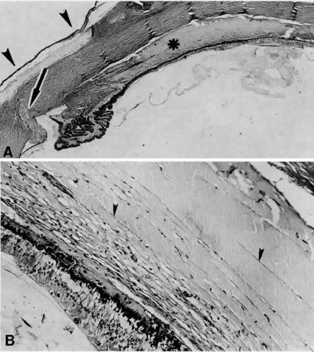
| Fig. 10. A. This extensive ciliochoroidal effusion (asterisk), which occurred 4 years after iridencleisis in a 70-year-old man,39 was mistaken for a malignant melanoma. The iris (arrow) is incarcerated in the limbal wound, and a flat, edematous infiltration bleb (arrowheads) is present (H E, × 25). B. Area of the ora serrata shows a ciliochoroidal effusion with proteinaceous material separating the tangentially oriented collagen fibers (arrowheads) that connect the choroid and ciliary body to the sclera (H E, × 115). |