Chapter 56
Medical Therapy of Glaucoma
MARC WEITZMAN and JOSEPH CAPRIOLI
Main Menu   Table Of Contents

Search

AQUEOUS HUMOR DYNAMICS
RECEPTOR PHYSIOLOGY
ADRENERGIC PATHWAYS
CHOLINERGIC PATHWAYS
INITIATING TREATMENT
TARGET PRESSURE
COMPLIANCE
DRUG DELIVERY
ONE-EYED THERAPEUTIC TRIALS
TREATMENT ALGORITHM
β-ADRENERGIC BLOCKING AGENTS
NONSELECTIVE α-ADRENERGIC AGONISTS
SELECTIVE α2-AGONISTS
THE SUSCEPTIBLE OPTIC NERVE
TREATMENT OUTCOMES
MECHANISMS OF DAMAGE
NEUROPROTECTION: NEW AVENUES FOR GLAUCOMA TREATMENT
OPTIC NERVE REGENERATION
CLINICAL PERSPECTIVES
REFERENCES

The definition of glaucoma has changed considerably over the past several decades. The disease is no longer defined as elevated intraocular pressure (IOP) but rather a condition comprising characteristic optic nerve head and visual field abnormalities that are frequently associated with IOP in the high-normal or elevated range.1 Major risk factors for the development of glaucomatous optic nerve damage include the level of IOP,2–5 increasing age,6 black race,7 and positive finding for the condition in the family history.8 IOP remains the only risk factor readily amenable to therapy; therefore, almost all currently used strategies for the treatment of glaucoma are aimed at lowering or preventing a rise in IOP.

The ultimate goal of most treatments in medicine is to improve the quality of life while minimizing associated side effects and costs. The goal of glaucoma treatment is to improve quality of life through the preservation of visual function. Medical treatment of glaucoma has associated side effects, complications, and costs. Most of this chapter discusses medications that reduce IOP in the setting of chronic glaucoma. In addition, medications that are useful in treating acutely elevated ocular pressure are reviewed, and newer, non-pressure-related neuroprotective strategies are mentioned.

Back to Top
AQUEOUS HUMOR DYNAMICS
Aqueous humor is actively secreted by the ciliary epithelial bilayer into the posterior chamber by an energy-dependent active process. The net fluid secreted is an osmotic consequence of Na+ , K+ -adenosine triphosphate (ATP)ase-driven sodium movement,9,10 and HCO3 generation.11 Formation of bicarbonate within the ciliary epithelium involves the reaction of OH and CO2 enzymatically promoted by carbonic anhydrase. In addition, amino acids and ascorbate are actively transported into the posterior chamber. In normal human eyes, aqueous humor formation averages 2.5 to 2.8 μl/min, which is enough to replace the entire anterior chamber volume once every 100 minutes. The rate of aqueous formation is independent of IOP, with the exception of extremely high pressures.11

Aqueous flows from the posterior chamber through the pupil into the anterior chamber and is drained from the eye by the iridocorneal chamber angle. In the normal human eye, about 90% of aqueous outflow exits through the trabecular meshwork into Schlemm's canal and enters the systemic circulation by way of intrascleral and episcleral veins. This outflow is commonly termed conventional outflow and is characterized by IOP dependency. Rate of conventional outflow is thus a function of both the hydrostatic pressure gradient and the resistance (R) across the trabecular meshwork. About 10% of aqueous humor in normal human eyes traverses the ciliary muscle to reach the suprachoroidal space and leaves the eye through the sclera or blood vessels12. This second type of outflow is called uveoscleral outflow and is IOP independent.13,14 Mathematical formulas that attempt to describe aqueous humor dynamics have been derived.15

Under steady-state conditions, aqueous inflow must equal total outflow. Thus,

(1)


Fin = Ftrab + Fu

where Fin equals inflow of aqueous into the posterior chamber, Ftrab equals conventional outflow, and Fu equals uveoscleral outflow. A direct relationship between pressure and flow generally holds true for conventional outflow, in which the pressure gradient is that between the IOP and the episcleral venous pressure, at least throughout the normal range of IOP. Uveoscleral outflow, however, when measured experimentally in monkeys, appears largely independent of pressure, averaging 0.5 μl throughout normal IOP ranges. (Total outflow is higher through the uveoscleral route in monkeys [up to 40%]16 than it is in humans [10%]).17 Thus, equation 1 can be written as follows:


Fin = (IOP - Pv)/R + Fu

or


IOP = (Fin - Fu) R + Pv

(2)

Conductivity, the inverse of resistance, is used by convention and is termed outflow facility (C). Thus, R = 1/C. Substitution yields what is often termed the modified Goldmann equation:


IOP = (Fin - Fu) C + Pv

(3)

Methods have evolved to measure most components in equation 3. IOP is measured directly with tonometry. Fin is estimated by fluorophotometry and averages 2.5 to 2.8 μl/min. Outflow facility is usually calculated with tonography, whereby corneal indentation is used to measure elevated IOP. The rate of recovery of IOP to baseline is inversely proportional to conventional outflow resistance.Pv, episcleral venous pressure, is typically around10 mmHg and is measured directly by several manometers. Uveoscleral outflow (Fu) can be calculated only in normal human eyes but has been estimated with invasive techniques in experimental animals. The microscopic pathways of uveoscleral outflow are poorly understood.

Elevated IOP is usually the result of a reduced outflow facility (C), rarely the result of elevated episcleral venous pressure (Pv), and essentially never the result of increased aqueous production (Fin).

Medical strategies to lower IOP manipulate three components of equation 3: aqueous inflow, conventional outflow, and uveoscleral outflow (Table 1). These three variables (Fin - Fu/C) together can be termed outflow pressure. This is easily estimated as outflow pressure = IOP - Pv. The fourth variable, episcleral venous pressure, cannot usually be altered pharmacologically and presents a theoretic limit for the amount of pressure lowering attainable through medical means. If, for example, Fin were almost completely suppressed, IOP would approach Pv. Of course, certain situations do exist in which IOP falls below Pv; however, these are usually pathologically or surgically induced. Pathologic examples include severe inflammation, phthisis bulbi, choroidal separation, and retinal detachments.


The outflow pressure concept is useful for predicting the amount of pressure reduction attained with specific medical strategies. This concept explains why given therapies do not lower IOP by a constant amount or percentage. For example, a strong miotic agent usually decreases outflow pressure by about 40% by increasing outflow facility. If pretreatment IOP was 40 mmHg and the Pv is10 mmHg, the outflow pressure of 30 mmHg wouldbe reduced by 40% down to 18 mmHg, and theIOP would be reduced to 28 mmHg. If the pretreat-ment IOP was 20 mmHg, the outflow pressure of10 mmHg would have been reduced by 40% to6 mmHg, for a resultant IOP of 16 mmHg.

Typically, the magnitude of IOP lowering of a given drug is greater when the drug is used singly than when it is used in combination with other pressure-lowering strategies. For example, if pretreatment IOP is 40 mmHg and Pv is 10 mmHg, a nonselective β-blocker or moderate miotic each can reduce outflow pressure by 40% when used alone. The β-blocker alone reduces the 30-mmHg outflow pressure by 40% down to 18 mmHg, for an IOP of 28 mmHg. Adding the miotic reduces the new18-mmHg outflow pressure to 11 mmHg, for a final IOP of 21 mmHg. Note that the first drug reduced the IOP by 12 mmHg, whereas the second drug dropped it only another 7 mmHg, with equal outflow pressure percentage reductions.

The modified Goldmann equation and the outflow pressure concepts predict that it is impossible to lower IOP below Pv if only Fin, C, and Fu are manipulated. In general, it is difficult to reduce IOP below about 10 mmHg through medical therapy.

Back to Top
RECEPTOR PHYSIOLOGY
Many commonly used antiglaucoma drugs are thought to act through sympathetic or parasympathetic pathways. A review of current concepts regarding adrenergic and cholinergic signal transduction is germane to understanding drug efficacy, interactions, and side effects. Norepinephrine is the neurotransmitter released at most sympathetic, postganglionic synapses, and the receptors that bind norepinephrine and other catecholamines are termed adrenergic receptors. In contrast, parasympathetic, postganglionic neurons release acetyl-choline (ACh), which is bound by cholinergic re-ceptors. Both systems involve interaction ofcell-surface receptors with regulatory proteins lo-cated within the cytoplasm that activate or inhibit specific cell functions. Agonist drugs activate receptors, whereas antagonists block the function of receptors.
Back to Top
ADRENERGIC PATHWAYS
Many medical agents used in glaucoma therapy act through adrenergic receptors. Classification of adrenergic receptors has evolved since 1948 when Ahlquist18 proposed a subdivision into α and β subgroups on the basis of relative agonist potency. For the β-adrenergic receptor, the division into β1 and β2 was originally based on relative potencies of epinephrine and norepinephrine. The β-adrenergic receptors, such as those mediating positive inotropic cardiac effects, are equally responsive to epinephrine and norepinephrine; β2-adrenergic receptors, such as those found in bronchial and vascular smooth muscle, are much more responsive to epinephrine.

The α-receptors are divided into α1 and α2 groups. The α1-receptors, the original postsynaptic α-receptor, are found in vascular smooth muscle, where they are noted to mediate the vasoconstrictor effect of sympathetic stimulation. Selective α1-agonists include phenylephrine and methoxamine. The α2-receptors were initially believed to be localized only to presynaptic norepinephrine synapses but have since been found in postsynaptic locations as well. Clonidine is a selective α2-agonist. Three subgroups each of the α-receptors (α1A, α1B, and α1C), α2-receptors (α2A, α2B, α2C), and β1-receptors (β1, β2, and β3) and five dopamine (DA1 through DA5) receptor subtypes have been described.19

Signal transduction refers to a series of specific cellular responses initiated by the binding of a hormone (signal) to its specific receptor binding site. Our understanding of signal transduction has expanded greatly since the discovery of coupling proteins, termed G-proteins, which regulate specific cellular enzymes or ion channels. The G-protein family contains three subunits; the alpha subunit determines receptor and effector specificity. All G-proteins are capable of binding guanylyl nucleotides and possess guanosine triphosphatase (GTPase) activity. When receptors are activated by agonist binding, they are able to catalyze the transfer of guanosine diphosphate (GDP) bound to the G-protein for cytoplasmic GTP. This transfer causes release of the active α-subunit, which affects its specific regulatory function. The active α-subunit activity is self-regulated because the inherently contained GTPase activity converts the recently bound GTP back into GDP, causing reassociation with the other subunits and functional inactivity.20

To date, three different G-proteins have been associated with adrenergic receptor subtypes. Stimulation of β-receptors activates the G-stimulatory (Gs) subfamily, which increases activity of adenylyl cyclase to produce the second messenger cyclic adenosine monophosphate (cAMP). CAMP-dependent protein kinase (A-kinase), through protein phosphorylation, seems to regulate many cellular activities, including calcium channels and chloride channels in epithelial cells. In some tissues, such as the heart, the active form of the G-protein itself can activate calcium channels.18

The α2-receptors are associated with the G-inhibitory (Gi) subfamily, some of which inhibit adenylyl cyclase. The α1-receptors are also associated with a Gi protein subfamily that can activate a specific enzyme, phospholipase C. Phospholipase C produces second messengers from polyphosphoinositide lipid precursors, inositol triphosphate, and diacylglycerol, which activate a different kinase, C kinase.

The effects of A and C kinases through protein phosphorylation on various cellular responses are not as well understood as is the generation of second messengers by G-proteins. In general, protein phosphorylation by second messenger-activated kinase can have wide-ranging cellular effects, including changes in enzymatic activity, changes in ion and other cell channels, changes in cell shape or surface properties, changes in the interaction of cells with their external environment, and changes in sensitivity to particular hormone signals.18

Various opinions exist regarding the relation between cAMP levels and aqueous formation. It is tempting to explain the effect of the β-blockers on reducing aqueous flow by means of an inhibition of β2-receptor activity, a reduction in Gs stimulation of adenylyl cyclase, and a lowering of cAMP levels. Undoubtedly, the mechanism is more complex because agents that increase cAMP, such as cholera toxin and forskolin, lower IOP.21,22

Back to Top
CHOLINERGIC PATHWAYS
Acetylcholine is the neurotransmitter released at autonomic preganglionic, parasympathetic postganglionic, a few sympathetic postganglionic, and somatic motor endings. ACh synthesis by cholineacetyltransferase is regulated by choline kinase. Acetylcholinesterase, the enzyme that degrades ACh, is located in cholinergic nerves, synapses, and neuroeffectors.

There are two general types of cholinergic receptors: those stimulated by muscarine and those stimulated by nicotine, hence the names muscarinic and nicotinic receptors. Both receptors are found in the central nervous system (CNS) and ganglia. Muscarinic receptors are also found on smooth muscle fibers, and nicotinic receptors are found on striated muscle fibers. In recent years, these two cholinergic subtypes have been further classified. Five muscarinic subtypes and two nicotinic subtypes have been described (M1 through M5, N1 and N2). The muscarinic receptors are similar to those described in the adrenergic section in that their action is coupled to specific G-proteins. M1, M3, and M5 activate phospholipase C through inositol phosphates and diacylglycerol and can stimulate the release of arachidonic acid. Stimulation of M2 and M4 inhibits adenylate cyclase.23

Some published studies suggest that all five muscarinic receptor subtypes are found within the ciliary muscle.24,25 The early hope of dissociating the accommodative and outflow effects of miotic agents has not yet been borne out. It appears that both effects result from stimulation of the M3 receptor subtype.26

Although second-messenger pathways for the adrenergic and cholinergic systems share similarities, it is important to understand that the specific surface receptors may be differentially distributed in various cell types. Levels of specific hormones may also vary because of regional differences in innervation. For example, parasympathetic fibers predominantly innervate the ciliary muscle and iris sphincter, but it is unclear if the ciliary epithelium receives significant sympathetic innervation.

In addition, seemingly identical signal transduction pathways can give rise to different responses in various cell types. For example, the β-receptor agonist isoproterenol, acting by way of cAMP pathways, can cause an increase in contractility in cardiac muscle and relaxation in other smooth-muscle locations.

Back to Top
INITIATING TREATMENT
That IOP should be lowered in most cases of untreated, diagnosed, chronic glaucoma generates little debate. Clearly, other risk factors exist, both major (age6,27,28, race,7 family history8) and minor (diabetes,29 hypertension, myopia).30,31 The significance of other findings is being evaluated (migraine headache, vasospasm, autoimmunity). Nevertheless, only IOP reduction has thus far been shown to slow glaucomatous visual field loss. The best evidence for the protective effect of pressure reduction is not based on controlled studies of medical therapy alone but rather on a comparison of outcomes of medical, surgical, and laser treatment modalities.32 Results of an early trial of phenytoin to protect the visual field of glaucoma patients showed that 20 of 65 eyes (32%) worsened after only 1 year of follow-up.33 Phenytoin was concluded not to protect the visual field, and this study is often used as a historical, essentially untreated, control group. Two long-term series of treated eyes found that 73% and 76% of eyes followed for 10 years and 7.6 years, respectively, developed visual field worsening.34,35 If one can accept the limitations of comparing these methodologically diverse studies and can assume that the rate of glaucoma progression remains constant in most patients, it appears that the treatment used in these two studies slowed the rate of progression from 32% per year to 7.3% to 10% per year.

There is a need for better evidence of a protective effect of pressure reduction. A study of IOP reduction in open-angle glaucoma funded by the National Eye Institute, with a no treatment control group, is under way. The value of IOP lowering in preglaucoma, ocular hypertension, or glaucoma suspects is much more controversial. Several small studies aiming to detect a protective effect from pressure lowering in ocular hypertension have yielded conflicting results.36–38 As a result, the multicenter Ocular Hypertension Treatment Study has been organized to study patients with elevated IOP without manifest glaucomatous damage. These patients are being randomized to pressure reduction or no intervention with close follow-up.

Back to Top
TARGET PRESSURE
For cases in which pressure lowering is indicated, how much is enough? Over the past several decades, the emphasis has shifted from normalizing a statistically elevated pressure to lowering a damaging pressure to a safe level.1 Prospective studies of specific target pressures using medical therapy only are not available. We must thus formulate target pressures based on studies designed for other purposes, such as studies comparing medical versus surgical treatment modalities. One problem with this process is that it neglects other important variables, such as IOP fluctuation and compliance. Trabeculectomy, when applied to a glaucomatous population, often lowers IOP to a mean level of about 15 mmHg, and most surgically successful cases have little diurnal or intervisit fluctuation in IOP. Medical therapy can also be used frequently to lower IOP to similar levels, but often with greater diurnal and intervisit variability. This greater IOP fluctuation may increase the progression of glaucomatous optic neuropathy.39–41 Another important difference between surgical and medical treatment is the issue of poor patient compliance. Patients who have had successful filtering surgery often do not depend on any medications for IOP control and probably have “real life” IOPs similar to those measured in the physician's office. It is well known that poor compliance with medical therapy often exists, and these poorly compliant patients probably have “real life” IOPs considerably higher than their optimized office levels.

Several important studies have influenced establishment of current target pressures. Chandler,42 in 1960, anecdotally noted that advanced glaucoma could often be stabilized if IOP was lowered to low-normal levels. In 1982, Grant and Burke43 expanded this concept with retrospective studies of patients declared legally blind from glaucoma at the Massachusetts Eye and Ear Infirmary in Boston. They concluded that stage I glaucoma (little visual field damage) could be stabilized with an IOP in the low 20s, stage II glaucoma (visual field loss in one hemifield) could be stabilized with an IOP in the high teens, and stage III glaucoma (visual field loss in both hemifields) could be stabilized with an IOP in the mid-teens. These observations were made during a time when kinetic perimetry and direct ophthalmoscopy were commonly used to evaluate glaucoma, and normal tension glaucoma was less likely to be diagnosed. Nevertheless, they support the generally accepted concept that advanced damage requires an IOP lower than that of early damage.

Mao and colleagues,44 in a retrospective study of early glaucoma (half these patients had normal visual fields), stratified pressure into three levels and found the chance of progression to be 100% if IOP was above 21 mm Hg, 53% if IOP was between 17 and 21 mm Hg, and 0% if IOP was below 17 mm Hg. Odberg,45 in a study of advanced glaucoma, also stratified IOP and found 33% progression if IOP was below 16 mm Hg, 47% progression if IOP was 10 to 20 mm Hg (mostly below 16 mm Hg), 82% progression if IOP was 10 to 20 mm Hg (mostly above 15 mm Hg), 84% progression with some IOPs above 20 mm Hg, and 100% progression with IOP always above 20 mm Hg. Other studies, including those of Kolker46 and Quigley and Maumenee,47 suggest that long-term IOP maintained in the mid-teens has a generally favorable prognosis.

A comparison of Mao and colleagues' and of Odberg's studies suggests that patients with more advanced damage require a lower IOP to stabilize their glaucoma. Solid evidence for this concept is still lacking. A second reason to favor the philosophy of more aggressive IOP lowering to treat more advanced glaucomatous damage is based on room for error. Patients with advanced field loss but good central acuity may have less room to progress without dire visual consequences compared with those with earlier disease.

The four studies on “high-tension” glaucoma patients (with their emphasis on absolute level of IOP achieved with treatment) are of less value in the patient who is discovered to have progressive cupping or visual field loss despite lower IOPs (below 22 mmHg). These “low-tension” glaucoma patients often exhibit more advanced damage in the eye with the higher IOP48,49 and have been shown in an early report to benefit from pressure lowering.50 In this population in particular, a target pressure based on a percentage of IOP lowering may be more appropriate.

Several different strategies to define target IOPs have been described. One simple goal (simple to define but not necessarily to achieve) is to target all high-tension glaucoma (IOP consistently above 25 mmHg before treatment) to an IOP in the low 20s if the damage is mild, the high teens if the damage is moderate, and the mid-teens or lower if the damage is severe. Another proposed target is a 30% to 50% reduction in the highest untreated pressure at which the patient suffers progressive damage.

The Collaborative Initial Glaucoma Treatment Study, a multicenter trial attempting to define the best treatment for newly discovered glaucoma, uses the following equation:


target pressure = 1 - (reference pressure + dvisual field score ÷ 100 × reference pressure

(4)

where the reference pressure represents the baseline pretreatment IOP and the visual field score is based on the Advanced Glaucoma Intervention Study technique for scoring visual field damage.51 These visual field scores range from 0 (no damage) to 20 (all sites deeply depressed). One problem with this technique is its requirement for reliable perimetry to provide the visual field score. In addition, the scoring process itself is somewhat complex and time consuming. A useful alternative technique uses the following equation:


target IOP = initial IOP


(100 - initial IOP/100) - D

(5)

where D is a constant based on the level of glaucomatous damage (perhaps modified by other risk factors). We recommend approximate D values of -6 for no evidence of damage, 0 for mild damage, 3 for moderate damage, and 6 for severe damage. These D values produce a pressure threshold in the high 20s for initiating therapy in an eye without evidence of damage (normal visual field and optic nerve head) (Table 2). In a patient with a reproducible pattern of visual field loss, the level of damage is probably best determined by perimetry (Table 3).52 In patients unable to provide reliable, repeatable quantitative perimetric data, emphasis should be placed on the status of the optic nerve head and nerve fiber layer combined with assessments of confrontation visual field testing and central acuity to determine the level of damage.


Although recommended for virtually all chronic glaucomas, establishment of an appropriate target pressure is probably of greatest importance in the patient with advanced disease with visual field loss impinging on central visual function. In these cases, any progression could cause significant functional compromise. In contrast, the patient with early disease can suffer subtle progression without a significant change in visual function.

ESTABLISHING A BASELINE

The derivation of a target pressure in a specific patient is multifactorial, individualized, and based on the current level of damage, pretreatment IOP range, the patient's expected life span, and specific risk factors. The goal of the process is to predict the IOP that will slow the disease enough to maintain acceptable visual function for the remainder of the individual's life. By definition, it is a prospective process (i.e., an educated guess) and will almost certainly be inadequate in selected patients in the sense of being either overly aggressive with unnecessary costs and side effects or relatively ineffective with an undesired rate of continued progression toward visual compromise.

It is important to continuously reassess the appropriateness of the target pressure in each patient. This process should include a constant search for ocular and systemic side effects, some of which can be extremely subtle. In addition, evidence of disease worsening should be explored. In general, detection of glaucoma progression involves repeatedly assessing the visual field status and optic nerve head and nerve fiber layer appearance. Perimetry, despite recent computerized advances, remains a subjective technique with sizable long-term fluctuations, particularly in moderately damaged portions of the visual field. The detection of subtle progression is best aided by performing several baseline visual field tests, especially in a perimetrically naive patient, to neutralize the learning effect and establish the patient's inherent variability. Subtle progression should, in general, be confirmed with additional follow-up studies to minimize regression to the mean and false-positive progression. Establishing a baseline level of optic disc damage typically involves obtaining high quality stereoscopic optic disc and nerve fiber layer photographs for future comparison purposes. Quantitative image analysis techniques have not yet proved superior to standardized stereo disc photographs for routine patient follow-up. If disc photography is impractical and not available, a careful disc drawing should be substituted, but this is almost certainly of lesser value for longitudinal comparison purposes.

Despite recent advances in the medical lowering of IOP, the practitioner is often faced with the situation of failing to achieve the target IOP despite maximum tolerated medical therapy. This should prompt a reassessment of the appropriateness of the target pressure, with consideration of the more invasive nature of laser and incisional surgery. If indications are firm for lowering IOP below the level obtained with maximum tolerated medical therapy (IOP unacceptably elevated), then surgery may be appropriate. For other patients in whom the target IOP is more nebulous and the treatment strategy has provided equivocal pressure control (IOP borderline), it may be appropriate to observe the patient closely during follow-up for evidence of further damage. Because target pressures are based on analysis of populations, individual patients who meet or exceed their target pressure reductions (IOP acceptable or optimal, respectively) with medical therapy can still suffer progressive optic nerve damage and demonstrate significant side effects. Therefore, it is important, even in these well-controlled patients, to assess the level of damage and constantly search for untoward adverse drug effects periodically.

Back to Top
COMPLIANCE
Compliance with medical therapy is critical if the therapy is to succeed. Several studies have suggested that poor compliance with glaucoma medication is a major problem. A study of patient compliance with systemic carbonic anhydrase inhibitors (CAIs) that involved monitoring serum bicarbonate levels suggested that 35% of patients were not taking the drug at all and another 22% were using the medication infrequently.50 A study of glaucoma patients who were supposedly using pilocarpine drops four times daily noted that of 99% of patients who claimed at interview to have instilled at least 75% of their drops, only 66% were actually this compliant, with 15% instilling the drops less than 50% of the time and 25% missing all four daily doses at least once a month.54,55

Several general principles can be applied to compliance with medical therapy of chronic glaucoma.

  • The more often a medication must be used, the greater the chance of noncompliance becomes.
  • Patients usually comply with morning doses better than evening doses.
  • In general, patients are less compliant than they claim.
  • Agents with side effects easily recognized by the patient, such as burning on instillation, dimming of vision, headache, and tingling fingers, are less likely to be used compliantly.
  • Multidrug regimens that require waiting a specified time between drops are less likely to be adhered to.
  • Patients who frequently miss appointments or show a poor understanding of their disease are often noncompliant.
  • Elderly patients may be particularly at risk for poor compliance because of poor hearing, slowed cognition, and a desire to reduce side effects.

Compliance is difficult to evaluate. It is a good idea to consider noncompliance in any case of progression despite seemingly good pressure control in the office. At times, it may be helpful to contact a family member to inquire about the use of medications.

Specific efforts to improve compliance include the following:

  • Limit dosing to once or twice daily if possible.
  • Advise use of medications that limit side effects. Nasolacrimal occlusion, prodrugs, and high-viscosity preparations limit systemic absorption.
  • Avoid confusing regimens. Carbachol three times daily, acetazolamide four times daily, and a β-blocker twice daily is a confusing regimen. Echothiophate iodide, sustained-release acetazolamide, and a β-blocker, all twice daily, is much easier to remember.
  • Use medication instruction sheets with dosing times clearly displayed.
  • Consider combination therapy with multiple drugs in a single bottle.
  • Consider drug interactions that can aggravate side effects.
  • Educate patients about and engage them in their disease. Tell them their IOP and target pressure, show them their visual field printouts, and discuss the optic disc findings.
Back to Top
DRUG DELIVERY
The eye affords us the ability to prescribe topical medicines. However, topical therapy does not eliminate systemic absorption and side effects, particularly from β-blockers. The capacity of the human conjunctival cul-de-sac is about 10 μl. The design of an eye drop bottle tip that delivers a small enough drop to saturate but not overflow the tear film creates essentially a micropipet, the shape of which presents a significant hazard to the cornea if accidental contact occurs. A “safe” tip design delivers 25 to 50 μl; this overflows the tear film into the lacrimal drainage system or onto the cheek. Of the 20% to 40% of the medication initially present in the cul-de-sac, about 15% per minute exits the tear film by new tear formation, blinking, and lacrimal drainage. Although the total dose of medications reaching the nasal mucosa may seem trivial compared with typical oral doses, medication absorbed by way of the nasal mucosa is not subjected to first-pass hepatic metabolism. Clinically significant blood levels can be achieved at target tissues, producing undesired adverse reactions. Efforts to maximize ocular penetration and minimize systemic absorption by way of the nasal mucosa generally limit lacrimal drainage. These efforts include increasing drop viscosity and using nasal lacrimal occlusion or eyelid closure for several minutes after instillation. If the ocular contact time can be increased with these techniques, it should be possible to lower the medication concentration, thus reducing costs and additional systemic side effects. Another powerful strategy to reduce systemic absorption is the use of prodrugs such as dipivefrin that enhance ocular penetration and reduce the total dose requirement. To maximize ocular absorption, a second topical agent should not be given for at least 10 minutes after the first.
Back to Top
ONE-EYED THERAPEUTIC TRIALS
Use of one-eyed therapeutic trials helps to define peak individual dose-response relations, minimizing overdosage with its attendant costs. Each ocular hypotensive agent is not uniformly efficacious in every patient because of multiple factors, including iris pigmentation, reflex tearing, circulating catecholamine levels, and adrenergic tone. High diurnal and intervisit fluctuations in IOP have been commonly noted in glaucoma and ocular hypertensive patients, which confounds the ability to assess drug response with bilateral administration. A useful technique to judge individual therapeutic efficacy is the one-eyed therapeutic trial whereby medication is instilled in one eye and the contralateral eye is used as an untreated control. Pretreatment IOP need not be bilaterally identical to use one-eyed trials; however, the difference in IOP must be fairly consistent. In addition, certain medications, such as the topical ss-blockers, do show a mild contralateral reduction in IOP with unilateral use that is believed to be secondary to systemic absorption.

Examples of the results of one-eyed trials include the following:

  • Pretreatment IOP right eye (RE) 28, left eye (LE) 28. RE only treated with miotic → RE 24, LE30 = IOP reduction of 6 mmHg
  • Pretreatment IOP RE 30, LE 25 (consistent5-mmHg difference). RE only treated with dipivefrin → RE 27, LE 27 = net IOP reduction of 5 mmHg
  • Pretreatment IOP RE 25, LE 23, RE treated with β-blocker → RE 22, LE 20 = net IOP reduction probably minimal
Back to Top
TREATMENT ALGORITHM
Introduction of new medications over the past decade has dramatically altered typical glaucoma treatment algorithms. In the 1980s, standard therapy was to start with a topical β-blocker or epinephrine and than, as second line, add a miotic agent. If more aggressive treatment was required, either a systemic carbonic anhydrase inhibitor or laser trabeculoplasty was used.

Over the past decade, three new classes of topical medications: the α2 agonists, CAIs, and prostaglandin (PG) analogues have been introduced. This has doubled the available classes of topical therapy to six. Most such classes are additive when used in combination, which provides the treating ophthalmologist myriad therapeutic options.

Currently, the two most used first line drugs are topical β-blockers and PG analogues. The β-blockers are the historical first-line drug because of their excellent efficacy, local tolerability, and in most cases ability to be dosed once a day. Serious side effects related to systemic β-blockade are uncommon although cautious review of many patients reveals subtle changes such as a reduced pulse and exercise tolerance, mild wheezing, increased cholesterol level, and CNS reactions including depression, fatigue, and impotence.

Concern over the systemic side effects of the β-blockers led to the increasing popularity of topical PGs as first line therapy. PGs are statistically slightly more effective at lowering IOP than β-blockers, are available for once daily dosing, and have minimal if any risk of systemic reactions. It is the local ocular side effects, such as permanent iris color changes, periocular pigmentation and rare aggravation of cystoid macular edema and uveitis that must be considered.

The α2 agonists are typically equally effective to β-blockers with fewer cardiopulmonary side effects and have been promoted as first-line drugs. Factors limiting their use as such include a need for twice daily or three times daily dosing, an approximately 5% rate of ocular allergy, frequent dry mouth, and occasionally significant CNS depression. This class may be more effective than topical β-blockers in patients already treated with a systemic β-blocker.56

Topical CAIs have been shown to be less effective than β-blockers and require twice daily or three times daily dosing. These drugs are still sometimes useful as first line treatment because of their good local and systemic tolerability. Oral CAIs are rarely used for chronic treatment because of their high incidence of systemic side effects. This class is occasionally helpful in patients allergic to multiple different eyedrops or unable to instill drops.

Nonselective adrenergic agonists such as epinephrine are uncommonly used because of their high rate of local intolerance, need for twice-daily dosing, and poor additivity to β-blockers. They are occasionally useful in patients in whom topical β-blockers are contraindicated, especially ifeconomic considerations favor a generic preparation.

The parasympathomimetics such as pilocarpine remain an important class for glaucoma treatment. Factors leading to their unpopularity in many phakic open angle glaucoma patients include common local tolerability problems such as induced myopia and miosis as well as a short duration of action that requires three times daily or four times daily dosing. Nevertheless, in patients with pseudophakia and relatively older patients less likely to be plagued by induced myopia the miotics can be very effective (and inexpensive) drugs. There may also be a specific role for this medication class in preventing pigment liberation in pigment dispersion or pseudoexfoliative glaucoma or in pulling the angle open in plateau iris syndrome and chronic angle closure glaucoma.

Our typical treatment algorithm for most open angle glaucoma patients is a β-blocker or PG first line with the other drug added second line. If a patient is concerned about iris color change, a β-blocker is used and either an α2 agonist or topical CAI added second line. Patients with contraindications to β-blocker use should be considered for first line treatment with either a PG compound or α2 agonist with either epinephrine or a topical CAI added next. Pseudophakic patients and the previously mentioned secondary glaucomas should be considered for miotic therapy.

Results of the Glaucoma Laser Trial demonstrated a favorable efficacy to complication ratio of argon laser trabeculoplasty (ALT) compared with medical therapy. Over the past decade many ophthalmologists treating glaucoma have escalated ALT's role from third or fourth line to first or second. ALT's relatively short lasting effect of 2 to 5 years must be discussed with the patient and laser remains an particularly helpful option in older patients where this length of control can be significant. Laser is often useful in noncompliant patients, patients with high rates of medication allergy, or patients unable to afford medications.

The indications and timing of incisional surgical treatment (e.g., trabeculectomy) vary widely. Factors involved in such decisions include the patient's preconceived notions regarding surgery, the surgeon's skill and experience, the likelihood of surgical versus medical success, and medical compliance and tolerability issues. The advent of wound healing modulation with 5-fluorouracil and mitomycin C as well as releasable suture techniques have improved surgical success although late onset bleb related infections and hypotony are of concern.

There is increasing evidence that chronic treatment with glaucoma topical agents can activate the subconjunctival fibroblast population, compromising surgical success. Some ophthalmologists have thus advocated earlier or even initial surgery. A multicenter, randomized trial of surgical versus medical treatment of newly diagnosed glaucoma is now underway as is a large scale survey of complications of glaucoma surgery using current surgical techniques. It is hoped that results of these studies may help clarify the frequently nebulous indications for surgical treatment.

Back to Top
β-ADRENERGIC BLOCKING AGENTS
The initial study of the effects of the sympathetic nervous system on IOP and aqueous flow was made in rabbits by Linnér and Prijot in 1955.57 They observed that excision of the superior cervical ganglion had a marked IOP lowering effect that lasted 24 hours. Because outflow resistance was similar to that in contralateral control eyes, it was concluded that aqueous secretion was reduced.

In 1958, Powell and Slater58 discovered the selective β-adrenergic antagonist dichloroisoproterenol (DCI). Sears and Bárány59 in 1960 reported that DCIs reduced IOP in ganglionectomized and control rabbits, providing the first evidence that this class of medications could act as an ocular hypotensive agent. Unfortunately, DCI had little effect on IOP in humans (M. L. Sears, personal communciation).

β-Adrenergic blocking agents were first reported to lower IOP in humans in 1967 when Phillips and coworkers60 described the ocular hypotensive effect of intravenous propranolol. In 1971, topical propranolol was also reported to reduce IOP.61 Unfortunately, the drug also produced corneal anesthesia, limiting its clinical usefulness. During the early 1970s, a search for a nonanesthetizing β-blocker ultimately led to the introduction of timolol in 1978.

β-Adrenergic antagonists reduce IOP by inhibiting aqueous humor formation by the ciliary epithelium.62–64 These drugs have a minimal effect on outflow facility.65 Topical β-blockers reduce aqueous formation by 24% to 48% in awake humans.66 Since timolol maleate was introduced for topical therapy in the late 1970s, this class of drug has become the standard first-line medication to lower IOP.67–70 The popularity of these drugs is no doubt due to the agents' long duration of action, efficacy, favorable local side effect profile, and relatively infrequent systemic adverse reactions when used appropriately. In fact, the topical β-blockers are the most frequently prescribed ophthalmic medications, accounting for over 70% of all glaucoma-directed prescriptions.71,72

PHARMACOLOGY

The ocular hypotensive effect of β-blockers seems to be largely mediated by the β2-receptor.73 Unlike other aqueous suppressants, such as CAIs or ss2-agonists, the β-blockers are ineffective during sleep,74 presumably because of reduced humoral or neural adrenergic tone (aqueous production normally falls by 45% during sleep).75

Most β-blockers are derivatives of the prototype β-adrenergic agonist isoproterenol and contain a benzene ring nucleus coupled to an ethylamine chain. A multitude of β-blockers are available for systemic use, and five are available for topical use (Table 4). These drugs differ in β-receptor selectivity, potency, intrinsic sympathomimetic activity (ISA), and membrane-stabilizing effects. The prototype nonselective β-blocker used systemically is propranolol (Inderal; Wyeth-Ayerst, Philadelphia, Pennsylvania), whereas metoprolol was one of the first “cardioselective” (β1 selective) agents. ISA refers to low-level stimulation of the adrenergic receptor as blocking occurs. Membrane stabilization prevents cellular depolarization, and some of these agents (such as propranolol) have local anesthetic properties, limiting topical ophthalmic use.2


CLINICALLY AVAILABLE AGENTS

Timolol

In 1976, timolol was reported to lower IOP in glaucomatous rabbits76 and normal human study subjects77 without significant ocular irritation. In 1977, the drug was noted to markedly reduce IOP in glaucoma patients.14,78

Commercially introduced in 1978, topical timolol maleate revolutionized the medical treatment of glaucoma and has become the most commonly used agent.79,80 Timolol is nonselective, inhibiting both β1- and β2-adrenergic receptor activity. The commercial preparations consist of the l-isomer. Timolol has minimal ISA and membrane-stabilizing effects and is about five times more potent than propranolol.81

EFFICACY.

Timolol significantly lowers IOP in most patients not undergoing other β-blocker treatment,including normal volunteers,82 ocular hypertensives,83–85 and chronic glaucoma patients.15,86–88 The percentage or absolute pressure reduction obtained with topical timolol varies from study to study because of differences in study populations (including pretreatment IOP and iris pigmentation) and study methodology. Nevertheless, most studies demonstrate a 20% to 28% reduction in IOP with topical timolol used as a single hypotensive agent. The additional IOP lowering effect is usually reduced in patients already receiving systemic, nonselective β-blocker treatment.89

The initial dose of timolol usually produces the greatest reduction in IOP, and over several weeks there is a partial loss of effect.90,91 This “short-term escape” may be caused by an increase in the number of β-adrenergic receptors within days of initiating therapy.92 Because of this short-term escape, the chronic IOP lowering effect of timolol should not be concluded until the completion of 2 to 4 weeks of use.26

A “long-term drift” has also been described. This is a timolol-induced reduction in aqueous flow that is lower after 1 year of treatment than it is after 1 week.93 Clinically, many patients with an acceptable IOP response after several initial weeks of therapy gradually lose pressure control after months to years of treatment.94 Whether this drift is due to further long-term adaptation of β-receptors, gradually declining outflow facility, or other factors is unknown. Long-term drift is not unique to the β-blockers but is seen with most medical modalities that lower IOP.

Timolol is usually more effective at lowering IOP than either epinephrine95,96 or pilocarpine,97 and it is this efficacy, combined with a favorable local side effect profile and a long duration of action, that accounts for its popularity as a first-line agent. Drugs such as oral CAIs3,4 and strong miotics have an efficacy approximately equivalent to that of timolol but more often produce adverse local or systemic side effects. As a first-line drug, the topical CAIs require three-times-daily dosing for optimal effect and are slightly less effective than topical timolol.98 Initial clinical experience with the chronic IOP lowering effect of the α2-adrenergic agonists, when used as single agents, appears to equal that of 0.5% timolol.99 Unfortunately, a significant number of apraclonidine-treated eyes develop local ocular allergies. Brimonidine is less allergenic however it must be dosed twice or three times daily. Topical latanoprost, a PG analogue, has been shown to be as effective as timolol and can be dosed once daily. It has minimal systemic side effects; however, local tolerance issues such as iris color change are often an issue. Latanoprost has become a commonly used first-line alternative to timolol.100–102

Even with efforts to limit systemic absorption, some blood levels of timolol usually occur. It is not uncommon to note a mild contralateral IOP lowering effect with unilateral use (crossover effect).15,21,23,103 This effect should be considered when using one-eyed therapeutic trials to evaluate β-blocker efficacy.

CONCENTRATION AND DOSE.

Timolol maleate is available in concentrations of 0.25% and 0.5%, both as solution (Timoptic; Merck & Co., Whitehouse Station, New Jersey) and as an ophthalmic gel-forming agent (Timoptic XE). Timoptic is available in bottles preserved with 0.01% benzalkonium chloride and in nonpreserved dropperettes. The gel-forming solution uses an anionic polysaccharide vehicle derived from gellan gum, which reacts with tear film cations to produce a high-viscosity gel, prolonging ocular contact time.104 The 0.5% concentration is at the top of the dose-response curve,21 and the 0.25% solution is often equally efficacious.105 Little difference in effectiveness was observed between the 0.25% and 0.5% solutions after 1 year of therapy in a population of open-angle glaucoma patients.106 Both concentrations decrease IOP maximally 2 hours after instillation and maintain a significant reduction for at least 24 hours.8,107 Although twice-daily use of timolol solution is recommended, once-daily administration is often clinically effective, thus cutting the cost in half.108 If the medication is used every morning a trough IOP measurement can be obtained by withholding drops on the day of an early morning pressure check.

Timoptic XE used once daily appears as effective as the same concentration of solution used bid; therefore, it is recommended for once-daily use. The cost of twice-daily solution and once-daily XE preparations is similar. Timoptic XE 0.5% once daily and Timoptic 0.5% solution once daily were compared in a small, 1-week, double-masked, parallel-group study that showed greater IOP reduction with the XE preparation. Longer trials involving more patients are needed before it can be concluded that Timoptic XE provides better once-daily IOP control. Because the cost of Timoptic solution when used once a day is approximately half that of Timoptic XE, it may be worth evaluating the response of the less expensive therapy in individual patients, particularly after encouraging nasolacrimal occlusion. Although Timoptic 0.5% XE used once daily is claimed to result in lower systemic plasma concentrations than Timoptic 0.5% used twice daily, differences in other systemic effects, such as pulse rate, blood lipids, and pulmonary function, have not yet been reported.109 There is some evidence that timolol is less effective in dark irides, possibly because of pigment binding.110 One study used home tonometry to compare (five times during the day) the mean IOP reductions of 0.5% timolol administered once daily in the morning or in the evening and 0.25% timolol administered in the morning; no significant difference was found among the three groups.42

Once-daily use of 0.5% timolol is substantially more economical than twice-daily use of the 0.25% concentration. Although compliance is often higher with early morning medications, bedtime use theoretically could reduce systemic side effects because peak plasma levels would occur during sleep. Soll's study108 of 0.25% Timoptic used at bedtime or twice daily demonstrated a greater reduction in pulse in the twice-daily group, presumably because the pulse rate was measured during daytime hours.45

Timolol is also available as the hemihydrate salt (Betimal, Ciba Vision Ophthalmics, Duluth, Georgia) in both 0.5% and 0.25% concentrations. Timolol hemihydrate solution when used once a day was similar in efficacy to timolol gel once a day.111 Despite what should be higher plasma levels compared with gel vehicle, the solution had similar effects on maximal pulse rate when exercising.112

Discontinuing or “washing out” timolol does not restore aqueous flow to normal levels for 2 to 6 weeks, and IOP requires at least 2 weeks to return to baseline levels.113 This is particularly important when stopping β-blockers before filtering surgery in an effort to limit early postoperative hypotony.

COMBINATION THERAPY.

In many patients, timolol therapy alone does not achieve target IOP lowering. Most of the other classes of ocular hypotensive agents, including the CAIs,5,114,115 miotics,116,117 α2-adrenergic agonists,118–120 and PGs121,122 produce anadditive effect. One apparent exception is thenonselective β-agonist class (epinephrine and dipivefrin), which typically provides little additional IOP lowering effect (approximately 2 mmHg).123–126 Nevertheless, wide patient variability is seen, and occasionally patients show more impressive additive effects, often best demonstrated in one-eyed therapeutic trials. Single bottle combinations of timolol-dorzolamide and timolol-latanoprost have been approved by the Food and Drug Administration (FDA) and may enhance patient compliance.

SYSTEMIC ADVERSE REACTIONS.

A 30-μl drop of0.5% timolol maleate contains about 0.2 mg of the drug, considerably less than the 30-mg maximum recommended oral dose for cardiovascular timolol use. Topical timolol is generally well tolerated.It must be remembered, however, that oral drugsare susceptible to first-pass hepatic metabolism, whereas nasal mucosal-absorbed medications travel to the pulmonary, cardiovascular, and CNS circulations more directly. Thus, seemingly minimal doses of topical ocular timolol can cause significant adverse effects in susceptible individuals.

Physicians in general and ophthalmologists in particular must be familiar with the potential systemic reactions of topical β-blockers. The effects are usually subtle and include fatigue, lethargy, mood changes, impotence, reduced exercise tolerance, shortness of breath, headache, and ankle edema. In 1985, topical timolol was the ninth most common drug of all medications prescribed to patients aged 75 or older. These elderly patients often use a host of other medications, which makes it difficult to identify significant timolol-related side effects. In addition, these reactions are commonly ascribed to aging or coexisting disease.127

Timolol can affect the cardiovascular, pulmonary, and CNSs. Cardiovascular (β1) effects of topical β-blockers can lead to reductions in pulse rate, cardiac contractility, and blood pressure. Timolol often reduces the resting pulse and blunts the exercise-induced increase in heart rate.128 Severe bradycardia, cardiac arrhythmias, heart block, congestive heart failure (CHF), and death are possible but are rare.129 Most β-blockers also alter plasma lipid profiles. Topical timolol 0.5% given twice daily to volunteers increased plasma triglycerides by 12% and decreased high-density lipoprotein cholesterol by 9%. This change in plasma lipids presents a theoretically significant risk factor for the development of coronary artery disease, although use of systemic timolol after myocardial infarction (MI) has been shown to reduce subsequent MI and mortality.130

Antagonism of β2-receptors in bronchi and bronchioles contracts smooth muscle, which can cause increased airway resistance, especially in patients with reactive forms of asthma or chronic obstructive pulmonary disease (COPD).131 Respiratory failure has been documented with timolol therapy, and the drug should not be used in patients with severe respiratory disease.64 Topical timolol therapy may adversely affect respiratory function in elderly patients without a history of known airway disease. In a study of 80 such patients older than age 60, topical timolol was replaced with either the β1-selective β-blocker betaxolol or the adrenergic agonist dipivefrin. There was a 13% increase and an 8% increase in mean peak flow rate and forced expiratory volume in 1 second (FEV1), respectively, when betaxolol was used, and a 14% increase and an 11% increase when dipivefrin was used. There was also improved exercise tolerance with both agents, although this was within the range of learning effect.132 An epidemiologic study of a largely white, inner-city population in the north of England found a 37% prevalence of airway obstruction in the over-65 age group.133

The CNS side effects of timolol are related to the drug's lipophilic structure and low protein binding, which enable it to cross the blood-brain barrier. These effects include fatigue, depression, anxiety, confusion, formed hallucinations, memory loss, psychosis, and disorientation.64,134,135 Timolol can decrease libido and cause impotence.136 Depression is more common in glaucoma patients than in those with ocular diseases of similar chronicity137 with prevalences ranging from 15% to 80%.138,139 One study concluded that the greatest single risk factor for falls in elderly glaucoma patients is the use of topical β-blockers.140

β-Blockers may blunt the responses to hypoglycemia-induced endogenous epinephrine release.141Because these responses normally produce hyperglycemia, sweating, and tremor, β-blocked hypoglycemic diabetic patients may have more pronounced hypoglycemia and be less aware of it.142 Timolol treatment has been reported to worsen myasthenia gravis143,144 and can mask the symptoms of hyperthyroidism.

Adverse local side effects with timolol are infrequent. In susceptible individuals, an allergic blepharoconjunctivitis can occur.64,74 This is occasionally secondary to preservatives and can be alleviated by use of the nonpreserved single-use vials. Local irritation of the corneal epithelium with blurring of acuity, conjunctival hyperemia, superficial punctate keratitis, and dry eye has been reported. Basal tear turnover has been reported to increase after a change to the nonpreserved preparation.140 Corneal anesthesia may also occur.146,147 Use of timolol as a sole agent in general has no effect on pupil size, but in combination with an adrenergic agonist it increases mydriasis.

Patients undergoing chronic timolol treatment show a decrease in goblet cell density148 and may develop subconjunctival fibroblast activation, altering the success of filtering surgery. In a cultured human Tenon's capsule fibroblast model, timolol, betaxolol, and levobunolol inhibited fibroblast proliferation. Some of this inhibition may be related to the benzalkonium chloride preservative.149

The influence of topical timolol on various components of ocular blood flow remains unclear, and there are conflicting reports in the literature.150–155 β-Blockers can cause vasoconstriction, and there have been reports of Raynaud's syndrome induced by timolol.156 If similar β-blocker-induced vasoconstriction occurs in the blood vessels supplying the optic nerve head, this class of medication, despite its IOP lowering ability, may be relatively contraindicated in glaucomas associated with poor optic nerve head perfusion.

CLINICAL INDICATIONS.

Timolol is FDA approved for use as a first-line agent in the treatment of patients with chronic open-angle glaucoma and patients with increased IOP who are thought to be at risk for the development of optic nerve damage or visual field loss. It is effective in reducing IOP in congenital glaucoma, although apnea has been reported in timolol-treated neonates. Timolol is contraindicated in patients with asthma, CHF, COPD, and myasthenia gravis. It is often administered during pregnancy, although its teratogenic effects have not been specifically studied.

Levobunolol

Levobunolol hydrochloride, the l-isomer of bunolol, is a nonselective β-adrenergic antagonist. It demonstrates neither ISA nor membrane-stabilizing properties and has been shown to significantly reduce IOP in normal subjects, ocular hypertensives,157 and glaucoma patients.158–161

EFFICACY.

In general, levobunolol is similar to timolol,162–165 although it possesses a slightly longer half-life.166 A double-masked study of 391 glaucoma and ocular hypertensive patients found a sustained mean reduction in IOP of 27% over 2 years, similar to the efficacy found in a timolol control group.167 This study found minimal long-term drift with levobunolol.

CONCENTRATION AND DOSE.

Levobunolol is commercially available as Betagan (Allergan, Irvine, California) and as generic preparations in concentrations of 0.25% and 0.5%. Maximum IOP reduction occurs within 2 to 6 hours,90,168 with measurable effects noted at 24 hours.90 The 0.5% concentration appears to be at the top of the dose-response curve, and several studies demonstrate equal efficacy for the 0.25% and 0.5% concentrations,93,96,169,170 even with once-daily dosing.93,171,172 A small contralateral pressure reduction has been noted, similar to that seen with timolol.90

COMBINATION THERAPY.

Combination of levobunolol with other classes of ocular hypotensive agents has not been as well studied as timolol combination therapy, but it appears to have a similar additive effect. There is little additive effect when levobunolol is combined with dipivefrin.168

ADVERSE REACTIONS.

Levobunolol does not differ significantly from timolol103 in terms of adverse reactions, and it is generally well tolerated by most patients.174 Vigilance for subtle side effects and cautious use in patients with reactive airway disease, bradycardia, CHF, and myasthenia gravis are recommended.

CLINICAL INDICATIONS.

In general, indications for levobunolol use are similar to those for timolol.

COST.

Several studies comparing the cost of levobunolol and timolol treatment have been performed. Cost comparison of the two agents should take into account the smaller drop volume dispensed with timolol. One study found that levobunolol bottles dispensed drops ranging in volume from 47 to61 μl, depending on temperature and bottle angle, compared with 33-μl drops from timolol bottles. Levobunolol therapy may be more expensive than timolol therapy. Although cost per similar-sized bottles is often the same, on a cost-per-drop basis timolol treatment is usually substantially less expensive (approximately two thirds of the cost).175 These studies do not take into account the compliance cap present on the Betagan bottle, which may eliminate extra doses if the patient forgets he or she has already administered the dose. In addition, these comparisons have not been performed with Timoptic XE. Levobunolol is available in generic form.

Metipranolol

Metipranolol is a nonselective β-adrenergic antagonist that lacks ISA and membrane-stabilizing ability. The drug is available in the United States as OptiPranolol (Bausch & Lomb, Tampa, Florida), a 0.3% preparation. It is available in Europe as a 0.6% preparation.

EFFICACY.

The 0.3% concentration of metipranolol decreased IOP by 21%, and the 0.6% concentration decreased IOP by 31%.176 It is generally given twice daily, although once-daily use may be effective. Metipranolol is typically as effective as the other nonselective topical β-blockers in reducing IOP.

CONCENTRATION AND DOSE.

In a double-masked crossover study, reducing the metipranolol concentration from 0.6% to 0.3% caused no difference in IOP reduction.177

ADVERSE REACTIONS.

In general, systemic adverse reactions are similar to those of the other nonselective topical β-blockers. Metipranolol 0.5% may have fewer cardiovascular effects than 0.5% timolol.178 An unusual local reaction unique to metipranolol is granulomatous uveitis, which has been reported in patients in Europe, particularly with the 0.6% concentration.179 In the United States, three patients to date have been reported to have developed granulomatous uveitis after topical use of 0.3% metipranolol.180,181 The uveitis recurred when one patient was rechallenged with the drug.

CLINICAL INDICATIONS.

In general, indications for metipranolol use are similar to those for the other nonselective ss-antagonists.

COST.

Metipranolol, on a cost-per-drop basis, is much less expensive than any other available topical β-blocker. Although actual cost varies from pharmacy to pharmacy, one study estimated that 1 year of twice-daily bilateral Timoptic therapy using10-ml bottles cost $112, whereas OptiPranolol over the same time period using the same bottle size cost $66.108

Carteolol

Carteolol is a nonselective β-blocker similar to the previously mentioned agents in its lack of membrane stabilization. Unlike these other drugs, however, carteolol demonstrates weak ISA, causing an early transient agonist response.182 This ISA theoretically could reduce bronchoconstriction and bradycardia and could improve blood flow, although this has not been clinically demonstrated.183

EFFICACY.

Two trials comparing carteolol and timolol found equivalent IOP reductions.184,185 No difference in effect was found between 1% and 2% concentrations.122,186 Carteolol 1% reduced IOP 11% to 14% versus placebo.187,188 This reduction is less than that usually reported for timolol; however, this was not a controlled study.

CONCENTRATION AND DOSE.

Carteolol 1% solution is commercially available as Ocupress (Otsuka American, Seattle, Washington). The half-life of an active metabolite, 8-hydroxy carteolol, is two to three times that of the parent compound.117

ADVERSE REACTIONS.

No evidence to date is available that substantiates a reduction in systemic side effects compared with timolol as predicted by ISA. A preliminary report suggests a reduction in adverse lipid profiles compared with timolol, although this effect was seen only in the initial portion of a crossover study and disappeared in the second half of the study.189 Similar effects of both drugs on pulse and blood pressure were seen in a double-masked, randomized comparison of carteolol and timolol.190

CLINICAL INDICATIONS.

In general, carteolol use is indicated for situations that are similar to those for which the other nonselective β-blockers are used. If the recently claimed lipid profile advantages are substantiated, the drug may be preferred in patients with coexisting hypercholesterolemia or coronary artery disease. The contraindications listed for the other nonselective ss-blockers apply to carteolol as well.

COST.

A recent survey of nine Connecticut pharma-cies revealed a similar mean price for a 10-ml bottle of Ocupress compared with Timoptic 0.5%. The price of Timoptic was $6 cheaper at one pharmacy, but it was $4 more at another pharmacy.

Betaxolol

Betaxolol is the only selective β-adrenergic antagonist available for topical ophthalmic use. It is often termed cardioselective because of its relative affinity for the β1 (cardiac) over the β2 (pulmonary) adrenergic receptor. The reduction in aqueous production by β-blockade is thought to involve primarily the β2-receptors that predominate in the ciliary nonpigmented epithelium.191,192 Although relatively β1-selective, betaxolol in the high ocular concentration achieved with topical use probably possesses enough β2-antagonist activity to reduce aqueous production. Alternatively, there may be a low level of β1-receptors in the nonpigmented epithelium, which accounts for the ocular hypertensive effect. Betaxolol must decrease aqueous flow because it has no effect on conventional or uveoscleral outflow facility.193

EFFICACY.

Topical betaxolol lowers IOP in normal and elevated states.194–197 When it is compared with timolol, most studies have shown betaxolol to be slightly less effective in reducing aqueous flow198 and decreasing IOP92,199–201. A multicenter, randomized trial found equivalent efficacy between betaxolol and dipivefrin, with both agents decreasing IOP by 14% to 17%.202 When patients are changed from timolol to betaxolol or randomized to one versus the other, IOP is often about 2 mm Hg higher in the betaxolol-treated eyes. One report suggests that despite a smaller reduction in IOP with betaxolol compared with timolol, betaxolol may better prevent visual field worsening in primary open-angle glaucoma patients. This report used mean sensitivity as the sole basis for following the visual fields and requires substantiation in larger cohorts of patients with more sophisticated visual field analysis.203 Other reports suggest that betaxolol may have beneficial ocular hemodynamic effects compared with timolol, particularly in low-tension glaucoma.204

CONCENTRATION AND DOSE.

Betaxolol hydrochloride is commercially available in a 0.5% solution as Betoptic (Alcon Laboratories, Fort Worth, Texas). It is also available in a 0.25% suspension (Betoptic S), which suspends the drug in microscopic polymeric resin beads. This effectively increases the ocular contact time and reduces stinging considerably. Both preparations have similar IOP lowering ability.205 Maximum drug effect is 2 hours after instillation and lasts at least 12 hours. There may be less systemic absorption with betaxolol compared with timolol.206

COMBINATION THERAPY.

Betaxolol appears similar to timolol in its additive effect with CAIs, miotics, and α2-agonists. The additivity of betaxolol to epinephrine-type drugs appears to be significantly greater than that of timolol, making this combination potentially clinically useful.133

SYSTEMIC ADVERSE REACTIONS.

Systemic cardioselectivity makes betaxolol theoretically a better drug than the nonselective ss-blockers in patients with mild pulmonary disease, although it certainly can provoke bronchospasm.207,208 Several studies suggest that betaxolol is better tolerated than nonselective β-blockers in patients with pulmonary dis-ease.143,209–212

In theory, there should be similar cardiac side effects when β1-selective agents are used compared with nonspecific β-blockers; betaxolol has been reported to result in bradycardia, sinus arrest,213 and CHF.214 Despite these potential problems, betaxolol does not appear to affect exercise-induced pulse rate increases in normal volunteers, perhaps because of decreased systemic absorption.63 Betaxolol may have a reduced propensity to cross the blood-brain barrier, producing fewer CNS side effects compared with those associated with timolol.68,215

LOCAL ADVERSE REACTIONS.

Betaxolol 0.5% solution therapy stings more than the other topical β-blockers. This problem has been reduced with the Betoptic S preparation.

INDICATIONS.

Betaxolol is indicated to reduce IOP in patients with ocular hypertension or glaucoma who are at risk for progressive optic nerve head damage. Its use may be preferred over the nonselective β-blockers in patients with mild cardiovascular, pulmonary, or CNS compromise. Significant systemic adverse reactions can still occur in these patients, and its use is strongly contraindicated in patients with more compromised cardiopulmonary systems.

COST.

Betoptic and Betoptic S typically cost slightly more than Timoptic but less than Betagan on a drop-by-drop basis.108 Because Betoptic S costs only slightly more than Betoptic and has comparable efficacy, reduced stinging, and possibly reduced systemic adverse reactions, it is the preferred agent.

Back to Top
NONSELECTIVE α-ADRENERGIC AGONISTS

CLINICALLY AVAILABLE AGENTS

Epinephrine

Epinephrine or adrenaline is a nonselective α- and β-adrenergic agonist. It is a natural hormone re-leased by the adrenal medulla into the systemic circulation during the “fight or flight” response. Norepinephrine is a neurotransmitter released at most sympathetic postganglionic junctions.

Epinephrine was reported to lower IOP in the 1920s,216,217 but early responses to the drug varied, no doubt because of a poor understanding of angle-closure mechanisms and impure preparations. The drug did not achieve widespread use until the 1950s when more stable preparations were available.218

The mechanisms of action of epinephrine in lowering IOP are complex and only partially understood, but they probably result from a balance of α- and β-adrenergic receptor stimulation. One major problem in understanding specific mechanisms underlying epinephrine's ocular hypotensive effect is the influence of receptor desensitization over time. Because the clinically used doses of epinephrine or epinephrine prodrug produce high ocular concentrations, changes in the normal signal transduction pathways probably occur. Thus, these drugs may lower IOP both acutely and chronically by differing mechanisms. In addition, the role of newer subtypes of adrenoreceptors, such as the β3 class, is not well understood in ocular tissues.

Any α stimulation in the ciliary processes may cause vasoconstriction, which would theoretically reduce the pressure gradient, producing ultrafiltration (if ultrafiltration is an important component of aqueous production). β-Adrenergic receptor stimulation in the ciliary epithelium acutely increases aqueous production,219,220 and in the ciliary muscle it may increase uveoscleral221 and trabecular outflow.222

Epinephrine measurably increases both conventional and uveoscleral outflow.223 The trabecular outflow effect of epinephrine can be blocked with the nonselective β-blocker timolol, but not with the β1-selective betaxolol, suggesting β2 mediation.199 This may explain why the addition of epinephrine to betaxolol lowers IOP significantly more than the addition of epinephrine to timolol. Epinephrine increases intracellular cAMP,224 which has been shown to mediate the epinephrine-induced increase in conventional outflow facility.

Epinephrine is commercially available as bitartrate (Epitrate 2.0%; Ayerst Laboratories, New York, New York), hydrochloride (Epifrin 0.5%, 1%, 2%, Allergan; Glaucon 1%, 2%, Alcon Laboratories), and borate salts (Eppy/N 1%, 2%; Barnes-Hind, Sunnyvale, California). The bitartrate preparation has about half the available epinephrine as equivalent concentrations of the other salts. A dose-response range of 0.12% to 1% is seen with maximum effect after 4 hours.225 Most clinicians use a twice-daily dosing schedule.

EFFICACY.

The long-term ocular hypotensive effect of epinephrine appears similar or slightly worse than that achieved with timolol, although timolol seems superior in the first year of treatment.226

COMBINATION THERAPY.

Epinephrine is additive to CAIs and miotics.227 Its combined effect with β-blockers is less impressive, but it may be greater when epinephrine is added to β1-selective betaxolol compared with nonselective agents.133,228–230 The epinephrine prodrug dipivefrin is additive to latanoprost.230a The ocular hypotensive effect of epinephrine is partially inhibited by oral indomethacin.231

SYSTEMIC ADVERSE REACTIONS.

Systemic reactions are common with epinephrine preparations and include tachycardia, hypertension, and arrhythmias.232 Cardiovascular side effects have been described in 25%233 of patients, and headache or brow ache in 10%.165 Caution should be exercised in patients using monoamine oxidase inhibitors, tricyclic antidepressants, and antihistamines, and in those with known cardiac problems or hyperthyroidism.

LOCAL ADVERSE REACTIONS.

More than 50% of patients started on epinephrine become intolerant to the drug over time, mostly because of local reactions.234 Conjunctival injection, tearing, and irritation are the most common local side effects with epinephrine use.235 After initial instillation, the conjunctiva blanches from vasoconstriction, but rebound hyperemia is often seen. The mydriatic effect may be enhanced by combined therapy with β-blockers,236 and acute angle-closure glaucoma may be precipitated in susceptible patients. Patients often develop local allergic reactions, with chronic topical epinephrine use often manifested as follicular conjunctivitis237 or periocular dermatitis.165 Epinephrine can be oxidized to adrenochrome, a melanin pigment,238 which may cause black conjunctival deposits in about 20% of patients undergoing chronic therapy.165 Adrenochrome can also discolor soft contact lenses.239 Corneal pigmentation occurs less frequently and is usually visually insignificant. Nasolacrimal duct obstruction may result from chronic epinephrine use.240 Corneal edema is rarely reported after long-term use.241

Epinephrine can cause blood-aqueous barrier breakdown, which exacerbates anterior uveitis.242 Cystoid macular edema (CME), resembling Irvine-Gass syndrome, has been reported in 10% to 20% of aphakic patients using epinephrine.243–245 Although this usually resolves on prompt discontinuation, chronic epinephrine-induced CME may be irreversible.175,176 The potential for CME in pseudophakic individuals with intact posterior capsules is less established.

CLINICAL INDICATIONS.

Epinephrine is indicated to reduce IOP in patients with chronic open-angle glaucoma and ocular hypertension who are at high risk for subsequent optic nerve damage. Caution should be exercised in prescribing epinephrine for patients with narrow angle glaucoma; those using monoamine oxidase inhibitors, tricyclic antidepressants, or antihistamines; and those with known cardiac disease or hyperthyroidism. In many patients, β-blockers are preferred primary agents because of their reduced local side effects. However, epinephrine is often a good first choice in patients with asthma.

Dipivefrin

Dipivefrin is a prodrug of epinephrine that enhances local bioavailability and reduces systemic side effects.246 The compound consists of two pivalyl acid chains esterified to epinephrine. This increases lipophilicity, enhancing corneal penetration 17-fold over epinephrine.247 After entry into the corneal stroma, esterases cleave the pivalyl chains, thus releasing free epinephrine into the anterior chamber.248 Because of its enhanced penetration, total concentration can be lowered to 0.1%, reducing systemic absorption and adverse systemic reactions. This enhanced penetration does not dissociate the adverse intraocular reactions from the therapeutic response.

EFFICACY.

When used twice daily as a single agent, dipivefrin decreases IOP 20% to 24% compared with baseline, which is similar to the effect of 1% epinephrine.249 A multicenter study found equivalent IOP lowering between betaxolol and dipivefrin when used as single agents.136 A concentration of 0.25% is at the top of the dose-response curve;250 however 0.1% produces less mydriasis.

CONCENTRATION AND DOSE.

Dipivefrin hydrochloride 0.1% is commercially available as Propine (Allergan) and is also available in generic form (Alcon Laboratories; Schein Pharmaceutical, Port Washington, New York). The maximal hypotensive effect is at 1 hour, and the drug is usually used twice daily.

COMBINATION THERAPY.

In general, the additive effect of dipivefrin with other classes of ocular hypotensive agents resembles that seen with epinephrine Dipivefrin has been shown to be additive to latanoprost.230a

SYSTEMIC ADVERSE REACTIONS.

Cardiovascular side effects are, for the most part, greatly reduced with dipivefrin compared with epinephrine use.166,251

LOCAL ADVERSE REACTIONS.

Dipivefrin may be less likely than epinephrine to produce local allergy, although long-term studies are lacking.252 Adrenochrome deposits are much less likely to occur with dipivefrin use.183 The amount of mydriasis is similar between the two drugs, and, in general, di-pivefrin should not be used in patients with narrow chamber angles. The incidence of aphakic CME theoretically should be similar to that found with epinephrine, although few studies are available.

CLINICAL INDICATIONS.

In general, dipivefrin indications are similar to those for epinephrine. The drug may be preferred over epinephrine because of reduced systemic reactions.

Back to Top
SELECTIVE α2-AGONISTS

CLINICALLY AVAILABLE AGENTS

Clonidine

The prototype α2-agonist is clonidine, which was introduced in 1962 as a nasal decongestant because of its pronounced vasoconstrictive abilities. It was coincidentally noted to have a systemic hypotensive effect,253 mediated largely by the CNS.254 Clonidine's lipophilicity allows it to readily penetrate the blood-brain barrier and stimulate vasomotor centers of the brain stem. These receptors reduce central sympathetic activity, producing reductions in resting heart rate, stroke volume, and total peripheral resistance.

In 1966, intravenous clonidine was noted to reduce IOP.255 In 1969, the same result was reported with topical application.256 Several investigators have since established the effectiveness of topical 0.25% and 0.5% clonidine in decreasing IOP, and these concentrations are available in Europe for glaucoma therapy.257–259 Unfortunately, these concentrations also markedly lower systemic blood pressure.189,191 Because of its intense vasoconstrictive effect, questions have been raised regarding clonidine's ability to reduce ocular blood flow. Varying effects on ophthalmic artery pressure have been reported.260,261

The α2-adrenergic agonists are potent inhibitors of aqueous production, reducing fluorophotometrically determined flow by 35% to 40% in awake humans.262–264 In normotensive subjects, IOP can fall to as low as 10 mmHg, raising the possibility of outflow effects as well.196 Tonographic studies show unaltered conventional outflow by α2-adrenergicagonists. Thus, elevated uveoscleral outflow and reduced episcleral venous pressure have been suggested as additional ocular hypotensive mechanisms.196

Reduction in aqueous flow occurs presumably by way of an α2-adrenergic stimulation in the ciliary epithelium. In epithelial cells, α2-receptors are of-ten negatively coupled, by way of G-proteins, toadenylyl cyclase.265 Theoretically, the α2 agonist-mediated reduction in aqueous flow could involve a prejunctional or postjunctional process (if the ciliary epithelium in humans receives significant sympathetic innervation) or could involve noninnervated receptors present on ciliary epithelial cells, or some combination of both. Presynaptic effects would be predicted to cause ciliary vasodilation, which has not been observed. In addition, in rabbits, transection of the superior cervical nerve trunk leads to increased ocular responses to apraclonidine266 suggesting postsynaptic supersensitivity. Thus, it appears that the presynaptic mechanisms are minimally involved in apraclonidine's ocular hypotensive effect.

Apraclonidine

Apraclonidine hydrochloride is a relatively selective α2-agonist created by adding an amide group to the clonidine benzene ring. This increases the molecule's ionization, limiting blood-brain barrier penetration, with reduced CNS and cardiovascular side effects.55,267,268 This ionization also reduces corneal penetration, and one study suggests that apraclonidine reaches the ciliary body primarily by way of transscleral routes.269

Similar to clonidine, apraclonidine decreases IOP by reducing aqueous production without altering tonographically determined outflow facility.194–196 A 35% suppression in aqueous flow has been measured after 4 hours.194 No significant effect on choroidal, retinal, or optic nerve blood flow has been found.270 Apraclonidine 1% has been shown to decrease conjunctival oxygen tension markedly, the significance of which is unknown.271

EFFICACY.

Apraclonidine 1% produces at least a 20% fall in IOP within 1 hour of administration.272 This effect is maximal at about 4 hours, with a 30% to 40% pressure reduction, and lasts at least 12 hours.196,199 This acute reduction in IOP is greater than that seen with timolol. Either the 0.25% or 0.5% concentration appears at the top of the dose-response curve.200,273 Apraclonidine was FDA approved in 1987 to prevent acute increases in IOP after anterior segment laser procedures. In one study, 18% of placebo-controlled eyes versus no apraclonidine treated eyes developed an IOP spike greater than 10 mmHg after ALT.274 In another study comparing the pressure spike blunting effect after 360-degree ALT of apraclonidine 1%, acetazolamide 250 mg, dipivefrin, pilocarpine 4%, and timolol 0.5%, only 3% of apraclonidine-treated eyes developed IOP spikes above 5 mmHg, compared with one third of the patients in each of the other groups.275 Apraclonidine has also been shown to blunt IOP spikes after YAG posterior capsulotomy,276,277 laser iridotomy,278 and cataract surgery.211

A 3-month double-masked trial found pressure lowered by equivalent amount with 0.25% apraclonidine three times daily, 0.5% apraclonidine three times daily, and timolol 0.5% three times daily34 when used chronically as a single agent.

FREQUENCY AND DOSING.

Apraclonidine is commercially available as Iopidine (Alcon Laboratories) in concentrations of 0.5% in 5-ml bottles for long-term use and 1% in 0.1-ml dropperettes for short-term administration. It was initially approved by the FDA to prevent pressure spikes after anterior segment laser, and instillation either before or after these procedures seems effective. Apraclonidine 1% given 1 hour before cataract extraction was found to reduce postoperative acute IOP elevation,274 whereas it was ineffective when given at the conclusion of surgery.280

For chronic medical treatment, the current recommendation is to instill 0.5% solution twice daily or three times daily, although there is no evidence to suggest greater clinical efficacy with the more frequent instillation.

COMBINATION THERAPY.

Apraclonidine 0.5% twice daily produced an additive IOP lowering effect when given to patients receiving chronic timolol 0.5% in a 3-month study.287 In a small double-masked, 3-month study, 0.125% apraclonidine used twice daily lowered pressure as well as the 0.25% concentration when added to timolol 0.5%.282 Interestingly, acute instillation of apraclonidine and timolol produces no greater pressure lowering than that seen with acute timolol alone.283

In 1993, the FDA approved apraclonidine 0.5% three times daily as an additive drug for patients receiving maximally tolerated antiglaucoma medical therapy. In a double-masked multicenter trial, apraclonidine 0.5% three times daily was given to patients on maximum therapy. At 90 days, 60% of the treated eyes and 32% of the placebo control eyes were able to avoid surgery. The mean pressure difference between the two groups at 90 days was approximately 2 mmHg. In this study, patients already receiving two aqueous suppressants did not demonstrate a significant pressure lowering effect compared with placebo and the addition of apraclonidine.284

SYSTEMIC ADVERSE REACTIONS.

The most common systemic effect of topical apraclonidine use is dry nose or mouth, which is noted in about 20% of patients.200 In a double-masked study, topical apraclonidine produced no greater fatigue than placebo.196,205 Apraclonidine minimally affects exercise-induced pulse increases in normal volunteers, with no resting pulse or mean arterial blood pressure changes.55,199,200,285 There was one case report of a patient with chest tightness after instillation of apraclonidine before laser iridotomy.286

LOCAL ADVERSE REACTIONS.

Most individuals receiving apraclonidine experience mild lid retraction, conjunctival blanching, and mydriasis.194,196,203 Significant acute local reactions are unusual. With chronic use, local allergic reactions manifesting as blepharoconjunctivitis or periocular dermatitis are unfortunately common and may limit the long-term usefulness in many patients. These responses may be dose and concentration dependent; at 90 days with three-times-daily therapy, 36% of the eyes treated with the 0.5% concentration compared with 9% of the eyes treated with the 0.25% concentration developed allergic blepharoconjunctivitis.34 In a different study using 0.5% twice daily, only 9% of patients exhibited this allergy.281

A mechanism for the high propensity of apraclonidine to produce local allergy has been proposed. It appears that bioactivation of the drug through oxidation occurs to a bis-iminoquinone which conjugates with proteins to form immunologically active aproclonidine-protein haptens. Brimonidine, another α2 agonist available for glaucoma treatment, has a much lower oxidative potential, which may explain brimonidine's reduced tendency to produce local allergic reactions.287,288

INDICATIONS.

Apraclonidine is FDA approved to prevent acute increases in IOP after anterior segment laser procedures. It may also be useful to blunt other acute pressure spikes, such as those after cataract surgery, vitreoretinal surgery,289 and cycloplegia.290

Apraclonidine is also FDA approved for topical administration as an additive drug for glaucoma patients receiving maximally tolerated medical therapy. It may be less effective in eyes already on two aqueous suppressants. Apraclonidine is occasionally useful as a second- or third-line agent; however, its high cost and frequent local allergic reactions are potential problems with chronic use.291

Brimonidine

Brimonidine tartrate is a selective α3-agonist that is more lipophilic than apraclonidine.201 It also has a reduced oxidative potential compared with apraclonidine.287,288 Brimonidine's ocular hypotensive efficacy is similar to timolol and its twice-daily dosing and low systemic side effects coupled with a reduced local allergy rate, compared with apraclonidine, have made it a popular second-line and frequently useful first-line agent.

EFFICACY.

Brimonidine's efficacy in reducing IOP both acutely and chronically is similar to that of apraclonidine.292 One study suggests that the predominant mechanism of IOP reduction with brimonidine is an aqueous suppressing effect initially but increased uveoscleral outflow chronically.293 It is as effective as apraclonidine in preventing acute IOP spikes following anterior segment laser procedures.294,295 As a chronic, first-line medication several studies have demonstrated that brimonidine tartrate 0.2% when dosed twice daily, reduces IOP similarly to timolol 0.5% twice daily.296–298 In patients already treated with a systemic β-blocker, brimonidine lowers IOP better than the addition of a topical β-blocker.298 The effect of brimonidine on IOP during sleep has not been published, but it probably acts similarly to apraclonidine to reduce IOP around the clock. This is a theoretical advantage over β-blockers, which are not effective during sleep.There is increasing evidence that nocturnal systemichypotension may reduce optic nerve perfusion pressure.299,300

FREQUENCY AND DOSING.

Brimonidine tartrate 0.2% is commercially available as Alphagan (Allergan Inc.). When used as monotherapy and dosed twice daily the trough IOP reduction is similar to timolol dosed twice daily. Nevertheless, the package insert recommends three times daily dosing.

COMBINATION THERAPY.

Brimonidine is additive to all other classes of ocular hypotensive agents. The combination of brimonidine and timolol was more effective than dorzolamide and timolol.301 In a study of 96 patients in whom brimonidine twice daily was added to otherwise maximally tolerated medical therapy, additional IOP reduction of 16% to 32% was a short-term finding depending on glaucoma subtype. Most such patients maintained IOP control beyond 6 months of follow-up.302 In a multicenter, retrospective analysis, addition of latanoprost to timolol lowered IOP by 6.3 mmHg whereas brimonidine added to timolol reduced IOP by 4.2 mmHg and dorzolamide added to timolol reduced IOP by 3.1 mmHg.303

SYSTEMIC ADVERSE REACTIONS.

Brimonidine has less effect on pulse rate than timolol.296,297 Brimonidine is also a safer medication than β-blockers in patients with reactive airway disease. CNS reactions such as fatigue, drowsiness and headache were similar to timolol in the Brimonidine Study Group.298 There is a case report of suspected brimonidine induced psychosis.304 There have been several reports of severe systemic toxicity in premature infants.305–307 Many patients notice dry mouth with chronic use. Brimonidine is contraindicated in patients treated with a monoamine oxidase inhibitor.

LOCAL ADVERSE REACTIONS.

The major local reaction seen with brimonidine use is an allergic conjunctivitis or periocular dermatitis. Several studies have demonstrated that this reaction is likely in only 9% to 23% of patients known to be allergic to apraclonidine. The mean time to the development of this allergy is 8 months. In a 1-year study comparing brimonidine twice daily with timolol twice daily, allergy developed in 11.5% of brimonidine-treated study subjects compared with 1% of timolol-treated study subjects.297 Brimonidine, like many other topical medications, can alter subconjunctival fibroblast populations-perhaps leading to reduced success if trabeculectomy is required.211,308–312

OCULAR PARASYMPATHOMIMETIC AGENTS

Parasympathomimetic agents, drugs that mimic the end effects of the postganglionic parasympathetic neurotransmitter ACh, are also termed cholinergic agents. This class of drug has been used to treat glaucoma since 1876, when Laqueur showed that physostigmine lowered IOP.313,314

Cholinergic agonists bind to ACh receptors and appear to act through G-protein-coupled second-messenger pathways.315 Glaucoma-related responses involve the muscarinic receptors. Although these receptors are found in the ciliary epithelium, cholinergic agents have little effect on aqueous production. Their usefulness in chronic ocular hypotensive therapy is derived from their ability to increase conventional outflow facility.

The most obvious ocular effects of cholinergic muscarinic agonists are caused by contraction of the iris sphincter and ciliary muscle, both of which are predominantly parasympathetically innervated smooth muscles. Iris sphincter contraction produces miosis, often pulling the peripheral iris away from the trabecular meshwork. Cholinergic drugs, when applied to the eye, are thus often termed miotics. Experiments on aqueous dynamics after removal of the iris demonstrate unchanged IOP and resting outflow facility, and pilocarpine induced changes in outflow facility.316 Contraction of the longitudinal ciliary muscle fibers is thought to create the cholinergically mediated increase in outflow facility, whereas contraction of the circular ciliary muscle leads to accommodation. The outflow facility increase is believed to be mediated primarily by the ciliary muscle pulling on the scleral spur, altering the cellular configuration of the trabecular meshwork and Schlemm's canal.317,318 Evidence for this includes abolition of pilocarpine's outflow response after disinsertion of the ciliary muscle from the scleral spur.319,320 This argues against evidence for a direct outflow effect of pilocarpine on the meshwork or Schlemm's canal.321–323

Although miotics often can pull the peripheral iris away from the trabecular meshwork by centripetal iris contraction, they also can increase relative pupil block. The accommodative effects tend to push the crystalline lens forward. In some cases, particularly with strong miotics, the increase in pupil block aggravates iris bombé and causes peripheral anterior chamber shallowing or angle closure.

Dissociation of the accommodative and outflow responses to miotics should reduce the induced myopia that makes cholinergic drugs so difficult to tolerate in young patients. To date, experiments using antagonists selective for muscarinic subtypes suggest that both the increase in outflow facility and the accommodation induced by pilocarpine are mediated by the M3 receptor subtypes.324 There is some evidence that aceclidine, a cholinergic agent available in Europe, enhances outflow facility with less of an accommodative effect than pilocarpine.325–328

The ocular parasympathomimetic drugs have been divided into two classes based on method of action. Direct-acting drugs mimic ACh, directly stimulating the cholinergic receptor and producing muscle contraction. Members of this group include ACh, pilocarpine, and carbachol. These direct-acting cholinergic agonists can be further subdivided based on structure. ACh and carbachol are choline esters and are degraded by the enzyme cholinesterase, whereas pilocarpine is non-choline ester resistant to this enzyme.

Indirect-acting cholinergics, also known as cholinesterase inhibitors, block the degradation of ACh, thereby enhancing cholinergic receptor stimulation. Included in this class are demecarium bromide (Humorsol; Merck, Sharpe & Dohme, West Point, Pennsylvania), echothiophate iodide (Phospholine Iodide; Wyeth-Ayerst), and diisopropyl fluorophosphate (DFP or Floropryl; Merck & Co.). Although the indirect-acting agents are used less frequently in glaucoma management, they are generally much more potent and longer-acting than the direct-acting agents.

The cholinesterase inhibitors can be further subdivided into reversible agents such as demecarium bromide and irreversible agents such as DFP and Phospholine Iodide.

DIRECT-ACTING AGENTS

Acetylcholine

The direct-acting agent ACh chloride (Miochol E; Ciba Ophthalmics, Atlanta, Georgia) is used intracamerally to produce rapid miosis during surgery. Its onset is within seconds, and its metabolism is relatively rapid because of cholinesterase susceptibility.

EFFICACY.

Rapid intraoperative miosis may help maintain intraocular lens positioning, demonstrate vitreous or iris incarceration in surgical wounds, and ease formation of peripheral iridotomy. ACh has been shown to blunt postcataract surgery IOP elevations.329–332

CONCENTRATION AND DOSE.

ACh must be prepared just before use because of its instability. It is packaged in a two-chambered vial that, when combined, forms 2 ml of a 1% concentration of ACh.

ADVERSE REACTIONS.

Corneal edema has been reported with intraocular ACh, but it is rare.333 Systemic side effects such as sweating, bradycardia, flushing, and hypotension are rarely noted.

Pilocarpine

Pilocarpine is the direct-acting cholinergic agonist used most often in the therapy of glaucoma. Derived from the plant Pilocarpus microphyllus, pilocarpine was first used by Brazilian natives to induce sweating. The ocular hypotensive effect was noted in 1877.334,335 The hydrochloride salt penetrates the lipophilic cornea much better than the pure compound.

Pilocarpine differs from ACh in its muscarinic cholinergic specificity.336,337 Stimulation of systemic muscarinic receptors often causes lacrimation and sweating. A less common systemic effect is a reduction in blood pressure and pulse.

Reliable dissociation of pilocarpine's outflow-enhancing effect from the induced myopia and miosis has not yet been achieved. Because of its relatively short half-life, frequent administration is required, making consistent spectacle correction of the induced myopia difficult. Efforts to increase the duration of pilocarpine effect with the use of gel polymers and controlled ocular release systems have been partially successful in improving local tolerability and compliance but have introduced additional local reactions (Table 5).


EFFICACY.

Pilocarpine hydrochloride solution is available in concentrations ranging from 0.25% to 10% (Adsorbocarpine, Alcon Laboratories; Isoptocarpine, Alcon Laboratories; Pilocar, Iolab Pharmaceuticals, Claremont, California; Pilocel, Professional Pharmacology), although the 1%, 2%, and 4% concentrations are used most often. In general, the 4% solution is at the top of the dose-response curve,338 although more concentrated preparations occasionally increase the effect in darkly pigmented eyes.247,339 The duration of 6 to 8 hours340 may be increased with nasal lacrimal occlusion or eyelid closure;341 however, the reliability of twice daily application in producing lasting pressure control must be established in the individual patient.

Controlled-release pilocarpine aqueous gel polymer (Pilopine HS Gel; Alcon Laboratories) contains 4% pilocarpine in a slow-release gel342,343 and prolongs ocular duration to 18 to 24 hours. When used at bedtime, much of the myopia and miosis will have dissipated by the time the patient awakens and will remain relatively constant throughout the day. Late afternoon and evening loss of pressure control has been described with pilocarpine gel polymer,247 as has the development of a chronic corneal film, which is usually visually insignificant.344,345

Controlled-release pilocarpine polymers (Ocusert; Alza, Palo Alto, California) release pilocarpine at either 20 or 40 μg/hr with essentially 0-order kinetics.346–349 The Ocusert Pilo-20 is roughly equivalent to pilocarpine 1% or 2%, whereas the Ocusert Pilo-40 approaches pilocarpine 3% to 4% in efficacy with a duration of 5 to 7 days.350 Ocuserts are particularly useful in younger glaucoma patients whovisually benefit from the relatively constant mildinduced myopia,351 can tolerate the low-grade miosis,352 and can readily adapt to placing and retaining the plastic ovoid disc in their conjunctival cul-de-sac. The occasional initial burst release of drug with Ocusert can be reduced by first soaking the Ocusert in a glass of water for an hour.

The reduction in pupillary variability produced with low-dose miotics is potentially useful in the treatment of pigment dispersion syndrome and pseudoexfoliation glaucoma, although long-term trials evaluating this effect are unavailable.

Low-dose pilocarpine is part of the recommended regimen for treating acute angle-closure glaucoma, where its benefit involves a reduction in iris bombé, pulling the peripheral iris away from the trabecular meshwork. Often, the initial high IOPs (more than 40 mmHg) associated with acute angle-closure glaucoma can render the iris sphincter ischemic and unresponsive to pilocarpine stimulation.353 The agent may be more useful in breaking an acute angle-closure attack after IOP has been brought below this ischemic level with aqueous suppressants and osmotic agents. Low-dose pilocarpine is also used as prophylaxis against primary angle closure until a peripheral iridotomy has been performed. Increasing either the concentration or the frequency of application of miotics can aggravate angle closure by forward displacement of the lens iris diaphragm and can increase systemic adverse reactions.354,355

CONCENTRATION AND DOSE.

Pilocarpine is usually started in concentrations of 0.5% to 1% in lightly pigmented eyes and 1% to 2% in darkly pigmented eyes and titrated upward in concentration and dose until the desired or maximal IOP lowering effect is achieved. Using pilocarpine drops at bedtime only for several initial doses may help reduce the significance of transient brow ache.

COMBINATION THERAPY.

Miotics are generally additive to all other classes of currently used ocular hypotensive agents. They are often used as second-line agents after β-blockers.356–358 Combination with a nonselective α-agonist may reduce miosis. When used in combination with other topical agents, the miotic drop should probably be administered second. This may allow the preservative in the first drop to partially disrupt the corneal epithelium, enhancing miotic ocular penetration and prolonging duration. Miotic-induced ciliary muscle contraction reduces uveoscleral outflow.359 Although this could theoretically limit the additivity of miotics and topical PGs, additivity has been reported.360

Although most clinicians continue pilocarpine use after ALT, one study suggests a minimal hypotensive effect with pilocarpine after successful ALT.361

SYSTEMIC ADVERSE REACTIONS.

Systemic side effects with pilocarpine are quite rare; however, frequent instillation of concentrated solutions may result in nausea, vomiting, diarrhea, sweating, bronchospasm, bradycardia, and CHF. Serious systemic reactions can be treated with systemic atropine.

LOCAL ADVERSE REACTIONS.

Local ocular side effects of pilocarpine are very common and often limit the drug's utility. Most often reported are miosis, myopia, and brow and temporal headache. The miosis is particularly troubling in patients with early central lens opacities in whom notable reductions in best-corrected acuity can occur. Miosis-related increased depth of focus can make vitreous floaters more apparent. A relative nyctalopia is produced from a reduction in retinal illumination. Some patients develop a pinhole effect from the miosis, resulting in a reduced dependence on eyeglasses. Pilocarpine-induced miosis can mimic true progression of glaucomatous visual field status. In general, patients on pilocarpine should have their pupils dilated before visual field testing. It may also be prudent to reestablish a new visual field baseline after prescribing pilocarpine.

The most common refractive change with pilocarpine is induced myopia resulting from circular ciliary muscle contraction, zonular relaxation, and forward displacement of the lens with increased lens axial diameter. Younger patients are particularly prone to developing this myopia, which is occasionally as high as 8 to 10 diopters.

Although low-dose miotics may be helpful in preventing primary acute angle-closure glaucoma by reducing iris bombé, higher concentrations may produce forward displacement of the lens iris diaphragm, aggravating pupillary block or mechanically pushing the peripheral iris toward the trabecular meshwork. Pilocarpine can worsen secondary angle-closure glaucoma. Chronic miotic therapy can also cause chronic angle-closure glaucoma.362–364 Gonioscopy is indicated yearly in patients on miotics and any time the concentration or dose is increased. Multiple cases of miotic-induced retinal detachments have been reported, including those occurring after the use of low-dose and sustained-release preparations.263,365–367 Most authorities recommend a peripheral retinal examination and prophylactic treatment of predisposing abnormalities before prescribing pilocarpine.368

Whether pilocarpine causes cataract formation, as the strong indirect cholinergic agents do, is unclear.369,370 In normal eyes, pilocarpine increases aqueous flare,371 and the drug may aggravate clinically significant disruptions in the blood-aqueous barrier. Although low-dose pilocarpine can occasionally be used cautiously in patients with mild uveitis and in some postoperative situations, it should probably be avoided in patients with more severe levels of inflammation. Additional local side effects include allergic reactions of the conjunctiva and eyelids, vascular injection, iris cysts, posterior synechiae formation, and poor diagnostic and therapeutic pupil dilation. Periodic pupil dilation (i.e., every 6 months) may reduce posterior synechiae and pupillary sphincter fibrosis.

INDICATIONS.

Pilocarpine is chronically indicated as an ocular hypotensive agent in chronic open-angle glaucoma and in patients with ocular hypertension at risk for the development of optic nerve or visual field loss. Its propensity for producing local side effects often limits its use to a second-line agent, and the recent availability of topical CAIs may further reduce its popularity. Nevertheless, it remains an inexpensive, effective agent that can often prove useful even with twice-daily therapy in motivated patients with clear lenses. Pilocarpine is particularly well tolerated in pseudophakic patients or older presbyopic phakic patients who possess clear media, and it should be considered in this population if there is a relative contraindication to a ss-blocker. Theoretically, pilocarpine may be preferred in early pigmentary glaucoma because it may reduce the liberation of pigment by immobilizing the pupil and by improving the clearance of trabecular pigment through its outflow facility enhancement.372 Pilocarpine is also chronically indicated in plateau iris syndrome.

Pilocarpine should be used for acute primary angle-closure glaucoma, for prophylaxis of primary angle-closure glaucoma until a peripheral iridotomy can be performed, and at the conclusion of eye surgery to blunt pressure spikes and maintain early postoperative miosis. Pilocarpine is contraindicated in most secondary angle-closure glaucomas, in active ocular inflammation, and in the presence of untreated retinal pathology predisposing to a retinal detachment.

Carbachol

Carbachol possesses a carbamoyl group rather than an acetyl group, providing resistance to degradation by cholinesterases.227,373 This provides enhanced potency and duration compared with pilocarpine. Unfortunately, the quaternary nitrogen portion of the carbachol molecule is charged at physiologic pH, reducing corneal penetration.374,375 In most ways, carbachol is similar to pilocarpine, although some carbamoyl-derived anticholinesterase activity may add a mild cholinesterase inhibiting effect. Carbachol may also cause the release of endogenous ACh from cholinergic nerve fiber terminals.376 Once in the eye, carbachol has a longer duration than pilocarpine285 or ACh; this makes intracameral carbachol (Miostat; Alcon Laboratories) popular during eye surgery.

EFFICACY.

Chronic topical carbachol decreases IOP by enhancing conventional outflow facility. The maximum IOP effect of 0.75% carbachol is equivalent to that of 1% pilocarpine; carbachol 3% is equivalent to pilocarpine 4% but has a longer duration of action. Several studies have reported success when changing patients from one agent to the other; however, the influence of regression on the mean effect is unclear.

Intraocular carbachol produces a more prolonged miosis and lowering of postoperative IOP than ACh.212,377 Postoperative pressure spikes are also better prevented compared with the other topical glaucoma medications and acetazolamide.

CONCENTRATION AND DOSE.

Intraocular carbachol 0.1% (Miostat) is available in 1.5-ml ampules, although the recommended maximum 0.5% mlshould be administered. For chronic topical administration, carbachol is available in concentrations of 0.75%, 1.5%, 2.25%, and 3%. The recommended frequency of administration is every 8 hours, although success with twice-daily dosing can often be achieved.

ADDITIVITY.

In general, carbachol is additive to the other classes of ocular hypotensive agents in a manner similar to that seen with pilocarpine. The application of carbachol, after other medications, with benzalkonium chloride preservative may enhance corneal penetration and prolong duration of action.

SYSTEMIC AND LOCAL ADVERSE REACTIONS.

Systemic and local adverse reactions with carbachol are similar in nature and greater in magnitude than those seen with topical pilocarpine.378

INDICATIONS.

Intraocular carbachol is indicated during eye surgery to produce miosis and blunt postoperative IOP spikes. Topical carbachol is indicated in the treatment of chronic open-angle glaucoma. It often produces a greater duration of action than pilocarpine and may be useful in patients unable to comply with dosing three or four times daily. Because of its greater propensity to cause anterior chamber shallowing, carbachol should not be used to treat angle-closure glaucoma or plateau iris syndrome. It is also contraindicated in patients with active uveitis and those predisposed to retinal detachment. Systemic relative contraindications include severe cardiopulmonary and gastrointestinal diseases.

INDIRECT-ACTING CHOLINERGIC AGENTS

In general, indirect-acting cholinergic agents are organophosphates initially developed for use as nerve gases.379 Many of these drugs are used as insecticides. The mechanism of action involves blocking cholinesterase activity, which enhances ACh neurotransmission and potentiates parasympathetic responses. It was noted in the 1950s that these drugs, when applied topically, could produce much stronger and longer IOP lowering than the direct-acting agents such as pilocarpine.

Echothiophate Iodide

Echothiophate iodide is the topical ocular cholinesterase inhibitor used most often. Its activity is often described as irreversible, with the drug binding acetylcholinesterase through relatively stable alkyl phosphorylation.

EFFICACY.

In general, this drug is much more potent and longer acting than topical pilocarpine in reducing IOP.380 Marked miosis usually occurs within 30 minutes of administration, and maximum IOP reduction occurs within 24 hours.381 The duration of IOP reduction is from several days to 2 weeks.382,383

CONCENTRATION AND DOSE.

Echothiophate iodide (Phospholine Iodide) is commercially available in four concentrations, 0.03%, 0.06%, 0.125%, and 0.25%. Often, the maximum effect is produced with the lower concentrations.384 The agent is unstable at room temperature and must be kept refrigerated. Instillation twice daily is commonly prescribed, although once-daily use or use as infrequent as two to three times per week often produces adequate pressure reduction.

ADDITIVITY.

Specific information is not available, but in general, the indirect-acting agents are additive to the other classes of ocular hypertensive agents.

SYSTEMIC ADVERSE REACTIONS.

Systemic side effects of cholinesterase inhibitors are similar to those seen with pilocarpine but are much more pronounced. These include nausea, vomiting, diarrhea, gastrointestinal activity, bronchoconstriction, slowed pulse, and lowered blood pressure.250,310 The agent pralidoxime chloride (Protopam; Wyeth-Ayerst), infused in a dose of 25 mg/kg over 2 hours, can release cholinesterase from the echothiophate phosphoryl group, serving as an antidote to systemic toxicity.385 An inhibition to plasma cholinesterase activity has been described within 3 to 4 weeks of beginning echothiophate iodide 0.25% twice daily.386 This can cause a prolonged effect of medications usually degraded by plasma cholinesterase, including ester local anesthetics such as procaine, tetracaine, and dibucaine, and the muscle relaxant succinyl choline.387

LOCAL ADVERSE REACTIONS.

Local ocular adverse reactions to echothiophate iodide are, in general, similar to those of other miotics but are more pronounced. Cataract formation is well documented, and use of the drug chronically is largely restricted to aphakic or pseudophakic patients.388,389 Iris pigment epithelial proliferation producing iris cyst formation is common and is possibly reduced by si-multaneous phenylephrine instillation.309 Retinal detachment is believed to be more common with cholinesterase inhibitors than with direct-acting cholinergic agents.390,391

Conjunctival allergic reactions and pseudopemphigoid392 have been reported, as has lacrimal punctate stenosis. Pronounced postoperative inflammatory reactions can occur if echothiophate iodide is not stopped several weeks before ocular surgery.

INDICATIONS.

Use of cholinesterase inhibitors for chronic glaucoma therapy is largely restricted to aphakic and pseudophakic patients who do not achieve adequate IOP lowering or compliance with a weaker miotic. The potential exists for once-daily dosing and greater pressure reduction with echothiophate iodide compared with pilocarpine in these patients. In general, echothiophate is underused by ophthalmologists. However, a careful peripheral retinal examination should be performed before its use is recommended.

Contraindications to echothiophate iodide use include angle-closure glaucoma, uveitis, retinal abnormalities predisposing to retinal detachment, and probably a clear crystalline lens or early cataract that is not yet visually significant. The drug should not be used if eye surgery is anticipated within several weeks.

OTHER INDIRECT-ACTING AGENTS

Two other cholinesterase inhibitors are commercially available for topical ophthalmic use. Deme-carium bromide (Humorsol) in concentrations of 0.125% and 0.25% is considered a reversible inhibitor because its interaction with cholinesterase is not as stable as that with the other indirect agents.393 Demecarium bromide is more stable in solution than echothiophate iodide and does not require refrigeration. The agent diisopropylfluorophosphate (Floropryl), an irreversible cholinesterase inhibitor, is less stable than echothiophate iodide and must be prepared in oil or gel, which limits ocular tolerability.

CARBONIC ANHYDRASE INHIBITORS

Friedenwald394,395 in 1949 theorized that aqueous humor formation involved the active secretion of alkaline fluid into the posterior chamber by the ciliary body. The enzyme carbonic anhydrase was found in the rabbit anterior uvea by Wistrand in 1951.396 Shortly thereafter, aqueous bicarbonate concentration was measured to be greater than that in plasma.397

Shortly after the introduction of acetazolamide as a diuretic, Becker demonstrated that oral doses of acetazolamide lowered IOP in humans.398 This effect was sustainable with chronic use,399 and the systemically administered CAIs became useful glaucoma medications for the following 40 years.

The enzyme carbonic anhydrase (CA) catalyzes the reversible conversion of carbon dioxide and hydroxide ion into bicarbonate. CA is found throughout the body. The greatest amount is found in red blood cells, and significant amounts are found in the kidneys, lungs, gastrointestinal tract, CNS, and secretory tissues.

Seven isoenzymes of CA have been described (CA I to CA VII).400,401 CA I and CA II are located within the cell cytoplasm. CA II appears to be the predominant ciliary epithelial subtype.402,403 The autosomal recessively inherited CA II deficiency abolishes acetazolamide-induced IOP reduction.404 There is evidence that CA IV present in the nonpigmented epithelial cell membrane may also be an important enzyme in aqueous secretion.405–407 The isoenzymes CA I and CA IV are also found in renal tubular cells.329

In the red blood cell, six times as much CA I as CA II is present; however, CA II is 10 times as active.408 CA II-deficient patients do not suffer from resting respiratory difficulties.409 Currently used systemic CAIs are CA I and CA II unselective, whereas some newer topical CAIs are more active against CA II.

Several studies using tonography, fluorophotometry, and photogrammetrically determined pupillary aqueous flow demonstrate that CAIs reduce aqueous humor formation by 20% to 40% with no significant change in outflow facility.410–418 A physiologic response to CAIs requires at least 99% inhibition of CA.419,420 Acetazolamide appears to reduce flow both day and night, in contrast to topical ss-blockers, which are ineffective during sleep.421

Systemic acidosis can decrease aqueous formation.422,423 This appears not to be a significant mechanism in the low-dose CAI-induced reduction of IOP.424,425 When high-dose systemic CAIs are used, and a systemic acidosis is produced, a further reduction in IOP may be seen.353

All available CAIs are sulfonamide derivatives. Because of the extensive side effects with systemic CAIs, researchers have long searched for a topically active agent. Acetazolamide, methazolamide, di-chlorphenamide, and ethoxzolamide have insufficient ocular penetration and potency to lower IOP when applied topically. Largely through the work of Maren,426 a series of sulfonamide derivatives with enhanced ocular penetration and potency have been developed, which led to FDA approval of dorzolamide in 1995 and brinzolamide thereafter. These topically active CAIs have mostly displaced the oral agents in the chronic treatment of glaucoma.

CLINICALLY AVAILABLE AGENTS

Acetazolamide

The prototype CAI, acetazolamide, has been used to reduce IOP for over 40 years. Acetazolamide is relatively nonselective in its inhibition of CA I and CA II. Acetazolamide is 93% plasma protein bound427,428 and is excreted in the urine largely unmetabolized.429

EFFICACY.

Oral acetazolamide reduces IOP by diminishing aqueous production within 1 hour. Its maximal effect is noted 2 to 4 hours after administration, and it has a duration of action of 6 to 8 hours.430,431 When given intravenously, an IOP reduction was noted within 2 minutes, with a peak effect noted by 10 to 15 minutes.357A dose response study suggests a near maximal peak pressure reduction at the 63-mg dose and a maximal reduction with a 250-mg dose, which also extends duration.357 Typical maximal doses are a total of 1 g per day divided into four 250-mg tablets or two 500-mg sustained-release capsules. After a 500-mg sustained-release capsule, a maximum IOP reduction of 30% to 41% was seen.358 Once-daily 500-mg sustained-release acetazolamide resulted in a decreased pressure response compared with twice-daily dosing.433

CONCENTRATION AND DOSE.

Acetazolamide (Diamox; Storz Ophthalmics, St Louis, MO) is available in 125- and 250-mg tablets and 500-mg sustained-release capsules (Diamox Sequels). The dose for chronic clinical use ranges from 125-mg by mouth twice daily to 250-mg by mouth four times daily. Alternatively, 500-mg sustained-release capsules can be given one or two times daily. One study compared 250 mg four times daily and 500 mg twice daily and found similar IOP and side effects.360 Intravenous acetazolamide is available in a 500-mg vial and should be reconstituted with at least 5 ml of sterile water. The recommended dose is 250 to500 mg initially followed by 250 mg every 4 hoursup to 1 g per day. In children, the dose is 5 to10 mg/kg every 6 hours.

ADDITIVITY.

CAIs are additive with all other classes of ocular hypotensive agents. IOP is reduced 3 to 4 mmHg when acetazolamide is added to topical timolol or betaxolol. The total outflow pressure reduction obtained with the combination of a β-blocker and CAI is approximately 50% to 60%.434–436

SYSTEMIC ADVERSE REACTIONS.

Approximately half the patients prescribed chronic systemic CAIs are unable to tolerate the drugs because of adverse reactions. In general, side effects are similar from agent to agent and are dose dependent.437 In some patients, switching drugs may improve the toler-ability.438

A study of patient compliance with acetazolamide using serum bicarbonate level suggested that 35% of patients were not taking the drug at all and 22% were taking it less frequently than prescribed.439

The most common adverse systemic reaction is a symptom complex of malaise, fatigue, weight loss, depression, anorexia, and loss of libido.440,441 Correcting systemic acidosis with sodium bicarbonate367 or sodium acetate366 may improve the tolerability. Patients also frequently note gastrointestinal distress with nausea and diarrhea.

Paresthesias of the extremities are very common, as is a reduction in the taste of carbonated beverages.355 Incidence of urolithiasis increases tenfold in patients receiving chronic acetazolamide,442 probably from metabolic acidosis with decreased urinary citrate and decreased solubilization of urinary calcium.443–445 Patients often note transient urinary frequency from the diuretic effect.

The most serious adverse reactions of CAIs are rare blood dyscrasias, including aplastic anemia. One third of these reactions are fatal.446 Most hematologic events are noted within 6 months of starting therapy, and reactions within 14 days have been reported.447 Aplastic anemia related to sulfonamide use appears to be idiosyncratic and not dose dependent. These reactions seem to be quite rare, and most glaucomatologists responding to a questionnaire do not monitor blood counts.448 However, individual physician responses range from periodic blood monitoring374 to careful inquiry regarding sore throat, fever, bruising, bleeding, and other symptoms of severe anemia.449

Acetazolamide in therapeutic doses often causes a metabolic and respiratory acidosis371 that can be particularly problematic in patients with diabetes, in patients with COPD,450 and in patients with sickle cell anemia and hepatic or adrenal insufficiency. Because of the renal excretion, patients with even mild renal failure can develop profound acidosis.451

Electrolyte abnormalities, particularly hypokalemia, are more common with acetazolamide initiation but may be compounded by coadministration with thiazide diuretics and systemic corticosteroids. Hypokalemia can be especially dangerous in patients using digoxin. Increased uric acid levels have been reported.370,452 Varying effects of acetazolamide on bone resorption have been reported.

The initial diuretic effect can be accompanied by a reduction in systemic blood pressure.453 CNS effects include an increase in cerebral blood flow and a reduction in cerebrospinal fluid (CSF) production, although a transient increase in CSF pressure has been reported in humans.454

Hypersensitivity reactions to acetazolamide, including rash and hepatic or renal effects, can occur in idiosyncratic fashions. CAIs are teratogenic in animals,455 producing a characteristic forelimb deformity.456 Small amounts of acetazolamide are excreted in breast milk.457 Another potential drug interaction is aspirin toxicity resulting in severe acidosis.458,459 Protein binding by nonsteroidal anti-inflammatory drugs may elevate acetazolamide plasma levels.463

LOCAL ADVERSE REACTIONS.

Ocular reactions to systemic CAIs are rare. Transient myopia has been described and is possibly related to ciliary body edema and lens swelling.461,462 Ciliochoroidal detachments have been described in patients with a history of previous glaucoma-filtering surgery who were given acetazolamide.463

INDICATIONS.

Oral acetazolamide is indicated for the acute and chronic reduction of IOP in all forms of glaucoma. The drug is rarely used chronically as a first-line agent because of its poor systemic side effect profile. Intravenous acetazolamide is indicated for acutely decreasing IOP in conditions such as acute angle-closure glaucoma and postsurgical IOP spikes.

CONTRAINDICATIONS.

Contraindications to CAI use include severe hepatic, renal, and COPDs, and in patients with known hypersensitivity to sulfonamide drugs. Other relative contraindications include sickle cell anemia, diabetes mellitus, mild renal disease, urolithiasis, and thiazide diuretic, corticosteroid, or digoxin use.

Methazolamide

The addition of a methyl group to the acetazolamide ring structure results in greater lipid solubility and lower plasma protein binding.355 Potency is thereby increased tenfold compared with acetazolamide. Methazolamide is also largely metabolized. Because it binds to red blood cells, its duration of action is also significantly greater than with acetazolamide.464,465

EFFICACY.

At low doses (25 to 50 mg bid), methazolamide reduced IOP 2 to 3 mmHg compared with a 5- to 6-mmHg reduction obtained with sustained-release acetazolamide 500 mg twice daily. The methazolamide reduction was accompanied by a minimal acidosis and fewer systemic adverse reactions.466 A dose-response study of 25 to 100 mg three times daily showed dose-dependent reductions in IOP of 5 to 8 mmHg and outflow pressure decreases of 18% to 31%.467 In this study, acetazolamide250 mg four times daily reduced IOP 7 mmHg. It was concluded that 100 mg twice daily approaches maximal response, although another study found a reduced IOP lowering with 100 mg methazolamide twice daily compared with acetazolamide 250 mg tid.468

CONCENTRATION AND DOSE.

Methazolamide is commercially available in 25- and 50-mg tablets (Neptazane, Storz Ophthalmics; MZM, Lederle, Wayne, New Jersey). The usual dose is started at 25 mg twice daily and titrated to 100 mg twice daily or tid.

ADDITIVITY.

In general, methazolamide is additive to other ocular hypotensive agents in a manner similar to that seen with acetazolamide.

SYSTEMIC ADVERSE REACTIONS.

Low-dose methazolamide may dissociate much of the IOP reduction from the systemic acidosis.355 This effect disappears at higher doses. Because of enhanced lipophilicity, methazolamide may be more likely to produce CNS side effects compared with those associated with acetazolamide. Other systemic reactions are also similar to those seen with acetazolamide.

LOCAL ADVERSE REACTIONS.

In general, the local adverse reactions resemble those seen with acetazol-amide.

INDICATIONS.

Methazolamide is indicated for chronic IOP reduction; the indications resemble those for acetazolamide. For acute situations in which rapid IOP lowering is desired, the longer onset with methazolamide makes the drug potentially less useful than acetazolamide

Dichlorphenamide

Dichlorphenamide (Daranide; Merck & Co.) is available in 50-mg tablets. The recommended dose is 25 to 50 mg one to three times a day. Dichlor-phenamide has never achieved popularity because of its slightly worse tolerability compared with the other systemic CAIs.469,470

Dorzolamide

Dorzolamide hydrochloride was approved by the FDA in 1995 for chronic topical administration. The drug is selective for CA II over CA I by a ratio of 3000:1, and for CA II over CA IV by 38:1.471 Dorzolamide is almost 20 times as potent as acet-azolamide in CA II activity398,472 and has a partition coefficient one order of magnitude greater.473

EFFICACY.

Dorzolamide 2% deceased aqueous flow in ocular hypertensive monkeys by 38% with no effect on aqueous outflow.474 In laser-induced ocular hypertensive monkeys, dorzolamide 2% decreased IOP at peak by 36%, which is comparable with that of 0.5% timolol. This effect was maintained 12 hours after administration, although timolol appears to have a longer duration of activity.475 After 5 days of twice-daily dosing, peak pressure lowering in monkeys increased to 10 mmHg.476

In humans, dorzolamide 2% produced a peak decrease in IOP of 17% to 29%.477–479 Studies using twice-daily dosing demonstrated morning trough pressure reductions of only 7% to 19%.418,480,481 Increasing the dosing to every 8 hours produced morning trough reductions of 13% to 21%418 and peak pressure decreases at 2 hours of 17% to 27%.418,482 There is some evidence that 0.7% dorzolamide given every 8 hours is near the top of the dose-response curve.483

A 4-week safety and efficacy study of dorzolamide 2% every 8 hours reported an 18% peak pressure reduction and a 13% trough pressure reduction.484 Corneal thickness, measured because of the potential for corneal endothelial dysfunction secondary to CA inhibition, increased minimally by 0.007 to 0.009 mm. No clinically significant changes in blood chemistries were noted. Other short-term studies have also failed to demonstrate clinically significant systemic biochemical effects.

In a large double-masked trial of 523 glaucoma and ocular hypertensive patients taking dorzol-amide 2% three times daily versus betaxolol 0.5% twice daily and timolol 0.5% twice daily, the peak pressure reduction levels at 2 hours postdose were 23%, 21%, and 25% for dorzolamide, betaxolol, and timolol, respectively.485 The 8-hour postdose reductions were 17%, 15%, and 20%, respectively, suggesting that dorzolamide is intermediate in efficacy between betaxolol and timolol.

CONCENTRATION AND DOSE.

Dorzolamide is commercially available in 2% concentration (Trusopt; Merck Sharpe & Dohme). The recommended frequency of administration is every 8 hours. A combination of dorzolamide 2% and timolol 0.5% (Cosopt, Merck and Co.) is also available.

ADDITIVITY.

As adjunctive therapy, dorzolamide 2% twice daily produced a 13% to 21% additional pressure reduction when given to 16 patients receiving timolol 0.5% twice daily.486 In a study of the additivity of dorzolamide 2% twice daily added to timolol 0.5% twice daily compared with pilocarpine 2% four times daily added to timolol 0.5% twice daily, comparable IOP lowering was seen in each group. Patients preferred dorzolamide to pilocarpine in a quality-of-life questionnaire in a ratio greater than 7:1.487 Most patients treated with oral CAIs in addition to other classes of topical hypotensive agents have switched from the oral CAI to topical dorzolamide without IOP increases.418,488,489 Other studies suggest that giving 2% dorzolamide twice daily or three times daily to patients receiving maximally tolerated topical therapy can produce additional pressure lowering. The addition of dorzolamide to timolol does not lower IOP as well as brimonidine or latanoprost.415,418,490

SYSTEMIC ADVERSE REACTIONS.

Because the idiosyncratic sulfonamide-related blood dyscrasias are not dose related, the potential exists for these reactions with topical dorzolamide. Nevertheless, although several million patients have now been treated worldwide with topical dorzolamide for about 5 years, there have been no case reports of bone marrow reactions. The risk of other adverse reactions with systemic CAIs appears to be greatly reduced, and perhaps eliminated, with dorzolamide.347,491,492 Dorzolamide binding to red blood cells may have a half-life of several months.418,493 Therefore, conclusions regarding systemic effects should not be reached until patients who have used the medication for at least 6 months are tested.

LOCAL ADVERSE REACTIONS.

Approximately 6% of patients discontinued dorzolamide because of drug-related conjunctival and lid reactions.409 There have been rare reports of corneal decompensation when dorzolamide is given to patients with marginal corneal clarity.494,495

INDICATIONS.

Dorzolamide is indicated to acutely or chronically lower IOP in patients with open- or closed-angle glaucoma and patients at risk for elevated pressure. Dorzolamide produces much of the IOP lowering effect while eliminating most of the systemic tolerability problems seen with oral CAIs. This breakthrough in the medical treatment of glaucoma has thus almost completely eliminated the use of systemic CAIs. As the risks of idiosyncratic blood dyscrasias have faded, topical dorzolamide, along with topical α2 agonists and PG analogues have replaced miotics as second-line therapy given to patients on topical β-blockers. In asthmatics and other β-blocker-intolerant patients, dorzolamide is sometimes useful as an alternative first-line agent although its efficacy is not quite as good as the PGs or α2 agonists.

Dorzolamide is relatively contraindicated in patients with known skin allergies to sulfa drugs, and it is more strongly contraindicated in patients with known hematologic, hepatic, and renal reactions to the sulfonamides.

Brinzolamide

Brinzolamide hydrochloride (Azopt, Alcon) is also a topically active CAI. Several studies have demonstrated efficacy and dosing similar to dorzolamide.496,497 Brinzolamide has a less acidic pH than dorzolamide which results in less stinging upon instillation.498

PROSTAGLANDINS

Prostaglandins are biologically active derivatives of arachidonic acid with diverse local responses that are tissue dependent. Arachidonic acid is bound to phospholipids in the membranes of most mammalian cells. The release of arachidonic acid is catalyzed by the enzyme phospholipase A2, and arachidonic acids are then converted into PGs by cyclooxygenase and PG synthetase.

PGs were initially evaluated in ocular disease in 1955 as possible mediators of inflammation. Ambache isolated a substance he termed irin from rabbit irides, which was able to cause feline499 and bovine500 miosis. Irin was subsequently found to contain a mixture of PGs, peptides, and lipid mediators.501

PGs are the most potent ocular hypotensive agents yet discovered. High doses of PGs were shown to increase IOP and blood-aqueous barrier breakdown when injected into rabbit anterior chambers.502,503 Inhibitors of PG synthetase, such as aspirin and indomethacin, were able to partially block rabbit blood-aqueous barrier breakdown after topical arachidonic acid administration,504 paracentesis,505 mechanical irritation,506 and iris laser burns.425 Elevated PG levels have also been isolated from patients with acute anterior iritis,507 Behçet's disease,508 and glaucomatocyclitic crisis.509

Not all early studies showed that PGs raised IOP. In rabbits injected with glass beads, C. B. Camras (personal communication, 1990) noted relative hypotony associated with the development of low-grade ocular inflammation. Other investigators noted increased outflow facility after PG administration, even during ocular hypertensive episodes.510–513 In 1977, topical PGs administered in rabbits were found to reduce IOP up to 7 mmHg for as long as 24 hours.514 Higher dose applications were noted to produce initial increases in pressure, which were eliminated by reducing the doses. These pressure reductions were explained by increased outflow facility.

Many PG compounds have since been tested in various nonhuman primates, demonstrating interspecies variability in hypotensive effect. Because monkeys most closely resemble humans, the laser-induced ocular hypertension monkey model has served as an important system to test PG efficacy. Multiple prostanoid receptors (e.g., DP, EP1 to EP4, FP, IP, and TP) have been identified based on studies using molecular biologic, second-messenger, radio-ligand binding, and functional techniques.515 In monkeys, it appears that agents selective for the EP3 and FP receptors reduce IOP.516–520

CLINICAL AGENTS

Latanoprost

Latanoprost, the 15R-epimer of 17 phenyl-substituted-PGF2a tromethamine salt (PhxA41), is a powerful ocular hypotensive agent that produces minimal systemic side effects. Although the drug may change the iris color, aggravate CME, and induce herpes simplex keratitis, latanoprost use to treat glaucoma has grown rapidly over the past several years, and it now vies with nonselective β blockers as a first-line drug.

Monkey studies have failed to completely explain IOP reduction obtained with PGs521–542 by either fluorophotometrically determined reductions in aqueous flow or tonographically determined outflow. This has raised the hypothesis of increased uveoscleral outflow, which has since been substantiated by invasive monkey studies using radiolabeled albumin or fluoresceinated dextran.525–527

In humans, latanoprost does not affect aqueous flow528,529 and shows either no effect451 or a slight increase452,530 in conventional outflow facility. With use of a fluorophotometric technique, latanoprost was found to elevate uveoscleral outflow.451

The mechanism underlying the enhanced uveoscleral outflow remains unclear. One hypothesis is that PGs may stimulate collagenase and other metalloproteinases to degrade the extracellular matrix531 between ciliary muscle bundles. One study has noted such dilated spaces.532 Another has found latanoprost increased monkey ciliary muscle metalloproteinase-2 and -3 activity and decreased collagen IV and VI after 10 days of treatment.533 A study of cultured human ciliary muscle found a reduction in collagen types I, III, and IV within 24 hours of exposure.534

EFFICACY.

The initial human latanoprost studies in normotensive volunteers used a 0.005% concentration once or twice daily for up to 1 month. A reduction in IOP of 20% to 40% was noted as long as 24 hours after dosing.535–540 Latanoprost 0.005% once daily has since been compared with timolol 0.5% twice daily in three double-masked studies performed in the United States,541 the United Kingdom,542 and Scandinavia.543 Two of these studies (those conducted in the United States and Scandinavia) found a substantially greater efficacy with latanoprost than with timolol, whereas the United Kingdom study found similar results with the two agents. All three studies found that latanoprost decreased IOP by 25% to 30% compared with baseline.

Latanoprost has been studied in several glaucoma subtypes. One study found latanoprost more effective than timolol in chronic angle closure glaucoma.544 Another found the drug effectively lowered IOP in steroid induced glaucoma.545 A one year comparison of latanoprost versus timolol in pigmentary glaucoma found a better IOP reduction in the latanoprost group.546 Several studies of latanoprost efficacy in childhood glaucomas have been published. These all reported the results of latanoprost when added to other therapies. In a study of 31 eyes with pediatric glaucoma only six eyes were conisdered to have responded to latanoprost treatment.547 A successful response to latanoprost treatment in Sturge-Weber glaucoma was noted in only 17% of eyes at 6 months.548 Another report of treating Sturge-Weber glaucoma with latanoprost noted two patients with juvenile onset glaucomas responding whereas four congenital onset patients did not respond.549

CONCENTRATION AND DOSE.

Latanoprost (Xalatan, Upjohn-Pharmacia, Kalamazoo, Michigan) has been tested in doses of 0.005% and is more effective with once daily than with twice daily dosing. Peak effect takes at least 12 hours, and one study suggests that evening dosing provides better daytime pressure reduction compared with morning dosing.466

ADDITIVITY.

Latanoprost's effect appears additive to that of timolol 0.5% twice daily.550,551 In one study, latanoprost provided a further pressure reduction of 2.5 to 4.5 mmHg (15% to 25%), and in another study it provided a further pressure reduction of 7 to 9 mmHg (30% to 35%) compared with the effect with timolol alone. When added to a nonselective β blocker, latanoprost lowered IOP more than brimonidine or dorzolamide.303 In patients inadequately controlled on timolol, switching to latanoprost was as effective as adding pilocarpine552 or switching to a timolol-dorzolamide combination.553

Latanoprost twice daily added to pilocarpine 2% three times daily resulted in an additional reduction in IOP of 2.7 to 3.3 mmHg (14% to 18%) compared with pilocarpine alone.362 Several other studies have also demonstrated the additivity of latanoprost to miotics.554,555 This is in contrast to monkey studies that failed to demonstrate additivity for latanoprost with pilocarpine.

A compassionate use, maximum tolerated medical therapy study of 160 eyes in which latanoprost was added found at least a 20% additional reduction in IOP in about 45% of eyes at 1 and 3 months and a 40% additional reduction in IOP in about 10% of eyes. Miotic use did not effect IOP additivity.556 There is some evidence that PG E2 agonists may be synergistic with latanoprost.557

SYSTEMIC ADVERSE REACTIONS.

No systemic side effects were demonstrated in the initial three large multicenter studies of latanoprost. This is likely because of the extremely low concentration of drug required to reduce IOP and the short plasma half life. In a study of tritiated latanoprost in cynomolgus monkeys, a single topical dose resulted in peak plasma levels at 5 minutes. The drug was rapidly hydrolyzed with a plasma elimination half-life of 10 minutes.558 Even with repeated use, there was no evidence of drug accumulation in plasma.559

LOCAL ADVERSE REACTIONS.

Earlier PG preparations demonstrated a propensity toward stinging, hyperemia, and irritation. Latanoprost was similar to timolol in the three comparative trials, except for a tendency toward mild hyperemia. There have been several case series of CME associated with latanoprost use.560–563 One study of patients undergoing cataract surgery found more angiographic CME in latanoprost treated eyes compared with placebo. Concurrent treatment with topical diclofenac, a non-steroidal anti inflammatory drug, reduced the amount of CME better than the mild corticosteroid, fluorometholone.564

One unique local adverse reaction seen with latanoprost is an increase in iris pigmentation. This striking effect has been noted only in patients with multicolored irides of the mixed blue-brown or green-brown type. In the United Kingdom study involving mostly white patients, 15 of 149 (10.1%) latanoprost-treated patients developed iris darkening. In these patients, darkening of the lighter colored peripheral iris stroma has been documented, producing a uniformly light brown iris. The mechanism underlying this change appears to involve stimulation of melanin synthesis within iris melanocytes and not an increase in melanocyte number. In a study of cultured human melanoma cell lines, latanoprost induced tyrosinase activity through a non-cAMP-dependent mechanism. A lack of a change in mitotic index is also consistent with no melanocyte division.565 Discontinuation of the drug does not result in reversal of iris color changes.566

There have been reports of periorbital pigmentation and hypertrichiasis associated with latanoprost use.567,568 Latanoprost use may aggravate herpes simplex keratitis.569,570

INDICATIONS.

Latanoprost is FDA approved as an agent to treat chronic glaucoma. Initial concerns that the iris color changes were precursors of more ominous side effects have been mostly resolved and the drug is currently widely used as a first- or second-line agent. Patients with iris color susceptible to change should be warned of the possibility of permanent iris darkening or heterochromia. The drug should be used with caution in patients with previously demonstrated iritis, CME, or herpes simplex keratitis. In addition, the clinical response to latanoprost seems to be more variable than other first line drugs such as β blockers. It is important to check IOP within several weeks or months after beginning latanoprost to verify IOP reduction. In some patients, IOP can actually rise with latanoprost use. Because of this variable response the use of one-eyed therapeutic trials is particularly important.

Unoprostone

Unoprostone isopropyl is another PG F2 analogue. The agent has been available in Japan since 1994. It is currently available in the United States (Rescula, Novartis Ophthalmics).

Several clinical studies of unoprostone versus timolol both used twice daily suggest similar or slightly less efficacy with unoprostone.571–573 Unoprostone was noted to be less effective than timolol when tested in glaucomatous monkey eyes.574 There have been case reports of iris color changes although most Japanese are probably not susceptible to this due to their typically dark brown irides at baseline.575 One side effect that seems to be more common in unoprostone compared with latanoprost is corneal epithelial toxicity.571

HYPEROSMOTIC AGENTS

Hyperosmotic agents, when administered systemically, cause a rapid increase in serum osmolality. This creates an osmotic concentration gradient for fluid to exit the eye, largely from the vitreous cavity by way of the ocular vasculature, thereby transiently reducing IOP. A reduction in vitreous weight of 3% to 4% has been measured after hyperosmotic use in animals.576 This translates to a 120- to 160-μl reduction in a typical 4-ml human eye. This volume is roughly half that of the typical anterior chamber. Because most of the fluid exits the vitreous, posterior movement of the lens iris diaphragm is also common, resulting in deepening of the anterior chamber.

Hyperosmotic substances were first described in 1904 as a treatment for glaucoma when Cantonnet577 published his results with oral lactose and sodium chloride. Hertel,578 10 years later, suggested intravenous concentrated saline as a hypotensive agent. In the late 1950s and 1960s, intravenous urea579,580 gained popularity, gradually followed by mannitol,581 glycerol,582 and isosorbide,583 which are the three most popular agents used today (Table 6).


IOP reduction seen with hyperosmotic agents depends on the osmotic gradient between the systemic circulation and the eye. This gradient is influenced by the dose and rate of the drug administered, the rate of absorption, the rate of drug removal, the volume of drug distribution, and the permeability of the blood-ocular barrier to the drug.

A second mechanism of hyperosmotic agent-induced ocular hypotension has been postulated to occur by way of central effects acting on the hypothalamus.584,585

Hyperosmotic agents can be administered orally or intravenously. In general, the oral agents are relatively slow and less effective, but they are also less likely to produce adverse cardiovascular reactions.

INTRAVENOUS AGENTS

Intravenous agents provide more rapid and greater IOP lowering than oral agents.586 They are more likely to rapidly overload the cardiovascular system. Intravenous agents are the agents of choice in patients with severe nausea or vomiting, coma, or the need for intraoperative administration.

Mannitol

Mannitol was demonstrated to reduce IOP in 1962. It is the intravenous hyperosmotic agent of choice. Mannitol has the highest molecular weight of all hyperosmotic agents, penetrates the eye poorly,480 and is secreted unmetabolized in the urine.587

EFFICACY.

Mannitol is the most effective ocular hypotensive medication available. It is not uncommon for IOP to fall 30 or more mmHg within approximately 30 minutes of mannitol administration.Advantages of mannitol include extracellular distribution, poor ocular penetration, and stable formulation.

CONCENTRATION AND DOSE.

Mannitol is commercially available (Mannitol 20%; Abbott Laboratories, Chicago, Illinois) in a 20% solution (20 g/100 ml, 100 g/500 ml). The recommended intravenous dose is 0.5 to 1.5 g/kg body weight481,588,589 delivered at 3 to 5 ml/min. Onset is typically 10 to 30 minutes, and peak effect occurs in 30 to 60 min-utes; the duration of action is approximately 6 hours.475,483,590,591

SYSTEMIC ADVERSE REACTIONS.

Several potentially life-threatening systemic adverse reactions occasionally result from intravenous mannitol use.592,593 Frail patients with cardiac or renal conditions may develop circulatory overload with angina, pulmonary edema, and CHF.594,595 Hyponatremia may develop from free-water overload.486 Cellular dehydration and potassium depletion may occur. In patients with renal failure, a prolonged hyperosmotic state may lead to CSF acidosis and neurologic compromise.489 Hemodialysis may be required to clear mannitol in patients with renal failure.489,596

A rapid reduction in cerebral volume may result in subdural hematomas from rupture of the veins between the sagittal sinus and the cerebral cortical surface. Anaphylactic reactions to mannitol have been reported.597 The most common adverse reactions are headache, back pain, and diuresis, which are present in most patients taking mannitol.

INDICATIONS.

Intravenous mannitol is indicated for situations requiring an urgent reduction in IOP for which an oral osmotic agent is contraindicated. It is commonly indicated for severe acute angle-closure glaucoma in which nausea and vomiting preclude use of oral agents, and before eye surgery. Vitreous dehydration and a reduction in IOP may be desired before or during cataract surgery, retinal detachment repair, corneal transplantation, and repair of ocular trauma.

Intravenous mannitol is relatively contraindicated in patients with renal disease, and nephrology consultation is probably warranted before use. Patients with a compromised cardiovascular status should also be treated very cautiously with intravenous mannitol.

ORAL AGENTS

In general, these agents are slower in onset and cause a less profound hypotensive effect than that seen with mannitol.480 They may be safer for in-office use, provided the patient's gastrointestinal tract can tolerate the medications.

Glycerol

This oral liquid hyperosmotic agent is readily metabolized by the liver, which may reduce the osmotic diuresis. Glycerol penetrates the eye poorly, distributes extracellularly, and is quite stable.

CONCENTRATION AND DOSE.

Glycerol is available as a 50% solution (Osmoglyn; Alcon Surgical) or a 75% solution (Glyrol, CooperVision, San Clemente, California). The usual dose is 1 to 1.5 g/kg body weight.598 Interestingly, 100-g glycerol solution occupies 80 ml volume because of the drug's high specific gravity. Onset of pressure reduction is typically 10 to 30 minutes, with a peak effect after 45 minutes to 2 hours and a duration of activity of 4 to 5 hours.476,492,599

SYSTEMIC ADVERSE REACTIONS.

Glycerol's sweet taste often produces or compounds nausea and vomiting. The drug may be better tolerated when given ice cold or mixed with juice. Glycerol is safer than mannitol in patients with renal failure because it is metabolized by the liver. The increased caloric load and dehydration may cause patients with diabetes to develop ketoacidosis.600

Isosorbide

Isosorbide, in general, has fewer side effects than glycerol because it is not metabolized.477,601,602 It distributes in total body water and penetrates the eye, so the total ocular dehydration may be reduced compared with that associated with glycerol, particularly after repeated doses.

CONCENTRATION AND DOSE.

Isosorbide is commercially available as a 45% (45 g/100 ml) solution (Ismotic; Alcon Surgical). The recommended dose is 1 to 1.5 g/kg body weight501,603 with onset, peak, and duration of action similar to those seen with glycerol.

SYSTEMIC ADVERSE REACTIONS.

Isosorbide is less likely to cause nausea and vomiting but more likely to cause diarrhea than glycerol.500,604 It is safe in patients with diabetes because it is not metabolized.477

INDICATIONS.

Glycerol and isosorbide are indicated when an urgent transient reduction in IOP is necessary for situations in which oral agents are likely to be tolerated (i.e, in the absence of severe nausea) and are not contraindicated (imminent surgery). These agents are particularly useful in the management of acute angle-closure glaucoma in which IOP is often markedly elevated. This elevated pressure can render the iris sphincter ischemic and relatively insensitive to pilocarpine. Hyperosmotic agents can often rapidly lower IOP, allowing miotics to open the angle and facilitate outflow, thereby medically breaking the acute attack. Hyperosmotic agents are also useful in malignant glaucoma and other situations with a shallow or flat anterior chamber. By dehydrating the vitreous, the anterior chamber often deepens, allowing restoration of more normal aqueous dynamics. Hyperosmotic agents are also useful for severe acute intraocular spikes, such as those seen after severe alkaline burns, cyclocryo-therapy, anterior segment laser procedures, cataract surgery, and occasionally cycloplegia.

Repeated doses of hyperosmotic agents usually lose effect, particularly in eyes with weakened blood-ocular barriers, such as those with neovascular glaucoma or uveitis. Nevertheless, several doses of these agents can often be used to “tide the patient over” until more definitive therapy can be accomplished.

Back to Top
THE SUSCEPTIBLE OPTIC NERVE
Perhaps the greatest challenge that faces physicians who treat glaucoma patients is the preservation of visual function in a patient who continues to worsen despite low IOPs. Such patients generally fall into one of three categories:
  • Relatively young patients (in their 50s or 60s) with normal-tension glaucoma associated with a generalized vasospastic disorder; vasospasm is often manifested by migraine headache or Raynaud's phenomenon.
  • Older patients (in their 70s or older) with normal-tension glaucoma; in at least some of these patients IOP lowering appears to be beneficial. Patients with advanced damage, incurred at high or low pressure, who continue to lose vision despite surgical intervention to lower IOP to subnormal levels.
  • Patients in the first two categories often have visual fields with dense localized scotomas, sometimes sudden and episodic progression, and focal abnormalities of the optic nerve head and nerve fiber layer (Fig. 1). Those in the third category, of course, have far advanced visual field loss with only small central or temporal visual islands.

Fig. 1. Optic disc photographs and visual fields of the right eye of a patient with progressive normal-pressure glaucoma. The right optic disc at baseline (A) and 6 years later (B). The visual field of the right eye at baseline (C) and after 6 years of follow-up (D). Multiple measurements of intraocular pressure, including diurnal curve measurements, never revealed pressures greater than 13 mmHg in this eye.

Back to Top
TREATMENT OUTCOMES
It is widely agreed that lowering IOP benefits most glaucoma patients, particularly those who have suffered damage at high IOP. The data that support this statement are too numerous to be cited here. The extent to which IOP should be lowered in an individual to prevent or slow additional damage, and the identification of those who benefit most, are questions without satisfactory answers. A retrospective report showed that glaucoma patients with a mean IOP higher than 21 mmHg during the follow-up period had progressive glaucomatous changes, those with mean pressures less than17 mmHg remained stable, and approximatelyhalf the eyes with mean pressures between 17 and21 mmHg had progressive glaucomatous changes.605 A 10-year follow-up study of corticosteroid treatment in trabeculectomy patients showed a direct correlation between level of IOP and stabilization of disc and field.606 Still, 10% of patients with a mean final IOP of 13 mmHg continued to show progression despite low IOPs during this period. Another outcomes-based study of trabeculectomy (i.e., those performed without antimetabolites) showed that progressive glaucomatous damage, by either disc or field, occurred in about one third of patients 6 years after trabeculectomy.607 In 12% (9 of 78), progression occurred at pressures that were deemed unlikely to be lowered by further surgical intervention. In a study of initial trabeculectomy for primary open-angle glaucoma, 40% of patients with advanced field loss continued to progress despite maximal medical or surgical treatment.608
Back to Top
MECHANISMS OF DAMAGE
There is overwhelming clinical evidence that IOP is an important risk factor for glaucomatous optic nerve damage.609 Even in normal-tension glaucoma, data suggest that IOP is still a risk factor.610–612 However, ample clinical indications exist that there are pressure-dependent and pressure-independent causes of glaucomatous optic neuropathy.613 Such mechanisms might operate over a wide range of IOP and could contribute to damage at both low and high pressures (Fig. 2). Different patterns of damage, as evidenced by visual field loss and pattern of optic nerve cupping, have been shown in subgroups of patients with low and high pressures.614–616 These data hint at the possibility of different predominant mechanisms in these subgroups.

Fig. 2. A schematic diagram that proposes a hypothesis regarding the relative roles of pressure-dependent and pressure-independent mechanisms of damage. Intraocular pressure is an important risk factor and may operate even in patients with low pressures. Pressure-independent components of damage may operate across a whole range of intraocular pressures in patients with high pressures and low pressures alike.

Back to Top
NEUROPROTECTION: NEW AVENUES FOR GLAUCOMA TREATMENT
The search for neuroprotective agents for glaucoma treatment is grounded in desperation: the desperation of continuing visual loss in some patients despite IOP reduction to quite low levels. Some cases continue to progress despite dramatic IOP lowering to 5 to 10 mmHg (see Fig. 1). This is not meant to imply that pressure-independent mechanisms of damage operate solely in the absence of pressure; pressure-dependent mechanisms (we do not yet know what those represent) may facilitate the induction of pressure-independent processes, and the reverse. The attack on pressure-independent mechanisms is, of course, impeded by the lack of elucidation of such mechanisms. What we present in this chapter are possible avenues for neuroprotection, given the likely players in the damage process (see Fig. 2). This topic is the subject of a published review.617

CALCIUM-CHANNEL BLOCKADE

Calcium-channel blockers have been used empirically to treat low-tension glaucoma for some years. Those patients with vasospastic conditions and normal-pressure glaucoma have been particularly targeted.618,619 Both nifedipine and nimodipine have been used for the treatment of normal-tension glaucoma. However, blockade of calcium channels at the neuronal cellular level, by interrupting the cascade of events that lead to death from ischemia, is also a reasonable rationale. The systemic lowering of blood pressure by calcium-channel blockers should be considered, because this could reduce perfusion pressure to the anterior optic nerve head and could potentiate ischemia.

Fifty-six patients with glaucoma who were concurrently taking calcium-channel blockers were compared with a control group not taking such medications in a retrospective clinical study with a mean follow-up period of 3.4 years.620 In patients with normal-tension glaucoma, there was a significant difference in the progression of visual field defects, with 11% (2 of 18) of patients taking calcium-channel blockers, compared with 56% (10 of 18) of controls whose condition worsened. Those authors concluded that calcium-channel blockers may be useful in the management of low-tension glaucoma. A short-term (6-month) prospective study of nifedipine in patients with normal-tension glaucoma suggested visual improvement in 24% (12 of 50); the younger patients with the lowest pressures responded the best.621 The long-term efficacy of this approach requires the scrutiny of a long-term clinical trial.

GLUTAMATE BLOCKADE

Neuronal injury from glutamate receptor-mediated neurotoxicity has been implicated as a central mechanism in a wide variety of CNS diseases, including ischemia, trauma, and some chronic neurodegenerative diseases. Excitotoxicity may also interact with other pathophysiologic processes to enhance or facilitate neuronal damage; this topic has been reviewed.622 The possibility that excitotoxicity may play a role in the chronic neurodegeneration of glaucomatous damage has been suggested.529,623

Considerable progress has been made in our understanding of the mechanisms of neuronal death in the CNS, but only recently has attention been focused on the importance of these mechanisms in the retina. In the CNS, endogenous excitatory amino acids are important agents of neuronal cell death,624 and early studies suggested that they also act in the retina.625 Hypoxia certainly plays a central role in retinal disease from diabetes and retinal vascular occlusion; its role in the damage of retinal ganglion cells in glaucoma is less certain. It is an important cause of central neuronal damage, and its effects appear to be mediated by excitatory amino acids.626 These excitotoxins, most notably glutamate, are important neurotransmitters in the inner retina627 and are present in high concentrations in retinal ganglion cells, enabling their potential role in pathologic retinal ganglion cell death.628,629 Reports of elevated levels of glutamate in the vitreous of glaucomatous monkeys and humans have provided additional fuel for this hypothesis.630,631 Whether the high vitreous levels of glutamate are a cause or result of damage is undetermined, but high concentrations of this neurotoxin would certainly be toxic to the inner retina.

The effects of hypoxia on retinal ganglion cell survival with use of an in vitro retinal ganglion cell culture model in the rat have been studied and have been compared with the effects of excitatory amino acid administration and the protective effect of an N-methyl-DL-aspartate (NMDA) antagonist.632 Retrograde labeling of ganglion cells was performed 2 days before dissociation by injection of the fluorescent dye DiI into the superior colliculus. Exposure of cultured ganglion cells to glutamate and NMDA showed a time- and concentration-dependent survival rate. Exposure of cells to hypoxia demonstrated a survival rate that was dependent on time and O2 concentration. Excitotoxic and hypoxic damage was entirely blocked by the noncompetitive NMDA blocker MK-801. Retinal ganglion cells cultured on Müller glia showed significantly better survival rates (p < 0.01) than those cultured on cortical astrocytes under hypoxia and exposure to 200-μmol glutamate. The results demonstrated that excitotoxic and hypoxic damage to cultured retinal ganglion cells is moderated by NMDA receptor blockade and by the presence of glial cells, especially retinal Müller cells. The importance of neuronal-glial interactions cannot be overestimated, and a primary defect in Müller cells cannot be ruled as a contributing factor in glaucomatous neuronal damage.

HEAT-SHOCK PROTEINS

Recent advances in the understanding of the biochemical and molecular biologic events that lead to neuronal cell death have suggested novel therapeutic approaches. Relatively little attention has been drawn to the importance of intrinsic neuroprotective events in the modulation of cell injury. In this context, heat-shock proteins (HSPs) are likely to play an important role in cell survival after a variety of metabolic insults. The production of HSPs increases neuronal tolerance to ischemic insults in the cerebral cortex and hippocampus.633 A protective role for HSPs against light-induced retinal injury has also been demonstrated in the rabbit retina.634 Antibodies that bind HSP can increase the rate of cell death after certain noxious insults, which suggests that these molecules may play an important role in cellular protection.635

One study showed that retinal ganglion cells express the 72-kd HSP after hyperthermia, sublethal hypoxia, and glutamate exposure in vitro.636 Furthermore, retinal ganglion cells in culture exposed to hyperthermia or sublethal hypoxia were much less susceptible to subsequent damage from excitotoxicity and anoxia. Inhibition of HSP synthesis by the addition of quercetin abolished the protective effect.637 The neuroprotective effect of the induction of HSP synthesis by hyperthermia and sublethal hypoxia suggests a role for HSP as a protective mechanism against ischemic and excitotoxic retinal ganglion cell death.

NITRIC OXIDE SYNTHASE INHIBITION

Nitric oxide is a rapidly diffusing gas with a short half-life in vivo. It has a vasodilatory action and may act as a nonconventional neurotransmitter in the brain. Calcium entry into the cell increases nitric oxide synthesis, which is generated from L-arginine through the action of nitric oxide synthase. The presence of nitric oxide activates cyclic guanosine monophosphate, whose effects are mediated through protein phosphorylation. Nitric oxide in sufficient concentrations is a potent neurotoxin. The exact place of nitric oxide in the cascade of events associated with ischemic CNS damage is not known, but it is almost certainly an important player. Inhibitors of nitric oxide synthase can protect neurons from nitric oxide toxicity.638 Cultured retinal ganglion cells are significantly less susceptible to damage from anoxia and excitotoxicity in the presence of nitric oxide synthase inhibitors.639

ANTIOXIDANTS

The reperfusion phase after ischemic injury produces highly reactive compounds called free radicals. These oxygen-containing molecules have unpaired electrons and react with lipids, nucleic acids, and proteins. They are thought to be important mediators of reperfusion injury. Free radicals may also facilitate the release of excitotoxins, and both may work together to bring about cellular death from ischemia.640 Free radicals have been implicated in the slow chronic neurodegeneration of amyotrophic lateral sclerosis,641 so their role in a chronic neural degeneration such as glaucoma is entirely feasible. Free-radical scavengers include endogenous enzymes such as catalase and superoxide dismutase, and the antioxidant vitamins, especially C and E. Therapy could take the form of turning on the synthesis of endogenous compounds or providing exogenous ones. Some level of antioxidation can be achieved through vitamin therapy, but well-controlled clinical studies are necessary to determine efficacy.

Back to Top
OPTIC NERVE REGENERATION
Impressive results reported by Aguayo and colleagues642 demonstrate the feasibility of CNS regeneration. Implantation of peripheral nerve sheath grafts into the eyes of rats promotes regrowth of axotomized retinal ganglion cells into the graft.643 These regenerated axons also have the ability to establish synaptic connections at target cells.642 The peripheral nerve sheath appears to confer on the central neurons the ability to regenerate by providing a suitable environment and growth factors. This approach may yield important molecular insights into neuroprotection or neuroregeneration, although we are unlikely to see any clinically applicable therapies in the near future.
Back to Top
CLINICAL PERSPECTIVES
Calcium-channel blockers are already in clinical use for the treatment of normal-tension glaucoma but not on an evidential basis; clinical trials are required to demonstrate their efficacy. The other approaches mentioned here are even more experimental and require significant animal studies to demonstrate safety and the potential for efficacy. Nonreversible glutamate blockers (e.g., MK-801) are problematic because of their CNS side effects, and nonselective blockers are also likely to have serious side effects on the inner retina. However, compounds with sufficient selectivity for the desired effects could become available to make this form of treatment feasible. If ischemia is an important process in some glaucoma patients, then inhibition of the potent neurotoxin nitric oxide may be helpful. Compounds for clinical use have not yet been developed, but the approach is being intensively studied for the CNS. Heat-shock protein therapy will probably require the ability to turn on gene translation therapeutically and transcription selectively. These treatments, although exciting, seem a long way off but within our reach. Approaches to facilitate neuroregeneration are feasible but they also will not be available at any time soon. The use of free-radical scavengers is still in the realm of animal experimentation. However, the antioxidant approach can be achieved immediately through vitamin therapy, but this requires well-controlled clinical studies to determine efficacy.

There should be a great deal of optimism, as well as caution, with respect to the future of neuroprotective treatment to prevent glaucomatous optic neuropathy. Intensive research must be directed to this area if we are to realize any of the potential benefits.

Back to Top
REFERENCES

1. Anderson DR: Glaucoma: The damage caused by pressure. XLVI Edward Jackson Memorial Lecture. Am J Ophthalmol 108:485, 1989

2. David R, Livingston DG, Luntz MH: Ocular hypertension: A long-term follow-up of treated and untreated patients. Br J Ophthalmol 61:668, 1977

3. Hollows FC, Graham PA: Intraocular pressure, glaucoma, and glaucoma suspects in a defined population. Br J Ophthalmol 50:570, 1966

4. Sommer A, Tielsch JM, Katz J et al: Relationship between intraocular pressure and primary open angle glaucoma in black and white Americans: The Baltimore Eye Study. Arch Ophthalmol 109:1090, 1991

5. Sommer A: Intraocular pressure and glaucoma. Am J Ophthalmol 107:186, 1989

6. Armaly MF, Krueger DE, Maunder LR et al: Biostatistical analysis of the collaborative glaucoma study. I. Summary report of the risk factors for glaucomatous visual-field defects. Arch Ophthalmol 98:2163, 1980

7. Tielsch JM et al: Racial variations in the prevalence of primary open angle glaucoma. The Baltimore Eye Survey. JAMA 266:369, 1991

8. Kolker AE: Glaucoma family study: ten-year follow-up (preliminary report). Isr J Med Sci 8:1357, 1972

9. Cole DF: Secretion of the aqueous humor. Exp Eye Res 25(Suppl):161, 1977

10. Cole DF: Evidence for active transport of chloride in ciliary epithelium of the rabbit. Exp Eye Res 8:5, 1969

11. Friedenwald JS: Formation of the intraocular fluid. Am J Ophthalmol 32:9, 1949

12. Bill A: Conventional and uveoscleral drainage of aqueous humor in the cynomolgus monkey (Macaca irus) at normal and high intraocular pressures. Exp Eye Res 5:45, 1966

13. Bill A: Physiology of the outflow mechanism. In Drance SM (ed): Applied Pharmacology in the Medical Treatment of Glaucomas. Orlando: Grune & Stratton, 1984:111

14. Maepae O, Bill A: The pressures in the episcleral veins, Schlemm's canal and trabecular meshwork in monkeys: Effects of changes in intraocular pressure. Exp Eye Res 49:645, 1989

15. Kaufman P, Mittag TW: Medical therapy of glaucoma. In Podos SM, Yanoff M (eds): Textbook of Ophthalmology. London: Mosby-Year Book, 1988:9.7

16. Bill A: Aqueous humor dynamics in monkeys (Macaca irus and Cercopithecus aethiops). Exp Eye Res 11:195, 1971

17. Bill A, Phillips C: Uveoscleral drainage of aqueous humor in human eyes. Exp Eye Res 12:275, 1971

18. Ahlquist RP: A study of the adrenotropic receptors. Am J Physiol 153:586, 1948

19. Mittag TW: Adrenergic and dopaminergic drugs in glaucoma. In Ritch R, Shields MB, Krupin T (eds): The Glaucomas. 2nd ed. St Louis: Mosby-Year Book, 1996:1409–1424.

20. Stryer L, Bourne HR: G-proteins: A family of signal transducers. Annu Rev Cell Biol 2:391, 1986

21. Gregory DS et al: Intraocular pressure and aqueous flow are decreased by cholera toxin. Invest Ophthalmol Vis Sci 20:371, 1981

22. Caprioli J et al: Forskolin lowers intraocular pressure by reducing aqueous outflow. Invest Ophthalmol Vis Sci 25:268, 1984

23. Erickson KA: Basic aspects of parasympathetic pharmacology. In Ritch R, Shields MB, Krupin T (eds): The Glaucomas. 2nd ed. St Louis: Mosby-Year Book, 1996:1385–1389.

24. Bonner TI: New subtypes of muscarinic acetylcholine receptors. In Levine RR, Birdsall NJM (eds): Subtypes of Muscarinic Receptors IV. Cambridge, UK: Elsevier, 1989

25. Lefkowitz RJ, Hoffman BB, Taylor P: Drug actions at synaptic and neuroeffector junctional sites. In Gilman AG et al (eds): Goodman and Gilman's the Pharmacological Basis of Therapeutics. 8th ed. New York: Pergamon Press, 1990

26. True-Gabelt B, Kaufman PL: Muscarinic receptor subtype antagonist inhibition of pilocarpine-stimulated outflow facility, accommodation, and pupil constriction in rhesus monkeys. Invest Ophthalmol Vis Sci 32(Suppl):1256, 1991

27. Bankes JL, Perkins ES, Tsolakis S et al: Bedford Glaucoma Survey. BMJ 1:791, 1968

28. Leibowitz HM, Krueger DE, Maunder LR et al: The Framingham Eye Study monograph: An ophthalmological and epidemiological study of cataract, glaucoma, diabetic retinopathy, macular degeneration, and visual acuity in a general population of 2631 adults, 1973-1975. Surv Ophthalmol 24(Suppl):335, 1980

29. Anderson DR: The management of elevated intraocular pressure with normal optic discs and visual fields. I. Therapeutic approach based on high risk factors. Surv Ophthalmol 21:479, 1977

30. Milton RC, Ganley JP: Risk of glaucoma in myopia: A population study. Invest Ophthalmol Vis Sci 16(Suppl): 85, 1977

31. Perkins ES, Phelps CD: Open angle glaucoma, ocular hypertension, low-tension glaucoma, and refraction. Arch Ophthalmol 100:1464, 1982

32. Migdal C, Gregory W, Hitchings RA: Long-term functional outcome after early surgery compared with laser and medicine in open-angle glaucoma. Ophthalmology 101:1651, 1994

33. Palmberg P: The rationale and effectiveness of glaucoma therapy. Presented at the annual meeting of the American Glaucoma Society, Miami, December 1988

34. Hart WM Jr, Becker B: The onset and evolution of glaucomatous visual field defects. Ophthalmology 89:268, 1982

35. Mikelberg FS, Schulzer M, Drance SM et al: The rate of progression of scotomas in glaucoma. Am J Ophthalmol 101:1, 1986

36. Epstein DL et al: A long-term clinical trial of timolol therapy versus no treatment in the management of glaucoma suspects. Ophthalmology 96:1460, 1989

37. Kass MA, Gordon MO, Hoff MR et al: Topical timolol administration reduces the incidence of glaucomatous damage in ocular hypertensive individuals: A randomized, double-masked, long-term clinical study. Arch Ophthalmol 107:1590, 1989

38. Schulzer M, Drance SM, Douglas GR: A comparison of treated and untreated glaucoma suspects. Ophthalmology 98:301, 1991

39. Bito L: Glaucoma: A physiologic perspective with darwinian overtones. J Glaucoma 1:193, 1992

40. Martinez-Bello C, Chauhan BC, Nicolela MT et al: Intraocular pressure and progression of glaucomatous visual field loss. Am J Ophthalmol 129:302, 2000

41. Asrani S, Zeimer R, Wilensky J et al: Large diurnal fluctuations in intraocular pressure are an independent risk factor in patients with glaucoma. J Glaucoma 9;134, 2000

42. Chandler PA: Long-term results in glaucoma therapy. Am J Ophthalmol 49:221, 1960

43. Grant WM, Burke JF Jr: Why do some people go blind from glaucoma? Ophthalmology 89:991, 1982

44. Mao LK, Steward WC, Shields MB: Correlation between intraocular pressure control and progressive glaucomatous damage in primary open-angle glaucoma. Am J Ophthalmol 111:51, 1991

45. Odberg T: Visual field prognosis in advanced glaucoma. Acta Ophthalmol 182(Suppl 65):27, 1987

46. Kolker AE: Visual prognosis in advanced glaucoma: A comparison of medical and surgical therapy for retention of vision of 101 eyes with advanced glaucoma. Trans Am Ophthalmol Soc 75:539, 1977

47. Quigley HA, Maumenee AF: Long-term follow-up of treated open-angle glaucoma. Am J Ophthalmol 87:519, 1979

48. Cartwright MJ, Anderson DR: Correlation of asymmetric damage with symmetric intraocular pressure in normal-tension glaucoma (low-tension glaucoma). Arch Ophthalmol 106:898, 1988

49. Crichton A, Drance SM, Douglas GR et al: Unequal intra-ocular pressure and its relation to asymmetric visual field defects in low-tension glaucoma. Ophthalmology 96:1213, 1989

50. Hitchings RA, Wu J, Poinoosawmy D et al: Surgery for normal tension glaucoma. Br J Ophthalmol 79:402, 1995

51. The Advanced Glaucoma Intervention Study Investigators: Advanced Glaucoma Intervention Study: 2. Visual field test scoring and reliability. Ophthalmology 101:1445, 1994

52. Hodapp E, Parrish RK, Anderson DR: The asymptomatic patient with elevated pressure. In: Clinical Decisions in Glaucoma. St Louis: CV Mosby, 1993:53

53. Alward PD, Wilensky JT: Determination of acetazolamide compliance in patients with glaucoma. Arch Ophthalmol 99:1973, 1981

54. Kass MA, Meltzer DW, Gordon MO: A miniature compliance monitor for eyedrop medication. Arch Ophthalmol 102:1550, 1984

55. Kass MA et al: Compliance with topical pilocarpine treatment. Am J Ophthalmol 101:515, 1986

56. Schuman JS: Effects of systemic beta-blocker therapy on the efficacy and safety of topical brimonidine and timolol. Brimonidine study groups 1 and 2. Ophthalmology 107:1171, 2000

57. Linnér E, Prijot E: Cervical sympathetic ganglionectomy and aqueous flow. Arch Ophthalmol 54:831, 1955

58. Powell CE, Slater IH: Blocking of inhibitory adrenergic receptors by a dichloro analog of isoproterenol. J Pharmacol Exp Ther 122:480, 1958

59. Sears ML, Bárány EH: Outflow resistance and adrenergic mechanisms. Arch Ophthalmol 64:839, 1960

60. Phillips CI, Howitt G, Rowlands DL: Propranolol as ocular hypotensive agent. Br J Ophthalmol 56:770, 1972

61. Musini A, Fabbri B, Bergamaschi M et al: Comparison of the effect of propranolol, lignocaine, and other drugs on normal and raised intraocular pressure in man. Am J Ophthalmol 72:773, 1971

62. Coakes RL, Brubaker RF: The mechanisms of timolol in lowering intraocular pressure. Arch Ophthalmol 96:2045, 1978

63. Yablonski ME, Zimmerman TJ, Waltman SR et al: A fluorophotometric study of the effect of topical timolol on aqueous humor dynamics. Exp Eye Res 27:135,1978

64. Dailey RA, Brubaker RF, Bourne WM: The effects of timolol maleate and acetazolamide on the rate of aqueous formation in normal human subjects. Am J Ophthalmol 93:232, 1982

65. Zimmerman TJ, Harbin R, Pett M et al: Timolol: Facility of outflow. Invest Ophthalmol 16:623, 1977

66. Brubaker RF, Nagataki S, Bourne WM: Effect of chronically administered timolol on aqueous humor flow in patients with glaucoma. Ophthalmology 89:280, 1982

67. Zimmerman TJ, Kaufman HE: Timolol: A beta adrenergic blocking agent for the treatment of glaucoma. Arch Ophthalmol 95:601, 1977

68. Zimmerman TJ, Kass MA, Yablonski ME et al: Timolol maleate: Efficacy and safety. Arch Ophthalmol 97:656, 1979

69. Sonntag JR, Brindley GO, Shields MB et al: Timolol and epinephrine: Comparison of efficacy and side effects. Arch Ophthalmol 97:273, 1979

70. Radius RL, Diamond GR, Pollack IP et al: Timolol: A new drug for management of chronic simple glaucoma. Arch Ophthalmol 96:1003, 1978

71. IMS National Disease Therapeutic Index, year ending June 1985

72. Bohn RL et al: Patterns of glaucoma medication use in the elderly: 1980-1990. J Glaucoma 3:259, 1994

73. Bromberg BB, Gregory DS, Sears ML: ss-adrenergic receptors in ciliary processes in the rabbit. Invest Ophthalmol Vis Sci 19:203, 1980

74. Topper JE, Brubaker RF: Effects of timolol, epinephrine, and acetazolamide on aqueous flow during sleep. Invest Ophthalmol Vis Sci 26:1315, 1985

75. Reiss GR, Lee DA, Topper JE et al: Aqueous humor flow during sleep. Invest Ophthalmol Vis Sci 25:776, 1984

76. LeDouarec JC et al: The effect of timolol, a beta-adrenergic blocking agent, on IOP in rabbits. Pharmacology 18:139, 1976

77. Katz IM, Hubbard WA, Getson AJ et al: Intraocular pressure decrease in normal volunteers following timolol ophthalmic solution. Invest Ophthalmol 15:489, 1976

78. Zimmerman TJ, Kaufman HE: Timolol: Dose response and duration of action. Arch Ophthalmol 95:605, 1977

79. Radius RL et al: Timolol: A new drug for management of chronic simple glaucoma. Arch Ophthalmol 96:1003, 1978

80. Novack GD: Ophthalmic beta blockers since timolol. Surv Ophthalmol 31:301, 1987

81. Zimmerman TJ: Topical ophthalmic beta blockers: A comparative review. J Ocul Pharmacol 9:373, 1993

82. Katz IM et al: Intraocular pressure decrease in normal volunteers following timolol ophthalmic solution. Invest Ophthalmol 15:489, 1976

83. Diamond GR et al: Extended clinical studies using timolol in patients with ocular hypertension and chronic open-angle glaucoma. Glaucoma 1:63, 1979

84. Zimmerman TJ, Canale P: Timolol: Further observations. Ophthalmology 86:166, 1979

85. Zimmerman TJ et al: Timolol maleate: Efficacy and safety. Arch Ophthalmol 97:656, 1979

86. Boger WP et al: Long-term experience with timolol ophthalmic solution in patients with open-angle glaucoma. Ophthalmology 85:259, 1979

87. Zimmerman TJ, Kaufman JE: Timolol: A ss-adrenergic blocking agent for the treatment of glaucoma. Arch Ophthalmol 95:601, 1977

88. Zimmerman TJ et al: Timolol plus maximum tolerated antiglaucoma therapy. Arch Ophthalmol 97:278, 1979

89. Gross FJ, Schuman JS: Reduced ocular hypotensive effect of topical ss-blockers in glaucoma patients receiving oral ss-blockers. J Glaucoma 1:174, 1992

90. Krupin T et al: One hour intraocular pressure response to timolol: Lack of correlation with long-term response. Arch Ophthalmol 99:840, 1981

91. Boger WP: Timolol: Short-term “escape” and long-term “drift.” Ann Ophthalmol 11:1239, 1979

92. Neufeld AH et al: Influences on the density of ss-adrenergic receptors in the cornea and iris-ciliary body of the rabbit. Invest Ophthalmol Vis Sci 17:1069, 1978

93. Steinert RF, Thomas JV, Boger WP: Long-term drift and continued efficacy after multiyear timolol therapy. Arch Ophthalmol 99:100, 1981

94. Lin LL et al: Long-term timolol therapy. Surv Ophthalmol 23:377, 1979

95. Moss AP et al: A comparison of the effects of timolol and epinephrine on intraocular pressure. Am J Ophthalmol 86:489, 1978

96. Sonntag JR et al: Timolol and epinephrine: comparison of efficacy and side effects. Arch Ophthalmol 97:273, 1979

97. Boger WP et al: Clinical trial comparing timolol ophthalmic solution to pilocarpine in open-angle glaucoma. Am J Ophthalmol 86:8, 1978

98. Strahlman ER et al: A controlled clinical trial of 2.0% dorzolamide (MK-507) compared to timolol and betaxolol. Invest Ophthalmol Vis Sci 35(Suppl):2177, 1994

99. Nagasubramanian S et al: Comparison of apraclonidine and timolol in chronic open-angle glaucoma. Ophthalmology 100:1318, 1993

100. Alm AA, Stjernschantz J: The Scandinavian Latanoprost Study Group: Effects of intraocular pressure and side effects of 0.005% latanoprost applied once daily, evening or morning. Ophthalmology 102:1743, 1995

101. Watson PG, Stjernschantz J: U.K. Latanoprost Study Group: Intraocular pressure (IOP) reducing effect and side-effects of latanoprost and timolol: A six-month double-masked 
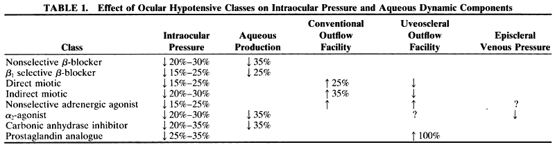
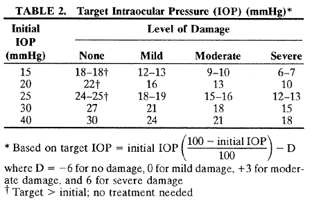
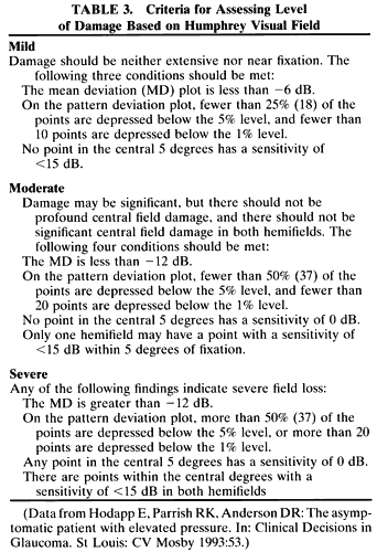
comparison. Ophthalmology 101(Suppl):80, 1994

102. Camras CB et al: Latanoprost, a potent ocular hypotensive prostaglandin analog, increases pigmentation in peripherally hypopigmented irides. Ophthalmology 101(Suppl):128, 1994

103. Glaucoma Laser Trial Research Group T: The Glaucoma Laser Trial (GLT). IV. Contralateral effects of timolol on the intraocular pressure of eyes treated with ALT. Ophthalmic Surg 22:324, 1991

104. Laurence J et al: A double-masked, placebo-controlled evaluation of timolol in a gel vehicle. J Glaucoma 2:177, 1993

105. Letchinger SL et al: Can the concentration of timolol or the frequency of its administration be reduced? Ophthalmology 100:1259, 1993

106. Mills KB: Blind randomized non-crossover long-term trial comparing topical timolol 0.25% with timolol 0.5% in the treatment of simple chronic glaucoma. Br J Ophthalmol 67:216, 1983

107. Katz IM, Hubbard WA, Getson AJ et al: Intraocular pressure decrease in normal volunteers following timolol ophthalmic solution. Invest Ophthalmol 15:489, 1976

108. Soll DB: Evaluation of timolol in chronic open angle glaucoma: once a day vs. twice a day. Arch Ophthalmol 98:2178, 1980

109. Sheddan AH: Timolol maleate in gel-forming solution. Chibret Intl J Ophthalmol 10:2, 1994

110. Katz IM, Berger ET: Effects of iris pigmentation on response of ocular pressure to timolol. Surv Ophthalmol 23:395, 1979

111. Stewart WC, Leland TM, Cate EA: Efficacy and safety of timolol solution once daily versus timolol gel in treating elevated intraocular pressure. J Glaucoma 7:402, 1998

112. Stewart WC, Cate EA, Stewart JA: Systemic beta-blockade with once daily Betimol or Timoptic-XE. J Ocular Pharm Ther 15:225, 1999

113. Schlecte LP, Brubaker RF: The effects of withdrawal of timolol in chronically treated glaucoma patients. Ophthalmology 95:1212, 1988

114. Calissendorff B et al: Timolol versus pilocarpine separately or combined with acetazolamide: Effects on intraocular pressure. Acta Ophthalmol 58:624, 1980

115. Kass MA et al: Timolol and acetazolamide: A study of concurrent administration. Arch Ophthalmol 100:941, 1982

116. Airaksinen PJ et al: A double-masked study of timolol and pilocarpine combined. Am J Ophthalmol 104:587, 1987

117. Smith RJ et al: Addition of timolol maleate to routine medical therapy: A clinical trial. Br J Ophthalmol 64:779, 1980

118. Blasini M, Shields MB: Apraclonidine hydrochloride as an adjunct to timolol maleate therapy. J Glaucoma 1:148,1992

119. Morrison J, Robin A: Adjunctive glaucoma therapy: A comparison of apraclonidine to dipivefrin when added to timolol maleate. Ophthalmology 96:3, 1989

120. Yaldo M et al: Additive effect of 1% apraclonidine hydrochloride to nonselective ss-blockers. Ophthalmology 98:1075, 1991

121. Rulo AH, Greve EL, Hoyng PF: Additive effect of latanoprost, a prostaglandin F3a analogue, and timolol in patients with elevated intraocular pressure. Br J Ophthalmol 78:899, 1994

122. Alm A et al: Latanoprost administered once daily caused a maintained reduction of intraocular pressure in glaucoma patients treated concomitantly with timolol. Br J Ophthalmol 79:12, 1995

123. Thomas JV, Epstein DL: Timolol and epinephrine in primary open-angle glaucoma: Transient, additive effect. Arch Ophthalmol 99:91, 1981

124. Goldberg I et al: Timolol and epinephrine: A clinical study of ocular interactions. Arch Ophthalmol 98:484, 1980

125. Keates EU, Stone RA: Safety and effectiveness of concomitant administration of dipivefrin and timolol maleate. Am J Ophthalmol 91:243, 1981

126. Knupp JA et al: Combined timolol and epinephrine therapy for open-angle glaucoma. Surv Ophthalmol 28:280, 1983

127. Semla TP, Schwartz A, Koch H et al: Patterns of drug prescribing. In Van Nostrand JF, Furner SE, Suzman R (eds): Health Data on Older Americans: United States, 1992: National Center for Health Statistics. Vital Health Stat 3:187, 1993

128. Atkins JM, Pugh BR, Timewell RM: Cardiovascular effects of topical ss-blockers during exercise. Am J Ophthalmol 99:173, 1985

129. Van Buskirk EM: Adverse reactions from timolol administration. Ophthalmology 87:447, 1980

130. The Norwegian Multicenter Study Group: Timolol-induced reduction in mortality and reinfarction in patients surviving acute myocardial infarction. N Engl J Med 304:801, 1981

131. Schoene RB, Abuan T, Ward RL et al: Effects of topical betaxolol, timolol, and placebo on pulmonary function in asthmatic bronchitis. Am J Ophthalmol 97:86, 1984

132. Diggory P et al: Avoiding unsuspected respiratory side-effects of topical timolol with cardioselective or sympathomimetic agents. Lancet 345:1604, 1995

133. Renwick DS, Connolly MJ: Prevalence and treatment of chronic airways obstruction (AO) in older adults. Age Ageing 23(Suppl 2):6, 1994

134. Duch S et al: Changes in depressive status associated with topical ss-blockers. Int Ophthalmol 16:331, 1992

135. McMahon CD et al: Adverse effects experienced by patients taking timolol. Am J Ophthalmol 88:736, 1979

136. Fraunfelder FT: Sexual dysfunction secondary to topical ophthalmic timolol. JAMA 253:3092, 1985

137. Caputo BJ, Katz LJ: The quality of life of a glaucoma patient in the light of treatment modalities. Curr Opin Ophthalmol 5:10, 1994

138. Bour J, Blanchard F, Segal A: Repercussion of glaucoma on a patient's life: Concerning 341 cases in Marne, France. J Fr Ophtalmol 16:380, 1993

139. Carrier P, Gentile S, Fusco R et al: Mood disorders in patients with chronic simple glaucoma. Psychiatry Res 36:33, 1991

140. Glynn R, Seddon J, Krug J et al: Falls in elderly patients with glaucoma. Arch Ophthalmol 109:205, 1991

141. Gerber SL, Cantor LB, Brater DC: Systemic drug interactions with topical glaucoma medications. Surv Ophthalmol 35:205, 1990

142. Velde TM, Kaiser FE: Ophthalmic timolol treatment causing altered hypoglycemic response in a diabetic patient. Arch Intern Med 143:1627, 1983

143. Coppeto JR: Timolol-associated myasthenia gravis. Am J Ophthalmol 98:244, 1984

144. Shaivitz SA: Timolol and myasthenia gravis. JAMA 252:1611, 1979

145. Kuppens EVMJ et al: Effect of timolol with and without preservative on the basal tear turnover in glaucoma. Br J Ophthalmol 79:339, 1995

146. Van Buskirk EM: Corneal anesthesia after timolol maleate therapy. Am J Ophthalmol 88:739, 1979

147. Weissman SS, Asbell PA: Effects of topical timolol (0.5%) and betaxolol (0.5%) on corneal sensitivity. Am J Ophthalmol 74:409, 1990

148. Herraras JM et al: Ocular surface alteration after long-term treatment with an antiglaucomatous drug. Ophthalmology 99:1082, 1992

149. Williams DE et al: Effects of timolol, betaxolol, and levobunolol on human Tenon's fibroblasts in tissue culture. Invest Ophthalmol Vis Sci 33:2233, 1992

150. Baxter GM, Williamson TH, McKillop G et al: Color Doppler ultrasound of orbital and ocular nerve blood flow: Effects of posture and timolol 0.5%. Invest Ophthalmol Vis Sci 33:604, 1992

151. Grunwald JE, Furubayashi C: Effect of topical timolol maleate on the ophthalmic artery blood pressure. Invest Ophthalmol Vis Sci 30:1095, 1989

152. Grunwald JE: Effect of topical timolol on the human retinal circulation. Invest Ophthalmol Vis Sci 27:1713, 1986

153. Grunwald JE: Effect of timolol maleate on the retinal circulation of human eyes with ocular hypertension. Invest Ophthalmol Vis Sci 31:521, 1990

154. Grunwald JE: Effect of two weeks of timolol maleate treatment on the normal retinal circulation. Invest Ophthalmol Vis Sci 32:39, 1991

155. Martin XD, Rabineau PA: Vasoconstriction effect of topical timolol on human retinal arteries. Graefes Arch Clin Exp Ophthalmol 227:526, 1989

156. Meuche C, Heidrich H, Blackman H: Raynaud syndrome following timolol containing eye drops. Fortschr Ophthalmol 87:45, 1990

157. Partamian LG, Kass MA, Gordon M: A dose-response study of the effect of levobunolol on ocular hypertension. Am J Ophthalmol 95:229, 1983

158. Bensinger RE et al: Levobunolol: A 3-month efficacy study in the treatment of glaucoma and ocular hypertension. Arch Ophthalmol 103:375, 1985

159. Long DA et al: Levobunolol and betaxolol: A double-masked, controlled comparison of efficacy and safety in patients with elevated intraocular pressure. Ophthalmology 95:735, 1988

160. Long D et al: Minimum concentration of levobunolol required to control intraocular pressure in patients with primary open-angle glaucoma or ocular hypertension. Am J Ophthalmol 99:18, 1985

161. Levobunolol Study Group T: Levobunolol: A 4-year study of efficacy and safety in glaucoma treatment. Ophthalmology 96:642, 1989

162. Berson FG et al: Levobunolol compared with timolol for the long-term control of elevated intraocular pressure. Arch Ophthalmol 103:379, 1985

163. Boozeman FW et al: Long-term evaluation of 0.25% levobunolol and timolol for therapy for elevated intraocular pressure. Arch Ophthalmol 106:614, 1988

164. Cinotti A et al: Levobunolol versus timolol for open-angle glaucoma and ocular hypertension. Am J Ophthalmol 99:11, 1985

165. Duzman E et al: A clinical evaluation of the effects of topically applied levobunolol and timolol on increased intraocular pressure. Am J Ophthalmol 94:318, 1982

166. McMahon CD, Shaffer RN, Hoskins HD Jr et al: Adverse effects experienced by patients taking timolol. Am J Ophthalmol 88:736, 1979

167. Levobunolol Study Group T: Levobunolol: A β-adrenoreceptor antagonist effective in the long-term treatment of glaucoma. Ophthalmology 92:1271, 1985

168. Duzman E et al: A clinical evaluation of the effects of topically applied levobunolol and timolol on increased intraocular pressure. Am J Ophthalmol 94:318, 1982

169. Derick RJ et al: Once daily versus twice daily levobunolol (0.5%) therapy. Ophthalmology 99:424, 1992

170. Novack GD et al: Plasma levobunolol levels following topical administration with reference to systemic side effects. Ophthalmologica 194:194, 1987

171. Rakofsky SI et al: A comparison of the ocular hypotensive effect of once daily and twice daily levobunolol treatment. Ophthalmology 96:8, 1989

172. Wandel T et al: Glaucoma treatment with once daily levobunolol. Am J Ophthalmol 101:298, 1986

173. Allen RC et al: A combination of levobunolol and dipivefrin for the treatment of glaucoma. Arch Ophthalmol 106:904, 1988

174. Rakofsky SI et al: Efficacy and safety of once daily levobunolol for glaucoma therapy. Can J Ophthalmol 24:2, 1989

175. Ball SF, Schneider E: Cost of β-adrenergic receptor blocking agents for ocular hypertension. Arch Ophthalmol 110:654, 1992

176. Serle JB, Lustgarten JS, Podos SM: A clinical trial of metipranolol, a noncardioselective β-adrenergic antagonist, in ocular hypertension. Am J Ophthalmol 112:302, 1991

177. Hickey-Dwyer M, Campbell SH, Harding S: Double-masked, three-period crossover investigation of metipranolol in control of raised intraocular pressure. J Ocul Pharmacol 7:277, 1991

178. Lyden M, Appelbaum HJ: Effect of ophthalmic metopranolol and timolol on exercise-induced tachycardia. J Glaucoma 4:124, 1995

179. Akingbehin T, Villada JR, Walley T: Metipranolol-induced adverse reactions. I. The rechallenge study. Eye 6:277, 1992

180. Melles RB, Wong IG: Metipranolol-associated granulomatous iritis. Am J Ophthalmol 118:712, 1994

181. Schultz JS, Hoenig JA, Charles H: Possible bilateral uveitis secondary to metipranolol (Optipranolol) therapy. Arch Ophthalmol 111:1607, 1993

182. Jasper JR, Michael MC, Insel PA: The ss-adrenoreceptor antagonist carteolol and its metabolite 8-hydroxycarteolol have different intrinsic sympathomimetic activities. Br J Clin Pharmacol 30:109S, 1990

183. Stewart WC: Carteolol, an ophthalmic ss-adrenergic blocker with intrinsic sympathomimetic activity. J Glaucoma 3:339, 1994

184. Kitazawa Y: Multicenter, double-blind comparison of carteolol and timolol in primary open-angle glaucoma. Adv Ther 10:95, 1993

185. Scoville B et al: A double-masked comparison of carteolol and timolol in ocular hypertension. Arch Ophthalmol 107:1777, 1989

186. Duff GR: A double-masked crossover study comparing the effects of carteolol 1% and 2% on intraocular pressure. Acta Ophthalmol 65:618, 1987

187. Duff GR, Graham PA: A double-crossover trial comparing the effects of topical carteolol and placebo on intraocular pressure. Br J Ophthalmol 72:27, 1988

188. Duff GR, Newcombe RG: The 12-hour control of intraocular pressure on carteolol 2% twice daily. Br J Ophthalmol 72:890, 1988

189. Freedman SF et al: Effects of ocular carteolol and timolol on plasma high-density lipoprotein cholesterol level. Am J Ophthalmol 116:600, 1993

190. Stewart WC et al: A 3-month comparison of 1% and 2% carteolol and 0.5% timolol in open-angle glaucoma. Graefes Arch Clin Exp Ophthalmol 229:258, 1991

191. Nathanson JA: Human ciliary process adrenergic receptor. Invest Ophthalmol Vis Sci 21:798, 1981

192. Wax MB et al: Characterization of β-adrenergic receptors in cultured human trabecular cells and in human trabecular meshwork. Invest Ophthalmol Vis Sci 30:51, 1989

193. Reiss GR, Brubaker RF: The mechanism of betaxolol, a new ocular hypotensive agent. Ophthalmology 90:1369, 1983

194. Berrospi AR, Leibowitz HM: Betaxolol: A new β-adrenergic blocking agent for treatment of glaucoma. Arch Ophthalmol 100:943, 1982

195. Caldwell DR, Salisbury CR, Guzek JP: Effects of topical betaxolol in ocular hypertensive patients. Arch Ophthalmol 102:539, 1984

196. Feghali JG, Kaufman PL: Decreased intraocular pressure in the hypertensive human eye with betaxolol, a β-adrenergic antagonist. Am J Ophthalmol 100:777, 1985

197. Radius RL: Use of betaxolol in the reduction of elevated intraocular pressure. Arch Ophthalmol 101:898, 1983

198. Gaul GR, Will NJ, Brubaker RF: Comparison of a non-cardioselective ss-adrenergic blocker and a cardioselective blocker in reducing aqueous flow in humans. Arch Ophthalmol 107:1308, 1989

199. Allen RC, Epstein DL: Additive effect of betaxolol and epinephrine in primary open-angle glaucoma. Arch Ophthalmol 104:1178, 1986

200. Collignon-Brach J: Long-term effect of ophthalmic ss-adrenoreceptor antagonists on intraocular pressure and retinal sensitivity in primary open-angle glaucoma. Curr Eye Res 11:1, 1992

201. Stewart RH, Kimbrough RL, Ward RL: Betaxolol versus timolol: A 6-month double-blind comparison. Arch Ophthalmol 104:46, 1986

202. Albracht DC et al: A double-masked comparison of betaxolol and dipivefrin for the treatment of increased intraocular pressure. Am J Ophthalmol 116:307, 1993

203. Kaiser HJ et al: Thirty-month visual field follow-up of glaucoma patients treated with ss-blockers. J Glaucoma 1:153, 1992

204. Harris A et al: Retrobulbar arterial hemodynamic effects of betaxolol and timolol in normal-tension glaucoma. Am J Ophthalmol 120:168, 1995

205. Weinreb RN et al: A double-masked, 3-month comparison between 0.25% betaxolol suspension and 0.5% betaxolol ophthalmic solution. Am J Ophthalmol 110:189, 1990

206. Vuori M et al: Plasma and aqueous humour concentrations and systemic effects of topical betaxolol and timolol in man. Acta Ophthalmol 71:201, 1993

207. Ananthanarayan CR, Vaile SJ, Feldman F: Acute episode of asthma following topical administration of betaxolol eyedrops. Can J Ophthalmol 28:80, 1993

208. Harris LS, Greinstein SH, Bloom AF: Respiratory difficulties with betaxolol. Am J Ophthalmol 102:274, 1986

209. Brooks AMV, Burdon JGW, Gillies WE: The significance of reactions to betaxolol reported by patients. Aust NZ J Ophthalmol 17:353, 1989

210. Schoene RB et al: Effects of topical betaxolol, timolol, and placebo on pulmonary function in asthmatic bronchitis. Am J Ophthalmol 97:86, 1984

211. Van Buskirk EM et al: Betaxolol in patients with glaucoma and asthma. Am J Ophthalmol 101:531, 1986

212. Weinreb RN et al: Long-term betaxolol therapy in glaucoma patients with pulmonary disease. Am J Ophthalmol 106:162, 1988

213. Zabel RW, MacDonald IM: Sinus arrest associated with betaxolol ophthalmic drops. Am J Ophthalmol 104:431, 1987

214. Ball S: Congestive heart failure from betaxolol. Arch Ophthalmol 10:320, 1987

215. Lynch MG et al: Topical ss-blocker therapy and central nervous system side effects: A preliminary study comparing betaxolol and timolol. Arch Ophthalmol 106:908, 1988

216. Knapp A: The action of adrenalin on the glaucomatous eye. Arch Ophthalmol 50:556, 1921

217. Gifford SR: Some modern preparations used in the treatment of glaucoma. Arch Ophthalmol 57:612, 1928

218. Becker B: Topical epinephrine in the treatment of the glaucomas. In: Symposium on Glaucoma, Transactions of the New Orleans Academy of Ophthalmology. St Louis: CV Mosby, 1967:152

219. Schenker HI et al: Fluorophotometric study of epinephrine and timolol in human subjects. Arch Ophthalmol 99:1212, 1981

220. Townsend DJ, Brubaker RF: Immediate effect of epinephrine on aqueous formation in the normal human eye as measured by fluorophotometry. Invest Ophthalmol Vis Sci 19:256, 1980

221. Becker B, Pettit TH, Gay AJ: Topical epinephrine therapy of open-angle glaucoma. Arch Ophthalmol 66:219, 1961

222. Robinson JC, Kaufman PL: Effects and interactions of epinephrine, norepinephrine, timolol, and betaxolol on outflow facility in the cynomolgus monkey. Am J Ophthalmol 109:189, 1990

223. Bill A, Inomata H, Smelser GK: Unconventional routes of aqueous humor outflow in cynomolgus monkey (Macaca irus). Am J Ophthalmol 73:893, 1972

224. Neufeld AH, Jampol LM, Sears ML: Cyclic AMP in the aqueous humor: The effects of adrenergic agents. Exp Eye Res 14:242, 1972

225. Obstbaum SA, Kolker AE, Phelps CD: Low-dose epinephrine: Effect on intraocular pressure. Arch Ophthalmol 92:118, 1974

226. Alexander DW, Betson FG, Epstein DL: A clinical trial of timolol and epinephrine in the treatment of primary open-angle glaucoma. Ophthalmology 95:247, 1988

227. Becker B, Ley AP: Epinephrine and acetazolamide in the therapy of the chronic glaucomas. Am J Ophthalmol 45:639, 1958

228. Cyrlin MN, Thomas JV, Epstein DL: Additive effect of epinephrine to timolol therapy in primary open-angle glaucoma. Arch Ophthalmol 100:414, 1982

229. Higgins RG, Brubaker RF: Acute effect of epinephrine on aqueous humor formation in the timolol-treated normal eye as measured by fluorophotometry. Invest Ophthalmol Vis Sci 19:420, 1980

230. Keates EU: Evaluation of timolol maleate combination therapy in chronic open-angle glaucoma. Am J Ophthalmol 88:565, 1979

230a. Widengard I, Maepea O, Alm A: Effects of latanoprostand dipivefrin, alone or combined, on intraocular pressure and on blood-aqueous barrier permeability. Br J Ophthalmol 82:404, 1988

231. Camras CB et al: Inhibition of the epinephrine-induced reduction of intraocular pressure by systemic indomethacin in humans. Am J Ophthalmol 100:169, 1985

232. Becker B, Morton WR: Topical epinephrine in glaucoma suspects. Am J Ophthalmol 62:272, 1966

233. Kerr CR et al: Cardiovascular effects of epinephrine and dipivalyl epinephrine applied topically to the eye in patients with glaucoma. Br J Ophthalmol 66:109, 1982

234. Becker B, Morton WR: Topical epinephrine in glaucoma suspects. Am J Ophthalmol 62:272, 1966

235. Kohn AN et al: Clinical comparison of dipivalyl epinephrine and epinephrine in the treatment of glaucoma. Am J Ophthalmol 87:196, 1979

236. Keates EU, Stone RA: Safety and effectiveness of concomitant administration of dipivefrin and timolol maleate. Am J Ophthalmol 91:243, 1981

237. Byron LT: Conjunctival reaction due to L-epinephrine bitartrate (Epitrate). Arch Ophthalmol 63:567, 1960

238. Corwin ME, Spencer WH: Conjunctival melanin depositions. Arch Ophthalmol 69:317, 1963

239. Miller D, Brooks SM, Mobilia E: Adrenochrome staining of soft contact lenses. Ann Ophthalmol 89:65, 1976

240. Spaeth GL: Nasolacrimal duct obstruction caused by topical epinephrine. Arch Ophthalmol 77:355, 1967

241. Waltman SR et al: Corneal endothelial changes with long-term topical epinephrine therapy. Arch Ophthalmol 95:1357, 1977

242. Miyake K, Miyake Y, Kuratomi R: Long-term effects of topically applied epinephrine on the blood-ocular barrier in humans. Arch Ophthalmol 105:1360, 1987

243. Kolker AE, Becker B: Epinephrine maculopathy. Arch Ophthalmol 79:552, 1968

244. Mackool RJ et al: Epinephrine-induced cystoid macular edema in aphakic eyes. Arch Ophthalmol 95:791, 1977

245. Thomas JV et al: Correlation of epinephrine use and macular edema in aphakic glaucomatous eyes. Arch Ophthalmol 96:625, 1978

246. Shell JW: Pharmacokinetics of topically applied ophthalmic drugs. Surv Ophthalmol 26:207, 1982

247. Mandell AI, Stentz F, Kitabchi AE: Dipivalyl epinephrine: A new pro-drug in the treatment of glaucoma. Ophthalmology 85:268, 1978

248. Anderson JA, David WL, Wei C: Site of ocular hydrolysis of a prodrug, dipivefrin, and a comparison of its ocular metabolism with that of the parent compound, epinephrine. Invest Ophthalmol Vis Sci 19:817, 1980

249. Krieglstein GK, Leydhecker W: The dose-response relationships of dipivalyl epinephrine in open-angle glaucoma. Graefes Arch Clin Exp Ophthalmol 205:141, 1978

250. Kaback MB et al: The effects of dipivalyl epinephrine on the eye. Am J Ophthalmol 81:768, 1976

251. Wandel T, Spinak M: Toxicity of dipivalyl epinephrine. Ophthalmology 88:259, 1981

252. Yablonski ME et al: Dipivefrin use in patients with intolerance to topically applied epinephrine. Arch Ophthalmol 95:2157, 1977

253. Van Zweiten PA: The central activity of antihypertensive drugs mediated via central α-receptors. J Pharm Belg 25:89, 1973

254. Hausler G: Cardiovascular regulation by central adrenergic mechanisms and its alteration by hypotensive drugs. Circ Res 37:223, 1975

255. Makabe R: Ophthalmological studies with dichlorophenyl-aminoimidazoline. Dtsch Med Wochenschr 91:1686, 1966

256. Hasslinger R: Catapres: A new drug lowering intraocular pressure. Klin Monatsbl Augenheilkd 154:95, 1969

257. Harrison R, Kaufmann CS: Clonidine: Effects of a topically administered solution on intraocular pressure and blood pressure in open-angle glaucoma. Arch Ophthalmol 95:1368, 1977

258. Heilmann K: Clonidine in glaucoma therapy: Interferences for therapy and obvious problems. Buch Augenarzt 3:56, 1974

259. Hodapp E et al: The effect of topical clonidine on intraocular pressure. Arch Ophthalmol 99:1208, 1981

260. Heilmann K: Studies on the effect of Catapres on intraocular pressure. Klin Monatsbl Augenheilkd 161:425, 1972

261. Krieglstein GK, Langham ME, Leydhecker W: The peripheral and central neural actions of clonidine in normal and glaucomatous eyes. Invest Ophthalmol Vis Sci 17:149, 1982

262. Gharagozolo NZ, Relf SJ, Brubaker RF: Aqueous flow is reduced by the α3-adrenergic agonist, apraclonidine hydrochloride (ALO 2145). Ophthalmology 95:1217, 1988

263. Lee DA, Topper JE, Brubaker RF: Effect of clonidine on aqueous humor flow in normal human eyes. Exp Eye Res 38:239, 1984

264. Robin AL: Short-term effects of unilateral 1% apraclonidine therapy. Arch Ophthalmol 106:912, 1988

265. Mittag TW, Tormay A: Drug responses of adenylate cyclase in iris-ciliary body determined by adenine labelling. Invest Ophthalmol Vis Sci 26:396, 1985

266. Thorig L, Bill A: Effects of BHG-920 in the eye and on regional blood flows in anesthetized and conscious rabbits. Curr Eye Res 5:565, 1986

267. Abrams DA et al: The safety and efficacy of topical 1% ALO 2145 (p-aminoclonidine hydrochloride) in normal volunteers. Arch Ophthalmol 105:1205, 1987

268. Jampel H et al: Apraclonidine: A 1-week dose response study. Arch Ophthalmol 106:1069, 1988

269. Chien DS et al: Corneal and conjunctival/scleral penetration of p-aminoclonidine, AGN 190342, and clonidine in rabbit eyes. Curr Eye Res 9:1051, 1990

270. Mansberger S et al: Acute effect of topical apraclonidine on perimacular and orbital hemodynamics. Invest Ophthalmol Vis Sci 35:2176, 1994

271. Serdahl CL, Galustian J, Lewis RA: The effects of apraclonidine on conjunctival oxygen tension. Arch Ophthalmol 107:1777, 1989

272. Robin AL, Coleman AL: Apraclonidine hydrochloride: An evaluation of plasma concentrations and a comparison of its intraocular pressure-lowering and cardiovascular effects to those of timolol maleate. Trans Am Ophthalmol Soc 88:149, 1990

273. Abrams DA et al: A limited comparison of apraclonidine's dose response in subjects with normal and increased intraocular pressure. Am J Ophthalmol 108:230, 1989

274. Robin AL et al: Effect of ALO-2145 on intraocular pressure following argon laser trabeculoplasty. Arch Ophthalmol 105:646, 1987

275. Robin AL: Argon laser trabeculoplasty: Medical therapy to prevent the intraocular pressure rise associated with argon laser trabeculoplasty. Ophthalmic Surg 22:31, 1991

276. Pollack IP et al: Effectiveness of apraclonidine in preventing the rise in intraocular pressure after neodymium:YAG posterior capsulotomy. Trans Am Ophthalmol Soc 86:461, 1988

277. Silverstone DE et al: Prophylactic use of apraclonidine for intraocular pressure increase after Nd:YAG capsulotomies. Am J Ophthalmol 113:401, 1992

278. Kitazawa Y, Taniguchi T, Sugiyama K: Use of apraclonidine to reduce acute intraocular pressure rise following Q-switched Nd:YAG laser iridotomy. Ophthalmic Surg 20:49, 1989

279. Wiles SB, MacKenzie D, Ide CH: Control of intraocular pressure with apraclonidine hydrochloride after cataract extraction. Am J Ophthalmol 111:184, 1991

280. Fry LL: Comparison of the postoperative intraocular pressure with Betagan, Betoptic, Timoptic, Iopidine, Diamox, Pilopine Gel, and Miostat. J Cataract Refract Surg 18:14, 1992

281. Stewart RH et al: The efficacy of apraclonidine as an adjunct to timolol therapy. Arch Ophthalmol 113:287, 1995

282. Blasini M, Shields MB: Apraclonidine hydrochloride as an adjunct to timolol maleate therapy. J Glaucoma 1:148, 1992

283. Kosela T, Brubaker RF: Apraclonidine and timolol: Combined effects in previously untreated normal subjects. Arch Ophthalmol 109:804, 1991

284. Robin AL, The Apraclonidine Maximum-Tolerated Medical Therapy Study Group: Short-term efficacy of apraclonidine hydrochloride added to maximum-tolerated medical therapy for glaucoma. Am J Ophthalmol 120:423, 1995

285. Robin AL, Ritch R, Shin DH: Short term efficacy of apraclonidine hydrochloride added to maximum-tolerated medical therapy for glaucoma. Am J Ophthalmol 120:423, 1995

286. King MH, Richards DW: Near syncope and chest tightening after administration of apraclonidine before argon laser iridotomy. Am J Ophthalmol 110:308,1990

287. Thompson CD, Vachaspati PR, Kolis SP et al: A proposed mechanism for p-aminoclonidine allergenicity based on its relative oxidative lability. Chem Res Toxicol 10:1032, 1997

288. Thompson CD, Macdonald TL, Garst ME et al: Mechanism of adrenergic induced allergy bioactivation and antigen formation. Exp Eye Res 64:767, 1997

289. Pulido JS, Sneed SR, Blodi SF: Apraclonidine hydrochloride in vitreoretinal surgery. Arch Ophthalmol 107:316, 1989

290. Hill RA et al: Apraclonidine prophylaxis for postcycloplegic intraocular pressure spikes. Ophthalmology 98:1083, 1991

291. Gieser SC, Juzych M, Robin AL et al: Clinical pharmacology of adrenergic drugs: In Ritch R, Shields MB, Krupin T (eds): The Glaucomas, Glaucoma Therapy. St Louis, Mosby-Year Book, 1996:1425

292. Schadlu R, Maus TL, Nau CB et al: Comparison of the efficacy of apraclonidine and brimonidine as aqueous suppressants in humans. Arch Ophthalmol 116:1441, 1998

293. Toris CB, Camras CB, Yablonski ME et al: Acute versus chronic effects of brimonidine on aqueous humor dynamics in ocular hypertensive patients. Am J Ophthalmol 128:8, 1999

294. Barnes SD, Campagna JA, Dirks MS et al: Control of intraocular pressure elevations after argon laser trabeculoplasty: Comparison of brimonidine 0.2% to apraclonidine 1.0%. Ophthalmology 106:2033, 1999

295. Chevrier RL, Assolian A, Dupeure J et al: Apraclonidine 0.5% versus brimonidine 0.2% for the control of intraocular pressure elevations following anterior segment laser procedures. Ophthalmic Surg Lasers 30:199, 1999

296. Javitt JC, Schiffman RM: Clinical success and quality of life with brimonidine 0.2% or timolol 0.5% used twice daily in glaucoma or ocular hypertension: A randomized clinical trial. Brimonidine Outcomes Study Group I. J Glaucoma 9:224, 2000

297. Katz LJ: Brimonidine tartrate 0.2% twice daily vs timolol o.5% twice daily: 1-year results in glaucoma patients. Brionidine Study Group. Am J Ophthalmol 127:20, 1999

298. Schuman JS, Horwitz B, Choplin NT et al: A 1-year study of brimonidine twice daily in glaucoma and ocular hypertension: A controlled, randomized, multicenter clinical trial. Chronic Brimonidine Study Group. Arch Ophthalmol 115:847, 1997

299. Hayreh SS, Zimmerman MB, Podhajsky P et al: Nocturnal arterial hypotension and its role in optic nerve head and ocular ischemic disorders. Am J Ophthalmol 117:603,1994

300. Graham SL, Drance SM: Nocturnal hypotension: Role in glaucoma progression. Surv Ophthalmol 43:S10, 1999

301. Centofanti M, Manni G, Gregori D et al: Comparative acute effects of brimonidine 0.2% versus dorzolamide 2% combined with beta-blockers in glaucoma. Graefes Archiv Clin Exp Ophthalmol 238:302, 2000

302. Theo Schwartzenberg OW, Buys YM: Efficacy of brimonidine 0.2% as adjunctive therapy for patients with glaucoma inadequately controlled with otherwise maximal medical therapy. Ophthalmology 106:1616, 1999

303. Stewart WC, Sharpe ED, Day DG et al: Comparison of the efficacy and safety of latanoprost 0.005% compared to brimonidine 0.2% or dorzolamide 2% when added to a topical beta-adrenergicblocker in patients with primary open-angle glaucoma or ocular hypertension. J Ocul Pharmacol Ther 16:251, 2000

304. Kim DD: A case of suspected alphagan-induced psychosis. Arch Ophthalmol 118:1132, 2000

305. Walters G, Taylor RH: Severe systemic toxicity caused by brimonidine drops in an infant with presumed juvenile xanthogranuloma. Eye 13:797, 1999

306. Carlsen JO, Zabriskie NA, Kwon YH et al: Apparent central nervous system depression in infants after the use of topical brimonidine. Am J Ophthalmol 128:255, 1999

307. Korsch E, Grote A, Seybold M et al: Systemic adverse effects of topical treatment with brimonidine in an infant with secondary glaucoma. Eur J Pediatr 158:685, 1999

308. Lark KK, Pasha AS, Yan X et al: The effect of latanoprost and brimonidine on rabbit subconjunctival fibroblasts.J Glaucoma 8:72, 1999

309. Serle JB et al: The effect of brimonidine tartrate in glaucoma patients on maximal medical therapy. Invest Ophthalmol Vis Sci 34(Suppl):1137, 1993

310. Derick RJ et al: Brimonidine tartrate: A one-month dose response study. Invest Ophthalmol Vis Sci 34(Suppl):1138, 1993

311. Norlund JR et al: The cardiovascular, pulmonary, and ocular hypotensive effects of brimonidine tartrate 0.2%. Arch Ophthalmol 113:77, 1995

312. Barnebey HS et al: The efficacy of brimonidine in decreasing elevation in intraocular pressure after laser trabeculoplasty. Ophthalmology 100:1083, 1993

313. Laqueur L: Ueber eine neue therapeutische Verwendung des Physostigmins. Zentrabl Med Wissenschr 24:421, 1876

314. Velhagen K: Die Grundlager der Okularen: Pharmakologie und Toxikologie des Carbaminocholins. Arch Augenheilkd 107:319, 1933

315. Bonner TI: Domains of muscarinic acetylcholine receptors that confer specificity of G protein coupling. Trends Pharmacol Sci 13:48, 1992

316. Kaufman PL: Aqueous humor dynamics following total iridectomy in the cynomolgus monkey. Invest Ophthalmol Vis Sci 18:870, 1979

317. Grierson I, Lee WR, Abraham S: Effect of pilocarpine on the morphology of the human outflow apparatus. Br J Ophthalmol 62:303, 1978

318. Lütjen-Drecoll E: Structural factors influencing outflow facility and its changeability under drugs: A study in Macaca arctoides. Invest Ophthalmol 12:280, 1973

319. Kaufman PL, Bárány EH: Loss of acute pilocarpine effect on outflow facility following surgical disinsertion and retrodisplacement of the ciliary muscle from the scleral spur in the cynomolgus monkey. Invest Ophthalmol Vis Sci 15:793, 1976

320. Kaufman PL, Bárány EH: Residual pilocarpine effect on outflow facility after ciliary muscle disinsertion 
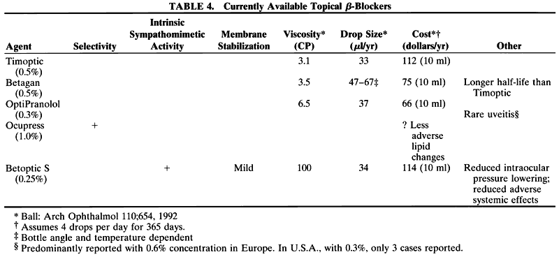
in the cynomolgus monkey. Invest Ophthalmol 15:558, 1976

321. Bárány EH: The mode of action of miotics on outflow resistance: a study of pilocarpine in the vervet monkey, Cercopithecus ethiops. Trans Ophthalmol Soc UK 86:539, 1966

322. Bárány EH: The mode of action of pilocarpine on outflow resistance in the eye of a primate (Cercopithecus ethiops). Invest Ophthalmol 1:712, 1962

323. Shaffer RN: In Newell FW (ed): Glaucoma: Transactions of the Fifth Conference. New York: Josiah Macy Jr Foundation, 1961

324. True-Gabelt B, Kaufman PL: Muscarinic receptor subtype antagonist inhibition of pilocarpine-stimulated outflow facility, accommodation, and pupil constriction in rhesus monkeys. Invest Ophthalmol Vis Sci 32(Suppl):1256, 1991

325. Lieberman TW, Leopold IH: The use of aceclidine in the treatment of glaucoma: Its effect on intraocular pressure and facility of aqueous humor outflow as compared to that of pilocarpine. Am J Ophthalmol 64:405, 1967

326. Drance SM, Fairclough M, Schulzer M: Dose response of human intraocular pressure to aceclidine. Arch Ophthalmol 88:394, 1972

327. Fechner PU, Teichmann KD, Weyrauch W: Accommodative effects of aceclidine in the treatment of glaucoma. Am J Ophthalmol 79:104, 1975

328. Pilza A, Lommatzsch P, Ulrich WD: Experimentelle und klinische Untersuchungen mit Aceclidin (Glaucostat). Ophthalmologica 168:37, 1974

329. McKinzie JW, Boggs MB Jr: Comparison of postoperative intraocular pressures after use of Miochol and Miostat.J Cataract Refract Surg 15:185, 1989

330. Wedrich A, Menapace R: Intraocular pressure following small-incision cataract surgery and polyHEMA posterior chamber lens implantation: A comparison between acetylcholine and carbachol. J Cataract Refract Surg 18:500, 1992

331. West J et al: Prevention of acute postoperative pressure rises in glaucoma patients undergoing cataract extraction with posterior chamber lens implantation. Br J Ophthalmol 76:534, 1992

332. Hollands RH, Drance SM, Schulzer M: The effect of acetylcholine on early postoperative intraocular pressure. Am J Ophthalmol 103:749, 1987

333. Grimmett MR et al: Corneal edema after Miochol. Am J Ophthalmol 115:236, 1993 (Letter)

334. Kronfeld PC: Eserine and pilocarpine: Our 100-year-old allies. Surv Ophthalmol 14:479, 1970

335. Weber A: Die Ursache des Glaukoms. Graefes Arch Ophthalmol 23:1, 1877

336. Goodman LS, Gilman A (eds): The Pharmacological Basis of Therapeutics. 5th ed. New York: Macmillan, 1985

337. Harris LS, Galin MA: Dose response analysis of pilocarpine-induced ocular hypotension. Arch Ophthalmol 93:42, 1975

338. Drance SM, Nash PA: The dose response in human intra-ocular pressure to pilocarpine. Can J Ophthalmol 6:9, 1971

339. Drance SM, Bernsted M, Schulzer M: Pilocarpine and intraocular pressure: Duration of effectiveness of 4% and 8% pilocarpine instillation. Arch Ophthalmol 91:104, 1974

340. March WF et al: Duration of effect of pilocarpine gel. Arch Ophthalmol 100:1270, 1982

341. Zimmerman TJ et al: Pilocarpine: A re-look at dose response and duration of action. Invest Ophthalmol Vis Sci 28(Suppl):377, 1987

342. Goldberg I et al: Efficacy and patient acceptance of pilocarpine gel. Am J Ophthalmol 88:843, 1979

343. Magder H, Boyaner D: The use of a longer-acting pilocarpine in the management of chronic simple glaucoma. Can J Ophthalmol 9:285, 1974

344. Nagasubramanian S, Stewart RH, Hitchings RA: Long-term effects of glaucoma therapy with 4% pilocarpine gel on cornea clarity and endothelial cell density. Int Ophthalmol 18:5, 1994

345. Johnson DH et al: A one-year multicenter clinical trial of pilocarpine gel. Am J Ophthalmol 97:723, 1984

346. Armaly MF, Rao KR: The effect of pilocarpine Ocusert with different release rates on ocular pressure. Invest Ophthalmol 12:491, 1973

347. Lee P, Shen Y, Eberle M: The long-term Ocusert pilocarpine system in the management of glaucoma. Invest Ophthalmol 14:43, 1975

348. Quigley HA, Pollack IP, Harbin TSJ: Pilocarpine Ocuserts: Long-term clinical trials and selected pharmacodynamics. Arch Ophthalmol 93;771, 1975

349. Worthen DM, Zimmerman TJ, Wind CA: An evaluation of the pilocarpine Ocusert. Invest Ophthalmol 13:296, 1974

350. Place VA et al: Comparative pharmacologic effects of pilocarpine administered to normal subjects by eye drops or by ocular therapeutic systems. Am J Ophthalmol 80:706, 1975

351. Hillman JS, Walker A, Davies EM: The management of chronic glaucoma with pilocarpine Ocusert. Trans Ophthalmol Soc UK 97:206, 1977

352. Brown HS et al: Visual effects of pilocarpine in glaucoma: Comparative study of administration by eyedrops or by ocular therapeutic systems. Arch Ophthalmol 94:1716, 1977

353. Tymer G, Scheie H: Mechanism of the miotic-resistant pupil with increased intraocular pressure. Arch Ophthalmol 50:572, 1953

354. Francois J, Goes F: Ultrasonographic study of the effect of different miotics on the eye components. Ophthalmologica 175:328, 1977

355. Francois J, Goes F: Comparative ultrasonographic study of the effect of pilocarpine 2% and Ocusert P 20 on the eye components. Am J Ophthalmol 86:233, 1978

356. Airaksinen PJ: The long-term hypotensive effect of timolol maleate compared with the effect of pilocarpine in simple and capsular glaucoma. Acta Ophthalmol 57:425, 1979

357. Boger WP, Puliafito CA, Steinert RF et al: Long-term experience with timolol ophthalmic solution in patients with open-angle glaucoma. Ophthalmology 85:259, 1978

358. Ashburn FS, Gillespie JE, Kass MA et al: Timolol plus maximum tolerated antiglaucoma therapy: A one-year follow-up study. Surv Ophthalmol 23:389, 1979

359. Bill A, Walinder P: The effects of pilocarpine on the dynamics of aqueous humor in a primate (Macaca irus). Invest Ophthalmol 5:170, 1966

360. Friström B, Nilsson SE: Interaction with PhXA41, a new prostaglandin analogue, with pilocarpine. Arch Ophthalmol 111:662, 1993

361. Quaranta L, Ripandelli G, Manni GL et al: Hypotensive effect of pilocarpine after laser trabeculoplasty. J Glaucoma 1:233, 1992

362. Wilkie J, Drance SM, Schulzer M: The effects of miotics on anterior chamber depth. Am J Ophthalmol 68:78, 1969

363. Abramson DH, Coleman DJ, Forbes M et al: Pilocarpine: Effect on the anterior chamber and lens thickness. Arch Ophthalmol 87:615, 1972

364. Abramson DH, Franzen LA, Coleman DJ: Pilocarpine in the presbyope: Demonstration of an effect on the anterior chamber and lens thickness. Arch Ophthalmol 89:100, 1973

365. Puustjari T: Retinal detachment during glaucoma therapy: A review of a case. Ophthalmologica 190:40, 1985

366. Weseley P, Liebmann J, Ritch R: Rhegmatogenous retinal detachment after initiation of Ocusert therapy. Am J Ophthalmol 112:458, 1991

367. Kalenak JW, Zavok ZN: Presumed sudden leakage of a pilocarpine Ocusert and rhegmatogenous retinal detachment. J Glaucoma 3:152, 1994

368. Kraushar MF, Steinberg JA: Miotics and retinal detachment: Upgrading the community standard. Surv Ophthalmol 35:311, 1991

369. Pietch RL, Bobo CB, Vallotton WW: Lens opacities and organophosphate cholinesterase-inhibiting agents. Am J Ophthalmol 73:236, 1972

370. Shaffer RN, Hetherington I: Anticholinesterase drugs and cataracts. Am J Ophthalmol 62:613, 1966

371. Mori M, Araie M, Sakurai M et al: Effects of pilocarpine and tropicamide on blood-aqueous barrier permeability in man. Invest Ophthalmol Vis Sci 33:416, 1992

372. Camras C, Campbell DG: Cases in controversy: Initial treatment of pigmentary glaucoma. J Glaucoma 2:44, 1993

373. Militor H: A comparative study of the effects of five choline compounds used in therapeutics: Acetylcholine chloride, acetyl-beta-methylcholine chloride, carbaminoyl beta-methylcholine chloride. J Pharmacol Exp Ther 58:337, 1936

374. O'Brien CS, Swan KC: Carbaminoylcholine chloride in the treatment of glaucoma simplex. Arch Ophthalmol 27:253, 1942

375. Smolen VF et al: Biophysical availability of ophthalmic carbachol. I. Mechanisms of cationic polymer- and surfactant-promoted miotic activity. J Pharm Sci 62:958, 1973

376. McKinstry DN, Koelle GB: Effects of drugs on acetylcholine release from the cat superior cervical ganglion by carbachol and by preganglionic stimulation. J Pharmacol Exp Ther 157:328, 1967

377. Hollands RH, Drance SM, Schulzer M: The effect of intracameral carbachol on intraocular pressure after cataract extraction. Am J Ophthalmol 104:225, 1987

378. Stocker FW: Experimental studies on the blood-aqueous barrier. Arch Ophthalmol 37:583, 1947

379. McCombie H, Saunders BC: Alkyl fluorophosphonates: Preparation and physiological properties. Nature 157:287, 1946

380. Reichert RW, Shields MB: Intraocular pressure response to the replacement of pilocarpine or carbachol with echo-thiophate. Graefes Arch Clin Exp Ophthalmol 229:252, 1991

381. Havener WH: Ocular Pharmacology. Vol 4. St Louis: CV Mosby, 1978

382. Barsam PC, Vogel HP: The effect of phospholine iodide on the diurnal variation of intraocular pressure in glaucoma. Am J Ophthalmol 57:241, 1964

383. Drance SM, Carr F: Effects of Phospholine Iodide (217MI) on intraocular pressure in man. Am J Ophthalmol 49:470, 1960

384. Harris LS: Dose-response analysis of echothiophate iodide. Arch Ophthalmol 86:502, 1971

385. Lipson ML, Holmes JH, Ellis PP: Oral administration of pralidoxime chloride in echothiophate iodide toxicity. Arch Ophthalmol 82:830, 1969

386. de Roetth A Jr et al: Blood cholinesterase activity in glaucoma patients with phospholine iodide. Am J Ophthalmol 62:834, 1966

387. Kalow W: Hydrolysis of local anesthetics by human serum cholinesterase. J Pharmacol Exp Ther 104:122, 1952

388. Axelsson U, Holberg A: The frequency of cataract after miotic therapy. Acta Ophthalmol 44:421, 1966

389. Thoft RA: Incidence of lens changes in patients treated with echothiophate iodide. Arch Ophthalmol 80:317, 1968

390. Alpar JJ: Miotics and retinal detachment. Ann Ophthalmol 11:395, 1979

391. Pape LG, Forbes M: Retinal detachment and miotic therapy. Am J Ophthalmol 85:558, 1978

392. Patten JT, Cavanagh HD, Allansmith MR: Induced ocular pseudopemphigoid. Am J Ophthalmol 72:917, 1971

393. Drance SM, Carr F: Effect of demecarium bromide (BC 48) on intraocular pressure in man. Arch Ophthalmol 62:673, 1959

394. Friedenwald JS: Carbonic anhydrase inhibition and aqueous flow. Am J Ophthalmol 40:159, 1955

395. Friedenwald JS: The formation of intraocular fluid. Am J Ophthalmol 32:9, 1949

396. Wistrand PJ: Carbonic anhydrase in the anterior uvea of the rabbit. Acta Physiol Scand 24:144, 1951

397. Kinsey VE: Comparative chemistry of aqueous humor in posterior and anterior chambers of rabbit eye. Arch Ophthalmol 50:401, 1953

398. Becker B: Decrease in intraocular pressure in man by a carbonic anhydrase inhibitor, Diamox. Am J Ophthalmol 37:13, 1954

399. Kupfer C, Lawrence C: Long-term administration of acetazolamide (Diamox) in the treatment of glaucoma. Am J Ophthalmol 40:673, 1955

400. Dodgson SJ: The carbonic anhydrases: Overview of their importance in cellular physiology and in molecular genetics. In Dodgson SJ et al (eds): The Carbonic Anhydrases: Cellular Physiology and Molecular Genetics. New York:Plenum Press, 1991

401. Wistrand PJ, Lindqvist A: Design of carbonic anhydrase inhibitors and the relationship between the pharmacodynamics and pharmacokinetics of acetazolamide. In Botré F, Gros G, Storey BT (eds): Carbonic Anhydrase from Biochemistry and Genetics to Physiology and Clinical Medicine. New York: VCH, 1991

402. Dobbs PC, Epstein DL, Anderson JP: Identification of isoenzyme C as the principal carbonic anhydrase in human ciliary processes. Invest Ophthalmol Vis Sci 18:867, 1979

403. Wistrand PJ, Carg LC: Evidence of a high-activity C type of carbonic anhydrase inhibitor in human ciliary processes. Invest Ophthalmol Vis Sci 18:802, 1979

404. Krupin T et al: Failure of acetazolamide to decrease intra-ocular pressure in patients with carbonic anhydrase II deficiency. Am J Ophthalmol 99:396, 1985

405. Maren TH, Wynns GC, Wistrand PJ: Properties of carbonic anhydrase (CA) IV, the membrane-bound enzyme in secretory tissues. Invest Ophthalmol Vis Sci 34(Suppl): 930, 1993

406. Murakami M et al: The loci of carbonic anhydrase activity in the ciliary epithelium of the rabbit eye: Electrophysiological study with isolated ciliary epithelial bilayer. Acta Histochem Cytochem 25:771, 1992

407. Schwam H et al: Identification and partial purification of a sodium dodecyl sulfate-resistant carbonic anhydrase activity from the rabbit ciliary process. Invest Ophthalmol Vis Sci 34(Suppl):930, 1993

408. Wistrand PJ: The importance of carbonic anhydrase B and C for the unloading of CO3 by the human erythrocyte. Acta Physiol Scand 113:517, 1981

409. Dodgson SJ et al: Carbonic anhydrase activity of intact carbonic anhydrase II-eficient human erythrocytes. J Appl Physiol 65:1472, 1988

410. Becker B: Use of methazolamide (Neptazane) in the therapy of glaucoma: Comparison with acetazolamide (Diamox). Am J Ophthalmol 49:1307, 1960

411. Becker B: Carbonic anhydrase and the formation of aqueous humor. The Friedenwald Memorial Lecture. Am J Ophthalmol 47:342, 1959

412. Becker B: The mechanism of the fall in intraocular pressure induced by the carbonic anhydrase inhibitor, Diamox. Am J Ophthalmol 39:177, 1955

413. Becker B: The effects of carbonic anhydrase inhibitor acetazolamide on the composition of the aqueous humor: Part II. Am J Ophthalmol 40:129, 1955

414. Holm O, Wiebert O: The effect of systemically given acetazolamide (Diamox) upon the formation of aqueous humour in the human eye, measured with a new photogrammetric method. Acta Ophthalmol 46:1243, 1968

415. Dailey RA, Brubaker RF, Bourne WM: The effect of timolol maleate and acetazolamide on the rate of aqueous humor formation in normal human subjects. Am J Ophthalmol 93:232, 1982

416. Topper JE, Brubaker RF: Effects of timolol, epinephrine, and acetazolamide on aqueous flow during sleep. Invest Ophthalmol Vis Sci 26:1315, 1985

417. Kupfer C, Gaasterland D, Ross K: Studies of aqueous humor dynamics in man. V. Effects of acetazolamide and isoproterenol in young and old normal volunteers. Invest Ophthalmol 15:349, 1976

418. Kupfer C, Gaasterland D, Ross K: Studies of aqueous humor dynamics in man. II. Measurements in young normal subjects using acetazolamide and L-epinephrine. Invest Ophthalmol 10:523, 1971

419. Maren TH: The relation between enzyme inhibition and physiological response in the carbonic anhydrase system.J Pharmacol Exp Ther 139:140, 1963

420. Wistrand PJ: The use of carbonic anhydrase inhibitors in ophthalmology and clinical medicine. Ann NY Acad Sci 429:609, 1984

421. McCannel CA, Heinrich SR, Brubaker RF: Acetazolamide but not timolol lowers aqueous humor flow in sleeping humans. Graefes Arch Clin Exp Ophthalmol 230:518, 1992

422. Bietti G et al: Acetazolamide, metabolic acidosis, and intraocular pressure. Am J Ophthalmol 80:360, 1975

423. Krupin T et al: Acidosis, alkalosis, and aqueous humor dynamics in rabbits. Invest Ophthalmol Vis Sci 16:997, 1977

424. Benedikt O, Zirm M, Harnocourt K: Die Beziehungen zwischen metabolischer Acidose und intraocularem Druck nach Carboanhyudrasehemmung mit Acetazolamid. Graefes Arch Clin Exp Ophthalmol 190:247, 1974

425. Friedman Z, Krupin T, Becker B: Ocular and systemic effects of acetazolamide in nephrectomized rabbits. Invest Ophthalmol Vis Sci 23:209, 1982

426. Maren TH: The development of ideas concerning the role of carbonic anhydrase in the secretion of aqueous humor: Relation to the treatment of glaucoma. In Drance SM, Neufeld AH (eds): Glaucoma: Applied Pharmacology in Medical Treatment. Orlando: Grune & Stratton, 1984:325

427. Maren TH: Carbonic anhydrase: Chemistry, physiology, and inhibition. Physiol Rev 47:595, 1967

428. Maren TH et al: The pharmacology of methazolamide in relation to the treatment of glaucoma. Invest Ophthalmol Vis Sci 16:730, 1977

429. Maren TH, Mayer E, Wadsworth BC: The pharmacology of Diamox: 2-acetylamino-1,3,4 thiadiazole-5-sulfonamide. Bull Johns Hopkins Hosp 95:199, 1954

430. Friedland BR, Mallonee J, Anderson DR: Short-term dose-response characteristics of acetazolamide in man. Arch Ophthalmol 95:1809, 1977

431. Lehmann B, Linnér E, Wistrand PJ: The pharmacokinetics of acetazolamide in relation to its use in the treatment of glaucoma and its effects as an inhibitor of carbonic anhydrases. In Rospe G (ed): Schering Workshop in Pharmacokinetics. Vol 5. New York: Pergamon Press, 1970

432. Linnér E, Wistrand PJ: The initial drop of the intraocular pressure following intravenous administration of acet-azolamide in man. Acta Ophthalmol 37:209, 1959

433. Berson FG et al: Acetazolamide dosage forms in the treatment of glaucoma. Arch Ophthalmol 98:1051, 1980

434. Berson FG, Epstein DL: Separate and combined effects of timolol maleate and acetazolamide in open-angle glaucoma. Am J Ophthalmol 92:788, 1981

435. Kass M et al: Timolol and acetazolamide: A study of concurrent administration. Arch Ophthalmol 100:941, 1982

436. Smith JP et al: Betaxolol and acetazolamide: Combined ocular hypotensive effect. Arch Ophthalmol 102:1794, 1984

437. Lichter PR et al: Patient tolerance to carbonic anhydrase inhibitors. Am J Ophthalmol 85:495, 1978

438. Lichter PR: Reducing side effects of carbonic anhydrase inhibitors. Ophthalmology 88:266, 1981

439. Alward PD, Wilensky JT: Determination of acetazolamide compliance in patients with glaucoma. Arch Ophthalmol 99:1973, 1981

440. Epstein DL, Grant WM: Carbonic anhydrase inhibitor side effects: Serum chemical analysis. Arch Ophthalmol 95:1378, 1977

441. Epstein RJ, Allen RC, Lunde MW: Organic impotence associated with carbonic anhydrase inhibitor therapy for glaucoma. Ann Ophthalmol 19:48, 1987

442. Kass MA et al: Acetazolamide and urolithiasis. Ophthalmology 88:261, 1981

443. Constant MA, Becker B: The effect of carbonic anhydrase inhibitors on urinary excretion of citrate by humans. Am J Ophthalmol 49:929, 1960

444. Higashihara E et al: Calcium metabolism in acidotic patients induced by carbonic anhydrase inhibitors: Response to citrate. J Urol 145:942, 1991

445. Parfitt AM: Acetazolamide and sodium bicarbonate-induced nephrocalcinosis and nephrolithiasis: Relationship to citrate and calcium excretion. Arch Intern Med 124:736, 1969

446. Fraunfelder FT, Bagby GC: Possible hematologic reactions associated with carbonic anhydrase inhibitors. JAMA 261:2257, 1989

447. Fraunfelder FT et al: Hematologic reactions to carbonic anhydrase inhibitors. Am J Ophthalmol 100:79, 1985

448. Mogk L, Cyrlin MN: Blood dyscrasias and carbonic anhydrase inhibitors. Ophthalmology 95:768, 1988

449. Zimran A, Beutler E: Can the risk of acetazolamide-induced aplastic anemia be decreased by periodic monitoring of blood cell counts? Am J Ophthalmol 104:654, 1987

450. Blocker ER, Rostand RA: Carbonic anhydrase inhibition in glaucoma: Hazard or benefit for the chronic lunger? Surv Ophthalmol 23:169, 1978

451. Goodfield M, Davis J, Jeffcoate W: Acetazolamide and symptomatic metabolic acidosis in mild renal failure. BMJ 284:422, 1982

452. Ayvazian JH, Ayvazian LF: A study of the hyperuricemia induced by hydrochlorothiazide and acetazolamide. J Clin Invest 40:196, 1961

453. Wettrell K, Pandolfi M: Propanolol vs. acetazolamide: A long-term, double-masked study of the effect on intraocular pressure and blood pressure. Arch Ophthalmol 97:280, 1979

454. Grant WM: Antiglaucoma drugs: Problems with carbonic anhydrase inhibitors. In Leopold IH (ed): Symposium on Ocular Therapy. Vol 6. St Louis: CV Mosby, 1973

455. Maren TH: Teratology and carbonic anhydrase inhibition. Arch Ophthalmol 85:1, 1971

456. Layton WM, Hallesy DW: Deformity of forelimb in rats: Association with high doses of acetazolamide. Science 149:306, 1965

457. Söderman P, Hartvig P, Fagerund C: Acetazolamide excretion into human breast milk. Br J Clin Pharmacol 17:599, 1984

458. Anderson CJ, Kaufman PL, Sturm RJ: Toxicity of combined therapy with carbonic anhydrase inhibitors and aspirin. Am J Ophthalmol 86:516, 1978

459. Coudon WL, Block AJ: Acute respiratory failure precipitated by a carbonic anhydrase inhibitor. Chest 69:112, 1976

460. Yablonski ME et al: Enhancement of the ocular hypotensive effect of acetazolamide by diflusinal. Am J Ophthalmol 106:332, 1988

461. Beasley FJ: Transient myopia and retinal edema during ethoxzolamide (Cardrase) therapy. Arch Ophthalmol 68:490, 1962

462. Galin MA, Baras I, Zweifash P: Diamox-induced myopia. Am J Ophthalmol 54:237, 1962

463. Vela A, Campbell DG: Hypotony and ciliochoroidal detachment following pharmacologic aqueous suppressant therapy in previously filtered patients. Ophthalmology 92:50, 1985

464. Bayne WF et al: Time course and disposition of methazolamide in human plasma and red blood cells. J Pharm Sci 70:78, 1981

465. Maren RH, Robinson B: The pharmacology of acetazol-amide as related to cerebrospinal fluid and the treatment of hydrocephalus. Bull Johns Hopkins Hosp 106:1, 1960

466. Stone RA et al: Low-dose methazolamide and intraocular pressure. Am J Ophthalmol 83:674, 1977

467. Dahlen K et al: A repeated dose-response study of methazolamide in glaucoma. Arch Ophthalmol 96:2214, 1978

468. Merkle W: Untersuchungen über die Wirkung von Methazolamid auf den intraokularen Druck. Klin Monatsbl Augenheilkd 176:181, 1980

469. Garrison L et al: A clinical comparison of three carbonic anhydrase inhibitors. Trans Pacific Coast Otolaryngol Ophthalmol Soc 48:137, 1967

470. Lichter PR et al: Patient tolerance to carbonic anhydrase inhibitors. Am J Ophthalmol 85:495, 1978

471. Sugrue MF et al: A study of the in vitro inhibition of human carbonic anhydrase isoenzymes I, II, and IV. Invest Ophthalmol Vis Sci 34(Suppl):1143, 1993

472. Baldwin J et al: Thienothiopyran-2-sulfonamides: Novel typically active carbonic anhydrase inhibitors for the treatment of glaucoma. J Med Chem 32:2510, 1989 (Letter)

473. Lippa EA: Topical carbonic anhydrase inhibitors. In Dodgson SJ et al (eds): The Carbonic Anhydrases: Cellular Physiology and Molecular Genetics. New York: Plenum Press, 1991

474. Wang RF et al: MK-507 (L-671, 152), a topically active carbonic anhydrase inhibitor, reduces aqueous humor production in monkeys. Arch Ophthalmol 109:1297, 1991

475. Sugrue MF, Funk H, Klotzbuecher C: Comparison of the topical carbonic anhydrase inhibitor L-671,152 and timolol in glaucoma monkeys. Invest Ophthalmol Vis Sci 31(Suppl):232, 1990

476. Wang RF et al: The ocular hypotensive effect of the topical carbonic anhydrase inhibitor L-671,152 in glaucomatous monkeys. Arch Ophthalmol 108:511, 1990

477. Hoffman HM et al: MK-507 (L-671,152): Lokale Verträglichkeit und Wirksamkeit eines neuen lokalen Karboanhydrasehemmers bei gesunden Probande. Fortschr Ophthalmol 88:513, 1991

478. Hoffman H et al: L-671,152: Local tolerability and activity of a new topical carbonic anhydrase inhibitor in normal volunteers. Presented at the Glaucoma Symposium of the XXVI International Congress of Ophthalmology, Singapore, March 17, 1990

479. Serle JB, Podos SM: Update on dorzolamide: A topical carbonic anhydrase inhibitor. In Krieglstein GK (ed): Glaucoma Update. Vol V. Heidelberg: Kaden Verlag, 1995:261

480. Bourgeois H et al: 2% L-671,152: Multiple-dose activity bid. Invest Ophthalmol Vis Sci 31(Suppl):233, 1990

481. Lippa E, Laibovitz R, Clineschmid C: L-671,152: A novel, active topical carbonic anhydrase inhibitor in patients. Presented at the Glaucoma Symposium of the XXVI International Congress of Ophthalmology, Singapore, 1990

482. Lippa EA et al: MK-507 vs. sezolamide: Comparative efficacy of two topically active carbonic anhydrase inhibitors. Ophthalmology 98:308, 1991

483. Lippa EA et al: Dorzolamide hydrochloride: Six-week, dose-response study of an active, topical carbonic anhydrase inhibitor. Invest Ophthalmol Vis Sci 34(Suppl): 931, 1993

484. Wilkerson M et al: Four-week safety and efficacy study of dorzolamide, a novel, active topical carbonic anhydrase inhibitor. Arch Ophthalmol 111:1343, 1993

485. Strahlman ER et al: A controlled clinical trial of 2.0% dorzolamide (MK-507) compared to timolol and betaxolol. Invest Ophthalmol Vis Sci 35(Suppl):2177, 1994

486. Nardin G et al: Activity of the topical CAI MK-507 bid when added to timolol bid. Invest Ophthalmol Vis Sci 32(Suppl):989, 1991

487. Laibovitz R et al: Comparison of quality of life and patient preference of dorzolamide and pilocarpine as adjunctive therapy to timolol in the treatment of glaucoma. J Glaucoma 4:306, 1995

488. Kitazawa Y et al: Topical dorzolamide hydrochloride can be a substitute for oral carbonic anhydrase inhibitors. Invest Ophthalmol Vis Sci 35(Suppl):2177, 1994

489. Serle JB et al: Compassionate case usage of dorzolamide (MK-507) in 16 glaucoma patients. Invest Ophthalmol Vis Sci 35(Suppl):2177, 1994

490. Kaplan BH et al: The use of MK-507 2% ophthalmic solution in patients with glaucoma or ocular hypertension. Invest Ophthalmol Vis Sci 35(Suppl):2177, 1994

491. Gervasoni JP et al: Absence of metabolic effects of a methyl-thienothiopyran-2-sulfonamide, a new carbonic anhydrase inhibitor (CAI), during a 2-week ocular administration. Clin Pharmacol Ther 49:192, 1991

492. Werner EB, Gerber DS, Yoder YI: Effect of a topical carbonic anhydrase inhibitor, 6-hydroxybenzo(b)thiophene-2-sulfonamide, on intraocular pressure in normotensive subjects. Can J Ophthalmol 22:316, 1987

493. Kitazawa Y: Long-term treatment with MK-507, a topical carbonic anhydrase inhibitor. Ophthalmology 100(Suppl):81, 1993

494. Konowal A, Morrison JC, Brown SV et al: Irreversible corneal decompensation in patients treated with topical dorzolamide. Am J Ophthalmol 127:403, 1999

495. Giasson CJ, Nguyen TQ, Boisjoly HM et al: Dorzolamide and corneal recovery from edema in patients with glaucoma or ocular hypertension. Am J Ophthalmol 129:144, 2000

496. Ingram CJ, Brubaker RE: Effect of brinzolamide and dorzolamide on aqueous humor flow in human eyes. Am J Ophthalmol 128:292, 1999

497. Sall K: The efficacy and safety of brinzolamide 1% ophthalmic suspension (Azopt) as a primary therapy in patients with open-angle glaucoma or ocular hypertension. Brinzolamide Primary Therapy Study Group. Surv Ophthalmol 44:S155, 2000

498. Silver LH: Ocular comfort of brinzolamide 1.0% ophthalmic suspension compared with dorzolamide 2.0% ophthalmic solution: Results from two multicenter comfort studies. Brinzolamide comfort study group. Surv Ophthalmol 44:S141, 2000

499. Ambache N: Irin, a smooth-muscle contracting substance present in rabbit iris. J Physiol 129:65P, 1955

500. Ambache N: Properties of irin, a physiological constituent of the rabbit iris. J Physiol 135:114, 1957

501. Bito LZ: Prostaglandins, old concepts and new perspectives. Arch Ophthalmol 105:1036, 1987

502. Beitch BR, Eakins KE: The effects of prostaglandins on the intraocular pressure of the rabbit. Br J Pharmacol 37:158, 1969

503. Waitzman MB, King CD: Prostaglandin influences on intraocular pressure and pupil size. Am J Physiol 212:329, 1967

504. Podos SM, Becker B, Kass MA: Indomethacin blocks arachidonic acid-induced elevation of intraocular pressure. Prostaglandins 3:7, 1973

505. Neufeld AH, Jampol LM, Sears ML: Aspirin prevents the disruption of the blood-aqueous barrier in the rabbit eye. Nature 238:158, 1972

506. Cole DR, Unger WG: Prostaglandins as mediators of the responses of the eye to trauma. Exp Eye Res 17:357, 1973

507. Eakins KE et al: Prostaglandin-like activity in ocular inflammation. BMJ 3:452, 1972

508. Masuda K, Izawa Y, Mishima S: Prostaglandins and uveitis: A preliminary report. Jpn J Ophthalmol 17:166, 1973

509. Camras CB, Chacko DM, Schlossman A: Posner-Schlossman syndrome. In Pepose JS, Holland GN, Wilhelmus KR (eds): Ocular Infection and Immunity. St Louis: CV Mosby, 1996:529

510. Casey WJ: Prostaglandin E3 and aqueous humor dynamics in the rhesus monkey eye. Prostaglandins 8:327, 1974

511. Green K, Kim K: Pattern of ocular response to topical and systemic prostaglandin. Invest Ophthalmol 14:36, 1975

512. Kass MA et al: Prostaglandin E and aqueous humor dynamics. Invest Ophthalmol 11:1022, 1972

513. Masuda K, Mishima S: Effects of prostaglandins on inflow and outflow of the aqueous humor in rabbits. Jpn J Ophthalmol 17:300, 1973

514. Camras CB, Bito LZ, Eakins KE: Reduction of intraocular pressure by prostaglandins applied topically to the eyes of conscious rabbits. Invest Ophthalmol Vis Sci 16:1125, 1977

515. Coleman RA, Smith WL, Narumiya S: Classification of prostanoid receptors: Properties, distribution, and structure of the receptors and their subtypes. Pharmacol Rev 46:205, 1994

516. Crawford KS et al: The DP-receptor agonist SQ27986 raises but does not lower intraocular pressure in ocular normotensive monkeys. J Glaucoma 1:94, 1992

517. Woodward DF et al: Marked species differences in the pharmacology of prostanoid induced ocular hypertension. Invest Ophthalmol Vis Sci 32(Suppl):1257, 1994

518. Woodward DF et al: Studies on the ocular hypotensive effects of prostaglandin F3a ester prodrugs and receptor selective prostaglandin analogs. J Ocul Pharmacol 10:177, 1994

519. Woodward DF et al: Intraocular pressure effects of selective prostanoid receptor agonists involve different receptor subtypes according to radioligand binding studies. J Lipid Mediat 7:545, 1993

520. Woodward DF et al: Definition of prostanoid receptor subtypes by radioligand binding and the effects of selective agonists on intraocular pressure. Exp Eye Res 55(Suppl): S91, 1992

521. Camras CB et al: Multiple dosing of prostaglandin F3a or epinephrine on cynomolgus monkey eyes: I. Aqueous humor dynamics. Invest Ophthalmol Vis Sci 28:463, 1987

522. Crawford K, Kaufman PL, Gabelt BT: Effects of topical PGF3a on aqueous humor dynamics in cynomolgus monkeys. Curr Eye Res 6:1035, 1987

523. Kaufman PL: Effects of intracamerally infused prostaglandins on outflow facility in cynomolgus monkey eyes with intact or retrodisplaced ciliary muscle. Exp Eye Res 43:819, 1986

524. Lee PY, Podos SM, Severin C: Effect of prostaglandin F3a on aqueous humor dynamics of rabbit, cat, and monkey. Invest Ophthalmol Vis Sci 25:1087, 1984

525. Gabelt BT, Kaufman PL: Prostaglandin F3a increases uveo-scleral outflow in the cynomolgus monkey. Exp Eye Res 49:389, 1989

526. Nilsson SFE et al: Increased uveoscleral outflow as a possible mechanism of ocular hypotension caused by prostaglandin F3a-1-isopropylester in the cynomolgus monkey. Exp Eye Res 48:707, 1989

527. Selen G et al: Effects of PhXA34 and PhDA100A, two phenyl substituted prostaglandin esters, on aqueous humor dynamics and microcirculation in the monkey eye. Invest Ophthalmol Vis Sci 32(Suppl):869, 1991

528. Toris CB, Camras CB, Yablonski ME: Effects of PhXA41, a new prostaglandin F3a analog, on aqueous humor dynamics in human eyes. Ophthalmology 100:1297, 1993

529. Ziai N et al: The effects on aqueous humor dynamics of PhXA41, a new prostaglandin F3a analogue, after topical application in normal and ocular hypertensive human eyes. Arch Ophthalmol 111:1351, 1993

530. Alm A, Villumsen J: PhXA34, a new potent ocular hypotensive drug: A study on dose response relationship and on aqueous humor dynamics in healthy volunteers. Arch Ophthalmol 109:1564, 1991

531. Tamm E, Rittig M, Lutjen-Drecoll E: Electromikroskopische and immunohistochemische Untersuchungen zur augendrucksenkender Wirkung von Prostaglandin F3a. Fortschr Ophthalmol 87:623, 1990

532. Lutchen-Drecoll E, Tamm E: Morphological study of the anterior segment of cynomolgus monkey eyes following treatment with prostaglandin F3a. Exp Eye Res 47:761, 1988

533. Ocklind A: Effect of latanoprost on the extracellular matrix of the ciliary muscle. A study on cultured cells and tissue sections. Exp Eye Res 67:179,1998

534. Lindsey JD, Kashiwagi K, Kashiwagi F et al: Prostaglandins alter extracellular matrix adjacent to human ciliary muscle cells in vitro. Invest Ophthalmol Vis Sci 38:2214, 1997

535. Alm A et al: Intraocular pressure-reducing effect of PhXA41 in patients with increased eye pressure: A one-month study. Ophthalmology 100:1312, 1993

536. Camras CB: Discussion of intraocular pressure-reducing effect of PhXA41 in patients with increased eye pressure: A one-month study. Ophthalmology 100:1316, 1993

537. Hotehama Y, Mishimi HK: Clinical efficacy of PhXA34 and PhXA41, two novel prostaglandin F3a-isopropyl ester analogues for glaucoma treatment. Jpn J Ophthalmol 37:259, 1993

538. Hotehama Y et al: Ocular hypotensive effect of PhXA41 in patients with ocular hypertension or primary open-angle glaucoma. Jpn J Ophthalmol 37:2790, 1993

539. Nagasubramanian S et al: Intraocular pressure-reducing effect on PhXA41 in ocular hypertension: Comparison of dose regimens. Ophthalmology 100:1305, 1993

540. Racz P et al: Maintained intraocular pressure reduction with once-a-day application of a new prostaglandin F3a analogue (PhXA41). Arch Ophthalmol 111:657, 1993

541. Camras CB, United States Latanoprost Study Group: Comparison of latanoprost and timolol in patients with ocular hypertension and glaucoma. Ophthalmology 103:138, 1996

542. Watson P, Stjernschantz J, Latanoprost Study Group: A six-month, randomized, double-masked study comparing latanoprost with timolol in open-angle glaucoma and ocular hypertension. Ophthalmology 103:126, 1996

543. Alm A, Stjernschantz J, Scandinavian Latanoprost Study Group: Effects of intraocular pressure and side effects of 0.005% latanoprost applied once daily, evening or morning: A comparison with timolol. Ophthalmology 102:1743, 1995

544. Aung T, Wong HT, Yip CC et al: Comparison of the intraocular pressure-lowering effect of latanoprost and timolol in patients with chronic angle closure glaucoma: A preliminary study. Ophthalmology 107:1178, 2000

545. Scherrer WJ, Hauber FA: Effect of latanoprost on intraocular pressure in steroid-induced glaucoma. J Glaucoma 9:179, 2000

546. Mastropasqua L, Carpineto P, Ciancaglini M et al: A 12-month, randomized, double-masked study comparing latanoprost with timolol in pigmentary glaucoma. Ophthalmology 106:550,1999

547. Enyedi LB, Freedman SF, Buckley EG: The effectiveness of latanoprost for the treatment of pediatric-glaucoma.J AAPOS: J Am Assoc Pediatr Ophthalmol Strabismus 3:33, 1999

548. Altuna JC, Greenfield DS, Wand M et al: Latanoprost in glaucoma associated with Sturge-Weber syndrome: Benefits and side-effects. J Glaucoma 8:199, 1999

549. Yang CB, Freedman SF, Myers JS et al: Use of latanoprost in the treatment of glaucoma associated with Sturge-Weber syndrome. Am J Ophthalmol 126:600, 1998

550. Alm A et al: Latanoprost administered once daily caused a maintained reduction of intraocular pressure in glaucoma patients treated concomitantly with timolol. Br J Ophthalmol 79:12, 1995

551. Rulo AH, Greve EL, Hoyng PF: Additive effect of latanoprost, a prostaglandin F3a analogue, and timolol in patients with elevated intraocular pressure. Br J Ophthalmol 78:899, 1994

552. Nordmann JP, Soderstrom M, Rouland JF et al: Comparison of the intraocular pressure lowering effect of latanoprost and a fixed combination of timolol-pilocarpine eye drops in patients insufficiently controlled with beta adrenergic antagonists. French latanoprost study group, and the Swedish latanoprost study group. Br J Ophthalmol 84:181, 2000

553. Emmerich KH: Comparison of latanoprost monotherapy to dorzolamide combined with timolol in patients with glaucoma or ocular hypertension. A 3-month randomised study. Graefes Arch Clin Exp Ophthalmol 238:19, 2000

554. Linden C, Alm A: Latanoprost and physostigmine have mostly additive ocular hypotensive effects in human eyes. Arch Ophthalmol 115:857, 1997

555. Shin DH, McCracken MS, Bendel RE et al: The additive effect of latanoprost to maximally-tolerated medications with low-dose, high-dose or no pilocarpine therapy. Ophthalmol 106:386, 1999

556. Patelska B, Greenfield DS, Liebmann JM et al: Latanoprost for uncontrolled glaucoma in a compassionate case protocol. Am J Ophthalmol 124:279, 1997

557. Wang RF, Podos SM, Serle JB et al: Effect of latanoprost or 8-iso prostaglandin E2 alone and in combination on intraocular pressure in glaucomatous monkey eyes. Arch Ophthalmol 118:74, 2000

558. Sjoquist B, Tajallaei S, Stjernschantz J: Pharmacokinetics of latanoprost in the cynomolgus monkey. 1st communication. Single intravenous, oral or topical administration on the eye. Arzneimittel-Forschung 49:225, 1999

559. Sjoquist B, Uhlin A Byding P et al: Pharmacokinetics of latanoprost in the cynomolgus monkey. 2nd communication. Repeated topical administration on the eye. Arz Forschung 49:234, 1999

560. Moroi SE, Gottfredsdottir MS, Schteingart MT et al: Cystoid macular edema associated with latanoprost therapy in a case series of patients with glaucoma and ocular hypertension. Ophthalmol 106:1024, 1999

561. Ayyala RS, Cruz DA, Margo CE et al: Cystoid macular edema associated with lataonoprost in aphakic and pseudophakic eyes. Am J Ophthalmol 126:602, 1998

562. Callanan D, Fellamn RL, Savage JA: Latanoprost-associated cystoid macular edema. Am J Ophthalmol 126:134, 1998

563. Warwar RE, Bullock JD, Ballal D: Cystoid macular edema and anterior uveitis associated with latanoprost use. Experience and incidence in a retrospective review of 94 patients. Ophthalmology 105:263, 1998

564. Miyake K, Ota I, Maekubo K et al: Latanoprost accelerates disruption of the blood-aqueous barrier and the incidence of angiographic cystoid macular edema in early postoperative-pseudophakia. Arch Ophthalmol 117:34, 1999

565. Dutkiewicz R, Albert DM, Levin LA: Effects of latanoprost on tyrosinase activity and mitotic index of cultured melanoma lines. Exp Eye Res 70:563, 2000

566. Camras CB, Neely DG, Weiss EL: Latanoprost-induced iris color darkening: a case report with long-term follow-up. J Glaucoma 9:95, 2000

567. Johnstone MA: Hypertrichosis and increased pigmentation of eyelashes and adjacent hair in the region of the ipsilateral eyelids of patients treated with unilateral topical latanoprost. Am J Ophthalmol 124:544, 1997

568. Kook MS, Lee K: Increased eyelid pigmentation associated with use of latanoprost. Am J Ophthalmol 129:804, 2000

569. Wand M, Gilbert CM, Liesegang TJ: Latanoprost and herpes simplex keratitis. Am J Ophthalmol 127:602, 1999

570. Kaufman HE, Yarnell ED, Thompson HW: Latanoprost increases the severity and recurrence of herpetic keratitis in the rabbit. Am J Ophthalmol 127:531, 1999

571. Linden C, Alm A: Prostaglandin analogues in the treatment of glaucoma. Drugs Aging 14:387, 1999

572. Nordmann JP, Rouland JF, Mertz BP: A comparison of the intraocular pressure-lowering effect of 0.5% timolol maleate and the docosanoid derivative of a prostaglandin F2 alpha metabolite, 0.12% unoprostone, in subjects with chronic open-angle glaucoma or ocular hypertension. Curr Med Res Opin 15:87, 1999

573. Stewart WC, Stewart JA, Kapik BM: The effects of unoprostone isopropyl 0.12% and timolol maleate 0.5% on diurnal intraocular pressure. J Glaucoma 7:388, 1998

574. Serle JB, Podos SM, Kitizawa Y et al: A comparative study of latanoprost (Xalatan) and isopropyl unoprostone (Rescula) in normal and glaucomatous monkey eyes. Jpn J Ophthalmol 42:95, 1998

575. Yamamoto T, Kitizawa Y: Iris-color change developed after topical isopropyl unoprostone treatment. J Glaucoma 6:430, 1997

576. Robbins R, Galin MA: Effect of osmotic agents on the vitreous body. Arch Ophthalmol 82:694, 1969

577. Cantonnet A: Essai de traitement du glaucome par les substances osmotiques. Arch Ophthalmol 24:1, 1904

578. Hertel E: Experimentelle Untersuchungen über die Abhangigkeit des Augendrucks von der Blutbeschaffenheit. Graefes Arch Clin Exp Ophthalmol 88:197, 1914

579. Javid M: Urea: New use of an old agent. Reduction of intracranial and intraocular pressure. Surg Clin North Am 38:907, 1958

580. Javid M, Settlage P: Effect of urea on cerebrospinal fluid pressure in human subjects: Preliminary report. JAMA 160:943, 1945

581. Weiss DI, Shaffer RN, Wise BL: Mannitol infusion to reduce intraocular pressure. Arch Ophthalmol 68:341, 1962

582. Virno M et al: Oral glycerol in ophthalmology: A valuable new method for the reduction of intraocular pressure. Am J Ophthalmol 55:1133, 1963

583. Becker B, Kolker AE, Krupin T: Isosorbide, an oral hyperosmotic agent. Arch Ophthalmol 78:147, 1967

584. Krupin T, Podos SM, Becker B: Effect of optic nerve transection on osmotic alterations of intraocular pressure. Am J Ophthalmol 70:214, 1970

585. Podos SM, Krupin T, Becker B: Effect of small dose hyperosmotic injection on intraocular pressure of small animals and man when optic nerves are transected and intact. Am J Ophthalmol 71:898, 1971

586. Galin MA, Davidson R, Shachter N: Ophthalmological use of osmotic therapy. Am J Ophthalmol 62:629, 1966

587. Smith EW, Drance SM: Reduction of human intraocular pressure with intravenous mannitol. Arch Ophthalmol 68:734, 1962

588. Brogliotti B, Marello M, Cembrano S: Azlione ioptonizzante del mannitolo in somministrazione endovenosa a bolo. Boll Ocul 57:547, 1978

589. O'Keefe M, Nabil M: The use of mannitol in intraocular surgery. Ophthalmic Surg 14:55, 1983

590. Adams RE, Kirschner RJ, Leopold IH: Ocular hypotensive effect of intravenously administered mannitol: A preliminary report. Arch Ophthalmol 69:55, 1963

591. Barry KG, Khoury AH, Brooks MH: Mannitol and isosorbide: Sequential effects on intraocular pressure, serum osmolality, sodium, and solids in normal subjects. Arch Ophthalmol 81:695, 1969

592. Grabie MT et al: Contraindications for mannitol in aphakic glaucoma. Am J Ophthalmol 91:265, 1981

593. D'Alena P, Ferguson W: Adverse effects after glycerol orally and mannitol parenterally. Arch Ophthalmol 75:201, 1966

594. Almog Y, Eyer O, Laser M: Pulmonary edema as a complication of oral glycerol administration. Ann Ophthalmol 18:38, 1986

595. Borges MF, Mochs J, Kjellstrand CM: Mannitol intoxication in patients with renal failure. Arch Intern Med 142:63, 1982

596. Whelan T et al: Acute renal failure associated with mannitol intoxication. Arch Intern Med 144:2053, 1984

597. McNeill IJ: Hypersensitivity reaction to mannitol. Drug Intell Clin Pharm 19:552, 1985

598. Drance SM: Effect of oral glycerol on intraocular pressure in normal and glaucomatous eyes. Arch Ophthalmol 72:491, 1964

599. Thomas RC: Glycerin: An orally effective osmotic agent. Arch Ophthalmol 70:625, 1963

600. Oakley DE, Ellis PP: Glycerol and hyperosmolar nonketotic coma. Am J Ophthalmol 81:469, 1976

601. Krupin T, Kolker AE, Becker B: A comparison of isosorbide and glycerol for cataract surgery. Am J Ophthalmol 69:737, 1970

602. Wisznia KI, Lazar M, Leopold IM: Oral isosorbide and intraocular pressure. Am J Ophthalmol 70:630, 1970

603. Mehra K, Singh R: Lowering of intraocular pressure by isosorbide: Effects of different doses of drug. Arch Ophthalmol 86:623, 1971

604. Mehra K et al: Lowering of intraocular tension: Effects of isosorbide and glycerin. Arch Ophthalmol 85:167, 1971

605. Mao LK, Stewart WC, Shields MB: Correlation between intraocular pressure control and progressive glaucomatous damage in primary open-angle glaucoma. Am J Ophthalmol 111:51, 1991

606. Araujo SV, Spaeth GL, Roth SM et al: A ten-year follow-up on a prospective, randomized trial of postoperative corticosteroids after trabeculectomy. Ophthalmology 102:1753, 1995

607. Nouri-Mahdavi K, Brigatti L, Weitzman M et al: Outcomes of trabeculectomy for primary open-angle glaucoma. Ophthalmology 102:1760, 1995

608. Jay JL, Alan D: The benefit of early trabeculectomy versus conventional management in primary open angle glaucoma relative to severity of disease. Eye 3:528, 1989

609. Sommer A: Intraocular pressure and glaucoma. Am J Ophthalmol 107:186, 1989

610. Cartwright MJ, Anderson DR: Correlation of asymmetric damage with asymmetric intraocular pressure in normal-tension glaucoma. Arch Ophthalmol 106:898, 1988

611. Chauhan BC, Drance SM: The influence of intraocular pressure on visual field damage in patients with normal-tension and high-tension glaucoma. Invest Ophthalmol Vis Sci 31:2367, 1990

612. Hitchings RA, Wu J, Poinoosawmy D et al: Surgery for normal tension glaucoma. Br J Ophthalmol 79:402, 1995

613. Schulzer M et al: Biostatistical evidence for two distinct chronic open angle glaucoma populations. Br J Ophthalmol 74:196, 1990

614. Caprioli J, Spaeth GL: Comparison of the optic nerve head in high- and low-tension glaucoma. Arch Ophthalmol 103:1145, 1985

615. Drance SM et al: Diffuse visual field loss in chronic open-angle and low-tension glaucoma. Am J Ophthalmol 104:577, 1987

616. Caprioli J, Sears M, Miller J: Patterns of early visual field loss in open-angle glaucoma. Am J Ophthalmol 103:512, 1987

617. Schumer RA, Podos SM: The nerve of glaucoma! Arch Ophthalmol 112:37, 1994

618. Flammer J, Guthauser U, Mahler F: Do ocular vasospasms help cause low tension glaucoma? In Greve EL, Heijl A (eds): Seventh International Visual Field Symposium, Amsterdam, September 1986. Dordrecht, The Netherlands: Martinus Nijhoff/Dr W. Junk Publishers, 1987:397

619. Rojanapongpun P, Drance SM: The response of blood flow velocity in the ophthalmic artery and blood flow of the finger to warm and cold stimuli in glaucomatous patients. Graefes Arch Clin Exp Ophthalmol 231:375, 1993

620. Netland PA et al: Calcium channel blockers in the management of low-tension and open-angle glaucoma. Am J Ophthalmol 115:608, 1993

621. Kitazawa Y et al: The effect of Ca2(+ )-antagonist on visual field in low-tension glaucoma. Graefes Arch Clin Exp Ophthalmol 227:408, 1989

622. Dugan LL, Choi DW: Excitotoxicity, free radicals, and cell membrane changes. Ann Neurol 35:S17, 1994

623. Cummins D, Takahashi N, Caprioli J: Electrophysiology of cultured retinal ganglion cells to investigate basic mechanisms of damage. In Krieglstein GK (ed): Glaucoma Update IV. Proceedings of the Glaucoma Society of the International Congress of Ophthalmology, Bali, March 1990. Berlin: Springer-Verlag, 1991:59

624. Olney J: Neurotoxicity of excitatory amino acids. In McGreer E, Olney J, McGreer P (eds): Kainic Acid as a Tool in Neurobiology. New York: Raven Press, 1978:95

625. Lucas D, Newhouse J: The toxic effects of sodium l-glutamate on the inner layers of the retina. Arch Ophthalmol 58:193, 1957

626. Choi D: Glutamate neurotoxicity and diseases of the nervous system. Neuron 1:623, 1988

627. Ehinger BZ: Glutamate as a retinal neurotransmitter. In Weiler R, Osbourne N (eds): Neurobiology of the inner retina. Berlin: Springer-Verlag, 1989:1

628. David P, Lusky M, Teichberg V: Involvement of excitatory neurotransmitters in the damage produced in chick embryo retinae by anoxia and extracellular high potassium. Exp Eye Res 46:657, 1988

629. Zeevalk G, Hyndman A, Nicklas W: Excitatory amino acid induced toxicity in chick retina: Amino acid release, histology and effects of chloride channel blockers. J Neurochem 53:1610, 1989

630. Dreyer EB, Lipton SA: Excitatory amino acids in glaucoma: A potentially novel etiology of neuronal loss. Invest Ophthalmol Vis Sci 33(Suppl):1093, 1992

631. Schumer RA, Podos SM, Lipton SA et al: Increased glutamate in the vitreous of monkeys with induced glaucoma. Invest Ophthalmol Vis Sci 35(Suppl):1484, 1994

632. Cummins D, Kitano S, Caprioli J: The effects of excitatory amino acids and ischemia on cultured rat retinal ganglion cells. Invest Ophthalmol Vis Sci 33(Suppl):1031, 1992

633. Kittigawa K: Hyperthermia-induced neuronal protection against ischemic injury in gerbils. J Cereb Blood Flow Metab 11:449, 1991

634. Barbe M, Tytell M, Gower D et al: Hyperthermia protects against light damage in the rat retina. Science 241:1817, 1988

635. Raibowol K, Mizzen L, Welch W: Heat shock is lethal to fibroblasts microinjected with antibodies against hsp70. Science 242:433, 1988

636. Kitano S, Caprioli J: Expression of 72kDa heat shock protein in cultured rat retinal ganglion cells: Ischemic tolerance and protective effect of hyperthermia. Invest Ophthalmol Vis Sci 34(Suppl):1430, 1993

637. Kitano S, Hori S, Koseki Y et al: The heat shock protein synthesis inhibitor, quercetin, blocks the protection effect of hyperthermia on anoxic rat retinal ganglion cells in culture. Invest Ophthalmol Vis Sci 35(Suppl):1868, 1994

638. Moncada S et al: Nitric oxide: Physiology, pathophysiology, and pharmacology. Pharmacol Rev 43:109, 1991

639. Koseki Y, Kitano S, Podos SM et al: A nitric oxide synthase inhibitor protects against anoxia in cultured rat retinal ganglion cells. Invest Ophthalmol Vis Sci 35(Suppl): 1968, 1994

640. Pellegrini-Giampietro D et al: Excitatory amino acid release and free radical formation may cooperate in the genesis of ischemia-induced neuronal damage. J Neurosci 10:1035, 1990

641. McNamara J, Fridovich I: Did radicals strike Lou Gehrig? Nature 362:20, 1993

642. Aguayo AJ et al: Synaptic connections made by axons regenerating in the central nervous system of adult mammals. J Exp Biol 153:199, 1990

643. Villegas-Perez MP, Vidal-Sanz M, Bray GM et al: Influences of peripheral nerve grafts on the survival and regrowth of axotomized retinal ganglion cells in adult rats.J Neurosci 8:265, 1988

Back to Top