Chapter 54G
Glaucoma in Aphakia and Pseudophakia
ALANA L. GRAJEWSKI
Main Menu   Table Of Contents

Search

CLASSIFICATION
INCIDENCE
POSTOPERATIVE COMPLICATIONS AND MANAGEMENT
ANGLE-CLOSURE GLAUCOMA ASSOCIATED WITH APHAKIA AND PSEUDOPHAKIA
REFERENCES

CLASSIFICATION
The terms “aphakic glaucoma” or “pseudophakic glaucoma” erroneously imply that the glaucoma is caused by the lens extraction or state of aphakia or pseudophakia. There are many causes that, alone or in combination, can result in elevated intraocular pressure (IOP). For this reason, these terms should be avoided and the glaucoma best described by the specific underlying mechanism responsible for the pressure elevation. The etiology of elevated IOP after cataract extraction is often multifactorial. In general, it is not unusual for there to be more than one coexisting mechanism for the glaucoma. Management of these concurrent problems is directed at each component individually. As the nature of cataract surgery has evolved from intracapsular cataract extraction (ICCE) to extracapsular cataract extraction (ECCE), the possible mechanisms for postoperative glaucoma have also changed. Management depends to a large degree on treating the primary mechanism. There are also some treatment considerations common to all categories. To categorize the pathogenesis of elevated IOP in aphakic or pseudophakic eyes, the broad classifications of open-angle or closed-angle are useful, as is the temporal relationship of the IOP elevation to surgery (e.g., early versus late, transient versus persistent).
Back to Top
INCIDENCE
The reported incidence of glaucoma after cataract extraction ranges widely. This is due in part to advances in microsurgical technique. In 1974, when Francois reported that the range of occurrence of postoperative glaucoma was from 0.7% to 7%, the suture material and method of closure was different than those used 2 years later, when the reported incidence was as high as 12%.1,2 In 1988, one large series reported that more than 50% of normal patients had an IOP of greater than 25 mmHg 2 to 3 hours after surgery.3 As the occurrence of microscopic leaks and hypotony diminished with improved sutures and tighter closure, the incidence of immediate IOP elevation after cataract surgery increased. Chronic secondary glaucoma, however, decreased as frequency of postoperative shallow chambers with peripheral anterior synechiae formation decreased. Today, the incidence in eyes undergoing standard ECCE is approximately 4%.4 Interestingly, several large series comparing ICCE with ECCE found no difference in the frequency of postoperative glaucoma when wound closure was similar.5,6

Both anterior and posterior chamber intraocular lens (IOL) implantation after cataract extraction has also been associated with postoperative glaucoma. Chronic secondary glaucoma complicated uneventful cataract extraction in 5.5% of eyes that received anterior chamber IOL implants, compared with 1.6% of eyes that had posterior chamber lens implants.7 Iris-plane or iris-fixed lenses, which are rarely seen today, often have been associated with prolonged IOP elevation.8 The incidence of postoperative glaucoma in eyes receiving secondary anterior chamber implants has been recorded to be as high as 11.3%.9

PREEXISTENT OPEN-ANGLE GLAUCOMA IN CATARACT EXTRACTION

Cataract and glaucoma frequently coexist in the same eye. The effect of cataract extraction on IOP control in patients with preexistent primary open-angle glaucoma can be divided into two categories:

  Transient effects: The immediate or acute elevation of IOP appears to occur more frequently in glaucomatous eyes than in nonglaucomatous eyes. Preoperative argon laser trabeculoplasty has been reported to be beneficial in postoperative IOP control,10,11 but Savage and associates12 and McGuigan and colleagues13 reported significant postoperative IOP spikes despite previous control with laser and topical medications.
  Persistent effects: In contrast, long-term or chronic IOP control is often unaffected by uncomplicated cataract extraction. A small decrease in IOP and improved IOP control after lens extraction has been reported.14,15 This improved IOP control may relate to the ability to use miotics after the elimination of a lens opacity. Undiagnosed preoperative pupillary block may be another factor in improved postoperative IOP control without the addition of new topical medications.

Coexistent Cataract and Glaucoma

The primary decision regarding management of postoperative IOP elevation in this group of patients begins preoperatively with the choice of surgery. If a patient has a cataract that is visually significant in the presence of glaucoma, the choice of surgery includes the following: (1) cataract surgery alone; (2) initial glaucoma filtering surgery, with cataract surgery planned as a future procedure; and (3) a combined glaucoma and cataract procedure. Opinions differ regarding each of these alternatives, and management of the patient with coexistent cataract and glaucoma is controversial.

The procedure of choice for each patient depends on several factors, including functional severity of the cataract, severity of the glaucoma (optic nerve and visual field damage), the patient's tolerance to current medication, and the feasibility of performing a second surgical procedure. Cataract extraction alone is indicated in patients who have good IOP control, are on a minimal amount of well-tolerated medication, and have mild glaucomatous damage. The advantage of performing cataract extraction alone is the likelihood of fewer intraoperative complications. The disadvantages are frequent postoperative IOP spikes and overall minimal IOP lowering.

A two-stage approach may be considered for a patient whose moderate or profound glaucomatous damage is uncontrolled despite maximally tolerated medication and whose cataract is mild to moderate. In this situation, filtration surgery is performed initially; if the cataract is marginal, the cataract surgery is scheduled for a future date. The advantage of this approach may be better long-term IOP control than cataract surgery combined with a standard trabeculectomy. Disadvantages include (1) a required second surgery, which can be more technically challenging as a temporal extraction or clear cornea approach; (2) the possibility of postoperative IOP spikes despite the presence of a functional filter16; and (3) the possibility that the patient will require a third operation if the initial filtering surgery fails after cataract extraction.

A combined ECCE or phacoemulsification and trabeculectomy is indicated for patients with advanced optic nerve and visual field damage.17 Advantages to this are the reduced number of surgical interventions and the decreased frequency of an early postoperative IOP elevation. Compared with the two-stage approach, a combined procedure offers early visual rehabilitation. Disadvantages include added surgical difficulty and an increased rate of surgical complications. Long-term IOP control may be compromised when filtration surgery is done in combination with cataract extraction.18–20 For this reason, several groups have attempted to enhance the filter function by using either intraoperative mitomycin C or postoperative subconjunctival 5-fluorouracil. Some of these reports have been encouraging, but long-term data have yet to be reported.21,22

Back to Top
POSTOPERATIVE COMPLICATIONS AND MANAGEMENT

TRANSIENT POSTOPERATIVE INTRAOCULAR PRESSURE ELEVATION

In the immediate postoperative period, a transient IOP elevation may accompany uncomplicated cataract extraction. Both ICCE23,24 and ECCE25,26 can be associated with significant and rapid changes in IOP after surgery. As stated previously, the incidence is similar between ICCE and ECCE.

The majority of early postoperative conditions are transient and related to outflow obstruction, but chronically elevated IOP may be a sequela of these early postoperative conditions.

Acute Infection

Endophthalmitis can cause an early IOP elevation, and its presence should be considered in the immediate postoperative period in patients with elevated IOP and inflammation. (A complete discussion of postoperative endophthalmitis can be found elsewhere in these volumes.)

Bacterial infection should be suspected whenever IOP elevation is associated with pain, decreased vision, and both intraocular and extraocular inflammation within 1 to 7 days after cataract extraction. Appropriate clinical and laboratory evaluation should be instituted.

MANAGEMENT. Attention to treatment of the infection takes precedence to managing the IOP. The mechanism of IOP elevation is most likely secondary to outflow obstruction from inflammatory cells and debris, and appropriate antimicrobial treatment should aid in clearing this particulate load. If the IOP remains elevated, other underlying mechanisms of IOP elevation should be looked for, identified, and treated as outlined in the remainder of this chapter.

Viscoelastic Substances

The transition from ICCE to ECCE has lead to a transition in postoperative complications. In particular, zonulolytic glaucoma induced by alpha-chymotrypsin has been replaced by transient early IOP elevation associated with viscoelastic substances, particularly sodium hyaluronate (Healon).27–29

IOP elevation can fluctuate as high as 60 mmHg and occurs by the first postoperative day.30 Spontaneous resolution can occur within 48 to 72 hours. Alternative viscoelastic substances, such as chondroitin sulfate and hyaluronic acid (Viscoat), have had similar postoperative IOP elevation problems.31 Methylcellulose 1% to 2% and hydroxypropyl methylcellulose 2% appear to offer some protection to corneal endothelium without raising the IOP.32

Mechanical obstruction of the trabecular meshwork with reduced outflow facility is the most likely primary mechanism of elevated IOP for all the viscoelastic agents.29 In enucleated human eyes, the outflow facility was decreased by 65%.33 Glasser and co-workers28 reported a decrease in postoperative IOP elevation after anterior chamber irrigation of sodium hyaluronate, but Stamper and associates34 found that aspiration of sodium hyaluronate did not significantly reduce the incidence of IOP elevation. Outflow facility in enucleated human eyes was not affected by either irrigation or aspiration of the sodium hyaluronate; it was improved by anterior chamber irrigation with hyaluronidase.

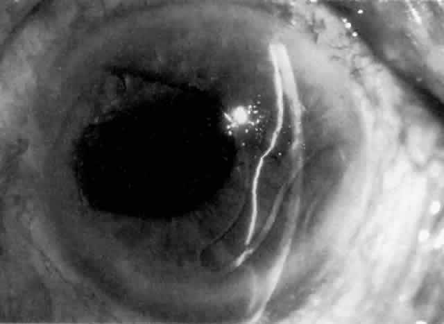
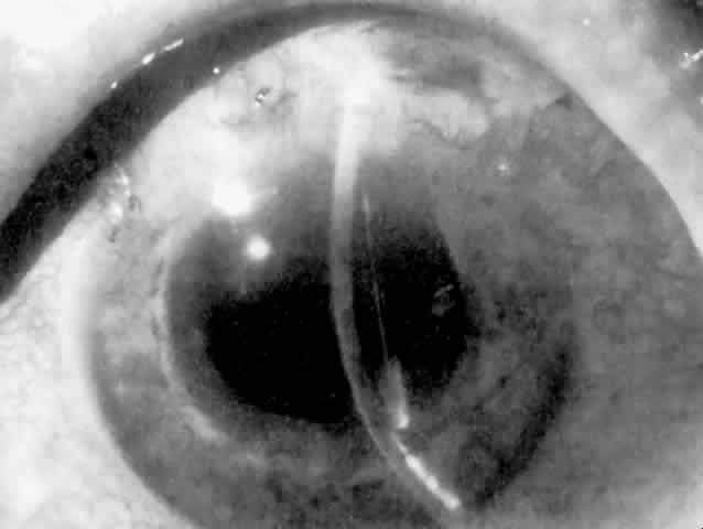
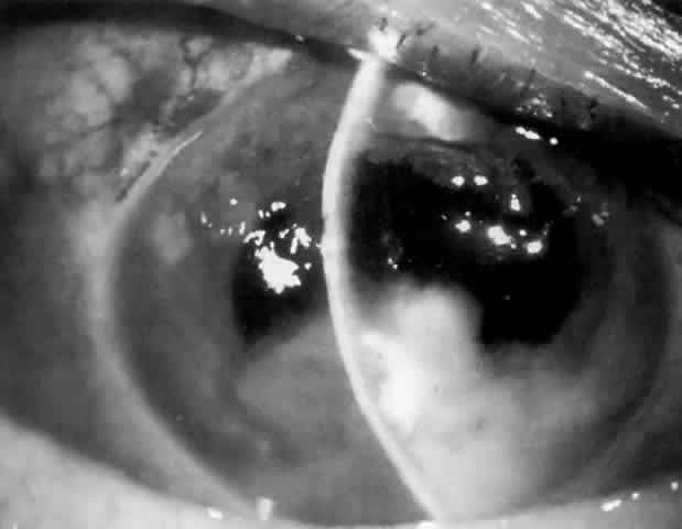
MANAGEMENT. The postoperative IOP elevation that occurs with the use of viscoelastic agents is exacerbated by cortical remnants and blood (Fig. 1). Careful cortical cleanup and aspiration of the viscoelastic agent may reduce the degree of IOP elevation. Prophylactic treatment of all patients (particularly those with preexistent glaucoma) with carbonic anhydrase inhibitors or beta-adrenergic blockers may decrease the incidence or severity of postoperative IOP elevation.35–38

Fig. 1. Hyphema and suspended blood in sodium hyaluronate (Healon) after extracapsular cataract extraction with posterior chamber lens implantation and trabeculectomy. Intraocular pressure is elevated, and there is microcystic corneal edema.

Hyphema

Most postoperative hemorrhages associated with cataract extraction or combined procedures are self-limited. The source of the hemorrhage can be the edge of the corneoscleral wound or the iridectomy. Unless there is a significant amount of blood, debris, or the presence of a viscoelastic substance in the anterior chamber, these early postoperative hyphemas are asymptomatic and not associated with elevated IOP. The mechanism of IOP elevation is obstruction of the trabecular meshwork by one or more of the following: blood, debris, macrophage, and clot. These substances are usually resorbed spontaneously without treatment.

MANAGEMENT. Unless resorption is delayed by excessive debris or compromised preexistent outflow, medical management is employed. Pharmacologic agents used are carbonic anhydrase inhibitors or beta-adrenergic blockers. To minimize iris movement and treat the often attendant intraocular inflammation, cycloplegics and corticosteroids can be of benefit. Surgical evacuation is necessary only if the glaucoma is unresponsive to tolerated medical therapy or there is risk of corneal blood staining.

Inflammation and Lens Particle Glaucoma

Cellular and chemical mediators of inflammation decrease trabecular meshwork function and outflow by a variety of mechanisms and in varying degrees of severity. Both cellular and particulate inflammatory debris can clog trabecular meshwork and compromise outflow.39,40 Additionally, the increased aqueous protein concentration caused by the breakdown of the blood-aqueous barrier also contributes to early postoperative IOP elevation. Each of these inflammatory components are further exacerbated by the presence of any residual viscoelastic substance in the anterior chamber.23 Contrary to decreasing outflow facility, the role that prostaglandins play in postoperative IOP elevation may relate to their effect on increasing aqueous production.

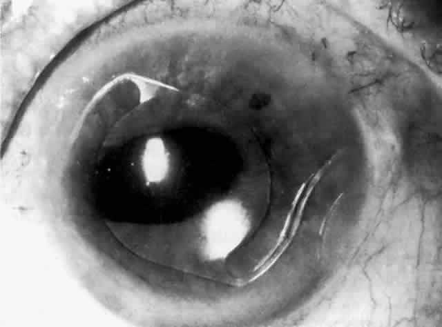
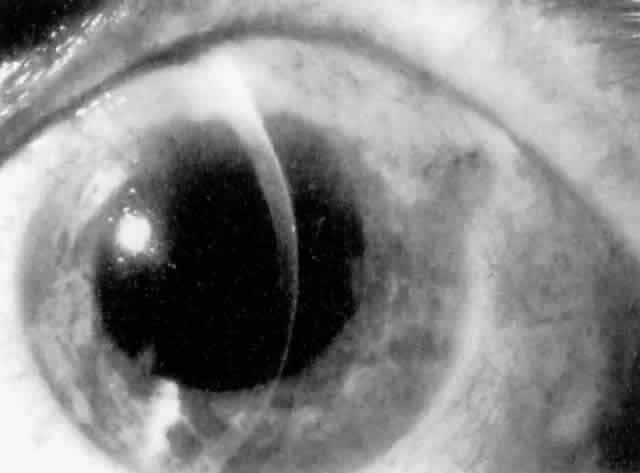
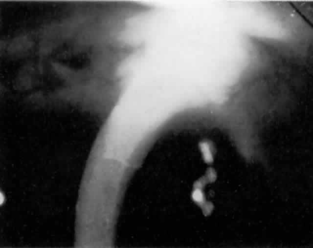
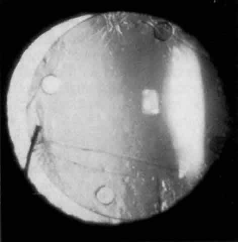
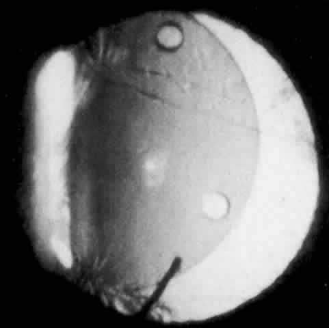
Cortical lens fragments retained in either the anterior chamber or the vitreous can also obstruct the trabecular meshwork in the form of free lens particles or macrophages swollen with lens material (Figs. 2 and 3). Glaucoma does not occur in all eyes that contain cortical remnants; the inflammatory response may be more pronounced and prolonged in eyes containing a higher amount of lens material. When inflammation is marked, keratic precipitates and sometimes a hypopyon may be present. Distinction between this sterile inflammatory endophthalmitis and infectious endophthalmitis can be difficult and may depend on the initial response to therapy. The presence or absence of IOP elevation is not helpful in making this distinction because IOP may be normal or elevated in both situations.

Fig. 2. Fluffy, hydrated cortical lens fragments retained in the anterior chamber after cataract extraction, resulting in inflammation and elevated intraocular pressure.

Fig. 3. Patient with marked inflammation and elevated postoperative intraocular pressure after phacoemulsification and lens implantation. Ultrasonographic evaluation revealed retained cortical and nuclear fragments seen on B-scan and a sharp spike on A-scan, indicating the highly reflective retained nuclear fragment. (Courtesy of Dr. E. Hodapp, Miami, FL.)

MANAGEMENT. Inflammation is likely to occur to some degree after cataract extraction. The degree to which this will affect postoperative IOP depends to some extent on preexistent trabecular meshwork function and the severity of the inflammatory response. In general, treatment is aimed at controlling the inflammation with corticosteroids. Aspirin and nonsteroidal anti-inflammatory agents (e.g., indomethacin) may also help to control postoperative inflammation.

Management of elevated IOP relies on treatment with beta-adrenergic blockers and carbonic anhydrase inhibitors. Because of the disruption of the blood-aqueous barrier, hyperosmotic agents are of limited utility in the postoperative patient with inflammation, and miotics should be avoided because they further disturb the blood-aqueous barrier and promote formation of posterior synechiae. Mydriatics or cycloplegics to decrease iris movement and prevent formation of posterior synechiae are recommended if inflammation is marked. If posterior synechiae do develop in the inflamed postoperative eye, attempts should be made to break these with dilation to avoid pupillary block as a result of a secluded pupil. If pharmacologic mydriasis is unsuccessful, peripupillary iridoplasty may achieve photomydriasis.41 Once iris bombé does develop, however, a laser iridotomy is indicated if vigorous mydriasis is unsuccessful.

Lens particle glaucoma treatment is similarly aimed at the underlying inflammation. The glaucoma usually resolves as the retained cortical material resorbs and inflammation decreases. Beta-adrenergic blockers and carbonic anhydrase inhibitors are of temporary help in controlling this transient IOP elevation. If spontaneous resorption is prolonged or medical therapy either fails or is not well tolerated, surgical removal of the residual cortex is necessary.

Vitreous in the Anterior Chamber

Anterior chamber vitreous prolapse can occur after an ICCE or after inadvertent rupture or dialysis of the posterior capsule during ECCE. In the case of phacoemulsification or planned ECCE, vitreous prolapse is often associated with other IOP-provoking elements, such as inflammatory debris or blood.42,43 The mechanism of open-angle postoperative glaucoma in this setting may be related to the degree of inflammation and blood in the anterior chamber; it may also be complicated by a direct obstruction of the trabecular meshwork by vitreous fibrils.44

MANAGEMENT. Management is most often medical rather than surgical. Beta-adrenergic blockers and carbonic anhydrase inhibitors are used to control the IOP elevation. Cycloplegics combined with hyperosmotics may help draw the vitreous from the anterior chamber angle. Many cases spontaneously resolve in several months as the vitreous recondenses and retracts from the angle. Anterior vitrectomy may be required if medical management is unable to control the IOP.

Trabecular Meshwork Dysfunction and Anterior Chamber Angle Distortion

The gonioscopic appearance of the corneoscleral incision after cataract extraction has been well described by Kirsch and co-workers.45,46 A white ridge resembling an “inverted snow bank” lines the inner margin of the corneoscleral incision. For approximately the first 2 weeks after cataract extraction, this ridge is seen to obscure the trabecular meshwork and to distort the anterior chamber angle. The decrease in outflow facility improves with resolution of the corneal scleral ledge.47 Clear corneal incisions do not develop this healing ridge. In one series of 95 cataract extractions, early postoperative IOP elevation with evidence of this inner ridge developed in 23% of cases involving limbal incisions; postoperative IOP was unaffected in cases involving cataract extractions with corneal incisions.48 Theories regarding the pathogenesis of this incisional ridge range from tight corneoscleral sutures to corneal stromal and trabecular meshwork edema.49

MANAGEMENT. Management of reduced outflow follows the route taken for inflammation, since some portion of this reduced outflow may recover as the inflammation resolves.

Alpha-Chymotrypsin

Alpha-chymotrypsin is an enzyme with a selective propensity to lyse lens zonules. The glaucoma induced by alpha-chymotrypsin has been referred to as zonulolytic glaucoma. In two studies on the effects of alpha-chymotrypsin on IOP, it was found that IOP elevation usually occurred 2 to 5 days after ICCE in 27% of patients in whom alphachymotrypsin was used for zonulolysis.50,51 IOP elevation is more common in patients with preexisting open-angle glaucoma. Tonographic studies have demonstrated a transient decrease in aqueous outflow facility.52–54 No long-term alteration in outflow has been demonstrated in eyes with zonulolytic glaucoma unless there was preexistent glaucoma. Histologic studies of animal eyes have suggested that accumulation of lens zonule fragments in the trabecular meshwork produces a transient outflow obstruction.55–57

MANAGEMENT. Zonulolytic glaucoma resolves within 48 to 72 hours. Medical therapy with beta-adrenergic blockers and carbonic anhydrase inhibitors is usually adequate. Preoperative or intraoperative administration of acetylcholine, mannitol, pilocarpine, or corticosteroids does not prevent zonulolytic glaucoma.58 Intraoperative irrigation of the anterior chamber to remove zonular fragments does not affect the degree of elevated IOP, but the use of minimum volumes of the enzyme can be effective in decreasing the risk of its occurrence.

PERSISTENT OR LATE POSTOPERATIVE INTRAOCULAR PRESSURE ELEVATION

Irreversible Trabecular Meshwork Damage

After cataract extraction, the IOP usually returns to normal within 1 week. If, however, the patient has a history of dysfunction of the trabecular meshwork (e.g., preexistent glaucoma) this resolution may be prolonged. In any patient, the IOP elevation can become sustained or permanent as a result of any of the multiple insults of cataract surgery on the trabecular meshwork.

MANAGEMENT. The chronic glaucoma in aphakia or pseudophakia should initially be evaluated for other possible treatable causes of sustained IOP elevation (e.g., steroids, recurrent hemorrhage, persistent inflammation, angle closure). Therapy should initially be directed at these problems if identified.

Patients whose persistent IOP elevation is not directly treatable should be managed medically. Miotics, beta-adrenergic blockers, and carbonic anhydrase inhibitors may be effective. Epinephrine compounds have been associated with macular edema in pseudophakic and aphakic patients and therefore should be used only with extreme caution.59 Postoperative laser trabeculoplasty may be effective, but it is generally considered more successful when performed before cataract extraction.10,11 Surgery is reserved for patients who cannot be managed on maximum tolerable medical therapy.

Therefore, management follows the route taken for simple open-angle glaucoma, with the exception of filtration surgery. Filtering surgery for aphakia and pseudophakia has a lower success rate than that reported for phakic eyes. The Multicenter 5-Fluorouracil Filtering Surgery Study reported that 28% of eyes that received postoperative subconjunctival 5-fluorouracil required reoperation during the first 3 postoperative years for IOP control, compared with 60% of the standard-treatment group. Antimetabolites, 5-fluorouracil, and mitomycin C have become proven adjuncts in glaucoma surgery for aphakia and pseudophakia.60–65

Persistent Inflammation

Persistent inflammation after cataract surgery may indicate one of several possibilities: low-grade infection, IOL-related inflammation, residual lens material, or epithelial downgrowth. (See previous section on Inflammation and Lens Particle Glaucoma for a discussion of the effects of residual lens material.) Chronic low-grade infections can include fungal causes; however, these are rare and are associated with marked inflammation. A persistent low-grade infection associated with persistent, subtle inflammation may be due to Propionibacterium acnes infection. This bacterium may be sequestered in residual cortex, and since anterior chamber taps and cultures are often negative, the diagnosis may be difficult to confirm.

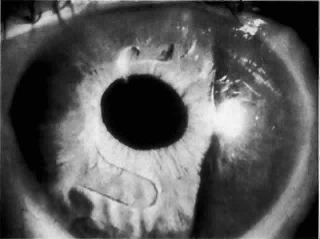
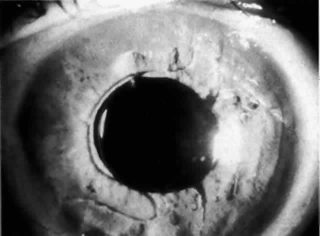
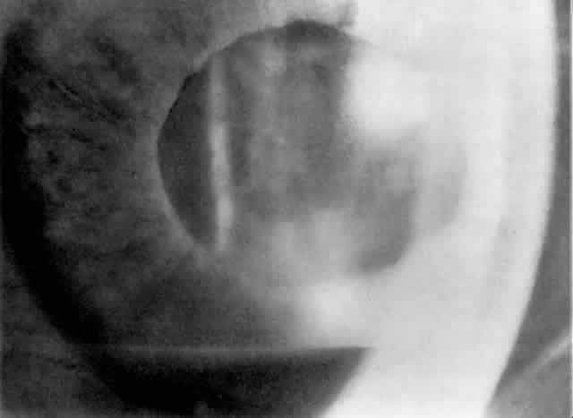
IOL-related inflammation is more commonly associated with some styles of lens implants than with others. Position of the lens or foreign material deposited on the lens during manufacture can also account for persistent inflammation. When the inflammation is associated with hyphema, it is termed UGH syndrome, which involves the triad of uveitis, glaucoma, and hyphema. UGH syndrome occurred more frequently in the 1970s, when iris-fixed lenses were commonly used.66,67 Today it is more often associated with anterior chamber lenses and is less likely to occur with posterior chamber lenses, although this has been reported.68,69 The mechanism is believed to be contact of the surface of the lens with the iris, causing mechanical irritation that may be enhanced by a poorly positioned or mobile lens implant. The loose posterior chamber lens may initially exhibit release of pigment due to friction on the posterior surface of the iris (pseudopigmentary glaucoma). This “windshield-wiper” effect alone can produce enough pigment to provoke an increase in IOP; however, it is the erosion into vascular tissue that results in hemorrhage, hyphema, and inflammation (Figs. 4 and 5).

Fig. 4. Posterior chamber lens implant with prolene haptics, with the patient's head in the primary position.

Fig. 5. Same posterior chamber lens implant as in Figure 4, but with the patient's head tilted, demonstrating mobility of the lens in the posterior chamber. (Courtesy of Dr. R. K. Parrish, Miami, FL.)

MANAGEMENT. Treatment of a P. acnes infection depends on the organism's being found or suspected when all taps and cultures are negative for other possible organisms. Removal of residual lens and capsule will usually resolve the situation, but if peripheral anterior synechiae have formed as a result of the persistent inflammation, the IOP elevation may require standard glaucoma management. IOL-related inflammation is also often reversible once the offending implant is removed. Some mild cases may be managed by minimizing iris movement against the lens caused by chronic use of mydriatics or cycloplegics. The IOP elevation will usually resolve unless persistent outflow obstruction exists in the form of synechiae closure or direct injury to the angle by the lens implant haptics.

Recurrent Hyphema

Hyphema without neovascularization of the iris can occur months or years after cataract extraction. This usually indicates vascularization of the cataract wound, Swan's syndrome,69 or erosion of a uveal vessel by an implant. Gonioscopic examinations can reveal a localized clot or vessels along the cataract incision site. These episodes are often transient and resolve with minimal treatment. The exception is IOL-related erosion of a uveal vessel, which can result in UGH syndrome.

MANAGEMENT. Management of each episode of acute IOP elevation in recurrent hyphema should be similar to that employed for hyphema in the immediate postoperative period. Treatment approaches include cycloplegia, corticosteroids, and aqueous suppressants. On rare occasions, if vascularization of the cataract wound is clear, argon laser ablation can be attempted. Treatment for IOL erosion consists of either cycloplegia to minimize iris movement or removal of the lens. It should be noted, however, that lens exchange or removal alone will not be sufficient to control the IOP when there is sufficient preexisting synechiae formation or trauma to the trabecular meshwork, in which case concomitant glaucoma surgery may need to be performed.

Ghost Cell Glaucoma

Ghost cell glaucoma can occur in both aphakic and pseudophakic eyes in which there is a vitreous hemorrhage and a disrupted vitreous face. Because of their reduced pliability compared with fresh red blood cells, tan or khaki cells obstruct outflow channels. IOP elevation, while self-limited, may persist longer than expected because of the large reservoir in the vitreous cavity. IOL-induced hemorrhage releasing blood cells into the vitreous cavity has resulted in ghost cell glaucoma, which can recur.70

MANAGEMENT. Ghost cell glaucoma is often self-limited with the exception of IOL-induced hemorrhage. Management requires aqueous suppressants, cycloplegia, and frequent examinations. Vitrectomy and anterior chamber irrigation may be necessary to eliminate the reservoir of ghost cells. Removing or repositioning the offending IOL implant may be combined with this procedure if the IOL is believed to have been the source of hemorrhage.

Persistent Release of Pigment: Pseudopigmentary Glaucoma

As mentioned in the Persistent Inflammation section, pseudopigmentary glaucoma is in many ways similar to the persistent inflammation that results from an IOL implant. The mechanism is believed to be contact of the surface of the lens with the iris. In persistent inflammation and UGH syndrome, typically either an anterior chamber lens or an iris-fixed lens is the culprit. With respect to pseudopigmentary glaucoma, however, the lens used is more likely to be a posterior chamber lens with the haptics (either one or both) positioned in the sulcus. In this way, the pigmented surface of the iris moves back and forth across the edge of the IOL, promoting release of pigment into the anterior chamber (see Figs. 4 and 5). The mechanism of IOP elevation is outflow obstruction from the excessive amounts of pigment granules and cell debris in the trabecular meshwork.

MANAGEMENT. Mild cases of pseudopigmentary glaucoma may be managed with aqueous suppressants and either mydriatics or miotics to minimize iris movement. Corticosteroids are not indicated. More severe cases may require IOL removal or replacement.

Back to Top
ANGLE-CLOSURE GLAUCOMA ASSOCIATED WITH APHAKIA AND PSEUDOPHAKIA

POSTOPERATIVE ANGLE CLOSURE WITH PUPILLARY BLOCK

Pupillary block glaucoma in aphakic or pseudophakic eyes usually develops in the immediate postoperative period. Adhesion of the pupil margin to the underlying IOL, capsule, or vitreous produces pupillary block. Apposition of the pupil to an overlying anterior chamber lens can also produce pupillary block without the development of adhesions. In an eye with pupillary block, the central chamber is of normal depth and the iris bulges forward from the pupil plane (iris bombé configuration). This is in contrast to ciliary block glaucoma, in which there is a shallow or flat central anterior chamber.

Pupillary Block and Intraocular Lens

Historically, iris-fixed lenses and anterior chamber lenses typically produced pupillary block in the early postoperative period. The presence of a surgical iridectomy may prevent pupillary block.71 It is recommended that at least one iridectomy be placed at the time of surgery if an anterior chamber lens is used. Diabetic patients may be predisposed to pupillary block even with the use of posterior chamber lens implants; thus, it is recommended that they have a surgically placed iridectomy. The mechanism of this predisposition in diabetic patients is unclear, but it may relate either to prolonged postoperative inflammation or to the increased thickness of the iris and ciliary body.72 The utility of routine iridectomy during cataract extraction with the placement of a posterior chamber lens implant is unclear. Many cataract surgeons advise against an iridectomy because of unnecessary trauma to the iris, which may be associated with hemorrhage or postoperative cystoid macular edema.73,74 It is unknown, however, how the potential benefits of routine primary surgical iridectomy weigh against the various risks.

MANAGEMENT. Laser peripheral iridectomy quickly resolves pupillary block (Figs. 6 and 7). Because of inflammation and iridocorneal contact peripherally, this procedure may be difficult to accomplish. If the view is inadequate or a laser is not available, pupillary dilation with phenylephrine will in some cases relieve pupillary block (Figs. 8 and 9). Ultimately the patient will need a laser peripheral iridectomy. If peripheral anterior synechiae have formed or the IOP elevation persists after relief of the pupillary block, the patient will require management similar to that used in chronic angle-closure glaucoma.

Fig. 6. Pupillary block with iris bombé around haptics of anterior chamber lens implant, resulting in elevated intraocular pressure.

Fig. 7. Resolution of pupillary block after laser peripheral iridectomy.

Fig. 8. Iris bombé nasal to anterior chamber lens implant before dilation (shadowed area on right). No peripheral iridectomy is present.

Fig. 9. Resolution of pupillary block and iris bombé after pupillary dilation. A subsequent laser peripheral iridectomy was performed.

Pupillary Block by Vitreous

The anterior hyaloid can occlude the pupillary aperture as well as an iridectomy.75,76 Often this results in the slow development of synechial closure and permanent angle-closure glaucoma. During the era in which ICCE was the predominant method of treatment, pupillary block by vitreous occurred frequently in cases that did not involve iridectomy. For this reason, basal iridectomies were usually placed at the time of surgery.77 With the advent of ECCE, pupillary block by vitreous has been described after neodymium:yttrium aluminum garnet (Nd:YAG) laser posterior capsulotomy.78,79

MANAGEMENT. Dilation rarely resolves pupillary block by vitreous. Laser iridotomy is frequently successful, but because the vitreous may occlude the iridectomy, it may become necessary to perform an anterior vitrectomy.80 If there are a significant number of recently formed peripheral anterior synechiae, this procedure may be combined with goniosyneolysis in an attempt to restore a functional angle. In rare cases, a pars plana vitrectomy is required.81

POSTOPERATIVE ANGLE CLOSURE WITHOUT PUPILLARY BLOCK

Ciliary Block

Ciliary block is also known as “malignant glaucoma” or “aqueous misdirection.” This condition results in angle closure caused by a misdirection of aqueous humor into the vitreous cavity as opposed to the posterior chamber due to resistance along the zonular-capsular-hyaloid face junction. In contrast to pupillary block, the central anterior chamber is shallow because the IOL or vitreous face is pushed forward, and a peripheral iridectomy will not prevent or resolve the acute angle closure. Ciliary block is more likely to occur in eyes with a small anterior segment and an anatomically narrow angle. Large posterior chamber IOLs (7-mm optic) in small, hyperopic eyes are also known to predispose patients to ciliary block.82

MANAGEMENT. Medical treatment consisting of cycloplegia and hyperosmotic agents is usually initiated first. Miotics are contraindicated. If the patient does not have a patent iridectomy, one should be performed to relieve any element of pupillary block. If medical management is ineffective, the posterior capsule and anterior hyaloid face should be disrupted with the Nd:YAG laser if possible (Figs. 10 and 11). If these maneuvers are not successful, pars plana vitrectomy is indicated.83,84 In a pseudophakic eye with ciliary block, IOL removal is not recommended.85

Fig. 10. Ciliary block after cataract extraction and posterior chamber lens implantation. The central chamber is shallow and the peripheral chamber flat. The intraocular lens is pushed forward, and the haptic posterior to the iris can be seen indenting the iris surface.

Fig. 11. After disruption of the posterior capsule and the anterior hyaloid face with the Nd:YAG laser, the chamber becomes immediately deep and the intraocular lens is no longer pressed against the iris. (Courtesy of Dr. E. Hodapp, Miami, FL.)

Suprachoroidal Hemorrhage

A large suprachoroidal hemorrhage may cause the ciliary body to rotate inward and rotate the iris-lens diaphragm forward. The subsequent angle closure is neither prevented nor relieved by iridectomy.

MANAGEMENT. Management is similar to that for ciliary block glaucoma, consisting of cycloplegia, hyperosmotics, and aqueous suppressants, as needed.

Epithelial Downgrowth

Epithelial downgrowth is characterized by a cellular membrane with an advancing edge on the posterior cornea and iris, elevated IOP, and large clumps of cells in the anterior chamber (Fig. 12). The mechanism of IOP elevation is outflow obstruction by the epithelial membrane and direct obstruction of the outflow channels by epithelial cells. The diagnosis can be confirmed with an argon laser to blanch the epithelium on the iris in the suspected area of involvement. Epithelial cells turn white when coagulated (Fig. 13).

Fig. 12. Advancing edge of epithelial growth appears as a faint line on the posterior cornea.

Fig. 13. Gonioscopic photograph of white, coagulated areas of epithelium on the surface of the iris after confirmatory treatment of the surface of the iris with argon laser. (Courtesy of Dr. E. Hodapp, Miami, FL.)

MANAGEMENT. Complex surgical procedures have been described for managing epithelial downgrowth. Treatment of the advancing epithelial membrane has been generally unsuccessful.86,87 Secondary glaucoma associated with downgrowth has recently benefited from the use of glaucoma drainage implants, such as the Molteno or Baerveldt implant.88

Back to Top
REFERENCES

1. Francois J: Aphakic glaucoma. Ann Ophthalmol 6:429, 1974

2. Duke-Elder S, Jay B: Post-operative glaucoma. In Duke-Elder S (ed): System of Ophthalmology, pp 716–723. St Louis, CV Mosby, 1976

3. Gross JG, Meyer DR, Robin AL et al: Increased intraocular pressure in the immediate post-operative period after extracapsular cataract extraction. Am J Ophthalmol 105:466, 1988

4. Kooner KS, Dulaney DD, Zimmerman TJ: Intraocular pressure following extracapsular cataract extraction and posterior chamber intraocular lens implantation. Ophthalmic Surg 19:471, 1988

5. Cinotti AA, Jacobson JH: Complications following cataract extraction. Am J Ophthalmol 36:929, 1953

6. Blodi FC: Failures of the cataract extractions and their pathologic explanation. J Iowa Med Soc 44:514, 1954

7. Hoskins HD: Management of pseudophakic glaucoma. In Greve EL (ed): Surgical Management of Coexisting Glaucoma and Cataract, p 41. Amsterdam, Kugler Publications, 1987

8. Worthen DM, Boucher JA, Burton JN et al: Interim FDA report on intraocular lenses. Ophthalmology 87:267, 1980

9. Kooner KS, Dulaney DD, Zimmerman TJ: Intraocular pressure following secondary anterior chamber lens implantation. Ophthalmic Surg 19:274, 1988

10. Brown SVL, Thomas JV, Belcher CD et al: Effect of cataract surgery on intraocular pressure reduction obtained with laser trabeculoplasty. Am J Ophthalmol 100:373, 1985

11. Thomas JV, Simmons RJ, Belcher CD: ALT in the presurgical glaucoma patient. Ophthalmology 89:187, 1982

12. Savage JA, Thomas JV, Belcher CD et al: Extracapsular cataract extraction and posterior chamber intraocular lens implantation in glaucomatous eyes. Ophthalmology 92: 1506, 1985

13. McGuigan LJB, Gottsch J, Stark WJ et al: Extracapsular cataract extraction and posterior chamber lens implantation in eyes with preexisting glaucoma. Arch Ophthalmol 104: 1301, 1986

14. Clayman HM, Jaffe NS, Light DS et al: Lens implantation, miosis and glaucoma. Am J Ophthalmol 87:121, 1979

15. Kooner KS, Dulaney DD, Zimmerman TJ: Intraocular pressure following ECCE and IOL implantation in patients with glaucoma. Ophthalmic Surg 19:570, 1988

16. Murchison JF Jr, Shields MB: An evaluation of three surgical approaches for coexisting cataract and glaucoma. Ophthalmic Surg 20:393, 1989

17. Shields MB: Combined cataract extraction and glaucoma surgery. Ophthalmology 89:231, 1982

18. Longstaff S, Wormald RPL, Mazover A, Hitchings RA: Glaucoma triple procedures: efficacy of intraocular pressure control and visual outcome. Ophthalmic Surg 21: 786, 1990

19. Naveh N, Kottass R, Glovinsky J et al: The long-term effect on intraocular pressure of a procedure combining trabeculectomy and cataract surgery, as compared with trabeculectomy alone. Ophthalmic Surg 21:339, 1990

20. Gillies WE, Brooks AMV: The results of combined cataract extraction and trabeculectomy using separate incisions. Aust NZ J Ophthalmol 20:239, 1992

21. Cohen JS: Combined cataract implant and filtering surgery with 5-fluorouracil. Ophthalmic Surg 21:181, 1990

22. Hennis HL, Stewart WC: The use of 5-fluorouracil in patients following combined trabeculectomy and cataract extraction. Ophthalmic Surg 22:451, 1991

23. Rich WJ: Further studies on early post-operative ocular hypertension following cataract extraction. Trans Ophthalmol Soc UK 89:639, 1969

24. Rich WJ, Radtke ND, Cohan BE: Early ocular hypertension after cataract extraction. Br J Ophthalmol 58:725, 1974

25. Hollands RH, Drance SM, Schulzer M: The effect of acetylcholine on early postoperative intraocular pressure. Am J Ophthalmol 103:749, 1987

26. Hollands RH, Drance SM, Schulzer M: The effect of intercameral carbachol on intraocular pressure after cataract extraction. Am J Ophthalmol 104:225, 1987

27. Cherfan GM, Rich WJ, Wright G: Raised intraocular pressure and other problems with sodium hyaluronate and cataract surgery. Trans Ophthalmol Soc UK 103:277, 1983

28. Glasser DB, Matsuda M, Edelhauser HF: A comparison of the efficacy and toxicity of and intraocular pressure response to viscous solutions in the anterior chamber. Arch Ophthalmol 104:1819, 1986

29. Passo MS, Ernest JT, Goldstick TK: Hyaluronate increases intraocular pressure when used in cataract extraction. Br J Ophthalmol 69:572, 1985

30. Handa J, Henry JC, Krupin T, Keates E: ECCE with posterior chamber lens implantation in patients with glaucoma. Arch Ophthalmol 105:765, 1987

31. Barron BA, Busin M, Page C et al: Comparison of the effects of Viscoat and Healon on postoperative intraocular pressure. Am J Ophthalmol 100:377, 1985

32. Aron-Rosa D, Cohn HC, Aron J-J, Bouquety C: Methylcellulose instead of Healon™ in extracapsular surgery with intraocular lens implantation. Ophthalmology 90:1235, 1983

33. Berson FG, Patterson MM, Epstein DL: Obstruction of aqueous outflow by sodium hyaluronate in enucleated human eyes. Am J Ophthalmol 95:668, 1983

34. Stamper RL, DiLoreto D, Schacknow P: Effect of intraocular aspiration of sodium hyaluronate on postoperative intraocular pressure. Ophthalmic Surg 21:486, 1990

35. Pape LG, Balasz EA: The use of sodium hyaluronate (Healon™) in human anterior segment surgery. Ophthalmology 87:699, 1980

36. Lewen R, Insler MS: The effect of prophylactic acetazolamide on intraocular pressure rise associated with Healon-aided intraocular lens surgery. Ann Ophthalmol 17:315, 1985

37. Percival SPB: Complications from use of sodium hyaluronate (Healonid) in anterior segment surgery. Br J Ophthalmol 66:714, 1982

38. Haiman MK, Phelps CD: Prophylactic timolol for the prevention of high intraocular pressure after cataract extraction: a randomized, prospective, double-blind trial. Ophthalmology 88:233, 1981

39. Melamed S: Alterations of trabecular aqueous flow after cataract extraction. Ophthalmic Surg 18:878, 1987

40. Gormaz A: Ocular tension after cataract surgery. Am J Ophthalmol 53:832, 1969

41. Naveh-Floman N, Rosner M, Blumenthal M: Pseudophakic pupillary block glaucoma with posterior-chamber intraocular lens. Glaucoma 7:262, 1985

42. Samples JR, Van Buskirk EM: Open-angle glaucoma associated with vitreous humor filling the anterior chamber. Am J Ophthalmol 102:759, 1986

43. Simmons RJ: The vitreous in glaucoma. Trans Ophthalmol Soc UK 95:422, 1975

44. Grant WM: Open-angle glaucoma with vitreous filling the anterior chamber following cataract extraction. Trans Am Ophthalmol Soc 61:196, 1963

45. Kirsch RE, Levine O, Singer JA: Ridge at internal edge of cataract incision. Arch Ophthalmol 94:2098, 1976

46. Kirsch RE, Levine O, Singer JA: Further studies on the ridge at the internal edge of the cataract incision. Trans Am Acad Ophthalmol Otolaryngol 83:224, 1977

47. Lee PF, Trotter RR: Tonographic and gonioscopic studies before and after cataract extraction. Arch Ophthalmol 58:407, 1957

48. Rothkoff L, Biedner B, Blumenthal M: The effect of corneal section on early increased intraocular pressure after cataract extraction. Am J Ophthalmol 85:337, 1978

49. Campbell DG, Grant WM: Trabecular deformation and reduction of outflow facility due to cataract and penetrating keratoplasty sutures. ARVO abstracts. Invest Ophthalmol Vis Sci 16(suppl):126, 1977

50. Kirsch RE: Glaucoma following cataract extraction associated with use of alpha-chymotrypsin. Arch Ophthalmol 72:612, 1964

51. Lantz JM, Quigley JH: Intraocular pressure after cataract extraction: effects of alpha-chymotrypsin. Can J Ophthalmol 8:339, 1973

52. Kirsch RE: Further studies on glaucoma following cataract extraction associated with the use of alpha-chymotrypsin. Trans Am Acad Ophthalmol Otolaryngol 69:1011, 1965

53. Galin MA, Barasch KR, Harris LS: Enzymatic zonulolysis and intraocular pressure. Am J Ophthalmol 61:690, 1966

54. Jocson VL: Tonography and gonioscopy before and after cataract extraction with alpha-chymotrypsin. Am J Ophthalmol 60:318, 1965

55. Ley AP, Holmberg AS, Yamashita T: Histology of zonulysis with alpha chymotrypsin employing light and electron microscopy. Am J Ophthalmol 49:67, 1960

56. Anderson DR: Experimental alpha chymotrypsin glaucoma studied by scanning electron microscopy. Am J Ophthalmol 71:470, 1971

57. Anderson DR: Scanning electron microscopy of zonulolysis by alpha chymotrypsin. Am J Ophthalmol 71:619, 1971

58. Bloomfield S: Failure to prevent enzyme glaucoma. Am J Ophthalmol 65:405, 1968

59. Kolker AE, Becker B: Epinephrine maculopathy. Arch Ophthalmol 79:552, 1968

60. Gressel MG, Parrish RK II, Folberg R: 5-Fluorouracil and glaucoma filtering surgery: I. An animal model. Ophthalmology 91:378, 1984

61. Heuer DK, Parrish RK II, Gressel MG et al: 5-Fluorouracil and glaucoma filtering surgery: II. A pilot study. Ophthalmol 91:384, 1984

62. Chen CW, Huang HT, Bair JS, Lee CC: Trabeculectomy with simultaneous topical application of mitomycin C in refractory glaucoma. J Ocul Pharmacol 6:175, 1990

63. The Fluorouracil Filtering Surgery Study Group: Three-year follow-up of the fluorouracil filtering surgery study. Am J Ophthalmol 115:82, 1993

64. Kitazawa Y, Kawase K, Matsushita H, Minobe M: Trabeculectomy with mitomycin: a comparative study with fluorouracil. Arch Ophthalmol 109:1693, 1991

65. Palmer SS: Mitomycin as adjunct chemotherapy with trabeculectomy. Ophthalmology 98:317, 1991

66. Choyce DP: Complications of the AC implants of the early 1950's and the UGH or Ellingson syndrome of the late 1970's. Am Intraocular Implant Soc J 4:22, 1978

67. Ellingson FT: The uveitis-glaucoma-hyphema syndrome associated with the Mark-VIII anterior chamber lens implant. Am Intraocular Implant Soc J 4:50, 1978

68. Percival SPB, Das SK: UGH syndrome after posterior chamber lens implantation. Am Intraocular Implant Soc J 9:200, 1983

69. Swan KC: Hyphema due to wound vascularization after cataract extraction. Arch Ophthalmol 89:87, 1973

70. Summers CG, Lindstrom RL: Ghost cell glaucoma following lens implantation. Am Intraocular Implant Soc J 9: 429, 1983

71. Cohen JS, Osher RH, Weber P: Complications of extracapsular cataract surgery: the indications and risks of peripheral iridectomy. Ophthalmology 91:826, 1984

72. Weinreb RN, Wasserstrom JP, Forman JS: Pseudophakic pupillary block and angle-closure glaucoma in diabetic patients. Am J Ophthalmol 1102:325, 1986

73. Schulze RR, Copeland JR: Posterior chamber intraocular lens implantation without peripheral iridectomy: a preliminary report. Ophthalmic Surg 13:567, 1982

74. Simel PF: Posterior chamber implants without iridectomy. Am Intraocular Implant Soc J 8:141, 1982

75. Shaffer RN: The role of vitreous detachment in aphakic and malignant glaucoma. Trans Am Acad Ophthalmol Otolaryngol 58:217, 1954

76. Reese AB: Herniation of the anterior hyaloid membrane following uncomplicated intracapsular cataract extraction. Trans Am Ophthalmol Soc 46:73, 1948

77. Swan KC: Relationship of basal iridectomy to shallow chamber following cataract extraction. Arch Ophthalmol 69:191, 1963

78. Ruderman JM, Mitchell PG, Draff M: Pupillary block following Nd:YAG laser capsulotomy. Ophthalmic Surg 14: 418, 1983

79. Shrader CE, Belcher CD III, Thomas JV: Acute glaucoma following Nd:Yag laser membranotomy. Ophthalmic Surg 14:1015, 1983

80. Cotlier E, Herman S: Aphakic flat anterior chamber: treatment by anterior vitriotomy. Arch Ophthalmol 86:506, 1971

81. Peyman GA, Sanders DR, Minatoya H: Pars plana vitrectomy in the management of pupillary block glaucoma following irrigation and aspiration. Br J Ophthalmol 62:336, 1978

82. Reed JE, Thomas JV, Lytle RA et al: Malignant glaucoma Induced by an intraocular lens. Ophthalmic Surg 21:177, 1990

83. Lynch MG, Brown RH, Michels RG: Surgical vitrectomy for pseudophakic malignant glaucoma. Am J Ophthalmol 102:149, 1986

84. Tomey KF, Senft SH, Antonio SR et al: Aqueous misdirection and flat chamber after posterior chamber implants with and without trabeculectomy. Arch Ophthalmol 105:770, 1987

85. Sheppard DD: The fate of eyes from which intraocular lenses have been removed. Ophthalmic Surg 10:58, 1979

86. Maumenee AE: Treatment of epithelial downgrowth and intraocular fistula following cataract extraction. Trans Am Ophthalmol Soc 62:153, 1964

87. Maumenee AE, Paton D, Morse PH: Review of 40 histologically proven cases of epithelial downgrowth following cataract extraction and suggested surgical management. Am J Ophthalmol 69:598, 1970

88. Fish LA, Heuer DK, Baerveldt G: Molteno implantation for secondary glaucoma associated with advanced epithelial ingrowth. Ophthalmology 97:557, 1990

Back to Top