Chapter 54B
Exfoliation Syndrome, Pigment Dispersion Syndrome, and the Associated Glaucomas
GREGORY L. SKUTA and REBECCA K. MORGAN
Main Menu   Table Of Contents

Search

EXFOLIATION SYNDROME AND EXFOLIATIVE GLAUCOMA
PIGMENT DISPERSION SYNDROME AND PIGMENTARY GLAUCOMA
REFERENCES

EXFOLIATION SYNDROME AND EXFOLIATIVE GLAUCOMA

HISTORY AND NOMENCLATURE

In 1917, Lindberg described grayish or bluish flakes of material on the pupillary border in some patients with glaucoma.1 Vogt later hypothesized that this material represented degenerative changes of the lens capsule followed by secondary desquamation and proposed the term senile exfoliation of the lens capsule.2 Busacca argued that the exfoliative material represented deposition of material formed elsewhere in the eye rather than degenerative changes of the lens capsule.3 Dvorak-Theobald subsequently showed that exfoliative material differed histochemically from lens capsule and, to differentiate this condition from true exfoliation of the lens capsule secondary to infrared exposure, suggested the term pseudoexfoliation of the lens capsule.4 Subsequent electron microscopic studies by Ashton and associates and Bertelsen and coworkers indicate that the anterior lens capsule was directly affected in this disorder.5,6 Bertelsen and associates suggest that preequatorial lens epithelial cells produced the abnormal fibrillar substance and recommend the term fibrillopathia epitheliocapsularis.6 Eagle and colleagues, who believe that the material represented abnormal basement membrane secretions, have called this condition basement membrane exfoliation syndrome.7

The terms exfoliation syndrome and pseudoexfoliation syndrome are now most commonly used to designate this disorder and are used interchangeably in current literature. However, since recent ultrastructural studies indicate that the material on the lens capsule is derived, at least in part, from the lens, it is proposed that the disorder be called exfoliation syndrome (XFS).8–10

EPIDEMIOLOGY

Although XFS has been well described in patients of Scandinavian heritage, it also has been documented in numerous populations, including other Europeans,11 the Japanese,12 Australian aborigines,13 Navaho Indians,14, individuals of Mediterranean origin,15 natives of India16 and Pakistan,17 and the southern Bantu tribe of South Africa.18 It is reported, but is relatively rare, among African Americans.19,20

Wide variations in the prevalence of XFS have been reported. These variations may result, at least in part, from differences in the definition of disease, populations studied, age and sex distributions, and examination techniques.21 In one study of 2958 patients older than 60 years of age examined by a single investigator in three countries, Aasved reports respective prevalence rate of exfoliation of 4.0% in England, 4.7% in Germany, and 6.3% in Norway.11 Among Australian aborigines older than 60 years of age, Taylor and coworkers note a 16.3% prevalence rate of exfoliative changes.13 In the United States, a prevalence rate of 1.6% among patients older than 30 years of age and 3.2% for those older than 60 years of age was noted in one study,22 and in another the prevalence rate was 0.7% for those aged 52 to 64, 2.6% for ages 65 to 74, and 5.0% for ages 75 to 85.21

The reported prevalence of increased intraocular pressures and glaucoma in XFS are variable, again depending on the populations evaluated. Kozart and Yanoff report that of 100 consecutive patients with XFS in Philadelphia, glaucoma was present in 7% and ocular hypertension (intraocular pressure above 22 mmHg without optic nerve cupping or visual field loss) was present in 15%.23 According to Henry and colleagues, the cumulative probability of elevated intraocular pressures developing in XFS eyes previously with normal intraocular pressures was 5.3% over 5 years and 15.4% over 10 years.24 Hensen and Sellevold note that glaucoma may develop in 7% to 20% of XFS eyes previously without glaucoma over 5 years and 9% to 24% of these eyes over 10 years.25 The lower figures refer to progression in women and the higher figures to progression in men. These investigators further note that in bilateral XFS and unilateral glaucoma, the second eye developed glaucoma within 5 years in 21% to 26% of cases.

Among patients with open-angle glaucoma, the prevalence rate of XFS varies widely with the geographic location. In the United States, reported figures range from 1% to 12%.9,20,23,26 A prevalence rate of 26% to 75% has been noted in various Scandinavian countries.27–29 Figures from other areas include a 46.9% prevalence rate in eastern Turkey,30 44.5% in northwestern Spain,31 and 1.4% in a South African white population.32

Exfoliation syndrome is predominantly a disorder of the elderly and affects both men and women. Although it has been described in patients as young as 17 years of age,33 it is rare among those younger than 40 years. The reported mean age of onset of this condition ranges from the late 60s to 70s, and the prevalence increases with increasing age.21,34 Although some studies show that exfoliation is more common among men,16,35 several large American and European series indicate that exfoliative changes are more common in women.18,19,34 However, among patients with exfoliative changes, the likelihood of the development of increased intraocular pressures and glaucoma appears to be greater in men.36–38

Some cases of exfoliation may be hereditary. However, no clear inheritance pattern of XFS has been described, and most cases appear to be sporadic.39,40 An epidemiologic study by Ringvold and coworkers reveals a higher than expected prevalence of XFS among both spouses of married couples, suggesting an environmental influence on the distribution of the disorder.41 Ringvold and colleagues also considered an infectious etiology, such as a slow virus, in the pathogenesis of exfoliation syndrome.10,41 Additionally, Küchle and Naumann report the occurrence of exfoliation in three eyes of two young patients 4 to 6 years after penetrating keratoplasty, raising the possibility that XFS is a transmissible disease (from donor buttons to recipients).42

CLINICAL FEATURES

Lens

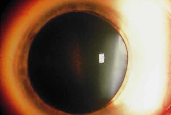
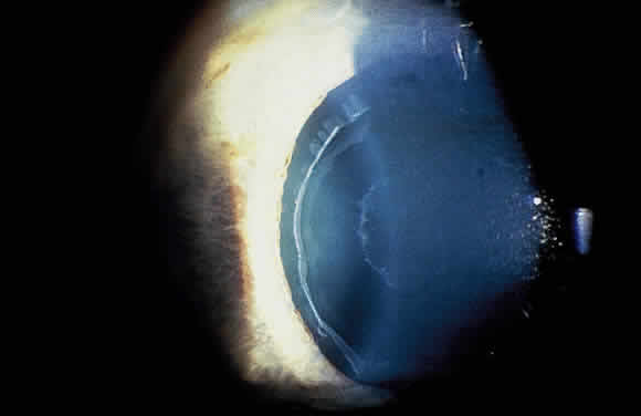
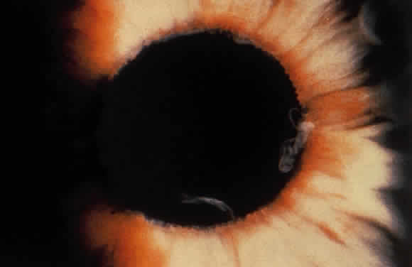
Deposition of exfoliative material on the anterior lens surface is the most commonly recognized feature of XFS and usually is best appreciated after pupillary dilation.43 A bull's-eye pattern generally is seen in which a translucent central zone and a granular peripheral zone of deposition are separated by an intermediate clear zone (Fig. 1). The translucent central zone of exfoliative material varies in diameter and may exhibit curled edges. In approximately 20% of patients with exfoliation, the central zone may be absent.38 The intermediate clear zone presumably results from lens contact with movement of the iris. The granular peripheral zone, which may be subtle but invariably is present in XFS, generally exhibits a well-delineated inner border and often shows numerous radial striations (Fig. 2). Occasionally, a bridge of exfoliative material may cross the clear zone to join the central zone and the granular peripheral zone.38,44

Fig. 1. Typical bull's-eye pattern of exfoliation on lens surface showing separation of central and peripheral zones of deposition by intermediate clear zone.

Fig. 2. Granular peripheral zone with well-defined inner border and numerous radial striations.

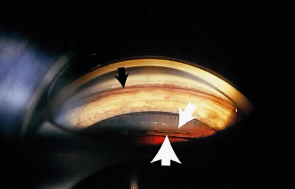
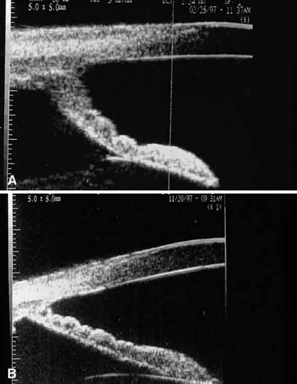
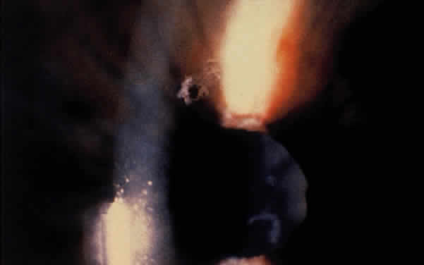
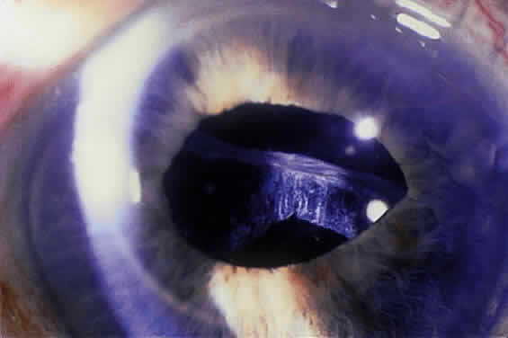
Some patients with XFS exhibit phacodonesis and subluxation of the lens, apparently resulting from degenerative changes in the zonular fibers.45–47 (Fig. 3) Spontaneous dislocation of the lens into the vitreous has been reported.48 In 1970, Bartholomew described 22 spontaneously displaced lenses in 19 patients with the disorder.45 Sixteen of the lenses were subluxated inferiorly. In these eyes, he visualized superior zonular fibers that were coated with exfoliative material and further noted that the zonular breaks generally were present at the ciliary body attachments and not at the lens, leading him to hypothesize that the main degenerative process occurred at the insertion of the zonular fibers to the basement membrane of the ciliary body epithelium. By electron microscopic examination, Schlötzer-Schrehardt and Naumann noted disruption of zonular fiber structure at the ciliary body insertion sites in eyes with XFS but also observed abnormalities at the attachments to the anterior lens capsule.46

Fig. 3. Subluxated lens in an eye with exfoliation syndrome. (Courtesy of Dr. Ronald Gross, Cullen Eye Institute, Houston, TX)

Bartholomew also describes phacodonesis in XFS and believes that this represents a probable sign of incipient lens displacement.49 On electron microscopic studies, Futa and Furuyoshi demonstrated marked degenerative changes in the lens zonules of XFS eyes with phacodonesis compared with normal zonular structure in those with no phacodonesis.47 Clinically, iridodonesis in association with phacodonesis may not necessarily be observed because of iris infiltration by exfoliative material or, perhaps, prolonged miotic therapy.

Iris and Pupil

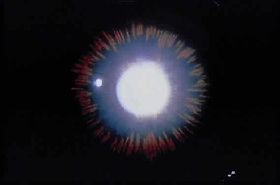
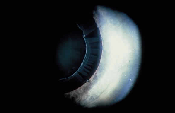
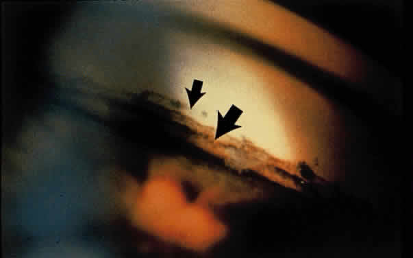
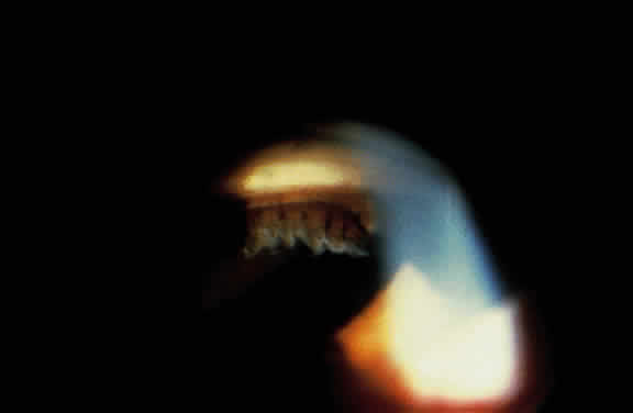
Exfoliative material frequently is observed at the pupillary border (Fig. 4) and should suggest the diagnosis of exfoliation even before the pupil is dilated.27 In addition, defects of the pigmented pupillary ruff are commonly seen. Aasved reports pupillary defects in 6.1% of eyes without XFS and in 74% of eyes with XFS.50 He further notes that in patients with unilateral XFS, pupillary ruff defects were twice as common in the affected eyes when compared with the fellow eyes. Iris transillumination defects in a moth-eaten pattern often are observed. Although these generally are limited to the region of the sphincter (Fig. 5), several patients also have diffuse midperipheral defects.51

Fig. 4. Accumulation of white exfoliative material at pupillary border. (Van Baskirk EM: Clinical Atlas of Glaucoma, p 53. Philadelphia, WB Saunders, 1986)

Fig. 5. Moth-eaten pattern of peripupillary iris transillumination defects seen in exfoliation syndrome.

Iris fluorescein angiographic studies in eyes with exfoliation disclose iris hypoperfusion, fewer radial iris vessels, and loss of the normal pattern of these vessels.52 Microneovascularization of the iris and associated fluorescein leakage also have been observed.53 These findings may result from tissue hypoxia caused by abnormal extracellular matrix formation or vascular abnormalities.54,55 Whether tissue hypoperfusion contributes to the development of these abnormalities or is a secondary effect of the XFS is not known. In support of the former are studies that show an increased frequency of XFS in eyes with a history of transient ischemic attacks56,57 and an increased prevalence in patients with a history of vascular disease.58

The iris hypoxia in this disorder may lead to atrophy of the iris pigment epithelium, resulting in particulate pigment deposition on the anterior iris surface seen in some eyes with exfoliation.55 Prince and Ritch note that the pigment often is found in a whorl-like pattern on the sphincter and is most commonly found over the inferior portion of the sphincter, although it also may be diffusely scattered over the peripheral iris surface.43

Typically, the pupil dilates poorly in an exfoliative eye. This likely is partly caused by atrophy and degeneration of the iris muscle cells55,59 and may be another association with iris hypoperfusion. Accumulated stromal iris exfoliative deposits also may contribute to poor mydriasis.

Cornea

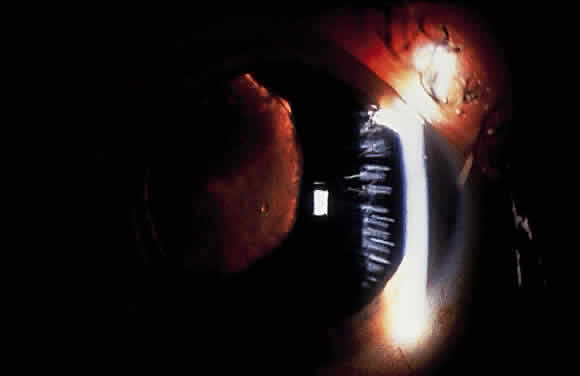
Slit-lamp examination of the cornea may reveal flakes or clumps of exfoliative material deposited on the endothelium43 (Fig. 6). On ultrastructural examination, Schlötzer-Schrehardt and associates note incorporation of the material into Descemet's membrane, leading them to believe that degenerated endothelial cells could be a source of the deposits.60 Pigment on the endothelium also may be observed. It usually is diffuse and unevenly distributed but rarely forms a Krukenberg spindle. Decreased endothelial cell counts and morphologic abnormalities of endothelial cells in eyes with exfoliation, as well as uninvolved fellow eyes of apparent unilateral cases, also have been reported.61–63

Fig. 6. Exfoliative material on the corneal endothelium.

Conjunctiva

Clinically, the conjunctiva does not appear to be involved in XFS. However, conjunctival biopsy specimens reveal the presence of exfoliative material,64–67 and fluorescein angiographic studies show loss of the normal limbal vascular patterns, congestion of the anterior ciliary vessels, and possible evidence of neovascularzation.68

Angle and Anterior Chamber

Exfoliation syndrome and pigment dispersion syndrome (PDS) are the two most common causes of increased pigmentation of the trabecular meshwork. The increased trabecular meshwork pigmentation in XFS tends to be less distinct and more patchy than the dense homogeneous deposition of trabecular pigment in PDS43 (Fig. 7). Clinically, the degree of pigmentation in XFS appears to be correlated with the severity of the glaucoma.26,69

Fig. 7. Patchy hyperpigmentation of trabecular meshwork (large arrow) in exfoliation syndrome. Faint pigment on Schwalbe's line represents a Sampaolesi's line (small arrow). Also notice exfoliative deposits in angle. (Van Baskirk EM: Clinical Atlas of Glaucoma, p 53. Philadelphia, WB Saunders, 1986)

Pigment also may be deposited on or anterior to Schwalbe's line (Sampaolesi's line). Occasionally, flakes of exfoliative material may be present in the anterior chamber angle (see Fig. 7). Although the anterior chamber angle usually is open in XFS, acute and chronic angle closure and occludable angles may be seen.9,69–71

Pupillary dilation in XFS may result in marked pigment dispersion into the anterior chamber. This may be accompanied by a substantial increase in the intraocular pressure,43 presumably resulting from decreased aqueous outflow caused by the dispersed pigment. Although pigment dispersion after dilation may occur in patients without XFS, Krause and colleagues have shown that the pigment released after dilation is substantially greater in XFS than in normal persons.72

The dispersed iris pigment on the anterior iris surface, in the angle, and in the anterior chamber after dilation arises from the iris pigment epithelium.73 As mentioned previously, iris hypoxia may result in pigment epithelial atrophy and subsequent pigment dispersion,55 but iris contact with a rough anterior lens surface also may contribute to this phenomenon.

Ciliary Body and Zonules

Exfoliative material also may be seen on the ciliary processes and zonules (Fig. 8). Using a modified method of gonioscopy known as cycloscopy, Mizuno and Muroi documented exfoliative material on the zonules and ciliary processes in all 31 eyes with clinical evidence of exfoliation.12 They further note that in patients with clinically unilateral exfoliation, more than half (17 of 31) of the fellow eyes demonstrated exfoliative material on the ciliary processes and zonules, thus establishing subclinical evidence of exfoliation in clinically “uninvolved” eyes.

Fig. 8. Exfoliative material on ciliary processes as seen through sector iridectomy in aphakic eye.

HISTOPATHOLOGY

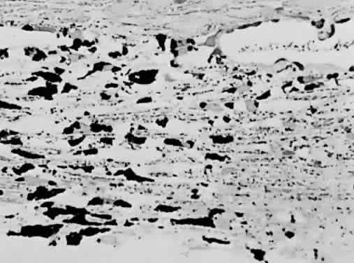
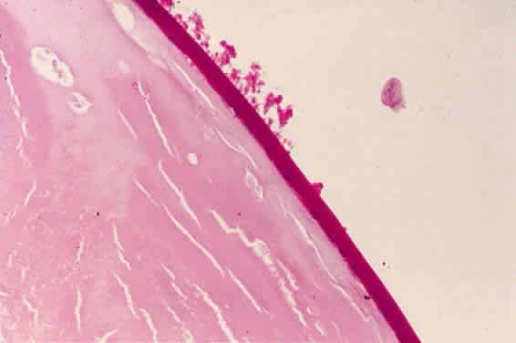
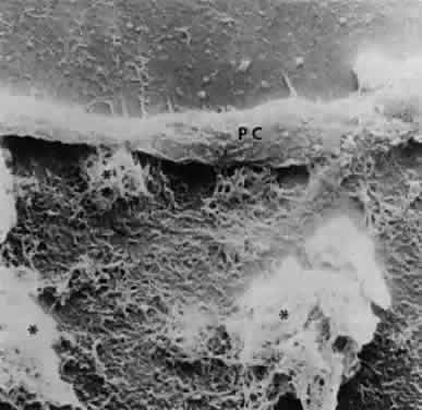
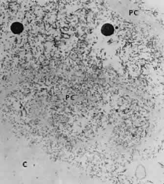
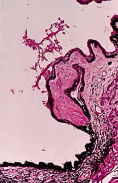
Accumulation of exfoliative material may be detected histopathologically throughout the anterior segment to include the lens, iris, trabecular structures, conjunctiva, ciliary body, and zonules. The deposition of fibrillar eosinophilic material on the anterior lens capsule is the classic histopathologic feature of XFS (Fig. 9). Ultrastructural studies also indicate some degree of actual exfoliative changes or peeling of the anterior lens capsule74 (Figs. 10 and 11). In addition, Ashton and associates describe a degenerative band containing exfoliative material within the inner half of the lens capsule.5 Bertelsen and coworkers independently noted projection of coarse fibrils from the lens surface into the deep portion of the lens capsule to form an amorphous layer of the lens capsule between the lens epithelium and normal lens capsule.6 Thus, studies indicate that exfoliative material accumulates on the anterior lens surface but also that the lens capsule itself may be involved, and that the underlying epithelial layer may at least partially contribute to the production of the exfoliative material.

Fig. 9. Deposition of fibrillar eosinophilic material on anterior lens capsule in exfoliation syndrome (PAS, × 250). (Courtesy of Dr. Marilyn C. Kincaid and Armed Forces Institute of Pathology, Washington, DC)

Fig. 10. Scanning electron micrograph showing layer of anterior lens capsule and exfoliative material (PC) peeling back from rest of capsule. Asterisks indicate exfoliative vegetations (× 5340). (Dark AJ, Streeten BW, Cornwall CC: Pseudoexfoliative disease of the lens: A study in electron microscopy and histochemistry. Br J Ophthalmol 61:466,1977)

Fig. 11. Transmission electron micrograph of peeling lens capsule (PC) rolled up in spiral, enclosing masses of exfoliative fibers and two melanin granules. C, main capsule (× 14,600). (Courtesy of Dr. Barbara Streeten)

A ground glass-appearing precapsular layer sometimes is observed on the anterior lens capsule of elderly patients. Ultrastructural studies show this layer to have a fibrillar structure similar to exfoliative material, indicating that this may be a precursor to the XFS.75,76

Exfoliative material also may be observed histopathologically on the pupillary border,8 on the anterior iris surface77, within the anterior iris stroma, around iris blood vessels,78–80 and on the posterior surface of the iris.3,4 The iris has been implicated as a source of the material,73,81 and essentially all iris cell types, including epithelial cells, fibrocytes, melanocytes, vascular endothelial cells, pericytes, and smooth muscle cells, may be involved in the production of exfoliative material.55 Exfoliative deposits also have been described on the zonules and ciliary processes (Fig. 12) and within the basement membrane of the nonpigmented ciliary epithelium.4,82 Observations by Mizuno and Muroi suggest that exfoliative material may accumulate on these structures before deposition elsewhere in the eye.12

Fig. 12. Light micrograph of exfoliative material overlying ciliary body process (PAS, × 100). (Courtesy of Dr. Marilyn C. Kincaid and Armed Forces Institute of Pathology, Washington, DC)

Exfoliative material and pigment have been described in the intertrabecular spaces, trabecular endothelium, juxtacanalicular tissue, and Schlemm's canal.3,4,8,83–85 An association between the extent of trabecular involvement by exfoliative material and intraocular pressure has been noted, with greater involvement producing higher intraocular pressures.86 Although most exfoliative material is carried to the trabeculum by aqueous, the material also may be produced by the trabecular endothelial cells.87,88

Exfoliative material generally is not found in the vitreous or posterior to the hyaloid face. However, it may accumulate on the hyaloid face after intracapsular cataract extraction.44,89

Exfoliative material also may be seen in extrabulbar tissues. As previously noted, exfoliative fibers have been seen in biopsy specimens of conjunctival tissue from eyes with XFS and also from fellow eyes that appeared to be clinically unaffected.64,65 Eagle and colleagues describe similar material in the walls of the short posterior ciliary arteries in the orbit.7 These authors suggest that exfoliative material is an abnormal basement membrane synthesized at multiple sites by aging cells. Histopathologic studies also reveal the material in orbital septa, extraocular muscles, vortex veins, and central retinal vessels.66,67

In 1992, Schlötzer-Schrehardt and associates,90 followed by Streeten and coworkers,91 reported electron microscopic findings of exfoliative fibers in more remote extraocular tissues. They demonstrated the material in lung, heart, liver, gallbladder, skin, kidney, and cerebral meninges of exfoliative patients. It was observed to be within the interstitial fibrovascular connective tissue in close proximity to elastic fibers, collagen fibers, fibroblasts, and vessel walls. This suggests that XFS is a systemic disorder involving generalized abnormal connective tissue metabolism. As noted previously, exfoliation may have an increased prevalence among individuals with systemic vascular conditions.56–58 However, no systemic clinical manifestations resulting from the presence of extraocular exfoliative material have been demonstrated.

NATURE OF EXFOLIATIVE MATERIAL

Exfoliative material consists of an irregular meshwork of fibers composed of fibrillar subunits.74,92 Masses of these fibers correspond to the material seen clinically (see Fig. 10). The individual fibrils, which measure 6 to 8 nm in diameter and exhibit cross-banding at 10- to 12-nm intervals, are hypothesized to consist of macromolecules with a protein backbone and polysaccharide side chains.93–95 In contrast to collagen, the amino acid content of exfoliative material lacks hydroxyproline.96 Ringvold and Husby noted histochemical, immunologic, and ultrastructural features that led them to classify exfoliative material as an amyloid-like substance.97 Repo and associates note Congo red-positive staining for amyloid in the vessel walls of 7 of 13 iris specimens from exfoliation patients and believe that these findings support the theory that XFS is associated with amyloid.59 Immunohistochemical studies by other investigators show heparin sulfate and chondroitin sulfate proteoglycans, laminin, entactin/nidogen, fibronectin, and amyloid P protein to be components of exfoliation material.98,99 Schlötzer-Schrehardt and coworkers suggest that the material may be an expression of a disordered extracellular matrix synthesis.98

As previously noted, Eagle and associates regarded exfoliative material as abnormal basement membrane material.7 In support of this assumption, other studies indicate that it may be a basement membrane proteoglycan.98,100 Streeten and coworkers report that, like zonular fibers, exfoliative material demonstrates staining characteristics similar to oxytalan, the microfibrillar component of elastic fibers.101 These investigators also have shown an immunologic relation to elastic microfibrils and elastin.95,102 The association of exfoliative material with elastosis in conjunctival biopsy specimens from XFS patients offers some support for the hypothesis that exfoliation represents a type of elastosis.103 In this elastosis, basement membrane-secreting cells appear to produce an abnormal protein-mucosubstance complex with elastic microfibrillar characteristics.

DIFFERENTIAL DIAGNOSIS

Pigment dispersion syndrome and true exfoliation of the lens capsule are included in the differential diagnosis of XFS. In contrast to the elderly population affected by XFS, PDS and, more specifically, pigmentary glaucoma tend to occur bilaterally in the eyes of young myopic patients.104 Peripheral radial iris transillumination defects are seen rather than the peripupillary atrophy of XFS. In addition, Krukenberg spindle generally is seen in pigmentary glaucoma, and the trabecular pigmentation in pigmentary glaucoma tends to be more dense and homogeneous than the patchy trabecular hyperpigmentation in XFS. The characteristic appearance of exfoliative material on the anterior lens capsule is not present in pigmentary glaucoma. However, XFS may occur in patients with PDS.105

In true exfoliation of the lens capsule secondary to intense heat,106 inflammation,107 trauma,107,108 or irradiation,107 the lens capsule is rolled up in characteristic clear, thin sheets. This condition, which also may be called capsular delamination,109 is only infrequently associated with glaucoma. Histopathologically, a prominent split in the anterior lens capsule is seen without associated deposition of exfoliative material.

LATERALITY

Clinically evident exfoliative changes initially are unilateral in a large percentage of patients with XFS, although these changes frequently become bilateral over time. In their series of 100 consecutive patients, Kozart and Yanoff describe 76 patients with unilateral pseudoexfoliation and 24 with bilateral changes.23 In patients who initially presented with unilateral exfoliation, Hansen and Sellevold noted the development of exfoliation in the fellow eyes in 31% of women and 40% of men over 5 years.25 Aasved describes similar progression in 43% of patients over 6 to 7 years.110 Henry and coworkers report a 5-year probability of 16.8% for developing XFS in the fellow eyes of initially unilateral patients.24 Patients with unilateral exfoliation tend to be younger than patients with bilateral exfoliation.37,38,44

These probabilities refer to the development of clinically evident exfoliation in fellow eyes of unilateral patients. As emphasized by Mizuno and Muroi, many, if not most, of fellow eyes in patients with clinically “unilateral” XFS may have early subclinical exfoliation.12 Results of conjunctival biopsies in fellow eyes of clinically unilateral patients that showed deposition of exfoliative material also support this conclusion.64,65

EXFOLIATION SYNDROME AND GLAUCOMA

When exfoliative changes are accompanied by glaucoma, the term exfoliative (or pseudoexfoliative) glaucoma is most commonly used. In eyes that develop exfoliative glaucoma, intraocular pressures generally are higher than in eyes with primary open-angle glaucoma.111–114 Presumably because of these higher intraocular pressures, optic nerve damage and visual field loss generally are greater in XFS when compared with primary open-angle glaucoma.87,115,116 Although a recent study by Jonas and Papastathopoulos found no significant variation in the disc appearance when they compared these two groups,117 comparative optic disc analysis performed by Tezel and Tezel reveals more diffuse neuroretinal rim damage in glaucomatous exfoliative eyes compared with more sectoral damage in primary open-angle glaucoma patients.118 Elastosis of the lamina cribrosa has been noted on electron microscopic studies in exfoliative eyes, suggesting that abnormal elastin may be a feature of the optic nerve composition in this disorder.119

The medical treatment of exfoliative glaucoma is similar to that for primary open-angle glaucoma and includes the use of topical β-blockers, alpha-adrenergic agonists, prostaglandin analogues, and topical or oral carbonic anhydrase inhibitors. Cholinergic agents and epinephrine compounds are less frequently used, since the introduction of newer ocular antihypertensives but represent additional medical options. The glaucoma in XFS tends to be less responsive to medical therapy than in primary open-angle glaucoma,9,115,120–123 and a higher percentage of XFS patients requires surgical intervention.110,114,115,120

Unlike patients with primary open-angle glaucoma, those with XFS respond to topical corticosteroids in a manner similar to normal persons.124,125 In a study of corticosteroid responsiveness, Pohjola and Horsmanheimo report that 11 of 39 XFS patients (28%) experienced rises in intraocular pressure of more than 6 mmHg after 4 weeks of treatment.124 This markedly contrasts to a much higher intraocular pressure response rate (above 90%) in patients with primary open-angle glaucoma.126–128

Eyes with XFS generally are agreed to be at risk for developing glaucoma and eyes with greater clinical evidence of XFS usually demonstrate higher intraocular pressures than eyes with lesser degrees of involvement. In a patient with unilateral exfoliation, for instance, the intraocular pressure generally is higher in the involved eye when compared with the uninvolved eye. Indeed, exfoliation should be strongly considered in the differential diagnosis of an elderly patient who presents with unilateral elevation of intraocular pressure.

Exfoliative glaucoma is considered a secondary glaucoma, since the accumulation of exfoliative material and pigment within the trabecular structures84,129,130 generally is believed to be associated with obstruction of aqueous outflow and a rise in intraocular pressure. That exfoliative glaucoma is a secondary rather than a primary glaucoma also is supported by the relative lack of intraocular pressure responsiveness to topical corticosteroids.124,125 However, many eyes with pigment dispersion and clinically evident exfoliation have normal intraocular pressures and do not develop glaucoma.24 Furthermore, some patients with clinically unilateral exfoliation have bilaterally elevated intraocular pressures. These findings suggest that, at least in some individuals, an underlying dysfunction of the trabecular meshwork may accompany exfoliative glaucoma,131 or that an underlying primary open-angle glaucoma also is present.

As described elsewhere, exfoliative glaucoma sometimes is associated with angle closure mechanism.9,69,71 Also notice that patients with exfoliation sometimes present with an acute and significant intraocular pressure rise, even in the presence of an open angle.

RESPONSE OF EXFOLIATIVE GLAUCOMA TO LASER AND INCISIONAL SURGICAL THERAPY

Because XFS patients are less responsive to medical treatment, surgical intervention often is required. Argon laser trabeculoplasty initially is effective in decreasing intraocular pressure in XFS.132–134 In one study of 34 XFS patients, the average intraocular pressure after argon laser trabeculoplasty fell from 28.5 to 16.1 mmHg, with an average follow-up of 5 months.134 Loss of effect with long-term follow-up has been described, sometimes with relative abrupt and unexpected rises in intraocular pressure.132

Glaucoma filtering surgery in exfoliative glaucoma appears to be at least as successful as that performed in eyes with primary open-angle glaucoma. Jerndal and Kriisa report that trabeculectomy (performed without use of adjunctive antifibrotics) effectively lowered intraocular pressure to less than 20 mmHg without medications in 42 of 52 patients (81%) with follow-up greater than 6 months.135 A prospective study by Konstas and associates shows a higher success rate after trabeculectomy among exfoliative glaucoma eyes (mean untreated postoperative intraocular pressure of 11.8 ± 4.4 mmHg) compared with the response in those with primary open-angle glaucoma (mean untreated postoperative intraocular pressure of 15.0 ± 4.6 mmHg) at 6 months of follow-up.114

Some observers note that intraocular pressures may drop after removal of the crystalline lens in eyes with XFS.136–138 It was originally hypothesized that removal of the lens also removed the source of exfoliative material,136,137 which is known to be untrue.89 Although lens removal often is necessary in patients with coexisting cataract and glaucoma, lens removal alone is not a recommended treatment for exfoliative glaucoma.

A new modality involving trabecular aspiration, in which trabecular debris and pigment are cleared from the anterior chamber angle of eyes with XFS to achieve improved aqueous outflow, has been reported by Jacobi and coworkers.139,140 In their most recent report, they observed that 54% of exfoliative patients who underwent the procedure in combination with cataract extraction and 45% of patients who underwent only the aspiration procedure had adequate control of intraocular pressures with no glaucoma medical therapy after 2 years of follow-up.140 They did note loss of response to the surgery over time, however, presumably because of reaccumulation of exfoliative material in the trabecular meshwork.

EXFOLIATION SYNDROME AND ANGLE-CLOSURE GLAUCOMA

Exfoliation generally is viewed as a secondary open-angle glaucoma. Bartholomew reports normal anterior chamber depths in XFS.141 However, Layden and Shaffer report a 23% prevalence rate of anterior chamber angles of grade II or less among patients with XFS and also report acute angle closure in four patients.9 Wishart and associates have recently described the presence of peripheral anterior synechiae in 14% of XFS patients and occludable angles in an additional 18%,69 and Gross and coworkers note occludable angles (but no acute angle-closure episodes) in 9.3% of exfoliative eyes.71

Dark proposes that angle closure in XFS may result from the development of iridocapsular adhesions and subsequent pupillary block.142 Other possible mechanisms of angle closure include anterior lens movement resulting from weak zonules and prolonged miotic therapy with forward displacement of the lens-iris diaphragm.143

EXFOLIATION SYNDROME, CATARACTS, AND CATARACT SURGERY

An increased prevalence of cataracts has been described in patients with XFS.21,26 It is unlikely that the exfoliation predisposes to cataract formation. Rather, both XFS and cataracts are conditions of the elderly that may coexist.

Problems during lens extractions in patients with XFS have been well described in the literature. Early reports note an increased incidence of intraoperative lens dislocation, vitreous loss, and capsular rupture in eyes with XFS undergoing intracapsular cataract extraction.144–146 Dark observes that synechia formation between the iris pigment epithelium and the peripheral anterior lens capsule can lead to capsular rupture during the intracapsular procedure.142 With extracapsular cataract extraction, several surgeons report an increased risk of zonular and capsular breaks in exfoliation eyes143,147–150 (Fig. 13). Guzek and coworkers, for example, report that the risk of zonular breaks in 241 patients with XFS during extracapsular cataract extraction was four times greater than in 736 non-XFS patients.148 Similarly, an increased frequency of intraoperative complications has been observed in exfoliative eyes undergoing phacoemulsification. Scorolli and associates found the risk of complications to be about five times greater when XFS was present.151 Droslum and coworkers note a 9.6% incidence rate of capsular tears, zonular tears, or vitreous loss during phacoemulsification procedures in XFS eyes compared with a 3.7% rate of the same complications in non-XFS cases.152

Fig. 13. Inferior and superior zonular dialyses that occurred during extracapsular cataract extraction in patient with exfoliation syndrome. (Skuta GL, Parrish RK II, Hodapp E et al: Zonular dialysis during extracapsular cataract extraction in pseudoexfoliation syndrome. Arch Ophthalmol 105:633, 1987)

The increased frequency of zonular breaks in exfoliative patients undergoing cataract procedures is believed to be primarily caused by degeneration of the zonular fibers.47,153 Other factors contributing to complicated cataract surgery in eyes with XFS include poor pupillary dilation150,153,154 and, as noted previously, synechiae between the iris pigment epithelium and anterior lens capsule.142

Intraoperative and postoperative posterior chamber intraocular lens implant subluxation also has been reported in association with XFS.155,156 Placement of the posterior chamber lens haptics into the ciliary sulcus rather than the capsular bag in XFS may enhance stability of the implant.148,156 Although the lens implant generally is well tolerated in XFS eyes that have undergone cataract extraction, an increased incidence of early postoperative fibrinoid iritis in these eyes has been reported.149,157 Zetterstrom and coworkers note a reduction of this iritis when heparin surface modified intraocular lenses are implanted.158

Back to Top
PIGMENT DISPERSION SYNDROME AND PIGMENTARY GLAUCOMA

HISTORY AND NOMENCLATURE

Although Krukenberg described vertical deposition of pigment on the corneal endothelium in 1899159 and others suggest that pigment may contribute to trabecular obstruction in some patients with open-angle glaucoma,160–162 the syndrome of pigmentary glaucoma was not described until the 1940s.163,164 Sugar and Barbour report two myopic glaucoma patients who exhibited anterior segment pigmentary changes, which included pigment deposition on the corneal endothelium, degeneration of the pigment epithelium of the iris (seen clinically as iris transillumination defects), and heavy pigmentation of the trabecular meshwork.164 When these anterior segment pigmentary changes are present in the absence of increased intraocular pressure or glaucomatous visual field and optic nerve changes, the condition is called PDS.

EPIDEMIOLOGY

Pigment dispersion syndrome is as common or more common in women compared with men.165–167 In contrast, pigmentary glaucoma is more common in men and typically affects young patients with mild to moderate myopia.165–169 This gender predilection may be related to a greater anterior chamber depth (the significance of which is described in a later section) in men. One study measured a mean depth of 3.22 ± 0.42 mm in men with PDS and a mean depth of 2.88 ± 0.38 mm in women with the syndrome.170 Also, the onset of glaucoma usually occurs earlier in men than in women. The average reported ages of patients at the time of diagnosis of pigmentary glaucoma range from 33.8 to 45.9 years in men and 45.8 to 53.4 years in women.165,166,171

The prevalence of PDS has not been well described. In one gonioscopic series of 817 white patients, 2.5% of them showed moderate to marked trabecular pigmentation.172 However, this study did not evaluate the presence or absence of Krukenberg spindles or iris transillumination defects. In another series, Ritch and coworkers note a prevalence of PDS in 2% to 4% of a white population between the ages of 20 and 40 years.173

Increased intraocular pressure in PDS may be delayed or may never occur. Indeed, the reported prevalence of glaucoma and ocular hypertension among eyes with PDS is variable and depends on the population studied. Among 43 patients with Krukenberg spindles and no glaucomatous visual field defects followed for an average of 5.8 years, Wilensky and associates described only two patients who developed visual field defects.159 Evans and others report a 6% prevalence rate of glaucoma among 202 patients with Krukenberg spindles,174 whereas Scheie and Cameron note glaucoma in 25.6% of 407 patients with PDS.165

Some studies indicate that autosomal recessive175 or dominant inheritance176–179 may contribute to the development of PDS and pigmentary glaucoma. Andersen and associates found the syndrome to be inherited as an autosomal dominant trait in four affected pedigrees and mapped the responsible gene in these families to the region of q35-q36 on the long arm of chromosome 7.179 In most patients with PDS, however, the possible role of hereditary factors remains inconclusive.

CLINICAL FEATURES

Pigment dispersion syndrome and pigmentary glaucoma are characterized by a generally vertical accumulation of pigment on the central corneal endothelium known as a Krukenberg spindle (Fig. 14). The usual pattern of pigment deposition results from a linear vertical junction of aqueous convection currents from the nasal and temporal halves of the anterior chamber.166 The pigment that accumulates on the corneal endothelium then undergoes phagocytosis by these cells. Although pleomorphism and polymegathism of corneal endothelial cells has been demonstrated on specular microscopic examination in eyes with PDS, the pigment does not appear toxic, since cell counts and corneal thicknesses remain normal in these eyes.180,181

Fig. 14. Vertical accumulation of pigment in central corneal endothelium (Krukenberg spindle) in pigmentary glaucoma.

Iris changes include deposition of pigment on the anterior iris surface and midperipheral iris transillumination defects of the iris pigment epithelium.166 If pigment deposition on the anterior iris surface is asymmetrical, heterochromia may be noted.166,182 In some asymmetrical cases, greater pigment dispersion has been noted in eyes with angle recession183 and in the cataractous eye in patients with unilateral lens opacities.184 The transillumination defects, which vary in severity from a few spokelike changes to 360° of involvement, can be seen by retroillumination achieved by directing a light through the pupil or by scleral transillumination166,185,186 (Fig. 15). A technique known as infrared transillumination videography allows visualization and documentation of iris defects that may not be visible on slit-lamp examination.187

Fig. 15. Spokelike midperipheral iris transillumination defects in pigmentary glaucoma as seen by retroillumination.

Pigment also may be deposited on the lens zonules and on the posterior lens capsule, particularly where the posterior zonules attach to the capsule182,185,188 (Fig. 16). The posterior lens capsular pigmentation often is called a Scheie's stripe or Scheie's line.

Fig. 16. Gonioscopy in patient with pigmentary glaucoma showing pigment on lens capsule (large white arrow) and on the zonules (small white arrow). Also shown is a dense homogeneous pigmentation of the trabecular meshwork typical of pigmentary glaucoma (black arrow).

On gonioscopic study, a dense homogeneous band of pigment is seen over the entire circumference of the trabecular meshwork166,182 (see Fig. 16). This contrasts to the patchy pigmentation seen in the XFS. Like XFS, pigment also may be observed on or anterior to Schwalbe's line to form a Sampaolesi's line. Another frequent gonioscopic finding is concavity of the midperipheral iris, which may be important in the pathogenesis of pigment dispersion.189 In addition, more iris processes than would be expected have been described in some patients with pigmentary glaucoma.167,190

An increased number of aqueous melanin granules has been observed in eyes with PDS. Using a laser flare-cell meter, Küchle and associates have detected reproducible counts of these granules and found them to be statistically significant in number when compared with non-PDS eyes.191 They observed a high correlation between the number of granules and degree of Krukenberg spindle formation and iris transillumination defects, and proposed using this quantification for assessment of the status of pigment dispersion and its response to treatment.

Pigment dispersion may occur with exercise and pupillary dilation and may be accompanied by an elevation of intraocular pressure in some patients with PDS or pigmentary glaucoma.192–195 In most cases, however, this increase is not significant.194,196,197 Epstein and associates note that increased pigment dispersion or a pressure elevation of greater than 2 mmHg after dilation with 10% phenylephrine is uncommon.194 Moreover, Smith and coworkers report that strenuous exercise, even with activation of pigment dispersion, may be associated with a significant decrease in intraocular pressure in patients with pigmentary glaucoma.196

Patients with PDS and pigmentary glaucoma appear to be at increased risk for the development of retinal detachments. In their series of 407 patients, Scheie and Cameron report a 6.6% incidence rate of retinal detachment among patients with pigment dispersion and normal intraocular pressures and a 7.6% incidence rate among patients with pigmentary glaucoma.165 A 20.0% incidence rate of lattice degeneration and an 11.7% incidence rate of full-thickness retinal breaks was reported by Weseley and coworkers in a series of 60 patients with pigment dispersion or pigmentary glaucoma.198

DIFFERENTIAL DIAGNOSIS

The differential diagnosis of pigmentary glaucoma includes XFS, uveitis, ocular melanosis, intraocular melanoma, and primary open-angle glaucoma with excessive pigmentation. Features differentiating pigmentary glaucoma from XFS are described earlier in this chapter. XFS and other entities cited earlier generally are distinguished by careful clinical examination.

HISTOPATHOLOGY AND PATHOGENESIS

Histopathologic findings in pigmentary glaucoma largely reflect the changes seen clinically, since pigment is deposited within the corneal endothelium; on the surfaces of the iris, lens, and zonules; and within the trabecular meshwork199–201 (Fig. 17). Focal defects of the iris pigment epithelium correspond to the iris transillumination seen at the slit-lamp biomicroscope. Scanning electron microscopic study of early pigment epithelial defects shows disruption of the cell membranes and dispersion of pigment granules.189,202 In large defects, the entire iris pigment epithelial layer is absent. Hyperplasia of the dilator muscle may be seen in many eyes with pigmentary glaucoma,199–201 although atrophy and hypoplasia of the dilator muscle have also been described.203

Fig. 17. Light micrograph of trabecular meshwork in pigmentary glaucoma showing dense accumulations of pigment particles. The canal of Schlemm is seen at the upper right (toluidine blue, × 1200). (Courtesy of Dr. Victor Elner and Armed Forces Institute of Pathology, Washington, DC)

It has been theorized that the dispersion of pigment results from a fundamental abnormality of the iris pigment epithelium,199–201 from the radial folds of iris pigment epithelium rubbing against the lens capsule to release pigment,204 or both. Brooks and Gillies suggest that hypovascularity of the iris also may contribute to onset of the disorder.205

Campbell describes concavity of the midperipheral iris in patients with pigmentary glaucoma and hypothesizes that pigment dispersion may be caused by anterior packets of zonules rubbing against the peripheral iris in the large myopic eyes.189 This theory is supported by histopathologic findings,189 by photogrammetric studies of anterior chamber dimensions,206 and by ultrasound biomicroscopic study.207,208 Photogrammetric studies of eyes with PDS have shown deeper anterior chambers, particularly in the midperiphery, than could be attributed to age, sex, or refractive error. Posterior bowing of the iris with iridozonular contact has been demonstrated on anterior segment ultrasound biomicroscopic examination of patients with pigmentary dispersion or pigmentary glaucoma (Fig. 18A).

Fig. 18. Ultrasound biomicroscopic image of concave iris in a patient with pigment dispersion syndrome. A. Notice the concave iris and iris contact with lens/zonules. B. Image of same eye after laser iridotomy with resolution of iris concavity and diminished iridolenticular contact. (Courtesy Dr. Robert Ritch)

A “reverse pupillary block” has been proposed as the explanation for this posterior bowing.207–209 In 1991, Campbell reported flattening of the iris and reflux of pigment into the posterior chamber when he performed laser peripheral iridotomies in 10 eyes of seven patients with pigmentary dispersion.210 Others have made similar observations,207–209 and have also described flattening of the iris with miotic therapy208 and with reduced blinking.211 It is postulated that a pressure gradient between the anterior and posterior chambers develops when aqueous is forced into the anterior chamber (perhaps by ocular movement) and cannot flow back into the posterior chamber because of a one-way valve effect at the junction of the pupillary iris and lens. This pressure gradient then pushes the iris posteriorly. Not all patients with PDS demonstrate iris flattening after laser peripheral iridotomy,212 and in one case report, laser iridotomy did not eliminate exercise-induced pigment dispersion.213 These studies suggest that other factors also may play a role in this disorder.

When elevations of intraocular pressure occur in PDS and pigmentary glaucoma, they are believed to result from alteration of the trabecular beams by excessive pigment with subsequent obstruction of aqueous outflow.104,214,215 This belief is supported by Grant's demonstration that pigment particles perfused into enucleated human eyes produced increased resistance to aqueous outflow.216

Richardson has proposed that the trabecular endothelial cells may phagocytose dispersed pigment particles and other debris and then undergo autolysis, migration away from the trabecular beams, or both.104 Transient elevations of intraocular pressure may occur during episodes of pigment dispersion, although the trabecular damage may be reversible as long as a steady state exists between the loss and replacement of the trabecular endothelial cells. Recurrent insults to the trabecular tissues could denude the trabeculae and lead to breakdown and collapse of the intertrabecular spaces with subsequent degeneration and sclerosis of the trabeculae. Richardson theorizes that the inability of the trabecular meshwork and endothelial cells to repair damaged trabecular sheets may explain, at least in part, sustained pressure elevations and the variability of the intraocular pressure response to pigment dispersion.

This variability also may be explained by a predisposition toward primary open-angle glaucoma. In support of this hypothesis, Becker and Podos report an intraocular pressure response to corticosteroids among patients with pigment dispersion similar to that seen in relatives of patients with primary open-angle glaucoma.217 However, a subsequent in vitro study using corticosteroids to inhibit transformation of peripheral blood lymphocytes did not demonstrate the cellular sensitivity to corticosteroids seen in primary open-angle glaucoma.218 Also, a congenitally anomalous angle structure may predispose to increased intraocular pressure in some patients,219 although this hypothesis is not widely accepted.

CLINICAL COURSE AND MANAGEMENT

Although pigmentary glaucoma generally is a bilateral condition, it may be asymmetrical.220 Richter and coworkers have demonstrated that the more severely involved eye in asymmetrical pigmentary glaucoma shows a greater degree of pigment dispersion.221 They also have correlated elevation of intraocular pressure with clinically active dispersion of pigment as characterized by an increase in iris transillumination defects, corneal pigmentation, or the appearance of pigment granules on the anterior lens surface in the undilated pupil.

Pigment dispersion is uncommon in African Americans. The thicker, more compact iris structure in the black population may prevent the midperipheral iris sagging present in PDS.104 However, in one study of 111 PDS and pigmentary glaucoma patients (four of whom were African American), black race was identified as a risk factor for the development and severity of glaucoma.169 Semple and Ball report a series of 20 African American patients with PDS who exhibited increased intraocular pressure and heavy pigment deposition on the cornea with no other ocular abnormalities.222 In contrast to the typical presentation of PDS in young, myopic white male patients, this homogeneous group of individuals was typified by an older age distribution, a preponderance of hyperopia, female gender, an absence of iris transillumination defects, and a flat iris insertion to the ciliary body.

As a PDS or pigmentary glaucoma patient becomes older, pigment dispersion may diminish. With aging, a smaller pupil and enlarging lens may decrease zonule-iris apposition and thus decrease pigment dispersion.189 Several studies document a decline in pigment dispersion over time with improvement of intraocular pressure control,167,189 and Ritch describes nonprogressive low-tension glaucoma in four elderly patients with signs of presumably inactive pigment dispersion.223 However, Richter and coworkers emphasize that patients older than 65 years of age still may demonstrate active pigment dispersion and worsening of glaucoma.221

The medical treatment of pigmentary glaucoma is similar to that for primary open-angle glaucoma, although intraocular pressures in pigmentary glaucoma may fluctuate widely and often may be more difficult to control.224 These intraocular pressure elevations may be associated with halos or blurred vision.167,182 Topical β-adrenergic antagonists, α2adrenergic agonists, carbonic anhydrase inhibitors, and prostaglandin analogues can be effective topical agents for control of ocular hypertension and glaucoma associated with pigment dispersion. Cholinergic agents, epinephrine compounds, and oral carbonic anhydrase inhibitors also may be helpful but are not as well tolerated.

Theoretically, miotics such as pilocarpine may be useful in treating pigmentary glaucoma, since they increase aqueous outflow and decrease contact between the zonules and the iris.189,208 Indeed, a decrease in iris transillumination defects with miotic therapy has been documented.189,222 However, pilocarpine in young patients with pigmentary glaucoma also may produce markedly increased myopia and visual difficulty. Sustained-release systems of miotic agents (e.g., ocular insert delivery systems or long-acting gels) may minimize potential adverse effects while enhancing their therapeutic effects. The alpha-adrenergic antagonist thymoxamine may produce miosis without inducing ciliary spasm and increased myopia189 but is not available in the United States. Dapiprazole, another α-blocker, is commercially available for reversal of pupillary dilation after ocular examinations. However, one study revealed this agent to be less effective than Üfy10ÝZ·n% pilocarpine for constriction of the pupil or relief of iris concavity in pigment dispersion patients.225

SURGICAL TREATMENT

As already noted, laser peripheral iridotomy may effectively eliminate the posterior bowing of the iris in many eyes with pigmentary dispersion207–210 (see Fig. 18A and B). Theoretically, this could reduce iridozonular contact and thereby decrease dispersion of pigment and avoid the subsequent development of glaucoma. However, laser iridotomy has not consistently resulted in iris flattening or resolution of pigment dispersion in all cases.212,213 Even in eyes in which the iris contour has improved, the long-term effects still are not well known. Further controlled studies are necessary to determine the efficacy of this mode of treatment for PDS and its impact on the development or progression of pigmentary glaucoma.

Argon laser trabeculoplasty, on the other hand, often is effective in reducing intraocular pressure in pigmentary glaucoma, although it may lose effect within months. In one report of 13 eyes in 10 patients with pigmentary glaucoma, the average initial drop in intraocular pressure was 10.6 mmHg.226 However, in five of the eyes, intraocular pressures higher than their baseline developed by 9 months after the procedure. In a 28- to 38-month follow-up series of argon laser trabeculoplasty by Ritch and associates, life table analysis of 32 eyes of 32 pigmentary glaucoma patients showed a cumulative success of 80% at 1 year, 62% at 2 years, and 45% at 6 years.227 The initial response to laser trabeculoplasty appears to be short lived in older patients or those who have had glaucoma for a longer period of time.226–229 This presumably could result from a more compromised trabecular meshwork.

Scheie and Cameron note that a greater percentage of eyes with pigmentary glaucoma (23.5%) require glaucoma filtering surgery when compared with eyes with primary open-angle glaucoma (14.5%).165 Other investigators indicate that the response to glaucoma filtering surgery appears to be similar to success rates for treatment of primary open-angle glaucoma.167–169,204

PSEUDOPHAKIC PIGMENT DISPERSION

Pigment dispersion and associated glaucoma have been described after extracapsular cataract extraction and placement of a posterior chamber intraocular lens. Samples and Van Buskirk have report six such patients in whom iris transillumination defects developed in association with intraocular lens hapticiris contact, increased intraocular pressure, and increased pigment deposition within the anterior segment.230 These investigators suggest that placement of the lens implant in the ciliary sulcus is more likely to cause pigment dispersion than implant placement within the lens capsule.

Back to Top
REFERENCES

1. Lindberg JG: Kliniska undersokningar over depigmentering av pupillarranden och genomylysbarkefav iris vid fall av alderstarr samit i normala ogon hos gamla personer. Doctoral thesis, Diss Helsingfors, 1917

2. Vogt A: Ein neues Spaltlampenbid des Pupillargebietes: Hellblauer Pupilearsaumfillz mit Nautchenbildunz aus der Lisenvorderkapsel. Klin Monatsbl Augenheilkd 75:1, 1925

3. Busacca A: Struktur und Bedeutung der Hautschennieder-Schlaze in der vorderen und hinteren Augendammer. Graefes Arch Clin Exp Ophthalmol 119:13335, 1927

4. Dvorak-Theobald G: Pseudoexfoliation of the lens capsule: Relation to “true” exfoliation of the lens capsule as reported in the literature and role in the production of glaucoma capsulocuticulare. Trans Am Ophthalmol Soc 51: 387, 1953

5. Ashton N, Shakib M, Collyer R, Blach R: Electron microscopic study of pseudoexfoliation of the lens capsule. I. Lens capsule and zonular fibers. Invest Ophthalmol 4:141, 1965

6. Bertelsen TI, Drablos PA, Flood PR: The so-called senile exfoliation (pseudoexfoliation) of the anterior lens capsule, a product of the lens epithelium. Acta Ophthalmol 42: 1096, 1964

7. Eagle RC Jr, Font RL, Fine BS: The basement membrane exfoliation syndrome. Arch Ophthalmol 97:510, 1979

8. Sunde OA: Senile exfoliation of the anterior lens capsule. Acta Ophthalmol 45(suppl):1, 1956

9. Layden WE, Shaffer RN: Exfoliation syndrome. Am J Ophthalmol 78:835, 1974

10. Tarkkanen A, Forsius H, eds: Exfoliation syndrome. Acta Ophthalmol 66(suppl 184):1, 1988

11. Aasved H: The geographical distribution of fibrillopathia epitheliocapsularis, so-called senile exfoliation or pseudoexfoliation of the anterior lens capsule. Acta Ophthalmol 47:792, 1969

12. Mizuno K, Muroi S: Cycloscopy of pseudoexfoliation. Am J Ophthalmol 87:513, 1979

13. Taylor HR, Hollows FC, Mann D: Pseudoexfoliation of the lens in Australian aborigines. Br J Ophthalmol 61:473, 1977

14. Faulkner HW: Pseudoexfoliation of the lens among the Navajo Indians. Am J Ophthalmol 72:206, 1971

15. Meyer E, Haim T, Zonis S et al: Pseudoexfoliation: Epidemiology, clinical and scanning electron microscopic study. Ophthalmologica 188:141, 1984

16. Sood NN, Ratnaraj A: Pseudoexfoliation of the lens capsule. Orient Arch Ophthalmol 6:62, 1968

17. Khanzada AM: Exfoliation syndrome in Pakistan. Pakistan J Ophthalmol 2:7, 1986

18. Bartholomew RS: Pseudocapsular exfoliation in the Bantu of South Africa. II. Occurrence and prevalence. Br J Ophthalmol 57:41, 1973

19. Gradle HS, Sugar HS: Glaucoma capsulare. Am J Ophthalmol 30:12, 1947

20. Ball SF, Graham S, Thompson H: The racial prevalence and biomicroscopic signs of exfoliation syndrome in the glaucoma population of southern Louisiana. Glaucoma 11:169, 1989

21. Hiller R, Sperduto RD, Krueger DE: Pseudoexfoliation, intraocular pressure and senile lens changes in a population-based survey. Arch Ophthalmol 100:1080, 1982

22. Cashwell LF Jr, Shields MB: Exfoliation syndrome: Prevalence in a southeastern United States population. Arch Ophthalmol 106:335, 1988

23. Kozart DM, Yanoff M: Intraocular pressure status in 100 consecutive patients with exfoliation syndrome. Ophthalmology 89:214, 1982

24. Henry JC, Krupin T, Schmitt M et al: Long-term follow-up of pseudoexfoliation and the development of elevated intraocular pressure. Ophthalmology 94:545, 1987

25. Hansen E, Sellevold OJ: Pseudoexfoliation of the lens capsule. II. Development of the exfoliation syndrome. Acta Ophthalmol 47:161, 1969

26. Roth M, Epstein DL: Exfoliation syndrome. Am J Ophthalmol 89:477, 1980

27. Ohrt V, Nehen JH: The incidence of glaucoma capsulare based on a Danish hospital material. Acta Ophthalmol 59:888, 1981

28. Lindbloom B, Thorburn W: Observed incidence of glaucoma in Halsingland, Sweden. Acta Ophthalmol 60:353, 1982

29. Blika S, Ringvold A: The occurrence of simple and capsular glaucoma in middle-Norway. Acta Ophthalmol 65(suppl 182):11, 1987

30. Yalaz M, Othman I, Nas K, Eroglu A et al: The frequency of pseudoexfoliation syndrome in the eastern Mediterranean area of Turkey. Acta Ophthalmol 70:209, 1992

31. Moreno-Montañ és J, Serna A, Paredes A: Pseudoexfoliative glaucoma in patients with open-angle glaucoma in the northwest of Spain. Acta Ophthalmol 68:695, 1990

32. Luntz MH: Prevalence of pseudoexfoliation syndrome in an urban South African clinic population. Am J Ophthalmol 74:581, 1972

33. Kontas A, Ritch R, Bufidis T et al: Exfoliation syndrome in a 17-year-old girl. Arch Ophthalmol 115:1063, 1997

34. Aasved H: Mass screening for fibrillopathia epitheliocapsularis, so-called senile exfoliation or pseudoexfoliation of the anterior lens capsule. Acta Ophthalmol 49:334, 1971

35. Taylor HR: The environment and the lens. Br J Ophthalmol 64:303, 1980

36. Brooks AMV, Gillies WE: The presentation and prognosis of glaucoma in pseudoexfoliation of the lens capsule. Ophthalmology 95:271, 1988

37. Hansen L, Sellevold OJ: Pseudoexfoliation of the lens capsule. I. Clinical evaluation with special regard to the presence of glaucoma. Acta Ophthalmol 46:1095, 1968

38. Tarkkanen A: Pseudoexfoliation of the lens capsule: A clinical study of 418 patients with special reference to glaucoma, cataract, and changes of the vitreous. Acta Ophthalmol 71(suppl):98, 1962

39. Pohjanpelto P, Hurskainen L: Studies in relatives of patients with glaucoma simplex and patients with pseudoexfoliation of the lens capsule. Acta Ophthalmol 50:255, 1972

40. Jerndal T, Svedbergh B: Goniodysgenesis in exfoliation glaucoma. Adv Ophthalmol 71(suppl):1, 1962

41. Ringvold A, Bilka S, Elsa°s T et al: The middle-Norway eye screening study. I. Epidemiology of the pseudo-exfoliation syndrome. Acta Ophthalmol 66:652, 1988

42. Küchle M, Naumann G: Occurrence of pseudoexfoliation following penetrating keratoplasty for keratoconus. Br J Ophthalmol 76:98, 1992

43. Prince AM, Ritch R: Clinical signs of the pseudoexfoliation syndrome. Ophthalmology 93:803, 1986

44. Gifford H Jr: A clinical and pathologic study of exfoliation of the lens capsule. Am J Ophthalmol 46:5508, 1958

45. Bartholomew RS: Lens displacement associated with pseudocapsular exfoliation: A report on 19 cases in the southern Bantu. Br J Ophthalmol 54:744, 1970

46. Schlötzer-Schrehardt U, Naumann G: A histopathologic study of zonular instability in pseudoexfoliation syndrome. Am J Ophthalmol 118:730, 1994

47. Futa R, Furuyoshi N: Phakodonesis in capsular glaucoma: A clinical and electron microscopic study. Jpn J Ophthalmol 33:311, 1989

48. Freissler K, Küchle M, Naumann G: Spontaneous dislocation of the lens in pseudoexfoliation syndrome: A case report. Arch Ophthalmol 113:1095, 1995

49. Bartholomew RS: Phakodonesis: A sign of incipient lens displacement. Br J Ophthalmol 54:663, 1970

50. Aasved H: Incidence of defects in the pigmented pupillary ruff in eyes with and without fibrillopathia epitheliocapsularis (so-called senile exfoliation or pseudoexfoliation of the anterior lens capsule). Acta Ophthalmol 51:710, 1973

51. Repo L, Teräsvirta M, Tuovinen E: Generalized peripheral iris transluminance in the pseudoexfoliation syndrome. Ophthalmology 97:1027, 1990

52. Vannas A: Fluorescein angiography of the vessels of the iris in pseudoexfoliation of the lens capsule, capsular glaucoma and other forms of glaucoma. Acta Ophthalmol 105(suppl):37, 1969

53. Brooks AMV, Gillies WE: The development of microneovascular changes in the iris in pseudoexfoliation of the lens capsule. Ophthalmology 94:1090, 1987

54. Ringvold A, Davanger M: Iris neovascularisation in eyes with pseudoexfoliation syndrome. Br J Ophthalmol 65: 138, 1981

55. Asano, N, Schlötzer-Schrehardt U, Naumann G: A histopathologic study of iris changes in pseudoexfoliation syndrome. Ophthalmology 102:1279, 1995

56. Repo L, Teräsvirta M, Koivisto K: Generalized transluminance of the iris and the frequency of the pseudoexfoliation syndrome in the eyes of transient ischemic attack patients. Ophthalmology 100:352, 1993

57. Repo L, Suhonen M, Teräsvirta M, Koivisto K: Color Doppler imaging of the ophthalmic artery blood flow spectra of patients who have had a transient ischemic attack: Correlations with generalized iris transluminance and pseudoexfoliation syndrome. Ophthalmology 102:1199, 1995

58. Mitchell P, Wang J, Smith W: Association of pseudoexfoliation syndrome with increased vascular risk. Am J Ophthalmol 124:685, 1997

59. Repo L, Naukkarinen A, Paljarvi L, Terasvirta ME: Pseudoexfoliation syndrome with poorly dilating pupil: A light and electron microscopic study of the sphincter area. Graefes Arch Clin Exp Ophthalmol 234:171, 1996

60. Schlötzer-Schrehardt U, Dörfler S, Naumann G: Corneal endothelial involvement in pseudoexfoliation syndrome. Arch Ophthalmol 111:666, 1993

61. Vannas A, Setala K, Ruusuvaara P: Endothelial cells in capsular glaucoma. Acta Ophthalmol 55:951, 1977

62. Miyake K, Matsuda M, Inaba M: Corneal endothelial changes in pseudoexfoliation syndrome. Am J Ophthalmol 108:49, 1989

63. Knorr H, Jünemann A, Händel A, Naumann G: Morphometrische und qualitative veränderungen des Hornhautendothels bei pseudoexfoliationssyndrom. Fortsachr Ophthalmol 88:786, 1991

64. Speakman JS, Ghosh M. The conjunctiva in senile lens exfoliation. Arch Ophthalmol 94:1757, 1976

65. Prince AM, Streeten BW, Ritch R et al: Preclinical diagnosis of pseudoexfoliation syndrome. Arch Ophthalmol 105: 1076,1987

66. Küchle M, Schlötzer-Schrehardt U, Naumann G: Occurrence of pseudoexfoliative material in parabulbar structures in pseudoexfoliation syndrome. Acta Ophthalmol 69:124, 1991

67. Schlötzer-Schrehardt U, Küchle M, Naumann G: Electron-microscopic identification of pseudoexfoliation material in extrabulbar tissue. Arch Ophthalmol 109:565, 1991

68. Laatikainen L: Fluorescein angiographic studies of the peripapillary and perilumbal regions in simple, capsular, and low-tension glaucoma. Acta Ophthalmol 111(suppl):3, 1971

69. Wishart PK, Spaeth GL, Poryzees EM: Anterior chamber angle in the exfoliation syndrome. Br J Ophthalmol 69: 103, 1985

70. Ritch, R: Exfoliation syndrome and occludable angles. Trans Am Ophthalmol Soc 92:845, 1994

71. Gross FJ, Tingey D, Epstein DL: Increased prevalence of occludable angles and angle-closure glaucoma in patients with pseudoexfoliation. Am J Ophthalmol 117:333, 1994

72. Krause U, Helve J, Forsius H: Pseudoexfoliation of the lens capsule and liberation of iris pigment. Acta Ophthalmol 51:39, 1973

73. Sugar S: Pigmentary glaucoma and the glaucoma associated with the exfoliation-pseudoexfoliation syndrome: Update. Ophthalmology 91:307, 1984

74. Dark AJ, Streeten BW, Cornwall CC: Pseudoexfoliative disease of the lens: A study in eletron microscopy and histochemistry. Br J Ophthalmol 61:462, 1977

75. Dark AJ, Streeten BW: Precapsular film on the aging human lens: Precursor of pseudoexfoliation? Br J Ophthalmol 74:717, 1990

76. Tetsumoto K, Schlötzer-Schrehardt U, Küchle M et al: Precapsular layer of the anterior lens capsule in early pseudoexfoliation syndrome. Graefes Arch Clin Exp Ophthalmol 230:252, 1992

77. Ringvold A: Light and electron microscopy of the anterior iris surface in eyes with and without pseudoexfoliation syndrome. Graefes Arch Clin Exp Ophthalmol 188:131, 1973

78. Ghosh M, Speakman JS: The iris in senile exfoliation of the lens. Can J Ophthalmol 9:289, 1974

79. Ringvold A: Electron microscopy of the walls of iris vessels in eyes with and without exfoliation syndrome. Virchows Arch 348:328, 1969

80. Kontas AG, Marshall GE, Cameron SA, Lee WR: Morphology of iris vasculopathy in exfoliation glaucoma. Acta Ophthalmol 72:751, 1993

81. Sugar HS: Onset of the exfoliation syndrome after intracapsular lens extraction: Case report. Am J Ophthalmol 89: 601, 1980

82. Ghosh M, Speakman JS: The ciliary body in senile exfoliation of the lens. Can J Ophthalmol 8:394, 1973

83. Ringvold A, Davanger M: Notes on the distribution of pseudoexfoliation material with particular reference to the uveoscleral route of aqueous humor. Acta Ophthalmol 55:807, 1977

84. Benedict O, Roll P: The trabecular meshwork of a non-glaucomatous eye with the exfoliation syndrome. Virchows Arch 384:347, 1979

85. Harnisch JP: Exfoliation material in different sections of the eye. Graefes Arch Clin Exp Ophthalmol 203:181, 1977

86. Ringvold A, Vegge T: Electron microscopy of the trabecular meshwork in eyes with exfoliation syndrome (pseudoexfoliation of the lens capsule). Virchows Arch 353:110, 1971

87. Layden WE: Exfoliation syndrome. In Ritch R, Shields MB (eds): The Secondary Glaucomas, p 115. St Louis, CV Mosby, 1982

88. Schlötzer-Schrehardt U, Naumann GO: Trabecular meshwork in pseudoexfoliation syndrome with and without open-angle glaucoma: A morphometric, ultrastructural study. Invest Ophthalmol Vis Sci 36:9, 1995

89. Sugar HS: Das Exfoliations syndrome: Ursache fibrillaren Materials auf der Linsenkapsel [English abstract]. Klin Monatsbl Augenheilkd 169:1, 1976

90. Schlötzer-Schrehardt UM, Roca MR, Naumann GO, Volkholz H: Pseudoexfoliation syndrome: Ocular manifestation of a systemic disorder? Arch Ophthalmol 110:1752, 1992

91. Streeten BW, Li Z, Wallace BS et al: Pseudoexfoliative fibrillopathy in visceral organs of a patient with pseudoexfoliation syndrome. Arch Ophthalmol 110:1757, 1992

92. Davanger M: The pseudoexfoliation syndrome: A scanning electron microscopic study. I. The anterior lens surface. Acta Ophthalmol 53:809, 1975

93. Dabvanger M: A note on the pseudoexfoliation fibrils. Acta Ophthalmol 56:114, 1978

94. Davanger M: Studies on the pseudoexfoliation material. Graefes Arch Clin Exp Ophthalmol 208:65, 1978

95. Streeten BW, Gibson SA, Dark AJ: Pseudoexfoliative material contains an elastic microfibrillar-associated glycoprotein. Trans Am Ophthalmol Soc 84:304, 1986

96. Ringvold A: A preliminary report on the amino acid composition of the pseudoexfoliation material. Exp Eye Res 15: 37, 1973

97. Ringvold A, Husby G: Pseudoexfoliation material: An amyloid-like substance. Exp Eye Res 17:289, 1973

98. Schlötzer-Schrehardt U, Dörfler S, Naumann GO: Immunohistochemical localization of basement membrane components in pseudoexfoliation material of the lens capsule. Curr Eye Res 11:343, 1992

99. Konstas AG, Marshall GE, Lee WR: Immunogold localisation of laminin in normal and exfoliative iris. Br J Ophthalmol 74:450, 1990

100. Harnisch JP, Barrach HJ, Hassell JR, Sinha PK: Identification of a basement membrane proteoglycan in exfoliation material. Graefes Arch Klin Ophthalmol 215:273, 1981

101. Streeten BW, Dark AJ, Barnes CW: Pseudoexfoliative material and oxytalan fibers. Exp Eye Res 38:52, 1984

102. Li ZY, Streeten BW, Wallace RN: Demonstration of elastin on pseudoexfoliative fibers in ocular tissues by immunoelectron microscopy. Invest Ophthalmol Vis Sci 29(suppl):34, 1988

103. Streeten BW, Bookman L, Ritch R et al: Pseudoexfoliative fibrillopathy in the conjunctiva: A relation to elastic fibers and elastosis. Ophthalmology 94:1439, 1987

104. Richardson TM: Pigmentary glaucoma. In Ritch R, Shields MB (eds): The Secondary Glaucomas, pp 84-98. St Louis, CV Mosby, 1982

105. Layden WE, Ritch R, King DG, Teekhasaenee C: Combined exfoliation and pigment dispersion syndrome. Am J Ophthalmol 109:530, 1990

106. Elschnig A: Ablosung der Zonulalamelle bei Glasbasern. Klin Monatsbl Augenheilkd 69:732, 1922

107. Butler TH: Capsular glaucoma. Trans Ophthalmol Soc UK 68:575, 1938

108. Kraupa E: Linsenkapselrisse ohne Wundstar. Z Augenheilkd 48:93, 1922

109. Brodrick JD, Tate GW Jr: Capsular delamination (true exfoliation) of the lens: Report of a case. Arch Ophthalmol 97:1693, 1979

110. Aasved H: The frequency of fibrillopathia epitheliocapsularis (so-called senile exfoliation or pseudoexfoliation) in patients with open-angle glaucoma. Acta Ophthalmol 49: 194, 1971

111. Aasved H: Intraocular pressure in eyes with and without fibrillopathia epitheliocapsularis. Acta Ophthalmol 49: 601, 1971

112. Hansen E, Sellevold OJ: Pseudoexfoliation of the lens caspsule. III. Ocular tension in eyes with pseudoexfoliation. Acta Ophthalmol 48:446, 1970

113. Futa R, Shimizu T, Furuyoshi N et al: Clinical features of capsular glaucoma in comparison with primary open-angle glaucoma in Japan. Acta Ophthalmol 70:214, 1992

114. Konstas AG, Lay JL, Marshall GE, Lee WR: Prevalence, diagnostic features, and response to trabeculectomy in exfoliation glaucoma. Ophthalmology 100:619, 1993

115. Aasved H: The frequency of optic nerve damage and surgial treatment in chronic simple glaucoma and capsular glaucoma. Acta Ophthalmol 49:589, 1971

116. Konstas AG, Stewart WC, Stroman GA, Sine CS: Clinical presentation and initial treatment patterns in patients with exfoliation glaucoma versus primary open-angle glaucoma. Ophthalmic Surg Lasers 38:111, 1997

117. Jonas JB, Papastathopoulos KI: Optic disk appearance in pseudoexfoliation syndrome. Am J Ophthalmol 123:174, 1997

118. Tezel G, Tezel TH: The comparative analysis of optic disc damage in exfoliative glaucoma. Acta Ophthalmol 71: 744, 1993

119. Netland PA, Ye H, Streeten BW, Hernandez MR: Elastosis of the lamina cribrosa in pseudoexfoliation syndrome with glaucoma. Ophthalmology 102:878, 1995

120. Tarkkanen A: Treatment of chronic open-angle glaucoma associated with pseudoexfoliation. Acta Ophthalmol 43: 514, 1965

121. Aasved H. Seland JH, Slagsvold JE: Timolol maleate in treatment of open-angle glaucoma. Acta Ophthalmol 57: 700, 1979

122. Bilka S, Saunte E: Timolol maleate in the treatment of glaucoma simplex and the glaucoma capsulare: A three-year follow-up study. Acta Ophthalmol 60:967, 1982

123. Konstas A, Mantziris DA, Stewart WC: Diurnal intraocular pressure in untreated exfoliation and primary open-angle glaucoma. Arch Ophthalmol 115:182, 1997

124. Pohjola S, Horsmanheimo A: Topically applied corticosteroids in glaucoma capsulare. Arch Ophthalmol 85:150, 1971

125. Gilles WE: Corticosteroid-induced ocular hypertension in pseudoexfoliation of the lens capsule. Am J Ophthalmol 70:90, 1970

126. Armaly MF: Effect of corticosteroids on intraocular pressure and fluid dynamics. II. The effect of dexamethasone in the glaucomatous eye. Arch Ophthalmol 70:492, 1963

127. Becker B, Mills DW: Corticosteroids and intraocular pressure. Arch Ophthalmol 70:500, 1963

128. Becker B, Hahn KA: Topical corticosteroids and heredity in primary open-angle glaucoma. Am J Ophthalmol 57: 543 1964

129. Sampaolesi R, Argento C: Scanning electron microscopy of the trabecular meshwork in normal and glaucomatous eyes. Invest Ophthalmol Vis Sci 16:302, 1977

130. Rodrigues MM, Spaeth GL, Sivvalingam E, Weinreb S: Value of trabeculectomy specimens in glaucoma. Ophthalmic Surg 9:29, 1978

131. Pohjanpelto PEJ: The fellow eye in unilateral hypertensive pseudoexfoliation. Am J Ophthalmol 75:216, 1973

132. Ritch R, Podos S: Laser trabeculoplasty in the exfoliation syndrome. Bull NY Acad Med 59:339, 1983

133. Pohjanpelto P: Late results of laser trabeculoplasty for increased intraocular pressure. Acta Ophthalmol 61:988, 1983

134. Thomas JV, Simmons RJ, Belcher D III: Argon laser trabeculoplasty in the presurgical glaucoma patient. Ophthalmology 89:187, 1982

135. Jerndal T, Kriisa V: Results of trabeculectomy for pseudooexfoliative glaucoma. Br J Ophthalmol 58:927, 1974

136. Gillies WE: Secondary glaucoma associated with pseudoexfoliation of the lens capsule. Trans Ophthalmol Soc UK 98:96, 1978

137. Gillies WE: Effect of lens extraction in pseudoexfoliation of the lens capsule. Br J Ophthalmol 57:46, 1973

138. Bartholomew RS: Effect of cataract extraction on the intraocular pressure in eyes with pseudoexfoliation of the lens. Trans Ophthalmol Soc UK 99:312, 1979

139. Jacobi PC, Krieglstein GK: Trabecular aspiration: A new mode to treat pseudoexfoliation glaucoma. Invest Ophthalmol Vis Sci 26:2270, 1995

140. Jacobi PC, Dietlein TS, Krieglstein GK: Bimanual trabecular aspiration in pseudoexfoliation glaucoma: An alternative in nonfiltering glaucoma surgery. Ophthalmology 105:886, 1998

141. Bartholomew RS: Anterior chamber depth in eyes with pseudoexfoliation. Br J Ophthalmol 64:322, 1980

142. Dark AJ: Cataract extraction complicated by capsular glaucoma. Br J Ophthalmol 63:465, 1979

143. Skuta GL, Parrish RK II, Hodapp E et al: Zonular dialysis during extracapsular cataract extraction in pseudoexfoliation syndrome. Arch Ophthalmol 105:632, 1987

144. Sugar HS: The Glaucomas, pp 329–331. New York, Paul B Hoeber, 1957

145. Gradle HS, Sugar HS: Concerning the chamber angle. II. Exfoliation of the zonular lamella and glaucoma capsulare. Am J Ophthalmol 23:982, 1940

146. Irvine R: Exfoliation of the lens capsule (glaucoma capsularis): Forty cases of exfoliation. Arch Ophthalmol 23: 138, 1940

147. Awan KJ, Humayun M: Extracapsular cataract surgery risks in patients with exfoliation syndrome. Pakistan J Ophthalmol 2:79, 1986

148. Guzek JP, Holm M, Coter JB et al: Risk factors for intraoperative complications in 1000 extracapsular cataract cases. Ophthalmology 94:461, 1987

149. Zetterström C, Olivestedt G, Lundvall A: Exfoliation syndrome and extracapsular cataract extraction with implantation of posterior chamber lens. Acta Ophthalmol 70:85, 1992

150. Lumme P, Laatikainen L: Exfoliation syndrome and cataract extraction. Am J Ophthalmol 116:51, 1993

151. Scorolli L, Scorolli L, Campos EC et al: Pseudoexfoliation syndrome: A cohort study on intraoperative complications in cataract surgery. Ophthalmologica 212:278, 1998

152. Drolsum L, Haaskjold E, Sandvig K: Phacoemulsification in eyes with pseudoexfoliation. J Cataract Refract Surg 24:787, 1998

153. Chijiwa T, Araki H, Ishibashi T, Inomata H: Degeneration of zonular fibrils in a case of exfoliation glaucoma. Ophthalmologica 109:16, 1989

154. Carpel EF: Pupillary dilation in eyes with pseudoexfoliation syndrome. Am J Ophthalmol 105:692, 1988

155. Raitta C, Setala K: Intraocular lens implantation in exfoliation syndrome and capsular glaucoma. Acta Ophthalmol 64:130, 1986

156. Tarkkanen AHA: Exfoliation syndrome. Trans Ophthalmol Soc UK 105:233, 1986

157. Drolsum L, Haaskjold E, Davanger M: Results and complications after extracapsular cataract extraction in eyes with pseudoexfoliation syndrome. Acta Ophthalmol 781:771, 1993

158. Zetterström C, Lundvall A, Olivestedt G: Exfoliation syndrome and heparin surface modified intraocular lenses. Acta Ophthalmol 70:91, 1992

159. Wilensky JT, Buerk KM, Podos SM: Krukenberg's spindles. Am J Ophthalmol 79:220, 1975

160. Von Hippel E: Zur pathologischen Anatomie des Glaukoms. Arch Ophthalmol 52:498, 1901

161. Koeppe L: Die Rolle des Irispigment beim Glaukom. Ber Etsch Ophthalmol Ges 40:478, 1916

162. Jess A: Zur Frage des Pigmentglaukoms. Klin Monatsbl Augenheilkd 71:175, 1923

163. Sugar HS: Concerning the anterior chamber angle. I. Gonioscopy. Am J Ophthalmol 23:853, 1940

164. Sugar HS, Barbour FA: Pigmentary glaucoma: A rare clinical entity. Am J Ophthalmol 32:90, 1949

165. Scheie HG, Cameron JD: Pigment dispersion syndrome: A clinical study. Br J Ophthalmol 75:264, 1981

166. Sugar HS: Pigmentary glaucoma: A 25 year review. Am J Ophthalmol 62:499, 1966

167. Lichter PR, Shaffer RN: Diagnostic and prognostic signs in pigmentary glaucoma. Trans Am Acad Ophthalmol Otolaryngol 74:984, 1970

168. Migliazzo CV, Shaffer RN, Nykin R, Magee S: Long-term analysis of pigmentary dispersion syndrome and pigmentary glaucoma. Ophthalmology 93:1528, 1986

169. Farrar SM, Shields MB, Miller KN, Stoup CM: Risk factors for the development and severity of glaucoma in the pigment dispersion syndrome. Am J Ophthalmol 108:223, 1989

170. Orgül S, Hendrickson P, Flammer J: Anterior chamber depth and pigment dispersion syndrome. Am J Ophthalmol 117:575, 1994

171. Berger A, Ritch R, McDermott J et al: Pigmentary dispersion, refraction, and glaucoma. Invest Ophthalmol Vis Sci 28(suppl):134, 1987

172. Spaeth GL: The normal development of the human anterior chamber angle: A new system of descriptive grading. Trans Ophthalmol Soc UK 91:709, 1971

173. Ritch R, Steinberger D, Liebmann J: Prevalence of pigment dispersion syndrome in a population undergoing glaucoma screening. Am J Ophthalmol 115:707, 1993

174. Evans WH, Odom RI, Wenaas EJ: Krukenberg's spindle: A study of 202 collected cases. Arch Ophthalmol 26:1023, 1941

175. Stankovic I: Ein Beitrag zur Kenntnis der Vererbung des Pigmentglaukoms. Klin Monatsbl Augenheilkd 139:165, 1961

176. Olander KW, Mandelkorn R, Zimmerman T: The pigment dispersion and open-angle glaucoma. Ann Ophthalmol 14:809, 1982

177. McDermott J, Ritch R, Berger A. Wang RF: Familial occurrence of pigmentary dispersion syndrome. Invest Ophthalmol Vis Sci 28(suppl):136, 1987

178. Paglinauan C, Haines JL, Del Bono E et al: Exclusion of Chromosome 1q21-q31 from linkage to three pedigrees affected by the pigment-dispersion syndrome [letter]. Am J Hum Genet 56:1240, 1995

179. Andersen JS, Pralea AM, DelBono, EA et al: A gene responsible for the pigment dispersion syndrome maps to chromosome 7q35-q36. Arch Ophthalmol 115:384, 1997

180. Murrell WJ, Shihab Z, Lamberts DW, Avera B: The corneal endothelium and central corneal thickness in pigmentary dispersion syndrome. Arch Ophthalmol 104:845, 1986

181. Lehto I, Ruusuvaara P, Setälä K: Corneal endothelium in pigmentary glaucoma and pigment dispersion syndrome. Acta Ophthalmol 68:703, 1990

182. Lichter PR: Pigmentary glaucoma: Current concepts. Trans Am Acad Ophthalmol Otolaryngol 78:309, 1974

183. Ritch R, Alward WLM: Asymmetric pigmentary glaucoma caused by unilateral angle recession [letter]. Am J Ophthalmol 116:765, 1993

184. Ritch R, Chaiwat T, Harbin TS: Asymmetric pigmentary glaucoma resulting from cataract formation. Am J Ophthalmol 114:484, 1992

185. Scheie HG, Fleischhauer HW: Idiopathic atrophy of the epithelial layers of the iris and ciliary body: A clinical study. Arch Ophthalmol 59:216, 1958

186. Donaldson D: Transillumination of the iris. Trans Am Ophthalmol Soc 72:89, 1974

187. Alward WLM, Munden PM, Verdick RE et al: Use of infrared videography to detect and record iris transillumination defects. Arch Ophthalmol 108:748, 1990

188. Zentmayer W: Association of an annular band of pigment on posterior capsule of lens with a Krukenberg spindle. Arch Ophthalmol 20:52, 1938

189. Campbell DG: Pigmentary dispersion and glaucoma: A new theory. Arch Ophthalmol 97:1667, 1979

190. Lichter PR, Shaffer RN: Iris processes and glaucoma. Am J Ophthalmol 70:905, 1970

191. Küchle M, Mardin CY, Nguyen NX et al: Quantification of aqueous melanin granules in primary pigment dispersion syndrome. Am J Ophthalmol 126:425, 1998

192. Schenker HI, Luntz MH, Kels B, Podos SM: Exercise-induced increase of intraocular pressure in the pigmentary dispersion syndrome. Am J Ophthalmol 89:598, 1980

193. Kristensen P: Mydriasis-induced pigment liberation in the anterior chamber associated with acute rise in intraocular pressure in open-angle glaucoma. Acta Ophthalmol 43:714, 1965

194. Epstein DL, Boger WP III, Grant WM: Phenylephrine provacative testing in the pigmentary dispersion syndrome. Am J Ophthalmol 85:43, 1978

195. Haddad R, Strasser G, Heilis P, Jurecka W: Decompensation of chronic open-angle glaucoma following mydriasis-induced pigmentary dispersion into the aqueous humor: A light and electron microscopic study. Br J Ophthalmol 65:252, 1981

196. Smith DL, Kao SF, Rabbani R, Musch DC: The effects of exercise on intraocular pressure in pigmentary glaucoma patients. Ophthalmic Surg 20:561, 1989

197. Haynes WL, Johnson AT, Alward WLM: Effects of jogging exercise on patients with the pigmentary dispersion syndrome and pigmentary glaucoma. Ophthalmology 99: 1096, 1992

198. Weseley P, Liebmann J, Walsh JB, Ritch R: Lattice degeneration of the retina and the pigment dispersion syndrome. Am J Ophthalmol 114:539, 1992

199. Fine BS, Yanoff M, Scheie HG: Pigmentary “glaucoma”: A histologic study. Trans Am Acad Ophthalmol Otolaryngol 78:314, 1974

200. Kupfer C, Kuwabara T, Kaiser-Kupfer M: The histopathology of pigmentary dispersion syndrome with glaucoma. Am J Ophthalmol 80:857, 1975

201. Rodrigues MM, Spaeth GL, Weinreb S, Sivalingam E: Spectrum of trabecular pigmentation in open-angle glaucoma: A clinicopathologic study. Trans Am Acad Ophthalmol Otolaryngol 81:258, 1976

202. Kampik A, Green WR, Quigley HA, Pierce LH: Scanning and transmission electron microscopic studies of two cases of pigment dispersion syndrome. Am J Ophthalmol 91:573, 1981

203. Yanoff M, Fine BS: Ocular Pathology: A Text and Atlas, p 769. Philadelphia, Harper & Row, 1982

204. Sugar S: Pigmentary glaucoma and the glaucoma associated with the exfoliation-pseudoexfoliation syndrome: Update. Ophthalmology 91:307, 1984

205. Brooks AMV, Gillies WE: Hypoperfusion of the iris and its consequences in anterior segment pigment dispersal syndrome. Ophthalmic Surg 25:307, 1994

206. Davidson JA, Brubaker RF, Ilstrup DM: Dimensions of the anterior chamber in pigment dispersion syndrome. Arch Ophthalmol 101:81, 1983

207. Pavlin CJ, Macken P, Trope G et al: Ultrasound biomicroscopic features of pigmentary glaucoma. Can J Ophthalmol 29:187, 1994

208. Potash SD, Tello C, Liebmann J, Ritch R: Ultrasound biomicroscopy in pigment dispersion syndrome. Ophthalmology 101:332, 1994

209. Karickhoff JR: Pigmentary dispersion syndrome and pigmentary glaucoma: A new mechanism concept, a new treatment, and a new technique. Ophthalmic Surg 23:269, 1992

210. Campbell DG: Pigmentary Glaucoma: Past, Present, and Future. American Glaucoma Society Lecture, San Diego, 1991

211. Liebmann LM, Tello C, Chew S et al: Prevention of blinking alters iris configuration in pigment dispersion syndrome and in normal eyes. Ophthalmology 102:446, 1995

212. Jampel HD: Lack of effect of peripheral laser iridotomy in pigment dispersion syndrome: Case report. Arch Ophthalmol 111:1606, 1993

213. Haynes WL, Alward WLM, Tello C et al: Incomplete elimination of exercise-induced pigment dispersion by laser iridotomy in pigment dispersion syndrome. Ophthalmic Surg Lasers 26:484, 1995

214. Richardson TM, Hutchinson BT, Grant WM: The outflow tract in pigmentary glaucoma: A light and electron microscopic study. Arch Ophthalmol 95:1015, 1977

215. Alvarado JA, Murphy CG: Outflow obstruction in pigmentary and primary open-angle glaucoma. Arch Ophthalmol 110:1769, 1992

216. Grant WM: Experimental aqueous perfusion in enucleated human eyes. Arch Ophthalmol 69:732, 1963

217. Becker B, Podos SM: Krukenberg's spindles and primary open-angle glaucoma. Arch Ophthalmol 76:635, 1966

218. Zink HA, Palmberg PF, Sugar A et al: Comparison of in vitro corticosteroid response in pigmentary glaucoma and primary open-angle glaucoma. Am J Ophthalmol 80:478, 1975

219. Jerndal LT: Goniodysgenesis and pigmentary glaucoma. Acta Ophthalmol 47:424, 1969

220. Kaiser-Kupfer MI, Kupfer C, McCain L: Asymmetric pigment dispersion syndrome. Trans Am Ophthalmol Soc 81:310, 1983

221. Richter CU, Richardson TM, Grant WM: Pigmentary dispersion syndrome and pigmentary glaucoma: A prospective study of the natural history. Arch Ophthalmol 104:211, 1986

222. Semple HC, Ball SF: Pigmentary glaucoma in the black population. Am J Ophthalmol 109:518, 1990

223. Ritch R: Nonprogressive low tension glaucoma with pigmentary dispersion. Am J Ophthalmol 94:190, 1982

224. Shields MB: Textbook of Glaucoma, 4th ed, p 245. Baltimore, Williams & Wilkins, 1998

225. Haynes WL, Thompson HS, Johnson AT, Alward WLM: Comparison of the miotic effects of dapiprazole and dilute pilocarpine in patients with the pigment dispersion syndrome. J Glaucoma 4:379, 1995

226. Lunde MW: Argon laser trabeculoplasty in pigmentary dispersion syndrome with glaucoma. Am J Ophthalmol 96: 721, 1983

227. Ritch R, Liebmann J, Robin A et al: Argon laser trabeculoplasty in pigmentary glaucoma. Ophthalmol 100:909, 1993

228. Hagadus J, Ritch R, Pollack IP et al: Argon laser trabeculoplasty in pigmentary glaucoma. Invest Ophthalmol Vis Sci 25:94, 1984

229. Lehto I: Long-term follow up of argon laser trabeculoplasty in pigmentary glaucoma. Ophthalmic Surg 23:614, 1992

230. Samples JR, Van Buskirk EM: Pigmentary glaucoma associated with posterior chamber intraocular lenses. Am J Ophthalmol 100:385, 1985

Back to Top