Chapter 51
The Developmental Glaucomas
TERRI-DIANN PICKERING, PATRICIA C. WONG, CHRISTOPHER J. DICKENS and H. DUNBAR HOSKINS, JR.
Main Menu   Table Of Contents

Search

NORMAL INFANT EYE
EFFECTS OF ELEVATED INTRAOCULAR PRESSURE ON THE INFANT EYE
ANATOMIC CLASSIFICATION OF DEVELOPMENTAL GLAUCOMAS
CLINICAL PRESENTATION
EXAMINATION
PRIMARY CONGENITAL GLAUCOMA
LATE-DEVELOPING INFANTILE GLAUCOMA
GLAUCOMA ASSOCIATED WITH CONGENITAL ANOMALIES
SECONDARY GLAUCOMA IN INFANTS
GUIDELINES FOR GLAUCOMA TREATMENT
REFERENCES

The developmental glaucomas are a group of diseases characterized by maldevelopment of the eye's aqueous outflow system. The resulting elevated intraocular pressure may occur at birth or anytime thereafter. Early recognition and therapy can significantly improve a child's visual outcome.

This chapter uses the Shaffer-Weiss disease classification, which divides the developmental glaucomas into the more common primary congenital glaucoma and glaucoma associated with other ocular or systemic congenital anomalies (Table 1).1 The recognition of associated syndromes can be helpful in genetic counseling. A third category seen in infancy consists of glaucomas that are not truly congenital but are acquired, secondary forms of glaucoma. These will be mentioned briefly.


TABLE 1. Shaffer-Weiss Classification of Congenital Glaucoma Patients


  I. Primary congenital glaucoma
 A. Late-developing primary congenital glaucoma
II. Glaucoma associated with congenital anomalies
 A. Aniridia
 B. Sturge-Weber syndrome
 C. Neurofibromatosis
 D. Marfan syndrome
 E. Pierre Robin syndrome
 F. Homocystinuria
 G. Goniodysgenesis (Axenfeld's anomaly and syndrome, Rieger's anomaly and syndrome, Peters' anomaly)
 H. Lowe syndrome
 I. Microcornea
 J. Microspherophakia
 K. Rubella
 L. Chromosomal abnormalities
 M. Rubinstein-Taybi (broad thumb) syndrome
 N. Persistent hyperplastic primary vitreous
III. Secondary glaucoma in infants
 A. Retinopathy of prematurity
 B. Tumors
  1. Retinoblastoma
  2. Juvenile xanthogranuloma
 C. Inflammation
 D. Trauma


An anatomic classification defines the actual developmental defect that is clinically evident at the time of examination (Table 2).2–4 The identification of anatomic defects can be useful in determining appropriate therapy and prognostic factors.


TABLE 2. Hoskins' Anatomic Classification of the Developmental Glaucomas


I. Isolated trabeculodysgenesis: malformation of trabecular meshwork in absence of iris or corneal anomalies
 A. Flat iris insertion
  1. Anterior insertion
  2. Posterior insertion
  3. Mixed insertion
 B. Concave (wraparound) iris insertion
 C. Unclassified
II. Iridotrabeculodysgenesis: trabeculodysgenesis with iris anomalies
 A. Anterior stromal defects of the iris
  1. Hypoplasia
  2. Hyperplasia
 B. Anomalous iris vessels
  1. Persistence of tunica vasculosa lentis
  2. Anomalous superficial vessels
 C. Structural anomalies
  1. Holes
  2. Colobomas
  3. Aniridia
III. Corneotrabeculodysgenesis: usually associated with iris anomalies
 A. Peripheral
 B. Midperipheral
 C. Central
 D. Corneal size


Normal variations in the infant eye as well as the secondary changes from elevated intraocular pressure are discussed first to distinguish them from anatomic defects associated with developmental glaucoma.

Back to Top
NORMAL INFANT EYE
At birth, the normal infant eye has a clear cornea approximately 10 mm in diameter. The intraocular pressure is usually below 16 mm Hg. The iris has a thin anterior stroma in the periphery that is not hypoplasia of the anterior stroma but incomplete development of the iris; this thickens as the child matures.

In the newborn, the iris inserts into the ciliary body, posterior to the scleral spur. The ciliary body is seen as a distinct band immediately anterior to the iris insertion. The presence of this band usually distinguishes the normal infant eye from one with congenital glaucoma associated with trabeculodysgenesis. Insertion of the infant iris into the angle wall is flat compared to the adult angle recess configuration. Recession of the angle, which turns the iris posteriorly before inserting into the ciliary body, does not occur until the first 6 to 12 months of life.

Back to Top
EFFECTS OF ELEVATED INTRAOCULAR PRESSURE ON THE INFANT EYE
During the first 3 years of childhood, the collagen fibers of the eye are more elastic than later in life. Elevation of the intraocular pressure causes rapid enlargement of the globe with progressive enlargement of the cornea and increase in axial length.5 As the cornea enlarges, stretching leads to ruptures of Descemet's membrane, epithelial and stromal edema, and corneal clouding. The iris is stretched so that the stroma appears thinned. The scleral ring, through which the optic nerve passes, also enlarges with elevated intraocular pressure. This results in rapid cupping of the optic nerve, which can quickly reverse if the intraocular pressure is normalized. This reversal in cupping is not seen in adult eyes and is probably related to the greater elasticity of the optic nerve head connective tissues in the infant.6,7

Infant eyes with advanced disease demonstrate atrophic changes throughout all structures. The iris, trabecular meshwork, ciliary body, choroid, and retina are thinned and degenerated. The cornea is scarred because of chronic edema and ruptures of Descemet's membrane. The optic nerve is completely cupped with little to no nerve tissue remaining.

Back to Top
ANATOMIC CLASSIFICATION OF DEVELOPMENTAL GLAUCOMAS
Maldevelopment of the anterior segment is the hallmark of developmental glaucoma. It may involve one or more of the angle structures: the trabecular meshwork, the iris, and/or the cornea.

ISOLATED TRABECULODYSGENESIS

In more than 50% of infants and juvenile patients presenting with glaucoma, isolated trabeculodysgenesis is the only developmental ocular anomaly seen. This anatomic defect is most commonly seen in primary congenital glaucoma.

There are no anomalies of the iris or cornea, although stretching effects secondary to elevated intraocular pressure may be seen. This trabecular maldevelopment presents in two forms. In the more common form, the iris inserts flatly into the trabecular meshwork at or anterior to the scleral spur (Fig. 1). This insertion usually obscures the ciliary body, although portions of the anterior ciliary body may be seen through a thickened trabecular meshwork if the angle is viewed obliquely. The level of iris insertion may vary along the angle circumference with portions of iris inserting behind the scleral spur and other portions inserting anterior to it. The surface of the trabecular meshwork may have a stippled, orange-peel appearance, and the peripheral iris stroma may appear thinned because of stretching from enlargement of the eye.

Fig. 1 Flat anterior iris insertion in trabeculodysgenesis. The iris inserts flatly and abruptly into the trabecular meshwork. Small iris processes are often visible on the trabecular surface. (Hoskins HD Jr., Shaffer RN, Hetherington J: Anatomical classification of the development of glaucomas. Arch Ophthalmol 102:1331, 1984. Copyright © 1984, American Medical Association.)

The second form of isolated trabeculodysgenesis has a concave iris insertion. The plane of the iris is posterior to the scleral spur. However, the anterior stroma continues over the trabecular meshwork, obscuring the scleral spur and ending just posterior to Schwalbe's line (Fig. 2).

Fig. 2 Concave iris insertion in trabeculodysgenesis. Superficial iris tissue wraps around the angle recess and covers the internal surface of the trabeculum. This may take the form of dense sheets (left) or an arborizing network (right). This is different from the small processes seen in the anterior iris insertion. (Hoskins HD Jr, Shaffer RN, Hetherington J: Anatomical classification of the development of glaucomas. Arch Ophthalmol 102:1331, 1984. Copyright © 1984, American Medical Association.)

Some authors suggest that a proliferation of collagen fibers and microfibrillar material in the inner wall of Schlemm's canal contribute to the developmental defect in primary congenital glaucoma.8,9

IRIDOTRABECULODYSGENESIS

Iris anomalies may occur in addition to trabecular malformation. They may involve the anterior stroma, iris vessels, or full thickness of the iris. The trabecular meshwork appearance is similar to that found in isolated trabeculodysgenesis.

Anterior Stromal Defects

Hypoplasia of the anterior iris stroma is the most common iris defect associated with developmental glaucoma (Fig. 3). This disorder demonstrates malformation of the collarette with absence or marked reduction of the crypt layer. The pupillary sphincter may appear prominent. Hypoplasia of the anterior iris stroma may be seen in Axenfeld's, Rieger's, and Peters' anomalies.

Fig. 3 Hypoplasia of the anterior iris stroma. The collarette is absent, and the sphincter is prominent. Note occasional strands of anterior stromal tissue above, and iris hole without sphincter involvement at the 9 o'clock position. (Surgical iridectomy at the 1 o'clock position.)

In hyperplasia of the anterior iris stroma, there is a thickened, velvety, pebbled appearance. This is an uncommon condition and has only been seen in association with Sturge-Weber syndrome with glaucoma.

Anomalous Iris Vessels

Vascular anomalies of the iris can present as persistence of the tunica vasculosa lentis or as irregularly wandering superficial anomalous iris vessels. The first condition exhibits a regular arrangement of vessels looping into the pupillary axis in front of or behind the lens. Normal radial vessels on the iris surface are also prominent be-cause there is usually hypoplasia of the anterior iris stroma.

In the second condition, anomalous superficial iris vessels wander irregularly over the iris surface with an often distorted pupil (Fig. 4). The iris surface has a whorled appearance and the anterior iris stroma is often hypoplastic. These vascular anomalies differ from the normal exposure of radial iris vessels seen in lightly pigmented eyes. Anomalous iris vessels are seen most frequently in eyes that present with glaucoma and cloudy corneas at birth, and are not associated with any particular syndrome. It is unclear whether they represent an earlier onset of primary congenital glaucoma or an entirely different syndrome. They do indicate a more severe malformation of the anterior segment and a grave prognosis, with such cases requiring multiple surgeries.

Fig. 4 Anomalous iris vessels. Vessels course irregularly over the iris surface. On the right, small vessels are seen crossing the pupillary margin. The pupil is distorted at the 10 o'clock position. Radial iris fibers are curved and irregularly arranged.

Structural Iris Defects

These defects may present as full-thickness holes through the iris with or without sphincter involvement. A more extensive defect with the majority of iris absent is seen in aniridia (Fig. 5).

Fig. 5 Aniridia with cataract. The absence of iris tissue is made more obvious by the lens opacity.

CORNEOTRABECULODYSGENESIS

Corneal stretching and clouding secondary to increased intraocular pressure are not considered congenital defects. Congenital corneal defects include peripheral, midperipheral, and central defects, as well as abnormalities in corneal size. There are associated iris abnormalities in the majority of cases.

Peripheral corneal lesions extend no more than 2 mm into clear cornea and usually involve the entire corneal circumference. The most common abnormality is Axenfeld's anomaly, which is characterized by posterior embryotoxon and adherent iris tissue (Figs. 6 and 7).

Fig. 6 Posterior embryotoxon. Note prominent Schwalbe's line with iris attachments. (Courtesy of Robert N. Shaffer)

Fig. 7 Axenfeld's anomaly. Gonioscopic view of iris adherent to Schwalbe's line. (Courtesy of Robert N. Shaffer)

Midperipheral corneal lesions involve broad areas of iris adhesion. The cornea is frequently opacified in these areas of attachment. Pupillary anomalies and iris holes are often associated. These lesions are generally found in Rieger's anomaly.

Central corneal defects are usually opacified and thinned, with a zone of clear cornea between the defect and limbus. Strands of iris tissue from the collarette may attach to the leukoma. In some cases there may be lens apposition to the corneal defect. These findings are typically seen in Peters' anomaly (Fig. 8).

Fig. 8 Peters' anomaly showing forward extension of the iris attached centrally to the opacified cornea.

Abnormalities of corneal size present as microcornea or megalocornea. Microcornea may be seen in various congenital conditions, including microphthalmos, nanophthalmos, Rieger's anomaly, and persistent hyperplastic primary vitreous. Megalocornea may be seen in patients with Axenfeld's syndrome or as an X-linked recessive condition. Megalocornea must be distinguished from corneal enlargement caused by elevated intraocular pressure.

Back to Top
CLINICAL PRESENTATION
Signs and symptoms of glaucoma in an infant differ from those of an older child or adult because of the elastic response of the infant eye to elevated intraocular pressure. Infants often present with epiphora, photophobia, and blepharospasm because of corneal stretching, ruptures of Descemet's membrane, and edema.10 The child may become irritable and so photophobic that he or she will bury his or her face to avoid light. Parents may notice haziness and enlargement of the eye and may initially consider the large eyes an attractive feature rather than a warning sign of glaucoma.
Back to Top
EXAMINATION
A thorough examination is usually best performed under general anesthesia. Delivery of anesthesia by mask with an oral airway is usually adequate for short examinations. However, if prolonged examination or surgery is required, endotracheal intubation should be performed.

After anesthesia is administered, the intraocular pressure should be checked as soon as is safely possible, because most general anesthetics lower intraocular pressure. The intraocular pressure may be measured with a hand-held electronic tonometer (Tono-pen), a Perkins hand-held applanation tonometer, a pneumotonometer, or a Schiötz tonometer.11,12 A useful upper limit is 21 mm Hg. However, this value is not absolute, and normal tolerable levels may vary among patients. Corneal changes caused by edema, calcification, or irregular corneal curvature can cause misleading intraocular pressure measurements.

The normal horizontal corneal diameter in a full-term newborn is 10 to 10.5 mm and increases to the adult diameter of approximately 11.5 to 12 mm by 2 years of age. A diameter greater than 12 mm in an infant is highly suggestive of congenital glaucoma.13 Measurements of the cornea are made in the horizontal meridian with calipers (Fig. 9). These recordings are accurate to 0.5 mm except when the limbus is stretched as a result of enlargement of the globe.

Fig. 9 Horizontal measurement of corneal diameter with calipers.

Corneal clouding and breaks in Descemet's membrane are also important in the evaluation of congenital glaucoma (Fig. 10). When corneal clouding is marked, it may be necessary to remove edematous corneal epithelium to view the other ocular structures. Developmental anomalies of the cornea and iris must be noted because they may alter diagnosis and treatment.

Fig. 10 Enlarged cornea with breaks in Descemet's membrane.

Gonioscopy is performed by use of a gonioprism or a Koeppe lens with a Barkan light and binocular microscope. The Koeppe lens also aids in viewing the lens, vitreous, and fundus. It neutralizes irregular corneal surfaces and improves the view of the optic nerve through a small pupil, allowing the entire optic nerve head to be seen within one field.

Optic nerve evaluation is the most important part of the glaucoma evaluation. Glaucomatous disc changes occur more rapidly in infants and at lower pressures than in older children or adults. Cup-to-disc ratios greater than 0.3 are rare in normal infants and must be considered highly suspicious of glaucoma (Table 3).14


TABLE 3. Cup/Disc Ratios, Birth to Three Years


C/D Glaucomatous Eyes (Total, 95) C/D Normal Eyes (Total, 46)
0.2 1 0–0.1 33
0.3 3 0.2 3
0.4 12 0.3 4
0.5 12 0.4 4
0.6 14 0.5 2
0.7 17 0.6 0
0.8 18    
0.9 18    

Cup to disc ratios greater than 0.3 are rare in normal infants and common in infants with glaucoma. (Hoskins HD Jr, Hetherington J, Shaffer RN, et al: Developmental glaucomas: Diagnosis and classification. In New Orleans Academy of Ophthalmology: Symposium on Glaucoma, 1981. St. Louis, CV Mosby, 1981)


Asymmetry of optic nerve cupping is also suggestive of glaucoma, particularly differences greater than 0.2 between the two eyes. The glaucomatous cupping may be oval in configuration but is more often round and central. Enlargement of the cup is circumferential, with the nerve retaining its pink rim until advanced stages of cupping (Fig. 11).

Fig. 11 Disc of glaucomatous infant with deep cupping. Pink tissue remains at the rim until the cupping is advanced. Note deep central cup with pink rim.

The cup size will remain stable or decrease with successful intraocular pressure control. Such a reduction in cup size occurs most commonly in the first year of life. An increasing cup size indicates inadequate intraocular pressure control and progression of glaucoma. Therefore, it is very important for the clinician to make careful drawings or take photographs for future comparison. Some children may be followed by visual fields when they are older.15

Cycloplegic refraction and treatment of amblyopia should also be performed to allow the child the best possible visual outcome. There may be significant differences in refractive error, especially in cases of unilateral glaucoma. Greater myopia or less hyperopia is generally found in the involved eye.

Back to Top
PRIMARY CONGENITAL GLAUCOMA
Primary congenital glaucoma is the most common form of infantile glaucoma. The frequency of this disease is 1 in 12,000 to 18,000 and it represents 1% to 5% of all glaucomas.16 It is characterized by isolated trabeculodysgenesis and is not associated with other ocular or systemic diseases. Seventy-five percent of cases occur bilaterally, and 65% occur in males. More than 80% of cases present before 1 year of age.

Sporadic occurrence is observed in approximately 90% of cases. The remainder demonstrate an autosomal recessive pattern with variable penetrance or a polygenic inheritance pattern.17,18 Siblings of an affected child should be examined for glaucoma. Advances have recently been made in the identification of genetic mutations associated with congenital glaucoma, including two chromosomal localizations on 2p21 (GLC3A) and 1p36 (GLC3B).19–22 For some CLC3A-linked families, various mutations in the cytochrome P4501B1 (CYP1B1) gene are found.23,24 One theory is that CYP1B1 is involved in the metabolism of a molecule important for the normal development and function of the anterior segment.19

Primary congenital glaucoma presents clinically with epiphora, photophobia, and blepharospasm. Examination often reveals increased corneal diameter, corneal cloudiness (Fig. 12), and breaks in Descemet's membrane. Other findings include an intraocular pressure greater than 21 mm Hg, isolated trabeculodysgenesis on gonioscopy, and increased optic nerve cupping.

Fig. 12 Primary congenital glaucoma. Buphthalmos and corneal clouding.

A differential diagnosis of these signs and symptoms should be considered before the physician diagnoses primary congenital glaucoma. Other causes of similar corneal changes include megalocornea, metabolic diseases, corneal dystrophies, trauma, and keratitis. Epiphora or photophobia may occur in nasolacrimal duct obstruction, keratitis, iritis, trauma, and corneal dystrophies. Optic nerve anomalies simulating a glaucomatous nerve include optic pits, colobomas, and hypoplasia. Generally it is not difficult to make the diagnosis of primary congenital glaucoma when all of the signs and symptoms are considered together.

Surgery is the preferred treatment for primary congenital glaucoma. There is generally a poor response to medications and numerous potential side effects in infants. Surgery provides success rates as high as 90% and demonstrates low complication rates.

Goniotomy is the preferred surgical procedure for infants younger than 2 to 3 years of age.25,26 A single goniotomy controls intraocular pressure in the majority of cases. However, recurrence of elevated pressure occurs in 20% to 30% of cases and requires a repeat of the procedure.27 Trabeculotomy is a reasonable alternative to goniotomy for surgeons who are more comfortable with this approach and when the angle is poorly visible because of marked corneal edema. Removal of the corneal epithelium by scraping or with alcohol can improve the view for goniotomy.

Back to Top
LATE-DEVELOPING INFANTILE GLAUCOMA
Late-developing infantile glaucoma occurs after infancy and may be difficult to distinguish from juvenile open-angle glaucoma. It occurs in children whose intraocular pressures are not elevated enough to cause corneal clouding or enlargement but are high enough to cause optic nerve damage. The angle may have the appearance of isolated trabeculodysgenesis or show partial development of the angle recess.
Back to Top
GLAUCOMA ASSOCIATED WITH CONGENITAL ANOMALIES

ANIRIDIA

Aniridia is a bilateral congenital anomaly in which the iris is markedly underdeveloped (see Fig. 5). The term aniridia is a misnomer because there is generally a rudimentary iris stump of variable extent visible on examination of the angle.

Two thirds of cases are autosomal dominant with a high degree of penetrance. This form has been linked to a mutation on the short arm of chromosome 11 (PAX6 gene).28 The remainder are sporadic, and approximately 20% are associated with Wilms' tumor.29 However, only 1% of patients with Wilms' tumor have aniridia. A deletion of the short arm of chromosomes 11 and 13 has been demonstrated in the association of Wilms' tumor and sporadic aniridia.30,31

Associated ocular conditions include keratopathy, cataract, ectopia lentis, foveal hypoplasia, and optic nerve hypoplasia. Optic nerve hypoplasia occurs in 75% of aniridic patients and may be related to poor macular development.32 Photophobia, nystagmus, decreased vision, and strabismus are common manifestations in aniridia. The visual acuity is generally no better than 20/200 because of the foveal hypoplasia and the accompanying nystagmus.

Progressive corneal opacification and pannus are commonly seen. These occur circumferentially in the periphery and extend centrally over time. In rare cases they may completely opacify the cornea.

Cataracts occur in 50% to 85% of patients.33 These cataracts may progress and require surgical removal by the second or third decade of life. In addition, the lens may be displaced with a segmental absence of zonules.

In most cases of glaucoma associated with aniridia, the glaucoma does not develop until later in childhood or in early adulthood.33 Therefore, the clinical signs such as buphthalmos, megalocornea, and tears of Descemet's membrane are rare. The glaucoma may be the result of trabeculodysgenesis or to progressive closure of the trabecular meshwork by the residual iris stump.34

If glaucoma develops during infancy, a goniotomy or trabeculotomy may be indicated. It has been suggested that early goniotomy may prevent the progressive adherence of the residual peripheral iris to the trabecular meshwork.35

In older children, medical therapy to control intraocular pressure should be attempted first. Any form of surgery has the risk of injuring the unprotected lens and zonules, and filtering procedures have an increased risk of vitreous incarceration. Cyclodestructive procedures may be necessary in certain patients with uncontrolled advanced glaucoma.

STURGE-WEBER SYNDROME (ENCEPHALOTRIGEMINAL ANGIOMATOSIS)

In Sturge-Weber syndrome (Figs. 13 and 14), the facial hemangioma follows the distribution of the trigeminal nerve. The facial hemangioma is usually unilateral but may be bilateral. Facial hemihypertrophy is commonly seen in the area of involvement. An ipsilateral leptomeningeal hemangioma may produce a seizure disorder. Conjunctival, episcleral, and choroidal hemangiomas are also common abnormalities. Diffuse uveal involvement has been termed the “tomato-catsup” fundus.36 No clear hereditary pattern has been established.

Fig. 13 Sturge-Weber syndrome. Facial hemangioma involving eyelids.

Fig. 14 Sturge-Weber syndrome. Episcleral vascularity.

Glaucoma more often occurs when the ipsilateral facial hemangioma involves the lids and conjunctiva (Fig. 15). Glaucoma may occur in infancy, late childhood, or young adulthood. The glaucoma that occurs in infancy looks and behaves similar to glaucoma associated with isolated trabeculodysgenesis and responds well to goniotomy. The glaucoma that appears later in life is probably related to elevated episcleral venous pressure from arteriovenous fistulas.37 In older children, medical therapy should be attempted first. However, if this is not successful, trabeculotomy or trabeculectomy should be considered.

Fig. 15 Sturge-Weber syndrome in an 8-year-old patient with a hemifacial hemangioma.

Filtering surgery is accompanied by the risk of rapid expansion of the choroidal hemangioma. Expansion probably occurs when the intraocular pressure level decreases below that of arterial blood pressure. This results in effusion of choroidal fluid into surrounding tissues and shallowing or flattening of the anterior chamber. Posterior sclerotomy should be performed in an attempt to drain fluid from the suprachoroidal space and allow anterior chamber reformation. If this is unsuccessful, the filtering site should be closed to raise intraocular pressure and allow the expansion to subside. Filtering surgery can be reconsidered later because the choroidal hemangioma may scar.

Klippel-Trenaunay-Weber syndrome is similar to, or may be a form of, Sturge-Weber syndrome.38 It is like Sturge-Weber syndrome except that the hemangioma involves the body and limbs, with associated hypertrophy of soft tissues and bone on the affected side.

NEUROFIBROMATOSIS (VON RECKLINGHAUSEN'S DISEASE)

Neurofibromatosis is characterized by multiple café au lait spots and neurofibromas of the skin. Systemic manifestations may include absence of the greater wing of the sphenoid bone, visceral neurofibromas, and neurofibromas of the peripheral and central nervous system. The inheritance pattern is autosomal dominant with variable expressivity because of a mutation on chromosome 17.39

Ocular features include neurofibromas of the eyelids (Fig. 16), conjunctiva, iris, ciliary body, and choroid. Lisch nodules, hamartomas of the iris, are usually present. Ectropion uvea (Fig. 17), retinal astrocytic hamartomas, and optic nerve gliomas are also associated findings. Proptosis may be due to optic nerve glioma or to dysplasia of the sphenoid bone, which presents as pulsating exophthalmos.

Fig. 16 Neurofibromatosis. Plexiform neurofibroma involving the lid.

Fig. 17 Neurofibromatosis. Gonioscopic appearance of the iris displaced anteriorly in an area of ectropion. Note iris neurofibromatous nodules.

Glaucoma is more likely to occur when neurofibromas involve the upper eyelid. The possible mechanisms of glaucoma include:

  1. Isolated trabeculodysgenesis;
  2. Synechial closure of the anterior chamber angle caused by thickening of the ciliary body and choroid;
  3. Neurofibromatous infiltration of the angle; and
  4. An avascular membrane in the angle40.

The treatment of choice in infants is usually goniotomy. Trabeculotomy is recommended if iris adhesions are prominent.

MARFAN SYNDROME

Marfan syndrome is characterized by musculoskeletal abnormalities such as arachnodactyly, hyperextensible joints, and scoliosis, and by cardiovascular disease and ocular abnormalities. Transmission is autosomal dominant with high penetrance, although approximately 15% of cases are sporadic.

Ocular features include ectopia lentis, microphakia, megalocornea, myopia, keratoconus, hypoplasia of the iris stroma and dilator muscle, retinal detachment, and glaucoma.41 The zonules are often attenuated and broken, leading to upward subluxation of the lens (Fig. 18). The lens may also become dislocated into the pupil or anterior chamber, leading to pupillary-block glaucoma.41

Fig. 18 Marfan syndrome. Dislocated lens in a patient with arachnodactyly. (Courtesy of Klaus Heilmann)

Open-angle glaucoma may also develop, frequently in childhood or adolescence. It is associated with congenital abnormalities of the anterior chamber angle. Dense iris processes bridge the angle recess, inserting anterior to the scleral spur. This iris tissue sweeping across the recess may have a concave configuration.42 Although open-angle glaucoma is rare in infancy, it may be treated with goniotomy or trabeculotomy. When the glaucoma occurs in older childhood, medical therapy should be attempted first.

PIERRE ROBIN SYNDROME

Pierre Robin syndrome is characterized by micrognathia, cleft palate, and glossoptosis. Ocular features include microphthalmos, proptosis, ptosis, high myopia, congenital glaucoma, retinal detachment, cataracts, and strabismus.43

The mechanism of glaucoma in this syndrome is not well understood. If isolated trabeculodysgenesis is present, goniotomy is the initial procedure of choice. Some cases of open-angle glaucoma can be managed by medical therapy, but miotics should be avoided because of the risk of retinal detachment. Neovascular glaucoma may occur in association with chronic retinal detachment.

HOMOCYSTINURIA

Homocystinuria is an autosomal recessive trait leading to an elevation of homocysteine in the blood and urine. Patients characteristically are lightly pigmented with blond hair and blue eyes. In habitus, they resemble patients with Marfan syndrome. Systemic manifestations include mental retardation, seizures, skeletal deformities with osteoporosis, and vascular lesions. General anesthesia can precipitate thromboembolism, adding to the hazards of surgery.

Ocular abnormalities include ectopia lentis and retinal detachment. Bilateral lens subluxation usually occurs inferiorly. Anterior lens subluxation or dislocation can lead to pupillary-block glaucoma. The glaucoma is treated by dilation of the pupil, peripheral iridectomy, and lens extraction if the lens has dislocated into the anterior chamber. General anesthesia should be avoided because of the risk of thromboembolism.

GONIODYSGENESIS

Axenfeld's Anomaly and Syndrome

The term Axenfeld-Rieger syndrome can be used to describe a variety of overlapping phenotypes. At least three known genetic loci can cause these disorders: 4q25, 6p25, and 13q14.44,45 Axenfeld's anomaly is characterized by iris strands attaching to the posterior embryotoxon, which is a prominent, anteriorly displaced Schwalbe's line (see Figs. 6 and 7 and Fig. 19). Hypoplasia of the anterior iris stroma may be present, but more severe defects are not. The disease is usually bilateral and transmitted as an autosomal dominant trait.

Fig. 19 Axenfeld's anomaly. Angle with extensive iris processes to Schwalbe's line.

Axenfeld's syndrome includes glaucoma and occurs in 50% of patients with the anomaly. If glaucoma occurs in infancy, goniotomy, or trabeculotomy is often successful. If glaucoma occurs later, medical therapy should be tried initially, and filtering surgery should be used if needed.

Rieger's Anomaly and Syndrome

Rieger's anomaly demonstrates midperipheral iris adhesions to the cornea in addition to the peripheral changes seen in Axenfeld's anomaly. There may be marked anterior iris hypoplasia and true structural defects leading to polycoria and corectopia (Fig. 20). Microcornea or megalocornea may be present. The ocular abnormalities are usually bilateral and inherited as an autosomal dominant trait, but they may be sporadic.46

Fig. 20 Rieger's anomaly. Iris hypoplasia and corectopia. (Courtesy of Robert N. Shaffer)

Glaucoma occurs in approximately 50% of cases.47 It may occur in infancy but usually presents later. In infants, a goniotomy or trabeculotomy is indicated. Subsequent filtering surgery is required in many cases. In older children, medical therapy should be tried before surgical treatment.

The term Rieger's syndrome is used when there are extraocular developmental defects in addition to the ocular anomalies. They include dental and facial anomalies such as hypodontia, microdontia, anodontia, and malar hypoplasia.

Peters' Anomaly

Most cases of Peters' anomaly are bilateral and sporadic in transmission, although some autosomal recessive and irregularly dominant cases have been reported.47 A common characteristic of this condition is a central corneal leukoma associated with a defect in the posterior stroma and Descemet's membrane. Iris adhesions arising from the iris collarette attach to the cornea (see Fig. 8). There may be adherence between the posterior cornea and lens with shallowing of the anterior chamber. Corneal transplantation and cataract removal are often required to provide a clear visual axis.

Approximately half of patients with Peters' anomaly develop glaucoma, which may present at birth or later. Peripheral corneotrabeculodysgenesis may be found but glaucoma may be present even when the anterior chamber angle appears grossly normal. When glaucoma develops in infancy, it is treated with goniotomy, trabeculotomy, or trabeculectomy. In older children, medical therapy should be used initially.

LOWE SYNDROME (OCULOCEREBRORENAL SYNDROME)

Lowe syndrome is an X-linked recessive disease caused by a mutation on Xq24-q26, and is characterized by mental retardation, renal rickets, and aminoaciduria.48 The most common ocular abnormalities are cataracts and glaucoma.49 Glaucoma is related to abnormal anterior chamber angle development and can have the appearance of isolated trabeculodysgenesis. The glaucoma in some patients with this syndrome has been controlled successfully by goniotomy.

MICROCORNEA

In microcornea, the horizontal corneal diameter is less than 10 mm. The eye may otherwise be normal or the microcornea may be associated with other ocular conditions. These include rubella syndrome, persistent hyperplastic primary vitreous, Rieger's anomaly, Peters' anomaly, microphthalmos, and nanophthalmos.

Patients with microcornea are generally hyperopic because the cornea tends to be flat. The anterior chamber is often shallow with narrow angles. This predisposes the patient to angle-closure glaucoma, and treatment should be directed at relieving the angle closure.

MICROSPHEROPHAKIA

Microspherophakia may occur as an isolated disorder that is inherited as an autosomal recessive or dominant trait,50 or it may be associated with the Weill-Marchesani syndrome. The syndrome is characterized by short stature, brachydactyly, brachycephaly, and microspherophakia.

The lens is small and spherical and may move anteriorly, resulting in pupillary-block glaucoma (Fig. 21). This condition is worsened by treatment with miotics. Angle-closure glaucoma can be treated by mydriatics, iridectomy, or lens extraction. Glaucoma usually occurs in late childhood or early adulthood.

Fig. 21 Weill-Marchesani syndrome. This spherical lens floated freely from the posterior chamber into the anterior chamber.

RUBELLA

Congenital rubella can produce serious ocular and systemic defects. Systemic abnormalities involve the cardiac, auditory, and central nervous systems. Ocular manifestations may present as glaucoma, cataract, keratitis, uveitis, microcornea, or pigmentary retinopathy.

Glaucoma may occur during infancy with isolated trabeculodysgenesis and responds to goniotomy (Fig. 22). The inflammation accompanying active rubella infection may produce elevations in intraocular pressure that should be treated with aqueous suppressants rather than goniotomy.

Fig. 22 Rubella keratitis with glaucoma showing a cloudy corneal stroma with removed epithelium. The intraocular pressure was 27 mm Hg.

CHROMOSOMAL ABNORMALITIES

Congenital glaucoma has been associated with chromosomal abnormalities of 17 different autosomes.51 They include trisomy 21, trisomy 13–15 (Fig. 23), trisomy 17–18, Turner's syndrome, and trisomy 20. These chromosomal abnormalities are associated with multiple systemic and ocular abnormalities. Appropriate surgical or medical therapy for glaucoma should be administered according to the individual case.

Fig. 23 Trisomy D (13–15). Defective chromosomes were detected in this child with typical isolated trabeculodysgenesis. Cleft palate and cardiac and gastrointestinal abnormalities were present.

RUBINSTEIN-TAYBI (BROAD THUMB) SYNDROME

Rubinstein-Taybi syndrome is characterized by large thumbs and large first toes.52 Patients with this syndrome also demonstrate mental and motor retardation. Ocular features include hypertelorism, epicanthus, lid colobomas, and congenital glaucoma.53 The glaucoma may be successfully treated by goniotomy.

PERSISTENT HYPERPLASTIC PRIMARY VITREOUS

Persistent hyperplastic primary vitreous results from failure of the primary vitreous and its vasculature to regress. This condition occurs unilaterally and is associated with microphthalmia.54 A whitish mass can be seen in the pupil, however, the view may be obscured by the cataractous lens. Contracture of the retrolental fibrovascular mass often draws the ciliary processes into the pupillary space and pushes the lens forward. This anterior movement and swelling of the cataractous lens can produce angle-closure glaucoma. The recommended treatment is removal of the lens and fibrovascular mass.55 However, surgical treatment is associated with the development of glaucoma in up to 30% of cases.56

FAMILIAL HYPOPLASIA OF THE IRIS WITH GLAUCOMA

Familial hypoplasia of the iris with glaucoma is an inherited, autosomal dominant entity characterized by anterior iris stroma hypoplasia, prominent pupillary sphincter, trabeculodysgenesis, and glaucoma. Glaucoma may occur anytime from birth to late adulthood, but nearly 100% ultimately develop glaucoma.57,58 Childhood cases respond well to goniotomy or trabeculotomy. In adult cases, trabeculectomy, trabeculotomy, and trabeculoplasty have all been effective treatments.

Although Axenfeld's, Rieger's, and Peters' anomalies may also demonstrate iris hypoplasia, familial iris hypoplasia with glaucoma is distinguished by the lack of corneodysgenesis.

Back to Top
SECONDARY GLAUCOMA IN INFANTS
These forms of glaucoma are not truly developmental but are acquired forms caused by other ocular disease. The glaucoma occurs secondary to retinopathy of prematurity, tumors, inflammation, and trauma (Fig. 24). Inflammation and trauma produce glaucoma in infants just as they do in adults. Aphakic and pseudophakic children after congenital cataract surgery now comprise the second most common category of pediatric glaucoma cases.59,60 The mechanism is either secondary open-angle or angle-closure glaucoma.

Fig. 24 Forceps injury with corneal clouding and moderate transient intraocular pressure elevation.

In advanced stages of retinopathy of prematurity, hemorrhage, edema, and fibrovascular tissue in the posterior segment may displace the lens-iris diaphragm forward, leading to angle-closure glaucoma.61 There is a 30% incidence of glaucoma in stage V disease.62

Tumors produce glaucoma in infants in the same manner as they do in adults. Two types of tumors that are particular to infants are retinoblastoma and juvenile xanthogranuloma. In addition to the usual tumor-related causes of glaucoma, spontaneous hyphema in juvenile xanthogranuloma may lead to elevated intraocular pressures.

Back to Top
GUIDELINES FOR GLAUCOMA TREATMENT
Medical and surgical therapies are used in the treatment of the developmental glaucomas. Surgical therapy is preferable to medications when treating infants with primary congenital glaucoma and many of the glaucomas associated with congenital anomalies. However, medications are often helpful in reducing intraocular pressure until surgery can be performed and in cases where there is a limited or poor response to surgery.

MEDICAL THERAPY

Medical therapy controls the intraocular pressure by decreasing aqueous production or increasing aqueous outflow. Use of medications in pediatric patients is generally safe with a low incidence of reversible side effects, with one major exception.62–65 Brimonidine (Alphagan, Allergan, Irvine, CA) is an α-2 agonist that can cause bradycardia, hypotension, hypothermia, hypotonia and apnea in infants.66,67 Severe lethargy has also been noted in toddlers, which severely limits its use in pediatric patients. Another α-2 agonist, apraclonidine (Iopidine, Alcon, Fort Worth, TX) has less somnolence issues, but should be used with caution.62 As with any medication, the parents should be alerted to the potential ocular and systemic side effects of the drug prescribed. For example, there is a case report of increased iris pigmentation in an infant after use of latanoprost (Xalatan, Pharmacia & Upjohn, North Peapack, NJ)68 In addition, the Food and Drug Administration has not ruled on the safety and effectiveness of these medications in infants and children.

SURGICAL THERAPY

Surgery to Increase Aqueous Outflow

Surgical therapy reduces intraocular pressure by reducing aqueous production or increasing aqueous outflow. The types of surgeries that increase aqueous outflow include goniotomy, trabeculotomy, trabeculectomy, full-thickness filtering procedures, cyclodialysis, and insertion of drainage devices.

In primary congenital glaucoma with isolated trabeculodysgenesis, goniotomy is the indicated procedure. It has a 90% success rate and preserves the conjunctiva for future surgery. Repeat surgery is needed in 25% to 30% of cases to achieve success.69 However, goniotomy requires good visibility of the anterior chamber angle, and clear corneas are not always the case in severe congenital glaucoma. Endoscopic goniotomy is under investigation for these cases.70,71 Trabeculotomy is a reasonable alternative when corneal opacity prevents adequate visibility of the angle but has the drawback of damaging the conjunctiva.72 However, the procedure can be performed inferotemporally to minimize the effect on the superior conjunctiva.73 Success rates are similar to or better than goniotomy.74–76 Repeat surgery is needed in an untreated area of the angle in 20% to 30% of cases.77 Complications of goniotomy and trabeculotomy are similar and include hyphema, iridodialysis, cyclodialysis, and Descemet's detachment.69,59 In refractory uveitic glaucoma, trabeculodialysis is a useful procedure with a low complication rate, and a success rate of 56%.78

A trabeculectomy may be performed after attempts at goniotomy and trabeculotomy have failed. Trabeculectomy is less successful in children than in adults because of aggressive wound healing and scar formation. Without the use of antifibrotic agents, trabeculectomy in the pediatric population has success rates of 30% to 35% noted over extended follow-up.79,80 The addition of antifibrotic agents such as 5-fluorouracil and mitomycin-C improves the success of trabeculectomy in children.81,82 Success rates range from 52% to 95% at 1 year, and up to 65% at 2 years.83–85 Intraoperative subconjunctival application of these agents is preferable to postoperative subconjunctival injection in an infant or child because of the cooperation required.86,87 Although the success rate of trabeculectomy with mitomycin-C increases, so does the complication rate.85 Complications include bleb leaks, hypotony, retinal detachment, cataract, and late endophthalmitis. Aphakic children and those under 2 years of age have lower success rates.88 Recently several authors have reported good results with a procedure combining trabeculotomy and trabeculectomy.85,89–91 Success rates at 1 year range from 45% for patients with anterior segment anomalies, to 94% for children without anomalies.

Implantation of synthetic drainage devices should be considered when filtering procedures have failed.92 Intraoperative antifibrotic agents may also be beneficial in this setting by inhibiting fibrous scarring over the drainage reservoir. For the Molteno implant, success rates range from 54% to 95% at 1 year, and from 34% to 80% at 2 years.93–97 The Baerveldt implant has success rates ranging from 61% to 80% 94,98 For the Ahmed glaucoma valve, success rates are reported from 70% to 90% at 1 year, and from 58% to 64% at 2 years.99–101 The complications of glaucoma implant surgery include hypotony, choroidal effusion, choroidal hemorrhage, corneal decompensation, tube and/or plate erosion or extrusion, cataract, recurrent uveitis, and retinal detachment.95,98,99

Deep sclerectomy and variations of this technique are currently under investigation for treating adult and pediatric glaucoma.102,103 In a small study of deep sclerectomy in congenital glaucoma, 60% of cases developed complications including hyphema, vitreous hemorrhage, choroidal detachment, and lack of clear identification of Schlemm's canal leading to conversion to a trabeculectomy. Complications of deep sclerectomy in adults are similar, and the high complication rate may limit its application in pediatric patients.

Surgery to Decrease Aqueous Production

When surgeries to increase aqueous outflow have failed, it is appropriate to consider surgical procedures to decrease aqueous production. These procedures are directed at the ciliary body where aqueous humor is produced. Cyclocryotherapy and cyclophotocoagulation are the common techniques used for this purpose.

Cyclocryotherapy is applied directly to the sclera overlying the ciliary epithelium. In pediatric patients it has a success rate of 66% at 6 months and 44% at 4.8 years.104 Postoperatively there is often severe inflammation and pain. The lowering of intraocular pressure is variable and frequently requires repeat treatments.

Transscleral yttrium-aluminum-garnet (YAG) cyclophotocoagulation has been introduced more recently and uses thermal energy to treat the ciliary body.105 Treatment may be applied through contact106,107 and noncontact lasers.108 The noncontact laser requires patient cooperation, whereas the contact laser is easily applied to infants and children under mask inhalation anesthesia. Results in pediatric glaucoma have an overall success rate of 50%.104 YAG cyclophotocoagulation may have the advantage of less pain and inflammation than that seen in cyclocryotherapy.

Cyclophotocoagulation may also be delivered through other laser systems. Diode laser photocoagulation has been evaluated in the management of refractory pediatric glaucoma and has a success rate of 72% at 1 year.109 The endoscopic diode laser has been used recently to provide direct treatment to the ciliary body processes under direct visualization.62

Cyclodestructive procedures carry the risk of chronic hypotony, inflammation, phthisis, loss of vision, and, rarely, sympathetic ophthalmia.110,111 Therefore, they should be reserved for cases in which surgery to improve outflow has failed.

Surgery for Pupillary-Block Glaucoma

Pupillary-block glaucoma is treated by laser or surgical peripheral iridectomy. This creates a passage between the anterior and posterior chambers and allows aqueous humor to flow through, relieving the pupillary block. Argon or YAG laser iridotomy requires the cooperation of the patient and therefore is not feasible in a young child or infant. In these cases, a surgical iridectomy is performed.

TREATMENT OF AMBLYOPIA

Despite successful control of the glaucoma, vision may still be impaired because of amblyopia. Amblyopia may result from corneal opacifications in the visual axis or from anisometropia. A cycloplegic refraction should be performed to correct the refractive error. Patients often have a significant amount of myopia and astigmatism in the affected eye(s). Occlusion therapy may be necessary to optimize the child or infant's potential for vision.

Back to Top
REFERENCES

1. Shaffer RN, Weiss DI: Congenital and Pediatric Glaucomas. St. Louis: CV Mosby, 1970

2. Hoskins HD Jr, Hetherington J Jr, Shaffer RN, et al: Developmental glaucoma: diagnosis and classification. Proceedings of the New Orleans Academy of Ophthalmology Glaucoma Symposium. St. Louis: CV Mosby, 1981

3. Hoskins HD Jr, Shaffer RN, Hetherington J Jr: Anatomical classification of the developmental glaucomas. Arch Ophthalmol 102:1331, 1984

4. Luntz MH: Congenital, infantile, juvenile glaucoma. Ophthalmology 86:793, 1979

5. Law SK, Bui D, Caprioli J: Serial axial length measurements in congenital glaucoma. Am J Ophthalmol 132:926, 2001

6. Shaffer RN, Hetherington J: The glaucomatous disc in infants: a suggested hypothesis for disc cupping. Trans Am Acad Ophthalmol 73:923, 1969

7. Quigley HA: Childhood glaucoma: results with trabeculotomy and study of reversible cupping. Ophthalmology 89:219, 1982

8. Bakunowicz-Lazarczyk A, Sulkowska M, Sulkowski S, et al: Ultrastructural changes in the trabecular meshwork of congenital glaucoma. J Submicrosc Cytol Pathol 33:17, 2001

9. Tawara A, Inomata H: Developmental immaturity of the trabecular meshwork in congenital glaucoma. Am J Ophthalmol 92:508, 1981

10. deLuise VP, Anderson DR: Primary infantile glaucoma (congenital glaucoma). Surv Ophthalmol 28:1, 1983

11. Borcon AF, Katsumi O, Hirose T: Tonometry in pediatric patients: a comparative study among Tono-pen, Perkins, and Schiötz tonometers. J Pediatr Ophthalmol Strabismus 32:373, 1995

12. Eisenberg DL, Sherman BG, McKowan CA, et al: Tonometry in adults and children. A monometric evaluation of pneumatonometry, applanation, and TonoPen in vitro and in vivo. Ophthalmology 105:1173, 1998

13. Anderson J: Hydrophthalmia or Congenital Glaucoma: Its Causes, Treatment and Outlook. London: Cambridge University Press, 1939

14. Richardson KT, Shaffer RN: Optic nerve cupping in congenital glaucoma. Am J Ophthalmol 62:507, 1966

15. de Souza EC, Berezovsky A, Morales PHA, et al: Visual field defects in children with congenital glaucoma. J Pediatr Ophthalmol Strabismus 37:266, 2000

16. Shaffer RN: Genetics and the congenital glaucomas. Am J Ophthalmol 60:981, 1965

17. Jay MR, Phil M, Rice NSC: Genetic implications of congenital glaucoma. Metab Ophthalmol 2:257, 1978

18. Merin S, Morin D: Heredity of congenital glaucoma. Br J Ophthalmol 56:414, 1972

19. Michels-Rautenstrauss KG, Mardin CY, Zenker M, et al: Primary congenital glaucoma: three case reports on novel mutations in the GLC3A (CYP1B1) gene. J Glaucoma 10:354, 2001

20. Sarfarazi M, Akarsu AN, Hossain A, et al: Assignment of a locus (GLC3A) for primary congenital glaucoma (buphthalmos) to 2p21 and evidence for genetic heterogeneity. Genomics 30:171, 1995

21. Akarsu AN, Turacli ME, Aktan SG, et al: A second locus (GLC3B) for primary congenital glaucoma (buphthalmos) maps to the 1p36 region. Hum Mol Genet 5:1199, 1996

22. Polansky JR, Nguyen TD: The TIGR gene, pathogenic mechanisms, and other recent advances in glaucoma genetics. Curr Opin Ophthalmol 9:15, 1998

23. Bejjani BA, Lewis RA, Tomey KF, et al: Mutations in CYP1B1, the gene for cytochrome P4501B1, are the predominant cause of primary congenital glaucoma in Saudi Arabia. Am J Hum Genet 62:325, 1998

24. Mashima Y, Suzuki Y, Sergeev Y, et al: Novel cytochrome P4501B1 (CYP1B1) gene mutations in Japanese patients with primary congenital glaucoma. Invest Ophthalmol Vis Sci 42:2211, 2001

25. Shaffer RN: Prognosis of goniotomy in primary infantile glaucoma (trabeculodysgenesis). Trans Am Ophthalmol Soc 80:321, 1982

26. Hoskins HD Jr, Shaffer RN, Hetherington J: Goniotomy vs trabeculotomy. J Pediatr Ophthalmol Strabismus 21:153, 1984

27. Broughton WL, Parks MM: An analysis of treatment of congenital glaucoma by goniotomy. Am J Ophthalmol 91:566, 1981

28. Mattox C, Walton DS: Hereditary primary childhood glaucomas. Int Ophthalmol Clin 33:121, 1993

29. Fraumeni JH: The aniridia-Wilms' tumor syndrome. Birth Defects 5:198, 1969

30. Riccardi VM, Sujansky E, Smith AC, et al: Chromosomal imbalance in the aniridia-Wilms' tumor association: 11p interstitial deletion. Pediatrics 100:574, 1982

31. Riccardi VM, Hittner HM, Francke U, et al: The aniridia-Wilms' tumor association: the critical role of chromosome band 11p13. Cancer Genet Cytogenet 2:131, 1980

32. Layman PR, Anderson RA, Flynn JT: Frequent occurrence of hypoplastic optic disks in patients with aniridia. Am J Ophthalmol 77:513, 1974

33. Nelson LB, Spaeth GL, Nowinski TS, et al: Aniridia: a review. Surv Ophthalmol 28:621, 1984

34. Grant M, Walton D: Progressive change in the angle in cogenital aniridia with development of glaucoma. Am J Ophthalmol 78:842, 1974

35. Walton DS: Aniridic glaucoma: the result of goniosurgery to prevent and treat this problem. Trans Am Ophthalmol Soc 84:59, 1986

36. Susac JO, Smith JL, Scelfo RJ: The “tomato-catsup” fundus in Sturge-Weber syndrome. Arch Ophthalmol 92:69, 1974

37. Phelps CD: The pathogenesis of glaucoma in Sturge-Weber syndrome. Ophthalmology 85:276, 1978

38. Lindenauer SM: The Klippel-Trenaunay syndrome. Ann Surg 162:303, 1965

39. Barker D, Wright E, Nguyen K, et al: Gene for von Recklinghausen neurofibromatosis is in the pericentromeric region of chromosome 17. Science 236:1100, 1987

40. Grant WM, Walton DS: Distinctive gonioscopic findings in glaucoma due to neurofibromatosis. Arch Ophthalmol 79:127, 1968

41. Cross HE, Jensen AD: Ocular manifestations in Marfan's syndrome and homocystinuria. Am J Ophthalmol 75:405, 1973

42. Burian HM, Alien L: Histologic study of the chamber angle in patients with Marfan's syndrome. Arch Ophthalmol 65:323, 1961

43. Smith JL, Stowe FR: The Pierre Robin syndrome (glossoptosis, micrognathia, cleft palate): a review of 39 cases with emphasis on ocular lesions. J Pediatr 27:128, 1961

44. Alward WLM: Axenfeld-Rieger syndrome in the age of molecular genetics. Am J Ophthalmol 130:110, 2000

45. Craig JE, Mackey DA: Glaucoma genetics: where are we? Where will we go? Curr Opin Ophthalmol 10:26, 1999

46. Shields MB, Buckley E, Klintworth GK, et al: Axenfeld-Rieger syndrome. A spectrum of developmental disorders. Surv Ophthalmol 29:387, 1985

47. Waring GO III, Rodrigues MM, Laibson PR: Anterior chamber cleavage syndrome: a stepladder classification. Surv Ophthalmol 29:3, 1975

48. Wadelius C, Fagerholm P, Petterson U, et al: Lowe oculocerebrorenal syndrome: DNA-based linkage of the gene to Xq24–26, using tightly linked flanking markers and the correlation to lens examination in carrier diagnosis. Am J Hum Genet 44:241, 1989

49. Lowe CU, Terrey M, MacLachlan EA: Organic aciduria, decreased renal ammonia production, hydrophthalmos, and mental retardation: a clinical entity. Am J Dis Child 83:164, 1952

50. Johnson VP, Grayson M, Christian JC: Dominant microspherophakia. Arch Ophthalmol 85:534, 1971

51. Sarfarazi M, Stoilov I: Molecular genetics of primary congenital glaucoma. Eye 14:422, 2000

52. Rubinstein JH, Taybi H: Broad thumbs and toes and facial abnormalities. Am J Dis Child 105:588, 1963

53. Shihab ZM: Pediatric glaucoma in Rubinstein-Taybi syndrome. Glaucoma 3:288, 1984

54. Reese AB: Persistent hyperplastic primary vitreous. Am J Ophthalmol 40:317, 1955

55. Smith RE, Maumenee AE: Persistent hyperplastic primary vitreous: results of surgery. Trans Am Acad Ophthalmol Otol 78:911, 1974

56. Johnson CP, Keech RV: Prevalence of glaucoma after surgery for PHPV and infantile cataracts. J Pediatr Ophthalmol Strabismus 33:14, 1996

57. Weatherhill JR, Hart CT: Familial hypoplasia of the iris stroma associated with glaucoma. Br J Ophthalmol 53:433, 1969

58. Martin JP, Zorab EC: Familial glaucoma. Br J Ophthalmol 58:536, 1974

59. Taylor RH, Ainsworth JR, Evans AR, et al: The epidemiology of pediatric glaucoma: the Toronto experience. J AAPOS 3:308, 1999

60. Asrani S, Freedman S, Hasselblad V, et al: Does primary intraocular lens implantation prevent 'aphakic' glaucoma in children? J AAPOS 3:33, 2000

61. Hartnett ME, Gilbert MM, Richardson TM, et al: Anterior segment evaluation of infants with retinopathy of prematurity. Ophthalmology 97:122, 1990

62. Beck AD: Diagnosis and management of pediatric glaucoma. Ophthalmol Clin North Am 14:501, 2001

63. Hoskins HD Jr, Hetherington J Jr, Magee SD, et al: Clinical experience with timolol in childhood glaucoma. Arch Ophthalmol 103:1163, 1985

64. Boger WP III, Walton DS: Timolol in uncontrolled childhood glaucomas. Ophthalmology 88:253, 1981

65. Portellos M, Buckley EG, Freedman SF: Topical versus oral carbonic anhydrase inhibitor therapy for pediatric glaucoma. J AAPOS 2:43, 1998

66. Carlsen JO, Zabriskie NA, Kwon YH, et al: Apparent central nervous system depression in infants after the use of topical brimonidine. Am J Ophthalmol 128:255, 1999

67. Sztajnbok J: Failure of naloxone to reverse brimonidine-induced coma in an infant. J Pediatr 140:485, 2002

68. Brown SM: Increased iris pigment in a child due to latanoprost. Arch Ophthalmol 116:1683, 1998

69. Russell-Eggitt IM, Rice NSC, Jay B, et al: Relapse following goniotomy for congenital glaucoma due to trabecular dysgenesis. Eye 6:197, 1992

70. Bayraktar S, Koseoglu T: Endoscopic goniotomy with anterior chamber maintainer: surgical technique and one-year results. Ophthalmic Surg Lasers 32:496, 2001

71. Joos KM, Alward WLM, Folberg R: Experimental endoscopic goniotomy: a potential treatment for primary infantile glaucoma. Ophthalmology 100:1066, 1993

72. Meyer G, Scwenn O, Pfeiffer N, et al: Trabeculotomy in congenital glaucoma. Graefe's Arch Clin Exp Ophthalmol 238:207, 2000

73. Khaw PT: What is the best primary surgical treatment for the infantile glaucomas? Br J Ophthalmol 80:495, 1996

74. Mendicino ME, Lynch MG, Drack A, et al: Long-term surgical and visual outcomes in primary congenital glaucoma: 360 trabeculotomy versus goniotomy. J AAPOS 4:205, 2000

75. Anderson DR: Trabeculotomy compared to goniotomy for glaucoma in children. Ophthalmology 90:805, 1983

76. McPherson SD Jr, Berry DP: Goniotomy vs external trabeculotomy for developmental glaucoma. Am J Ophthalmol 95:427, 1983

77. McPherson SD, McFarland D: External trabeculotomy for developmental glaucoma. Ophthalmology 87:302, 1980

78. Williams RD, Hoskins HD, Shaffer RN: Trabeculodialysis for inflammatory glaucoma: a review of 25 cases. Ophthalmic Surg 23:36, 1992

79. Beauchamp GR, Parks MM: Filtering surgery in children: barriers to success. Ophthalmology 86:170, 1979

80. Gressel MG, Heuer DK, Parrish RK: Trabeculectomy in young patients. Ophthalmology 91:1242, 1984

81. Zalish M, Leiba H, Oliver M: Subconjunctival injection of 5-fluorouracil following trabeculectomy for congenital and infantile glaucoma. Ophthalmic Surg 23:203, 1992

82. Mockevac M, Erzurum S, Goldenfeld M, et al: Trabeculectomy with intraoperative mitomycin-C in pediatric patients. American Academy of Ophthalmology Abstract. Ophthalmology 100(suppl):134, 1993

83. Susanna R Jr, Oltrogge EW, Carani JCE, et al: Mitomycin as adjunct chemotherapy with trabeculectomy in congenital and developmental glaucoma. J Glaucoma 4:151, 1995

84. Mandal AK, Walton DS, John T, et al: Mitomycin C-augmented trabeculectomy in refractory congenital glaucoma. Ophthalmology 104:996, 1997

85. Al-Hazmi A, Zwaan J, Awad A, et al: Effectiveness and complications of Mitomycin C use during pediatric glaucoma surgery. Ophthalmology 105:1915, 1998

86. Sidoti PA, Belmonte SJ, Liebmann JM, et al: Trabeculectomy with Mitomycin-C in the treatment of pediatric glaucomas. Ophthalmology 107:422, 2000

87. Zalish M, Leiba H, Oliver M: Subconjunctival injection of 5-Fluorouracil following trabeculectomy for congenital and infantile glaucoma. Ophthalmic Surg 23:203, 1992

88. Azuara-Blanco A, Wilson RP, Spaeth GL, et al: Filtration procedures supplemented with mitomycin C in the management of childhood glaucoma. Br J Ophthalmol 83:151, 1999

89. Mullaney PB, Selleck C, Al-Awad A, et al: Combined trabeculotomy and trabeculectomy as an initial procedure in uncomplicated congenital glaucoma. Arch Ophthalmol 117:457, 1999

90. Elder MJ: Combined trabeculotomy-trabeculectomy compared with primary trabeculectomy for congenital glaucoma. Br J Ophthalmol 78:745, 1994

91. Mandal AK, Naduvilath TJ, Jayagandan A: Surgical results of combined trabeculotomy-trabeculectomy for developmental glaucoma. Ophthalmology 105:974, 1998

92. Munoz M, Tomey KF, Traverso C, et al: Clinical experience with the Molteno implant in advanced infantile glaucoma. J Pediatr Ophthalmol Strabismus 28:68, 1991

93. Eid TE, Katz LJ, Spaeth GL, et al: Long-term effects of tube-shunt procedures on management of refractory childhood glaucoma. Ophthalmology 104:1011, 1997

94. Netland PA, Walton DS: Glaucoma drainage implants in pediatric patients. Ophthalmic Surg 24:723, 1993

95. Hill RA, Heuer DK, Baerveldt G, et al: Molteno implantation for glaucoma in young patients. Ophthalmology 98:1042, 1991

96. Billson F, Thomas R, Aylward W: The use of two-stage Molteno implants in developmental glaucoma. J Pediatr Ophthalmol Strabismus 26:3, 1989

97. Nesher R, Sherwood MB, Kass MA, et al: Molteno implants in children. J Glaucoma 1:228, 1992

98. Donahue SP, Keech RV, Munden P, et al: Baerveldt implant surgery in the treatment of advanced childhood glaucoma. J AAPOS 1:41, 1997

99. Djodeyre MR, Calvo JP, Gomez JA: Clinical evaluation and risk factors of time to failure of Ahmed glaucoma valve implant in pediatric patients. Ophthalmology 108:614, 2001

100. Englert JA, Freedman SF, Cox TA: The Ahmed valve in refractory pediatric glaucoma. Am J Ophthalmol 127:34, 1999

101. Coleman AL, Hill R, Wilson MR, et al: Initial clinical experience with the Ahmed glaucoma valve implant. Am J Ophthalmol 120:23, 1995

102. Luke C, Dietlein TS, Jacobi PC, et al: Risk profile of deep sclerectomy for treatment of refractory congenital glaucomas. Ophthalmology 109:1066, 2002

103. Kozobolis VP, Christodoulakis EV, Tzanakis N, et al: Primary deep sclerectomy versus primary deep sclerectomy with the use of 
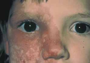
mitomycin C in primary open angle glaucoma. J Glaucoma 11:287, 2002

104. Wagle NS, Freedman SF, Buckley EG, et al: Long-term outcome of cyclocryotherapy for refractory pediatric glaucoma. Ophthalmology 105:1921, 1998

105. Hampton C, Shields MB: Transscleral neodymium:YAG cyclophotocoagulation: a histological study of human autopsy eyes. Arch Ophthalmol 106:1121, 1988

106. Trope GE, Steven MA: Mid-term effects of Nd:YAG transscleral cyclocoagulation in glaucoma. Ophthalmology 97:73, 1990

107. Phelan MJ, Higginbotham EJ: Contact transscleral Nd:YAG laser cyclophotocoagulation for the treatment of refractory pediatric glaucoma. Ophthalmic Surg Lasers 26:401, 1995

108. Schuman JS, Puliafito CA, Allingham RR, et al: Contact transscleral continuous wave neodymium:YAG laser cyclophotocoagulation. Ophthalmology 97:571, 1990

109. Kirwan JF, Shah P, Khaw PT: Diode laser photocoagulation: role in the management of refractory pediatric glaucomas. Ophthalmology 109:316, 2002

110. Sabates R: Choroiditis compatible with the histopathologic diagnosis of sympathetic 
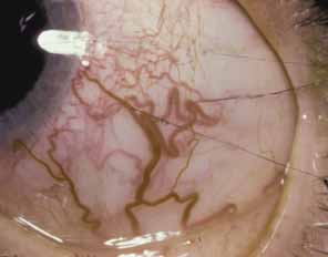
ophthalmia following cyclocryotherapy of neovascular glaucoma. Ophthalmic Surg 19:176, 1988

111. Brown SVL, Higginbotham E, Tessler SH: Sympathetic ophthalmia following Nd:YAG cyclotherapy. Ophthalmic Surg 21:736, 1990

Back to Top