Chapter 47
Tonometry
RICHARD F. BRUBAKER
Main Menu   Table Of Contents

Search

KINDS AND PROPERTIES OF TONOMETERS
CALIBRATION
TECHNIQUES OF TONOMETRY
ERRORS IN TONOMETRY
REFERENCES

Tonometry, in reference to the eye, is the noninvasive measurement of intraocular pressure.1,2 A tonometer is an instrument that exploits the physical properties of the eye to permit measurement of pressure without the need to cannulate the eye. An ideal tonometer must make accurate and repeatable measurements of intraocular pressure without affecting the pressure or without harming the eye. Of the large number of tonometers that have been invented, only a few have gained widespread use for clinical tonometry.

The physical properties of the normal cornea determine the limits of accuracy of tonometry. Numerous attempts have been made to design tonometers that are applied to the conjunctiva or lids, but these tonometers have never been satisfactory. Tonometry can be attempted in eyes with scarred or edematous corneas, but the tonometric data obtained in such eyes are never reliable and sometimes misleading.

The normal cornea is almost an ideal tissue through which to couple the pressure in the anterior chamber with the force of a tonometer. The cornea has good tensile strength. Its collagen is arranged in discrete layers, which in most species are not interwoven or cross-linked. Its ground substance is very slippery. For these reasons very little force is required to flatten or indent the normal cornea except to counteract the force of the intraocular pressure. However, the structural resistance of the cornea to deformation is not negligible and has been considered in the design of the most accurate tonometers.

When the cornea is deformed by a tonometer, the resulting fluid displacement causes the remainder of the globe to distend. Because of the tendency of the wall of the eye to resist stretching, deformation of the cornea raises the intraocular pressure. The greater the deformation, the greater the rise in intraocular pressure, although the relation is not linear. In the dead eye the relation between the volume of deformation and the change in pressure is relatively simple, but in the living eye this relation is complex and time-dependent. The intraocular pressure immediately prior to the application of a tonometer is symbolically represented as Po; the intraocular pressure during tonometry is symbolically represented as Pt. Po and Pt are never exactly equal for any tonometer. Spontaneous intraocular pressure without reference to tonometry is symbolically represented as Pi.

Back to Top
KINDS AND PROPERTIES OF TONOMETERS
Tonometers in which the intraocular pressure is negligibly raised during intraocular pressure measurements, for example less than 5%, are termed low-displacement tonometers. The Goldmann tonometer displaces only 0.5 μl of aqueous humor and raises intraocular pressure only 3%. The Krakau vibration tonometer raises intraocular pressure even less. For tonometers of this type Po and Pt are assumed to be equal. Tonometers that displace a large volume of fluid and consequently raise intraocular pressure significantly are termed high-displacement tonometers. In a normal eye intraocular pressure is more than doubled during tonometry with a Schi øtz tonometer. High-displacement tonometers are in most circumstances less accurate than low-displace-ment tonometers. An applanation tonometer is one that flattens a portion of the cornea. An indentation tonometer is one that indents the cornea with a shape other than a flat surface.

The relation between the physical forces involved in tonometry and the geometry of indentation or applanation can be deduced from simple physical principles, but the relation between Pt and Po can be deduced only from calibration tables of the species to be tested. These tables are derived from experiments comparing tonometric readings and pressure measurements ultimately derived by direct cannulation of the eye. These tables are based on mean values and cannot account for the myriad of variables that can affect the relation between Po and Pt in an individual eye. The dependency on calibration tables derived from such studies is an inherent disadvantage of high-displacement tonometry. This shortcoming applies to all high-displacement tonometers, both indentation and applanation.

A large number of tonometers have been invented and a moderate number have gained common usage. The number of interesting physical principles that have been exploited to carry out tonometry reflects the ingenuity of the inventors. Table 1 lists the categories into which tonometers can be classified. Of the wide variety of tonometers that have been described, those that are the most convenient to use, the simplest to calibrate, the most stable from day to day, the easiest to standardize, and the most free of maintenance problems have emerged as the most commonly used instruments in clinical practice. Several of these instruments are discussed below.


TABLE 1. Classification of Tonometers

  1. Invasive instruments
    1. Needles and cannulas (manometry)
    2. Transensors

  2. Noninvasive instruments (tonometers)
    1. Instruments that touch the eye
      1. Static instruments
        1. Applanation tonometers
          1. Constant area (Goldmann, Mackay-Marg)
          2. Constant force (Maklakoff)

        2. Indentation tonometers
          1. Constant indentation
          2. Constant force (Schi øtz)


      2. Dynamic instruments
        1. Ballistic tonometers
          1. Impact acceleration
          2. Impact duration
          3. Rebound velocity

        2. Vibration tonometers (Krakau)


    2. Instruments that do not touch the eye (noncontact tonometers) (Grolman)


The first practical tonometer (and also the simplest) was invented by Maklakoff in 1885.3 This applanation tonometer consisted simply of a flat-bottomed weight that could be brought to rest briefly on the cornea of a supine subject. The area of contact was estimated from the diameter of disturbance of a film of dye on the face of the tonometer. The Maklakoff tonometer was a high-displacement, fixed-force, variable-area tonometer. A number of variations of this tonometer have been described to improve its convenience.

Fick, in 1888, is credited with inventing a second applanation tonometer employing a fixed area produced by an adjustable force. This instrument was a forerunner of the Goldmann applanation tonometer (1954), which is today generally considered the most accurate clinical tonometer.

The Schi øtz tonometer is an old instrument that is still used extensively in clinical practice and is useful for performing tonography.4 The Schi øtz tonometer consists of three parts, a footplate assembly, a plunger assembly, and a handle. Its major advantage over the Maklakoff tonometer is that the readings from the scale of the instrument can be made quite rapidly and no special dye is required. The instrument can be used for rapid screening of large numbers of persons for elevated intraocular pressure as evidence of glaucoma. The Schi øtz tonometer is both an indentation tonometer and a high-displacement tonometer.

The mechanical complexity and unique shape of the plunger and footplate assembly of the Schi øtz tonometer have made standardization of manufacture mandatory. Seemingly negligible variances in the weight or dimensions of parts of this instrument can cause large differences in the relation between intraocular pressure (Po) and instrument scale reading (R) from one instrument to another. In the United States standardization, inspection, and certification of tonometers have been the task of the Committee on Standardization of Tonometers of the American Academy of Ophthalmology. Aside from its ease of use, the Schi øtz tonometer, in comparison with low-displacement instruments, is less sensitive to artifacts caused by lid malposition, lid pressure, or abnormal tension in the extraocular muscles, which can occur at the time tonometry is carried out. However, it is much more prone to artifacts caused by abnormal ocular rigidity such as occurs in myopic eyes, in eyes containing a compressible gas, or in eyes on which scleral-buckling procedures have been performed.

The Goldmann tonometer is an important instrument that permits an accurate clinical measurement of intraocular pressure in the human eye.5 The development of this instrument was based on careful consideration of the optimal area of the cornea to be flattened to minimize the inward force caused by surface tension of the tears and the outward force caused by the elasticity of the cornea. Goldmann's research led him to the conclusion that for the human eye, applanation diameters of 3 mm to 4 mm were most suitable. A fixed diameter of 3.06 mm (area = 7.35 mm2) was chosen to permit direct conversion of the force in decigrams to the pressure in the eye in millimeters of mercury. This area is large enough that the force produced by the intraocular pressure is easy to measure yet small enough to cause a negligible increase in intraocular pressure. Hand-held versions of Goldmann's instrument have been devised, notably the instrument of Draeger6 and the instrument of Perkins.7 The latter instrument, which is quite convenient, can be used in any position, is simple to calibrate, and has gained widespread use in clinical ophthalmology. The Perkins tonometer has the highest patient acceptance and permits examination of small children without general anesthesia.

The Bigliano tonometer8 (Durham tonometer, Applamatic tonometer) as modified by Webb9 (Pneumatonometer, Pneumatonograph) measures intraocular pressure from the complex interaction of a flowing stream of gas between a flat metallic footplate and a flexible membrane applied to the cornea. Its greatest use has been in research in which an objective measure of a change in pressure is required. If calibrated by the closed stopcock method for the specific species to be tested, it can be useful for tonometry in animals. Although claims have been made to the contrary, the Bigliano-Webb tonometer is a gravity-dependent, high-displacement tonometer that must be calibrated empirically. However, it provides an objective means of measuring intraocular pressure repeatedly and is a very sensitive method of measuring changes of intraocular pressure or ocular pulse pressure in a given eye.

An instrument that minimizes the effects of the tear attraction and the forces of corneal bending is the Mackay-Marg tonometer.10 In situations in which the cornea is modestly abnormal, this instrument can provide an estimate of intraocular pressure that is probably more reliable than that of other instruments. However, one can never be sure whether any instrument, including the Mackay-Marg tonometer, measures the intraocular pressure accurately in eyes with very thick or extensively scarred corneas.

An ingenious tonometer that measures intraocular pressure without direct contact with the cornea and without anesthesia is the Grolman noncontact tonometer11 (AO Noncontact Tonometer, NCT). The center of the cornea is aligned optically with the instrument. The operator depresses a button that causes a solenoid-driven piston to eject a jet of air at the corneal apex. The velocity of the jet increases linearly for 3 msec. The exact moment of corneal deformation in this 3-msec interval is detected optically. The higher the intraocular pressure the longer before the cornea indents. The time between triggering and deformation can be related empirically to intraocular pressure. The instrument is sufficiently accurate for clinical tonometry.

Back to Top
CALIBRATION
All tonometers must be calibrated in order to yield accurate results. Absolute calibration of tonometers always requires correlation between an instrument reading and simultaneous measurement of a manometric reading when the eye is cannulated. Two fundamentally different methods of tonometer calibration have been described, the open-stopcock method and the closed-stopcock method. In the closed-stopcock method, the pressure in the eye is first adjusted by an external manometer to a preselected pressure while the cannula to the eye is hydraulically connected to the manometer. The stopcock to the cannula is then closed so that the eye is temporarily disconnected from the manometer. Tonometry is performed immediately. The tonometric scale reading is correlated with the intraocular pressure just prior to closing the stopcock (Po). A second calibration procedure, the open-stopcock method, is performed in the same manner except that the stopcock is left open, allowing the tonometer and the manometric system to equilibrate. The pressure in the eye during tonometry (Pt ) is correlated with the instrument scale reading. For instruments that are used to measure Po, only the closed-stopcock system of measurement is suitable unless the tonometer is a low-displacement tonometer or an algorithm can be devised to relate Pt to Po for all scale readings of the tonometer.

It is important that intraocular pressure measurements be standardized so that studies conducted in one part of the world can be compared with studies conducted elsewhere. The need for standardization was most apparent in the years after the death of Hjalmar Schi øtz, whose laboratory in Oslo produced most of the Schi øtz tonometers that were available to the profession. In the years following his death, manufacturers began to vary a number of critical specifications of this instrument, leading to a chaotic situation in the discipline of tonometry. A number of attempts were made at standardization of these tonometers, culminating in 1942 with the establishment of the Committee on Standardization of Tonometers of the American Academy of Ophthalmology and Otolaryngology. Standardization is just as important for modern tonometers as it was for early tonometers at the turn of the century.

Back to Top
TECHNIQUES OF TONOMETRY
With few exceptions the clinician is interested in measuring the undisturbed steady-state pressure of the eye. It is necessary to begin with a good tonometer, carefully standardized and calibrated, and to perform tonometry with a properly executed technique. With the Schi øtz tonometer the subject can be permitted to sit in a reclining chair or lie in the supine position with his head and eyes directed vertically. A topical anesthetic such as 0.5% proparacaine hydrochloride is instilled into the conjunctival sac. The tonometer is first checked by placing its full weight on the test block supplied with the instrument. This test block permits the plunger to extend 0.05 mm below the position of the concave surface of the footplate. The upper and lower lid are separated gently, taking care not to press on the globe or to widen the palpebral fissure excessively. As the subject maintains steady vertical gaze with the aid of a fixation device, such as a blinking fixation light or the subject's own thumbnail, the instrument is lowered until it almost touches the cornea. It is advisable to hesitate several seconds to allow the subject to relax and to commence normal breathing before the instrument is lowered onto the cornea. The full weight of the tonometer (16.5 g when the 5.5-g plunger weight is used) must rest on the cornea before the scale is read. Care must be taken that the instrument is centered on the cornea, that the supporting sleeve is in its midposition, that the plunger is oriented vertically, and that the scale is easily visible to the examiner. The scale is marked in millimeters from 0 mm to 20 mm, representing a plunger protrusion of 0.05 mm to 1.05 mm. The examiner estimates the scale reading to the nearest 0.25 mm. Proper coupling of the instrument to the eye can be confirmed by observing oscillations of the pointer induced by the cardiac pulse. The instrument is removed from the eye, and the subject allowed to blink. The reading is repeated one or two times. The fellow eye is tested in the same way. The scale readings are averaged for each eye. The value of Po, which corresponds to the scale reading and the weight used, is then read from the 1955 scale supplied with the instrument. If the scale reading is 3 or less, one of the additional weights should be added to the plunger to improve the accuracy of tonometry. This precaution is especially important if an electronic Schi øtz tonometer is used, in which the terminal bore in the footplate has been enlarged to minimize plunger friction during tonography. The tonometer can then be disassembled and cleaned with water or alcohol or sterilized (in the case in which the examiner suspects that the subject's eye is infected with a transmissible organism).

The Goldmann tonometer is used in conjunction with a slit-lamp biomicroscope to which one of two types of Goldmann tonometers can be attached. The following description is written with the Haag-Streit slit lamp in mind. The principles may easily be applied to other biomicroscopes.

The slit lamp is adjusted for the examiner. Low-power oculars are preferable, and the objective lens is also set at its lowest power. The examiner may choose to align the optical portion of the tonometer with either the left objective or the right objective of the biomicroscope, depending on his own ocular dominance. The front surface of the prism is wiped with water and dried with a clean tissue. Hexachlorophene or other detergents should not be used, since residue on the prism may be toxic to the cornea. The biprism is adjusted such that the interface between the two prisms is oriented horizontally. If a high degree of corneal astigmatism is present in the subject's eye, the axis of the interface should be set 43° from the flattest axis of the cornea.

The examiner estimates the pressure in the eye and sets the dial of the tonometer to the estimated pressure. (The scale reading in grams multiplied by 10 equals the pressure in millimeters of mercury.)

The blue filter is introduced into the light path, and the slit is opened to its widest position. The light must be sufficiently oblique from either side to illuminate the tip of the prism and not cause interfering reflections.

Corneal anesthesia is required for accurate tonometry in most subjects. Proparacaine hydrochloride 0.5% is suitable. After instillation of the anesthetic, fluorescein can be added to the tear film by touching the everted lower tarsal conjunctiva with a moistened strip of fluorescein-impregnated paper. Alternatively one can use an anesthetic such as 0.4% benoxinate hydrochloride, which contains 0.25% fluorescein sodium. In either case it is not necessary to use more than a small amount of fluorescein to observe the inner margins of the meniscus of contact between the cornea and the tonometer. If argon laser treatment is to follow tonometry the same day, the least amount of fluorescein should be applied that permits visualization, and the fluorescein should be washed away with an irrigating solution immediately after tonometry. (Fluorescein absorbs the 488-nm wavelength of argon lasers.) If anesthetics cannot be instilled because of allergy, an experienced examiner can warm the tip with water to slightly above body temperature and perform tonometry without anesthesia.

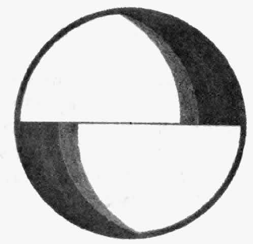
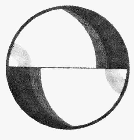
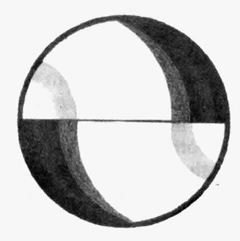
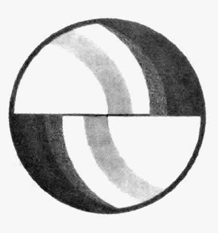
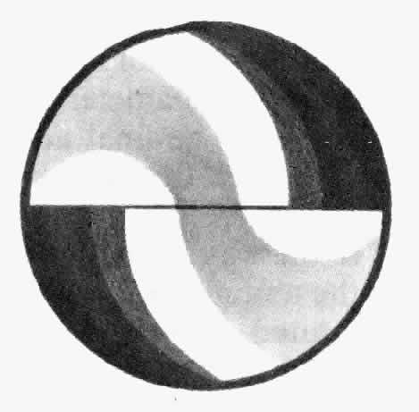
The slit lamp is adjusted so that the patient is comfortable. A tight collar or necktie should be loosened. The patient is asked to stare straight ahead and to resist the natural tendency to blink. The slit lamp is moved in order to approach the cornea along the optic axis. With the joystick held back, the entire examining unit of the slit lamp (including the tonometer) is moved to a position approximately 2 mm to 3 mm anterior to the cornea. The initial position should be inferior to the visual axis in order to make it possible to slip under the eyelashes of the upper lid without touching them. If necessary the upper lid should be lifted gently. The tonometer is then raised until the prism is centered. At this point the examiner begins to look through the biomicroscope. The reflected image of the tip of the tonometer can be used to guide the alignment of the tip even before contact is made. The image appears as hint purple arcs that move as the position is adjusted. When these arcs appear symmetric in the two halves of the biprism, the instrument is aligned correctly (Figs. 1 THROUGH 9). The joystick is moved forward slowly. Just before the corneal surface is touched, the arcs will meet.

Fig 1. View through the tonometry prism in the position of use.

Fig 2. When the tonometer is advanced within 7 mm of the cornea, faint purple arcs appear at the lateral edges of the prism.

Fig 3. As the tonometer comes closer, the arcs begin to focus and move closer together.

Fig 4. Appearance of the arcs as the tonometer moves closer.

Fig 5. Appearance of the arcs when the tonometer is still far enough away from the eye that vertical and horizontal adjustments of the tonometer are possible without contacting the cornea.

Fig 6. Appearance of the arcs just before contact. After contact, the purple arcs are replaced by green semicircles.

Fig 7. Appearance of the arcs when the tonometer is higher than the corneal apex.

Fig 8. Correct appearance of the green semicircles at the tonometric endpoint.

Fig 9. Appearance of the arcs when the tonometer is too far to the right of the apex of the cornea.

When the corneal surface is touched, two bright green semicircles appear. These semicircles should be in the proper position when they first appear. If not, the joystick should be pulled back, the tonometer wiped, and the process repeated. The subject must blink between applications to keep the cornea moist. This step is especially recommended for inexperienced examiners. If the cornea is only slightly out of line, the alignment can be adjusted without withdrawing the tonometer.

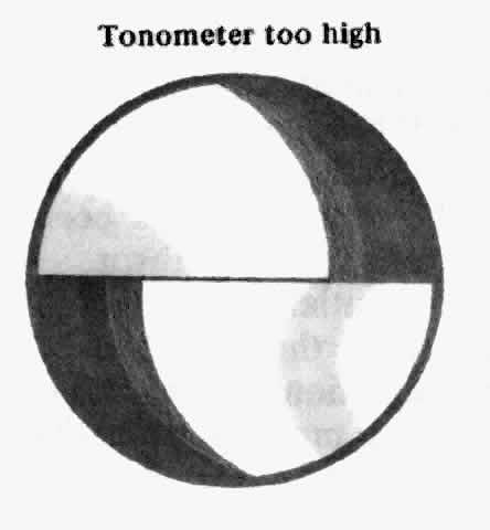
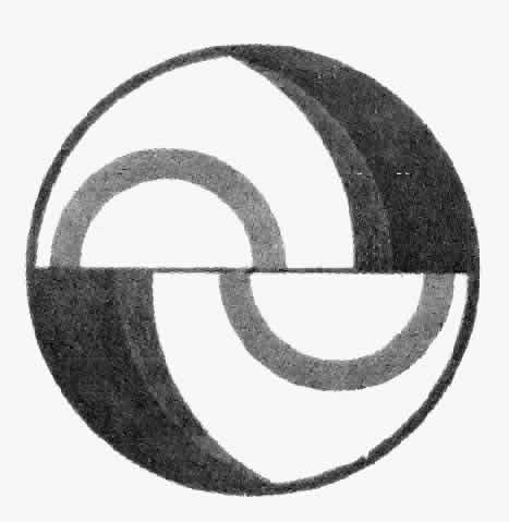
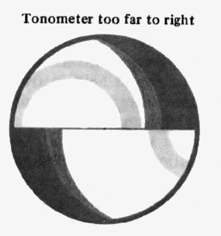
The two halves of the meniscus of contact, the inner margins of which delineate the flattened area, are observed. These semicircles should be symmetric, bright, and sharply focused. The tonometer dial is adjusted so that the inner edge of the superior semicircle meets the inner edge of the inferior semicircle exactly. Pulsatile changes in the diameter of the semicircles are usually observed. The final adjustment can be made halfway between the two extremes of the pulse.

If an eyelash has “strayed” in the way so that one of the semicircles is distorted, the tonometer is withdrawn and the examination repeated. If there is too much or too little dye present, the problem should be corrected. If a diffuse greenness is noted in the entire area where the prism is in contact with the cornea, the cornea may have been abraded and tonometry may not be accurate. If the semicircles overlap markedly and their size does not change as the tonometer dial is turned, the tonometer has been pushed too far forward and must be withdrawn.

It is recommended that after initial determination of the pressure of each eye is made the procedure be repeated. If the second determination of intraocular pressure differs by more than 1 mm Hg from the first, a third measurement is made. Consecutive measurements of a given eye should differ by no more than 1 mm Hg.

The prism is wiped with water and dried with a clean tissue. The tonometer is stowed so that no pressure is placed on the torsion balance. On certain slit lamps the tonometer can be swung to the side or the tonometer arm swiveled into its holder.

Use of the Perkins tonometer differs in certain particulars from use of the Goldmann instrument. The newcomer to Perkins tonometry will find that the intraocular pressure will seem to be slightly lower with the hand-held instrument than with the slit-lamp instrument. After a little practice, the two instruments can be made to agree fairly closely. It is advisable to use a slightly greater concentration of fluorescein when employing the hand-held instrument in order to make the inner margin of the tear wedge easier to observe. It is also mandatory that the room be darkened and that fresh batteries be used so that adequate illumination is available during tonometry. The examiner must be careful not to allow the meniscus of the biprism to mingle with either the superior or inferior tear meniscus of the lid margins. An excess of tears or mucus will obscure the end point and lead to large errors. This precaution is especially important when examining the eye in the first few days after surgery. It is also important that the lids and lashes not touch the tonometer. If the cornea is astigmatic, it is advisable to measure the pressure at two orthogonal orientations of the biprism and average them. In patients with abnormal extraocular muscles and deviations, such as dysthyroid ophthalmography, the Perkins tonometer can be used to measure the pressure in the position of greatest muscle relaxation and avoid artifacts due to ductions against retracted muscles.

The Mackay-Marg tonometer must be used skillfully with a technique that is not easy to master. It is important that the instrument be calibrated regularly against an absolute standard. This procedure is necessary to eliminate errors due to accumulation of residues between the sensitive plunger and the insensitive footplate. For daily use the instrument can be calibrated against the weight of its own plunger. These procedures assure the examiner that mechanical and electrical drift do not affect the accuracy of the reading. It is also helpful from time to time to compare Mackay-Marg readings with Goldmann readings in subjects with normal corneas. If the instrument is used regularly the technique can be mastered. If the baseline is set properly the instrument can be used in any position from vertical to horizontal.

The most common mistake when using the Mackay-Marg tonometer is to move the tip back and forth too rapidly. Rapid movements impart significant forces on the pressure-sensing transducer as a result of inertia. The rate of back and forth motion of the tonometer should be slow enough that if performed in air without touching the eye no visible oscillations will be present on the tonometric tracing. The instrument is then applied to the cornea as smoothly and evenly as possible and at right angles to the corneal surface. A slight indentation of the cornea is required to be certain that the instrument is properly coupled to the eye. Unsteadiness either of the patient or of the head of the examiner or too slow an application and withdrawal can produce artifacts in the tracing. These artifacts must be discriminated from true end points. A number of consistent end points must be observed before concluding that the reading is accurate.

Mackay-Marg readings will tend to be high in comparison with Goldmann readings. This situation is exaggerated when the cornea is abnormal or if the tonometer is used incorrectly. When the cornea is excessively scarred or thick, no tonometer can measure intraocular pressure with sufficient accuracy to satisfy clinical needs.

The Bigliano tonometer8 has been available to clinicians in two forms. The earlier form, marketed under the trade name Applamatic, was difficult to use and offered no advantages over the Mackay-Marg tonometer. An important modification was made by Webb,9 who devised a gas bearing and floating tip, which facilitated the application of the tonometer to the eye and permitted the tonometer to make a continuous recording of intraocular pressure. The instrument is available commercially under the trade names Pneumatonometer* and Pneumatonograph.


* Digitals, Cambridge, MA.
† Alcon Laboratories, Fort Worth, TX.

These instruments are easy to use for performing tonometry in humans. If calibrated according to the manufacturer's recommended method, tonometry can be performed in human subjects in the sitting position. The tonometer tip is brought against the anesthetized cornea until a high-pitched sound is heard and the pressure is read from an LED display or from a graph. In normal human eyes this tonometer tends to read high at low intraocular pressures and low at high intraocular pressures, and thus its range is compressed toward normal pressures.12

The reader is cautioned that a number of papers have been published in which it is concluded that this instrument is an applanation tonometer that follows the Fick-Imbert law, that it can be used in any position without a correction for gravity, and that it is a low-displacement tonometer. These conclusions have not been confirmed by the most meticulous studies of the instrument.12 Nevertheless this tonometer provides an objective reading, permits a continuous recording of intraocular pressure, and is simple to use. For these reasons it is among the best tonometers for clinical research in which an unbiased measurement of changes of intraocular pressure is needed in human or in animal eyes. When used for measurement of intraocular pressure in animal eyes, the instrument must be recalibrated. At the least, the instrument must be calibrated against the eye of the species to be tested using the closed-stopcock method. In some animals with very flexible corneas such as the rabbit, it is preferable to reduce the rate of gas flow by removing the cover of the instrument and adjusting the regulator until the gas bearing is barely functional. The recalibrated instrument is then more reliable for use in these species.

Back to Top
ERRORS IN TONOMETRY
A number of sources of tonometric error that are not related to the instrument itself should be considered. The steady-state intraocular pressure is partly dependent on body position. Most tonometry is performed in the sitting position, but some tonometers, such as the Schi øtz tonometer and the Maklakoff tonometer, are customarily employed in the supine position. The change of intraocular pressure that occurs after a change of body position is to some extent time dependent. However, the change in pressure is probably not as great as would be expected from the change in height between the eye and the right atrium. Lid pressure can produce a change of intraocular pressure. If the upper lid is elevated from the globe, intraocular pressure can be slightly lower. If the subject voluntarily widens his palpebral fissure to accommodate the tonometer, intraocular pressure can be somewhat higher. In persons with exophthalmus, lid malposition, or restrictions of the movement of extraocular muscles, transient changes of intraocular pressure can occur during tonometry. In persons with dysthyroid exophthalmopathy, intraocular pressure can increase on attempted upward gaze. A tight collar can affect the steady state of intraocular pressure, as can the subject's fear of the test procedure. With most tonometers the end point is somewhat subjective. Examiner bias can play an important role and can affect results. Examiner bias in tonometry is particularly important in unmasked clinical studies in which the examiner or the patient expects beneficial results from a drug or treatment procedure. Such studies are best performed by unbiased examiners or by appropriate masking techniques for the particular tonometer that is used. With some tonometers there are almost no sufficient methods to eliminate observer bias. This fact must be considered when designing a clinical study involving tonometry.
Back to Top
REFERENCES

1. Draeger J: Tonometry. Basel, S Karger, 1966

2. Gloster J: International Ophthalmology Clinics: Tonometry and Tonography. Boston, Little Brown & Co, 1965

3. Maklakoff: L'ophthalmotonometrie. Arch Ophthalmol 4:159, 1885

4. Schi øtz H: Ein neuer Tonometer; Tonometrie. Arch Augenheilkd 52:401, 1905

5. Goldmann H: Un nouveau tonometre applanation. Bull Mem Soc Fr Ophthalmol 67:474, 1954

6. Draeger J: Simple hand applanation tonometer. Am J Ophthalmol 62: 1208, 1966

7. Perkins ES: Hand-held applanation tonometer. Br J Ophthalmol 49:591, 1965

8. US Patent No. 3,099,262, Physiologic fluid pressure sensing head. Inventor, Robert P Bigliano, July 30, 1963

9. US Patent No. 3,714,819, Applanation tonometer comprising porous air bearing support for applanating piston. Inventor, Robert H Webb, February 6, 1973

10. Mackay RS, Marg E: Fast, automatic electronic tonometers based on exact theory. Acta Ophthalmol 37:495, 1959

11. US Patent No. 3,585,849, Method and apparatus for measuring intraocular pressure. Inventor, Bernard Grolman, June 22, 1971

12. Moses RA, Grodzki WJ Jr: The pneumatonograph, a laboratory study. Arch Ophthalmol 97:547, 1979

Back to Top