Chapter 35
Retinoblastoma
CYNTHIA A. BOXRUD and LA-ONGSRI ATCHANEEYASAKUL
Main Menu   Table Of Contents

Search

INCIDENCE
GENETICS
CLINICAL OBSERVATIONS
CLINICAL FEATURES
CLINICAL MANIFESTATIONS
DIAGNOSIS
GROWTH PATTERNS
DIFFERENTIAL DIAGNOSIS
PATHOGENESIS
TREATMENT
SECONDARY NONOCULAR TUMORS
TRILATERAL RETINOBLASTOMA
ACKNOWLEDGMENT
REFERENCES

Retinoblastoma is a highly malignant tumor of the eye. Early diagnosis and prompt treatment are important factors in a child's survival and can often salvage useful vision in one or both eyes. In the past 5 years, the molecular basis for retinoblastoma has become better understood. These scientific advances have led to preventive and prognostic strategies as well as innovative treatment protocols. New treatment modalities and trends have been successful. Combinations involving chemotherapy have had encouraging results. There has been a trend away from external beam radiotherapy (EBR), once the gold standard, as the primary treatment for retinoblastoma. Many of these issues will require prospective randomized clinical trials with long-term follow-up.
Back to Top
INCIDENCE
The incidence in various well-studied population groups around the world varies from 1 in 3,300 to 1 in 20,000 live births.1–4 Retinoblastoma has a worldwide distribution, with a higher incidence in some populations, such as in Haiti, Jamaica, Nigeria, and South Africa.5,6 The highest incidence was reported by Albert and colleagues,7 who found an incidence of 1:3300 in Haiti. In the United States, the annual incidence is 3.5 cases per million children younger than 15 years and account for approximately 2.5% to 4% of all cancers diagnosed in children younger than 15 years.8

The incidence of retinoblastoma decreases with age. Most tumors occur before age 2 years and are diagnosed before age 5 years. The median age at diagnosis in the absence of a family history is approximately 24 months for unilateral cases. With a family history, the age of diagnosis is significantly younger. Abramson and associates9 studied familial retinoblastoma and patients with a previously normal eye examination. Sixty-two percent of the first eyes were diagnosed by 6 months and 90% by 12 months. Retinoblastoma has also been encountered at birth, although it is rare. There has been some suggestion in the literature of a slight preponderance in boys, but most series find no sex predilection. No racial predilection has been observed.

Back to Top
GENETICS
Advanced techniques in the field of molecular biology have burgeoned in the past decade, and this new information has produced a greater understanding of oncogenesis, with the ultimate cloning of the retinoblastoma gene (RB1).

Bilateral cases of retinoblastoma can produce unilateral offspring, and unilateral cases can produce bilateral offspring. Bilateral cases are the product of a mutation in the genetic material transmitted in the gametes, which can be transmitted to subsequent generations. Unilateral cases are transmitted as hereditary cases in 8% to 10% of the patients.

In 1984, Murphree reviewed the data suggesting that a single locus existed for all of the forms of retinoblastoma located in the region 13q14.10 In 1983 and 1986, Cavenee and associates11–13 presented evidence that the development of retinoblastoma was related to the presence of a mutant allele in the 14 band of chromosome 13, which they later termed the retinoblastoma gene (RB1). Its genomic organization has been fully characterized, and the entire sequence of the 180-kilobase genomic locus has been completed.14,15

The retinoblastoma gene is composed of 27 exons ranging in size from 31 to 1889 base pairs (Fig. 1). It is the first representative of a class of cancer genes that restricts the uncontrolled growth of embryonic cells rather than produces cell growth. The gene product is a 110-kDa nuclear phosphoprotein of 928 amino acids that is normally present in all cells. This protein (pRB) acts as a cell cycle-regulatory growth suppressor in part by its ability to repress transcription.16 In its hypophosphorylated state, pRB binds to transcription factor (e.g., E2F) and inhibits cell proliferation. When pRB is absent, cells proliferate uncontrollably, leading to certain cancers. The interaction between pRB and E2F is regulated by other factors, including cyclin D1, cdk4, and p16.17 One normal copy of the gene is adequate to prevent tumor formation. Thus, retinoblastoma is one of a group of human cancers caused by loss-of-function mutations at distinct genetic loci, termed recessive oncogenes. Malignant transformation of a retinal cell occurs after both homologous copies to the retinoblastoma gene in that cell undergo loss-of-function mutations.

Fig. 1. Genetic map of the retinoblastoma gene and its neighbors. (Bowcock AM, Farrer LA, Hebert JM et al: Eight closely linked loci place the Wilson disease locus within 13q14-q21. Am J Hum Genet 43:664, 1988.)

These findings support Knudson's “two-hit” hypothesis, which postulates that the development of retinoblastoma requires at least two separate genetic events.18 His work was based on a comparison of the age at diagnosis in unilateral and bilateral retinoblastoma cases. The mechanism can best be explained by looking at hereditary retinoblastoma. Here, the germinal cells have two alleles: one normal “anticancer” allele and one defective allele. This defective allele corresponds to the first hit in Knudson's theory. The second, normal gene can still restrict the uncontrolled growth of tumors. At a later time, a mutagenic alteration hits the normal allele, suppressing its function as well; after this, a second-hit retinoblastoma develops. The functional loss is critical for tumor formation.

Inactivation of the retinoblastoma gene may contribute to the progression of other types of cancer, such as osteosarcoma,19 parathyroid carcinoma,20 and certain soft tissue sarcomas.21

Back to Top
CLINICAL OBSERVATIONS
A heritable tendency toward retinoblastoma is present in 40% of cases. These are characterized by multiple foci of tumor growth involving one or both eyes (Fig. 2).

Fig. 2. Foci of retinoblastoma growth.

Approximately 40% of all retinoblastomas are bilateral and 60% are unilateral. All of the bilateral cases are germinal mutations. Of the unilateral cases, 15% are germinal and 85% are somatic.

Family history is present in 8% to 10%, which implies that 92% are sporadic in nature. Of these sporadic cases, about 25% occur as a genetic or germinal mutation and 75% occur as a somatic mutation.

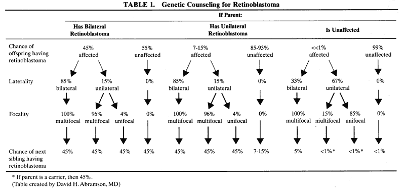
Germinal mutations are present in all patients with a family history. A child with bilateral retinoblastoma or a family history of the disease effectively has a 50% chance of passing the disease to his or her offspring. With a penetrance of 90%, the chance of passing retinoblastoma is 45%.

Patients with sporadic, unilateral retinoblastoma have only a 10% to 15% chance of transmitting the disease to their children. Although certain cases of unilateral retinoblastoma with a solitary tumor have been established as germinal mutations, most do not behave in this fashion.

When a child with retinoblastoma is born to normal parents who have no family history of the disease, there is approximately a 1% chance that subsequent children will have the disease and a 5% chance that there will be another affected child somewhere in the ensuing family line (Table 1).


Back to Top
CLINICAL FEATURES
In the absence of a known family history of the disease, the early months of retinoblastoma growth in a child's eye usually go undetected by both the family and the primary care physician. Even if the pediatrician or family practitioner performs a red reflex test or examines the optic nerve with a direct ophthalmoscope in the first year of life, he or she may well miss seeing the tumor and not suspect its presence unless the tumor is large enough or located in a part of the retina where it will reflect the light entering the pupil.

The tumor does not cause pain or discomfort as it enlarges and virtually never causes a red or inflamed eye early in the course of the disease. If the fovea is involved with tumor in one eye, strabismus may occur, but the unilateral visual loss caused by the tumor will not be apparent clinically because of the normal vision in the other eye.

Retinoblastoma is occasionally observed in premature infants, and there are a few reports of prenatal diagnosis.22 However, when retinoblastoma is diagnosed and the extent of disease is evaluated in 3-month-old patients, the percentage who have advanced disease is nearly the same as the percentage of those whose disease was diagnosed at 1 year of age.23

Bilateral retinoblastoma is most often asymmetric and is usually diagnosed at an earlier age than unilateral disease. The average age at diagnosis in bilateral disease is 12 months; in unilateral disease it is 24 months. During the evaluation of bilateral or unilateral disease, the presence of intraocular calcium, a classic feature but not mandatory, can help make the diagnosis.

Parents and relatives of children with retinoblastoma are routinely examined. When retinal tumors are seen in adults, they most commonly represent a benign manifestation of the RB1 gene called a retinoma. The tumor undergoes spontaneous regression, leaving a chorioretinal scar with flocculent residual calcium, visible either with the ophthalmoscope or with radiologic techniques, and the presence of a blood vascular system that is not part of the retinal or choroidal circulation.24 Although retinomas do not require treatment, they suggest the presence of a germline RB1 mutation and necessitate careful follow-up.25

Back to Top
CLINICAL MANIFESTATIONS
The presenting signs and symptoms of retinoblastoma are varied (Table 2). A white reflex in the pupil is the most common clinical sign (56%).26 In the German literature, this was called a cat's-eye reflex and is now termed leukocoria (Fig. 3). The differential diagnosis of retinoblastoma is the differential diagnosis of leukocoria.


TABLE 2. Presenting Signs and Symptoms of Retinoblastoma

  Laukocoria
  Strabismus
  Uveitis
  Hyphema
  Red eye
  Glaucoma/rubeosis irides
  Proptosis
  Endophthalmitis/panophthalmitis
  Orbital cellulitis
  Irritability
  Visual obscuration
  Miscellaneous


Fig. 3. Leukocoria or cat's-eye reflex in the pupil of a young boy with retinoblastoma.

When a tumor arises in the macular region, it presents as a white reflex while it is still relatively small (three to four disc diameters) (Fig. 4). It can readily be seen when the patient looks straight ahead at the observer. When a tumor arises in the retinal periphery, it may grow large before it demonstrates a white reflex, and the reflex is seen only when the child is looking in a particular direction.

Fig. 4. Small macular retinoblastoma with slightly dilated feeding vessels. (Courtesy of Carol L. Shields, MD, Philadelphia, PA.)

The second most common presenting sign of retinoblastoma is strabismus (23%).26 Any child with strabismus should have fundus examination to rule out intraocular pathology. The strabismus in retinoblastoma is due to involvement of the macula. Therefore, if the macula can be clearly seen with the ophthalmoscope, retinoblastoma can be ruled out as the cause of the strabismus. However, there are rare exceptions. One child was noted to have congenital esotropia and normal maculae with small multiple tumors in both eyes (personal communication, R.E. Ellsworth).

A red, painful eye, often accompanying glaucoma, can be a presenting sign of retinoblastoma. Children with these manifestations are frequently treated for uveitis before the correct diagnosis is made.

Poor vision can be the presenting symptom in two situations. The first scenario is a child with a slow-growing retinoblastoma that grows during a period of years and ultimately involves the macula. This leads to a spontaneous complaint of poor vision in an older child. Second, poor vision can be found in infants with massive bilateral tumors, producing loss of visual acuity, often associated with behavior problems.

Endophthalmitis, panophthalmitis, and orbital cellulitis that begin explosively over 1 or 2 days can be a sign of spontaneous necrosis in the tumor. The rapid development suggests an active inflammatory process, and adjacent sinus disease may be suspected. An aspect of this cellulitis is its rapid resolution with or without antibiotics. Because antibiotics are usually used in an effort to combat infection, a rapid response is taken to indicate an infectious origin when this is not actually the case. Once this error has been made, the true diagnosis may not be made for months or years, especially in an eye with opaque media.

Unilateral mydriasis is an occasional presenting sign. It generally reflects extensive tumor, with total retinal detachment.

Hyphema may be a presenting sign of retinoblastoma. If it clears, gray tumor remnants may be seen in the iris. These can be confused with juvenile xanthogranuloma during the first year of life. Temporization for a period of several weeks is reasonable. Here, ultrasonography can be useful in diagnosing a posterior tumor responsible for the hyphema.

A change in iris color, often from blue to a darker color, may be due to one of several factors. Pre-existing hyphema in the anterior chamber may lead to a greenish-brown discoloration of the iris stroma; in other cases, rubeosis irides produced by an advanced tumor may give the iris a darker hue.

White spots on the iris are an occasional first sign of the disease and represent implantation growths on the surface of the iris stroma or vitreous floaters that have come forward through the zonula to lodge in the chamber angle.

Even in the United States, signs and symptoms of metastasis may be the presenting picture of retinoblastoma, and failure to thrive along with such vague symptoms as somnolence or hyperirritability may be the initial clue. In many parts of the world, exophthalmos is the most common presenting sign of retinoblastoma. Although this is rare in the United States, it can occur. The presence of a mass in the posterior segment is the clue that would differentiate this from rhabdomyosarcoma or some other type of rapidly progressive exophthalmos in a child.

Back to Top
DIAGNOSIS
Patients with retinoblastoma fall into two groups: those in whom the tumor can readily be seen, and those in whom the tumor is not visible.

In the first group, the retinal tumor can be seen with the ophthalmoscope, projecting forward into the vitreous. The tumor is usually creamy pink, and neovascularization frequently is present on the surface. Microaneurysms and telangiectatic vessels are common in the vascular stroma of these tumors. Nutrient vessels may be so large that they mimic the afferent vessels of an angioma (Fig. 5). This presentation can be especially confusing if a portion of the retina is detached. About 5% of patients with retinoblastoma have spontaneous hemorrhages on the surface of the tumor, and approximately 5% show yellow, fatty exudate adjacent to the tumor or in the macular area. Approximately 10% of retinoblastomas have a pigmented, atrophic annulus around the circumference of the tumor, and this is interpreted as a sign of spontaneous regression. Several retinoblastomas have been seen to have a tremendous proliferation of the retinal pigment epithelium in a quadrant adjacent to the tumor, producing a uniform black appearance over a large area of the retina. The explanation for this phenomenon is not known.

Fig. 5. Large macular retinoblastoma with prominent dilated and tortuous feeding vessels. The tumor overhangs part of the optic disc. (Courtesy of Carol L. Shields, MD, Philadelphia, PA.)

In the second group of patients, a clear view of the tumor is precluded by vitreous hemorrhage, retinal detachment, or inflammatory reaction. In these situations, ultrasonography, computed tomography (CT), or magnetic resonance imaging (MRI) can be of particular value.

Two features of retinoblastoma are almost pathognomonic: calcification and seeding of tumor cells into the vitreous. CT can be used to confirm the presence of calcification or the extension of tumor (Fig. 6). We assess all patients for the presence of any pineal lesions associated with trilateral retinoblastoma. MRI has been shown to have remarkable sensitivity in distinguishing differences in tissue density. Its advantages are that it uses no external radiation and is a relatively noninvasive technique. With the use of a surface coil, spatial relations in the eye can be even more clearly delineated.

Fig. 6. Axial CT scan with contrast demonstrating calcification in a retinoblastoma of the left globe.

Calcification may occur as a result of any advanced hemorrhagic or inflammatory disease in the retina, but the peculiar fluffy pattern of calcification in retinoblastoma is unique. This calcification can be easily seen with the ophthalmoscope and has been verified by radiography to exist in 70% of tumors. When calcium is noted on the surface of a tumor, it is sharply demarcated and glistening white and resembles cottage cheese. When the calcium lies deeper in the tumor, it is seen as a gray-white translucent area with indistinct outlines. When the tumor expands into the vitreous, it can be confused with inflammation with vitreous floaters seen in toxocariasis, toxoplasmosis, and sarcoidosis.

Transillumination may be helpful in distinguishing ordinary retinal detachments from the solid detachments caused by retinoblastoma. The newer fiberoptic sources, which give cold and brilliant light, are far better than the old types of transilluminators. Fluorescein angiography shows a distinctive pattern when the tumor vascularization can be seen, but in these situations, angiography is usually unnecessary to confirm the diagnosis.

Four types of ultrasonographic patterns have been seen in retinoblastoma. The most common finding is a mixed tumor type.27 Other echographic findings include solid, cystic, and diffuse infiltrating tumor types. More than 80% of retinoblastoma patients demonstrate calcium in the tumor by ultrasonography. This is very helpful in differentiating a retinoblastoma from other intraocular pathology. The degree of calcification is a factor determining the internal reflectivity in the tumor. An attempt to correlate the echographic patterns and histopathologic findings28 showed no correlation between the degree of rosette differentiation, pseudorosettes, or necrosis and A-scan echographic pattern.

Intensive efforts have been made at finding a biochemical marker in diagnosing retinoblastoma. Levels of both lactate dehydrogenase and neuron-specific enolase have been shown to be elevated in the vitreous or aqueous humor of eyes with retinoblastoma.29–31 Further investigation needs to address this area.

Back to Top
GROWTH PATTERNS
Retinoblastomas have been divided macroscopically into two groups, endophytic and exophytic. Endophytic tumors appear to originate in the internal nuclear layers of the retina and extend into the vitreous cavity, where they can readily be seen with the ophthalmoscope (Fig. 7). This is a common pattern. An exophytic tumor arises in the external nuclear layer. It may grow into the subretinal space and consequently detach the retina (Fig. 8). Palazzi and associates32 reviewed the histopathology of 297 enucleated eyes from patients with unilateral retinoblastoma and discovered some interesting correlations. They stated that positive family history was seen in the endophytic type more than the exophytic type. Also, they found that glaucoma and choroidal invasion occurred more often in the exophytic type.

Fig. 7. A. Advanced endophytic retinoblastoma with floating, white retinoblastoma tumor seeds in the vitreous. (Courtesy of Carol L. Shields, MD) B. Cross section of an enucleated globe demonstrating a massive endophytic retinoblastoma. (Courtesy of Carol L. Shields, MD, and Ralph C. Eagle Jr, MD, Philadelphia, PA.)

Fig. 8. A. Massive exophytic retinoblastoma with total retinal detachment. (Courtesy of Carol L. Shields, MD) B. Cross section of an enucleated globe with the posterior cavity filled by a large exophytic-type retinoblastoma. (Courtesy of Carol L. Shields, MD, and Ralph C. Eagle Jr, MD, Philadelphia, PA.)

Eyes with retinoblastoma often have multiple tumor foci. In our analysis of 100 cases, 84 patients had more than one tumor in the involved eye, with the average number being five. This pattern probably represents true multicentric origin rather than tumor spread in the affected retina.

There are two possible explanations why retinoblastoma is more commonly observed at the ora serrata or in the far periphery. First, there is some suggestion, both clinical and histologic, that tumors can migrate in the potential subretinal space in exophytic retinoblastoma. At the ora serrata, where they can be trapped, tumefaction occurs. Second, the retina differentiates last in the far periphery, where one would expect a second mutation to result in tumor formation.

SEEDING

The collagenous and vascular stroma is poorly developed in retinoblastomas, and the cells are not particularly cohesive. As tumors grow larger in the eye, peripheral portions can break loose and float freely in the vitreous, producing the clinical picture of seeding. Seeding in connection with large tumors is an unfavorable prognostic sign. A limited amount of seeding is occasionally seen with relatively small tumors, and in these situations the prognosis is somewhat more optimistic.

A seed floating in the vitreous may remain viable for a long time, but such seeds do not grow rapidly. At any time, however, they may settle on the retina and produce implantation growths. The tumor seeds are affected by EBR but are not influenced by current chemotherapeutic agents, which penetrate poorly into the vitreous.

Seeding may occur in three circumstances. The first is with large tumors that break apart, as described earlier. The second is seen after EBR to undifferentiated tumors. The principal tumor mass becomes heavily calcified into a ball resembling cottage cheese, and portions of this mass break away and spread into the vitreous. These seeds are glistening white, very sharply demarcated, and almost always totally inactive. The third type of seeding is also a response to EBR. Certain tumors (perhaps those that are better differentiated) do not take on the glistening white calcific appearance after irradiation, but they do break apart. The vitreous becomes studded with large clouds of gray, translucent tumor remnants, which may contain viable retinoblastoma cells. This regression pattern is difficult to interpret and may lead to implantation growth. Therefore, children with this type of seeding must be examined under anesthesia at 1-month intervals.

CHOROIDAL INVASION

Choroidal invasion is almost always present in larger retinoblastomas, but it is by no means tantamount to systemic metastasis. Three clinical indications that should raise suspicion for choroidal invasion are rapid tumor growth over a period of days or weeks, high elevation, and yellow at the summit, suggesting that the lamina vitrea has been pushed forward ahead of the tumor.

Attempts have been made to determine the significance of choroidal invasion and relate it to the risk for metastasis.33–35 The data suggest that massive choroidal invasion significantly increases the risk for metastasis; systemic chemotherapy should be given in these patients. It is probable that eyes with isolated choroidal invasion do not benefit from chemotherapy.

The most common chain of histologic events preceding orbital extension is massive involvement of the posterior choroid and extension along the scleral emissaria, or scleral nodules. The presence of scattered tumor cells in the episclera is difficult to interpret histologically because it may be an artifact of sample preparation.

Howard36 suggested that photocoagulation may destroy a portion of the lamina vitrea that acts as a natural barrier to the choroid. This would facilitate tumor invasion into the choroid and sclera. However, the appropriate technique of laser photocoagulation can help avoid this complication.

OPTIC NERVE INVASION

Because the retina and optic nerve are one contiguous tissue, it is not surprising that retinoblastoma, a tumor of the retina, can extend through the lamina cribrosa into the optic nerve. It is common to see limited extension into the optic nerve, 2 to 3 mm posterior to the lamina cribrosa, by the time an eye with retinoblastoma must be removed. Therefore, it is very important to obtain as long a nerve as possible at the time of enucleation.

When the tumor has grown 10 to 12 mm backward into the optic nerve, to the point where the central retinal artery and vein exit through the subarachnoid space, it almost always gains access to the cerebrospinal fluid (CSF) and then slowly progresses into the meninges over the base of the brain and in the ventricles. When this occurs, the prognosis for life is poor.

When tumor is seen in the nerve at the line of section, it must be assumed that there is residual tumor beyond the stump of the nerve. The death rate in this group of patients exceeds 50%.37 Improvement in the chemotherapeutic regimen combined with a hematopoietic stem cell rescue might help prevent distant metastasis and increase the survival rate.38

METASTASIS

Metastasis is here considered to include not only remote metastasis but contiguous as well. The retinoblastoma cells in the eye can travel by numerous routes to various parts of the body. Hematogenous spread from blood vessels in the tumor or from choroidal extension is the most common pathway. The second most common pathway is involvement of the optic nerve, either by direct extension through the lamina cribrosa into the optic nerve or from choroid at the termination of Bruch's membrane into border tissue surrounding the nerve head. Extension then occurs into the CSF, with seeding into the meninges of the ventricles and over the base of the brain. Retinoblastoma can also spread through the lymphatic system after extension into the orbit. For unknown reasons, retinoblastoma is never encountered in the lungs (except from a marrow site in the rib), although tumor cells obviously pass across the lungs during circulation.

Back to Top
DIFFERENTIAL DIAGNOSIS
In a typical case, the diagnosis is straightforward. However, when a retinal detachment obscures the tumor or when the vitreous is cloudy with either hemorrhage or inflammation, the diagnosis can be most challenging. Here is where ultrasonography, CT, or MRI may be of value. On the rare occasion in which the diagnosis cannot be made by noninvasive investigations, fine-needle aspiration biopsy through the cornea (not the pars plana) may be useful.39,40 The differential diagnosis of intraocular retinoblastoma includes conditions that cause leukocoria (Table 3).


TABLE 3. Differential Diagnosis of Leukocoria

  Cataract
  Persistent hyperplastic primary vitreous
  Granulomatous uveitis
  Metastatic endophthalmitis
  Vitreous hemorrhage
  Toxocara granuloma
  Toxoplasmosis
  Viral retinitis
  Norrie disease
  Autosomal recessive retinal dysplasia
  Dominant exudative vit-
  reoretinopathy
  Juvenile X-linked retinoschisis
  Retinochoroidal coloboma
  Congenital retinal fold
  Myelinated nerve fibers
  High myopia
  Morning glory disc anomaly
  Astrocytic hamartoma
  Angiomatosis retinae (capillary hemangioma)
  Medulloepithelioma
  Coats' disease
  Retinopathy of prematurity
  Retinal detachment


PERSISTENT HYPERPLASTIC PRIMARY VITREOUS

Persistent hyperplastic primary vitreous is a congenital anomaly in which the primary vitreous fails to regress in utero. Highly vascular mesenchymal tissue nurtures the developing lens during intrauterine life. The mesenchymal tissue forms a mass behind the lens. A gray-yellow retrolental membrane may produce leukocoria, with the subsequent suspicion of retinoblastoma. The globe is white and slightly microphthalmic. Patients have no history of prematurity or oxygen administration. Retrolental fibrovascular tissue may invade the lens capsule, causing lens swelling and acute glaucoma. Although a persistent hyaloid artery may be present, affected eyes have a normal retina and may be capable of useful vision if the membrane can be opened.41

GRANULOMATOUS UVEITIS

Advanced retinoblastoma can produce a typical picture of toxoplasmic uveitis, with large muttonfat keratic precipitates and a cloudy vitreous with an underlying yellowish mass (masquerade syndrome).42 Apparent response of this condition to steroid therapy should not be taken as an indication of inflammatory origin, and a high index of suspicion should be maintained until a final diagnosis is made.

METASTATIC ENDOPHTHALMITIS

Septic emboli from a bacterial infection in other parts of the body can be the source of endogenous endophthalmitis in children.43,44 These patients might have leukocoria indistinguishable from retinoblastoma.

VITREOUS HEMORRHAGE

A vitreous filled with blood is an uncommon presenting sign of retinoblastoma.26 When it is encountered, careful temporization is in order, especially in very young infants. Persistent vessels of the hyaloid system can bleed after birth trauma; as they undergo regression, the blood usually clears to reveal a normal fundus. Hemorrhagic disease of neonates may also be accompanied by vitreous hemorrhage. Because unrecognized contusive trauma to the eye is probably the most common cause of vitreous hemorrhage in young children, the diagnosis may be quite difficult. Ultrasonography along with MRI and CT may be helpful. Hemorrhages occur in the retina of 20% to 30% of neonates after normal delivery but rarely in newborns delivered by cesarean section. These are usually self-limited and disappear during the first few months of life. However, if a massive hemorrhage occurs and becomes organized, an elevated, gray mass simulating retinoblastoma may occur.

TOXOCARA GRANULOMA

Larval granulomatosis is caused by a granulomatous reaction around the larva of the roundworm Toxocara canis. The clinical picture ranges from a quiet solitary retinal nodule (which can look remarkably like a retinoblastoma) to acute and widespread inflammation with a cloudy vitreous. The number of larvae that enter the eye and the route they take (whether to the choroid or the retina) seem to play an important part in the ocular manifestations. In many children, an overlying antigen-antibody reaction seems to occur. The larvae most commonly enter through the short ciliary arteries, and an eosinophilic granuloma containing a larval fragment is often present in the macular area. Should larvae enter the central retinal artery, they may lodge in the retinal periphery and produce characteristic granulomas in that location. We have encountered several such cases diagnosed as larval granulomatosis that have responded well to intensive cryotherapy of the peripheral lesions.

Two clinical clues suggest a diagnosis of larval granulomatosis. The first is the presence of a glistening white core in the center of a granuloma, and the second is the presence of long, straight, grayish-yellowish strands extending into the vitreous. These are uncommonly seen in retinoblastoma.

Serum Toxocara ELISA test or vitreous study with the ELISA reaction to Toxocara canis homologous antigen may be of value for the diagnosis.45 Serial observation during a period of weeks differentiates between larval granulomas (which remain constant in size) and retinoblastoma (which grows). A histologic diagnosis may be obscure because the larval fragments are digested and disappear entirely with time.

TOXOPLASMOSIS

The classical triad of congenital toxoplasmosis includes chorioretinitis, intracranial calcification, and hydrocephalus. It results from transplacental infection with Toxoplasma gondii during pregnancy. Chorioretinal scar is the most common eye finding and can be seen either in the peripheral retina or the macular area.46 The diagnosis of congenital toxoplasmosis can be made by serologic tests measuring antibodies to the parasites.47,48 The ocular lesions can be treated with sulfadiazine, pyrimethamine, and corticosteroid.

CATARACT

Pediatric cataract can be associated with many disorders, including intrauterine infection (congenital rubella syndrome, congenital toxoplasmosis), metabolic disease (galactosemia, mannosidosis, Refsum syndrome), renal disease (Lowe syndrome, Alport syndrome), connective tissue disease (Marfan syndrome, homocystinuria), dermatologic disorders (Cockayne syndrome, Rothmund-Thomson syndrome), and chromosomal abnormalities (trisomy, monosomy, deletion). With the use of slit-lamp biomicroscopy, the diagnosis of pediatric cataract can be made with increased certainty. In eyes with dense cataract obscuring the fundus view, ultrasound biomicroscopic examination can be of value in differentiating other abnormalities behind the cataract, including retinal detachment and intraocular tumor.49

VIRAL RETINITIS

Although uncommon, retinitis can occur after viral infection in immunocompetent children. For examples, after measles infection the retina may develop diffuse vasculitis with a macular star.50 In immunocompromised children, such as those with HIV infection, or after organ transplantation, the eye may develop cytomegalovirus retinitis, although this is not as common as in adult patients.51

NORRIE DISEASE

Norrie disease is a bilateral X-linked dysplasia of the retina. It is most commonly encountered in males who are blind from birth. The eyes usually become phthisical or progress from retinal dysplasia to total retinal detachment. Other clinical features include late-onset hearing loss, mental retardation, and microcephaly.

AUTOSOMAL RECESSIVE RETINAL DYSPLASIA

Retinal dysplasia is a nonspecific pathologic term that describes a syndrome producing retinal folds and dystrophic retina in association with other systemic anomalies. Some of these patients exhibit trisomy 13. The associated anomalies include cerebral agenesis, hydrocephalus, encephalocele, lissencephaly (smooth brain), cerebellar malformation, cleft lip and palate, malrotation of the gut, and anomalies of the heart and vascular system. As the syndromes accompanied by retinal dysplasia become better delineated, the term retinal dysplasia will become used as a histologic description rather than as a clinical entity.

DOMINANT EXUDATIVE VITREORETINOPATHY

The eye findings in this condition may be indistinguishable from retinopathy of prematurity (ROP) and Coats' disease.52 This is the disease of small peripheral vessels of the retina. Clinical findings include posterior vitreous detachment, snowflake vitreous opacity, recurrent vitreous hemorrhage, macular displacement, localized retinal detachment, retinal neovascularization, secondary cataract, and phthisis bulbi.

JUVENILE X-LINKED RETINOSCHISIS

The primary defect in this condition is thought to be in the Mueller cell, the principal glial cell of the retina. Clinical findings include cystic retinal degeneration, intraretinal splitting, retinal atrophy, retinal detachment, choroidal sclerosis, and hemorrhage in the schisis cavity or the vitreous. Foveal schisis is the most common finding and occurs in essentially all affected persons. The retinoschisis gene (XLRS1) has been found, and it may be involved in cell adhesion processes during retinal development.53

RETINOCHOROIDAL COLOBOMA

Retinochoroidal colobomas are defects in closure of the fetal fissure. The absence of retina and choroid is seen as a white reflex (bare sclera). Both of these defects are diagnosed by examination under anesthesia. Retinochoroidal coloboma can be an isolated finding or part of a condition called CHARGE association. Recent studies of retinochoroidal coloboma in children revealed a decreasing prevalence of retinal and choroidal detachment in these eyes compared with previous studies (from around 30% to less than 10%).54

CONGENITAL RETINAL FOLD

The simple form of congenital retinal fold is an isolated anomaly in differentiation of the retina. It extends from the nerve head into the retinal periphery; most cases are unilateral. Children with this condition are normal in all other aspects. When the fundus can be clearly seen, the condition is noted to be unlike retinoblastoma. During evaluation, it is important to recognize that similar folds can also be secondary to ROP, trisomy 13–15, foreign bodies, and granulomatous inflammation.

MYELINATED NERVE FIBERS

When myelinated nerve fibers are extensive and over the posterior pole, they may produce a white reflex in the pupil. When a child is adequately examined, the diagnosis is obvious.

HIGH MYOPIA

When marked thinning of the retina is accompanied by a large myopic crescent around the nerve, a white reflex may be seen. It leads to the suspicion of retinoblastoma. Careful examination with the indirect ophthalmoscope and retinoscopy reveals the correct diagnosis.

MORNING GLORY DISC ANOMALY

Optic nerve colobomas and the rare congenital anomaly called morning glory disc anomaly are congenital entities that result from abnormal development of the optic nerve. They can be an isolated anomaly or associated with central nervous system anomalies such as basal encephaloceles, agenesis of the corpus callosum, absent chiasm, or pituitary dysfunction.55

ASTROCYTIC HAMARTOMA

Retinal astrocytic hamartoma (astrocytoma) is a benign tumor of glial cell origin. It can occur as an isolated abnormality56,57 or can be part of a syndrome called tuberous sclerosis. Calcification may develop in the lesion, which makes it difficult to differentiate from retinoblastoma.58

ANGIOMATOSIS RETINAE (CAPILLARY HEMANGIOMA)

The retinal lesions of angiomatosis retinae are benign vascular anomalies. When the retinal tumor is associated with cerebellar tumors (in approximately 25% of cases), the condition is known as von Hippel-Lindau disease. At a certain stage in the evolution of angiomatosis retinae, the picture is very similar to retinoblastoma (Fig. 9). Both conditions are marked by large feeder vessels that appear to arise de novo in the tumor. When angiomas become gliotic and obscured by overlying detachment, they cannot be clearly differentiated from retinoblastoma. Again, ancillary tests can be useful to detect calcification. When diagnostic difficulty arises and there is no useful vision in the affected eye, the most reasonable course is enucleation.

Fig. 9. A. Capillary hemangioma of the retina with prominent afferent and efferent vessels. B. Fluoroscein angiography showing filling of the tumor with dye. (Courtesy of William Tasman, MD, Philadelphia, PA.)

MEDULLOEPITHELIOMA (DICTYOMA)

A dictyoma is the anterior counterpart of retinoblastoma arising from the medullary epithelium of the ciliary body. It can be benign or very rarely malignant and produces a myriad of clinical presentations. It rarely simulates retinoblastoma. Dictyomas tend to grow en plaque over the surface of the ciliary body, iris, lens, and anterior chamber angle. They are occasionally markedly cystic or diffusely involve the entire ciliary body. An elevated gray mass in the area of the ciliary body or anterior retina may be either a dictyoma or retinoblastoma. The clinical course of development during a period of time will lead to the correct interpretation.

COATS' DISEASE

Coats' disease is a nonhereditary condition found most commonly in males. It is a primary vascular anomaly of the retina characterized by telangiectatic vessels that leak lipoproteinaceous exudate into the retina and subretinal space (Figs. 10 and 11). Later, a detachment with a solid white or yellow-pink material may stimulate a retinoblastoma. Patches of telangiectatic and aneurysmal vessels strongly suggest Coats' disease but are also seen in retinoblastoma. The involved retina often has a characteristic greenish-yellow sheen, and the detached portion of the retina may be dark gray or black as a result of proliferation of the retinal pigment epithelium. Although this can also be a feature of retinoblastoma, it is relatively rare. Cholesterol crystals glistening in the subretinal space strongly suggest a later stage of Coats' disease. Although the vascular anomaly in Coats' disease is probably present at birth, the lesion does not cause symptoms until the retina detaches and central vision is lost, with cases being discovered at an average age of 6 to 8 years.

Fig. 10. A. Coats' disease with exudation in the posterior pole. B. Peripheral retinal telangiectasia responsible for the exudate. C. Fluorescein staining of the peripheral lesion. (Courtesy of William Tasman, MD, Philadelphia, PA.)

Fig. 11. A. Total retinal detachment with Coats' disease. B. Total retinal detachment with retinoblastoma. C. Comparison of CT scan between Coats' disease and retinoblastoma. D. Type I regression. E. Type II regression. F. Type III regression.

RETINOPATHY OF PREMATURITY

Retinopathy of prematurity occurs in premature infants as a result of the toxic effects of oxygen on avascular retina. Children who are of normal birth weight and who have not been given oxygen occasionally have a picture identical to ROP.59 Elevated gray areas are present in the periphery and may mimic retinoblastoma. In addition, when a complete retrolental membrane is present, it may produce a white reflex in the pupil. This condition is usually bilateral and symmetric. Long ciliary processes drawn into the retrolental membrane, along with a history of prematurity and oxygen administration, suggest the correct diagnosis.

RETINAL DETACHMENT

Retinal detachment in children can be part of several syndromes such as Stickler syndrome, Ehlers-Danlos syndrome, Kniest dysplasia, and incontinentia pigmenti.60 Recently, there was a report of retinal detachment causing leukocoria in a child with the ring chromosome 13 syndrome.61 A recent report of congenital retinal detachment suggested an autosomal recessive inheritance pattern.62

Back to Top
PATHOGENESIS

STEM CELL

The most common embryonic tumors in humans arise in tissues that continue developing past the normal process of birth. Embryonic tumors of the eye (retinoblastoma), kidney (Wilms tumor), and brain (neuroblastoma) are examples. Because these tumors have common embryonic origins, specific antitumor immunotherapies, such as vaccines, may be reactive against more than one form of cancer.63

During the normal maturation process, the human retina is not completely developed at birth. Animal studies have clearly illustrated that retinal development occurs beginning in the posterior pole and extends anteriorly to the ora serrata. The superior and nasal retina complete this process before the inferior and temporal retina.64 If the human retina differentiates in a similar fashion, this may explain why new foci of retinoblastoma tend to appear predominantly in the inferior and temporal periphery. Abramson and colleagues9 observed that the younger the age at diagnosis, the more likely it is for tumor to be seen in the posterior pole.

Much has been written about the cell of origin of retinoblastoma. Some authors argue that retinoblastoma arises from glial elements in the retina. These were initially thought to be the only cells capable of reproduction after differentiation of the retina was complete. There is now an increasing body of evidence that the cell of origin arises from neuronal cells.65–67

There are two histogenic models to explain the predominance of cone elements in retinoblastoma. In the first model, the tumorigenic (second) hit is presumed to occur in a retinoblast sublineage already committed to cone differentiation. Rod cell and Müller cell phenotypes would not be expressed in the resulting tumor clone of cells because their lineage had been bypassed. In the second model, the tumorigenic mutation takes place in the immature neural retinoblast. The cone phenotype would be favored by default because the extrinsic signals to induce rod differentiation might be missing in the tumor microenvironment. Retinal precursor cells persist in the retina until after birth and retain the capacity to differentiate into photoreceptors, neurons, and glial retina. These observations may define a window of opportunity for the second event that likely lasts from fetal week 12 until 4 to 5 years of age.

MICROSCOPIC TYPE

A typical retinoblastoma is composed of small, uniform, round or polygonal cells with scant cytoplasm and a large, chromatin-rich nucleus that stains deeply with hematoxylin. These cells grow luxuriantly around blood vessels, forming conspicuous patterns. Retinoblastomas exhibit a unique form of differentiation to produce elements similar to those seen in photoreceptor cells. Three cytologic features of rosette formation may be seen: fleurettes, Homer-Wright rosettes, and Flexner-Wintersteiner rosettes. Fleurettes show differentiation toward photoreceptors and may be seen in other tumors. Homer-Wright rosettes are tumor cells that send neural processes toward a central zone and have no central lumen. Flexner-Wintersteiner rosettes, or true rosettes, are nearly pathognomonic for retinoblastoma (Fig. 12). They are characterized by differentiated tumor cells arranged around a patent central space. Some data, however, suggest that retinoblastoma may be derived from a primitive stem cell of neuroectodermal tumor with the capacity for differentiation in both neuronal and neuroglial directions.68

Fig. 12. A. This well-differentiated retinoblastoma contains many Flexner-Wintersteiner rosettes (hematoxylin and eosin, × 100). B. FlexnerWintersteiner rosettes have a central lumen that corresponds to the subretinal space. The tumor cells surrounding the lumen are joined by intercellular junctions, analogous to the retina's external limiting membrane (hematoxylin and eosin, × 250). C. Low-magnification photomicrograph of retinoblastoma showing basophilic areas of viable tumor, eosinophilic zones of necrosis, and purple foci of dystrophic calcification within the necrotic zones. The viable cells form characteristic sleeves around vessels (hematoxylin and eosin, × 10). (Courtesy of Ralph C. Eagle Jr, MD, and Vitaliano B. Bernardino, MD, Philadelphia, PA.)

Historically, tumors composed mainly of rosettes have been described as neuroepitheliomatous, and the less differentiated tumors have been characterized as the retinoblastoma type. However, almost all retinoblastomas have some less differentiated areas.

There is no observed relation between cell type and either the tumor's responsiveness to irradiation or the prognosis in general. However, the cell type may determine the regression pattern, which can be seen ophthalmoscopically in the retina after EBR. The anaplastic retinoblastoma-type areas of the tumor respond dramatically to EBR therapy; the nuclear material precipitates out as a white, chalky complex that resembles cottage cheese. This material probably represents precipitated DNA from the large tumor nuclei. The well-differentiated portions of the tumor, containing well-developed rosettes and fleurettes, do not undergo this marked change on irradiation but instead assume a gray, translucent appearance, which has been described as resembling fish flesh. Because most of these tumors are a mix of these types, the typical regression pattern shows a combination, generally of cottage-cheese material surrounded by opaque to translucent gray areas.

In bilateral cases of retinoblastoma, the histologic type is the same in both eyes. If one eye in such a case were enucleated and the histologic type identified, the type of regression pattern induced by irradiation in the other eye might well be anticipated.

Retinoblastoma outside the eye, in the orbit or at metastatic sites, has a much different histologic picture. True rosettes and pseudorosettes are rarely if ever seen, and areas of necrosis and calcium deposition are absent. The cells are larger, with more cytoplasm, probably because cells are not as firmly compressed in these cases as they are in the eye, where they must grow against a rather high tissue pressure.

Back to Top
TREATMENT
When a child with retinoblastoma is seen for the first time, examination should be conducted under anesthesia with complete mydriasis. The size and location of all tumors should be carefully charted using the indirect ophthalmoscope. If a tumor is missed and then subsequently seen in follow-up, it must be assumed that a new tumor has arisen at that site, and further treatment must be undertaken. Such an error can lead to serious overtreatment, resulting in functional loss of the eye.

For nearly 40 years, we have used the Reese-Ellsworth classification (Table 4) to classify patients with intraocular retinoblastoma. This system was used to determine prognosis and treatment results based on the size and location of the original tumors. The system refers to the prognosis for survival of the treated eye, with useful vision; it does not apply to prognosis for life.


TABLE 4. Reese-Ellsworth Classification for Intraocular Retinoblastoma

  Group 1: Very Favorable

  1. Solitary tumor, <4 disc diameters in size, at or behind the equator
  2. Multiple tumors, none >4 disc diameters, at or behind the equator

  Group 2: Favorable
  1. Solitary tumor, 4---10 disc diameters, at or behind the equator
  2. Multiple tumors, 4---10 disc diameters, at or behind the equator

  Group 3: Doubtful
  1. Any lesion anterior to the equator
  2. Solitary lesions, >10 disc diameters, behind the equator

  Group 4: Unfavorable
  1. Multiple tumors, any of which are >10 disc diameters
  2. Any anterior lesion extending to the ora serrata

  Group 5: Very Unfavorable
  1. Massive tumors involving more than half the retina
  2. Vitreous seeding


During the past 10 years, there have been significant changes in the treatment plan and approach for patients with intraocular retinoblastoma.69,70 This is due to an increasing number of patients with early detection of small tumors including the peripheral ones, more knowledge in the adverse effects of some treatments, such as EBR,71 and advances in the use of systemic chemotherapy for childhood cancers.72

In June 1998, at the Ninth Symposium of the Retinoblastoma Society in Geneva, Switzerland, Dr A. Linn Murphree presented the newly proposed classification for intraocular retinoblastoma. The tumors were divided into five groups from A to E, depending on their size and location (Tables 5 and 6). This classification is easier to remember than the Reese-Ellsworth classification and demonstrates a good correlation between the eye findings and the prognosis for vision and also the prognosis to salvage the eye. The author also proposed the guidelines of treatment for each group.


TABLE 5. Newly Proposed Classification for Intraocular Retinoblastoma

  Group A
  One or more intraretinal tumors <le>3 mm in greatest diameter; none touching the optic nerve or impinging on the foveal avascular zone. No vitreous seeding or subretinal fluid.
  Group B
  Brachytherapy-eligible disease. Solitary retinoblastoma outside zone I with basal diameter no >10 mm or multiple closely spaced smaller tumors confined to a single retinal area no >10 mm in diameter. No diffuse vitreous seeding* or significant † retinal detachment (SRD).
  Group C
  Confined disease of a size requiring chemotherapy. One or more intraretinal or endophytic tumors, none >15 mm in greatest basal diameter. No local or diffuse vitreous seeding or significant retinal detachment. Small tumors (<3 mm) touching the optic nerve or involving the fovea.
  Group D
  Dispersed intraocular disease. Vitreous seeding or significant retinal detachment may be present. The total volume of the tumor does not exceed half the volume of the eye. No detectable extraretinal disease except for vitreous involvement. Potential for useful vision.
  Group E
  Extraretinal ‡ retinoblastoma or the presence of intraocular tumor volume less than half the volume of the eye. Primary enucleation is recommended. Anterior segment disease; glaucoma; hyphema; total detachment with fixed retinal folds.


*Localized vitreous seeding is allowed no >2 mm from the surface of the tumor.
†Significant retinal detachment is defined as an area of detachment equal to or greater than the retinal area occupied by the tumor.
‡Extraretinal retinoblastoma defines the disease extending beyond the retina and vitreous.


TABLE 6. Guidelines of Treatment for Newly Proposed Classification

  Group A
  Local modalities only; cryotherapy for peripheral disease, direct laser photocoagulation for posterior disease.
  Group B
  Primary brachytherapy (single site). If multiple brachytherapy sites are needed, downclassify to Intraocular group C. If brachytherapy is not available, treat like group C.
  Group C
  Three-drug chemoreduction (primary neoadjuvant chemotherapy) + local consolidation (cryotherapy, laser, or plaque). Lens-sparing EBR consolidation for tumors that have minimal response to chemotherapy. If age <1 year, consider plaque.
  Group D
  Four-drug chemoreduction (primary neoadjuvant chemotherapy) with prechemotherapy cryodisruption plus whole eye consolidation (whole eye EBR or intensitied chemotherapy). Primary enucleation for unilateral group D.
  Group E
  Primary enucleation is recommended.
  EBR, external-beam radiotherapy.


In most cases, unilateral retinoblastoma is treated by enucleation because the tumor has usually grown to a considerable size before diagnosis is made. In some unilateral cases, however, less drastic treatment may be considered. In hereditary cases and in those in which a small tumor involving the macula has led to strabismus, unilateral retinoblastoma can be treated with relative safety.

In bilateral cases, the eye that leads to the diagnosis is often far advanced and must be enucleated, but the remaining eye has a more favorable prognosis and is amenable to treatment. Death depends as much or more on spread from the more advanced eye before enucleation than on spread from the contralateral eye during treatment.

In rare cases, both retinas are totally involved with tumor, and there is no useful vision. In these situations bilateral enucleation is the wisest course.

The following modalities are currently used to treat intraocular retinoblastoma: enucleation, laser photocoagulation, laser hyperthermia, cryotherapy, radioactive plaque brachytherapy, EBR, and systemic chemotherapy.

ENUCLEATION

Unilateral retinoblastoma, unfortunately, is usually far advanced at the time of detection, and little normal retina is seen. In bilateral cases, the presentation is often strikingly asymmetric, with one eye involved by massive tumor and the opposite eye much less involved. In both of these situations, enucleation of the more advanced eye is the wisest choice.

The technique of enucleation is important, because rough or excessive manipulation of the globe may encourage extravasation of tumor cells into the bloodstream. If penetration of the eye occurs by any means (a needle or scissors), treatment should include either radiation to the orbit or systemic chemotherapy or both.

If the optic nerve cannot be seen with the ophthalmoscope, a long section of the nerve must be obtained at the time of enucleation. Preoperative CT or MRI may be helpful to suggest nerve involvement. Obtaining a long nerve in children with small orbits can be very difficult. However, if mersiline traction sutures (on a spatulated needle) are placed broadly through the insertions of the lateral and medial rectus muscles and clamped together on a straight hemostat, the eye may be drawn forward for easier access to the nerve. Straight scissors with a blunted tip are then passed down the medial wall of the orbit and pressed back toward the apex, pointing the tips of the scissors down to avoid the levator complex in an attempt to obtain a long nerve.

LASER PHOTOCOAGULATION

Indirect ophthalmoscopic laser photocoagulation is used as primary treatment for small retinoblastomas or as an adjunct to systemic chemotherapy for larger tumors. Tumors up to 3 mm in diameter can be treated with photocoagulation alone if they are not near the fovea or optic nerve head. Retinoblastomas depend on the retinal circulation for nutrition during a long period of their growth, and once this circulation has been interrupted, the tumor is gradually resorbed.

Combining systemic chemotherapy and laser photocoagulation may help avoid enucleation or EBR in some eyes with medium-sized tumors. In this scenario, systemic chemotherapy initiates a decrease in tumor volume (chemoreduction), and then laser photocoagulation is used to destroy the residual active lesion.73

Regression patterns in response to laser photocoagulation may be flat chorioretinal scars (type IV) or fish-flesh lesions (type II). Table 7 shows the regression patterns that can be seen after treatment for intraocular retinoblastoma.


TABLE 7. Regression Patterns After Treatment for Intraocular Retinoblastoma


Type ICalcification
Type IIFish-flesh lesion
Type IIIMixed calcification and fish-flesh lesion
Type IVFlat chorioretinal scar


LASER HYPERTHERMIA

Recently, laser hyperthermia in combination with systemic chemotherapy (thermochemotherapy) has been used to treat posterior pole tumors with successful results.73–75 Suggested laser parameters include diode laser power 300 to 400 mW, duration 15 to 20 minutes. Laser hyperthermia should be performed on the same day as systemic chemotherapy for the maximum effect.

CRYOTHERAPY

Cryotherapy destroys tumor cells by obliteration of the microcirculation to the tumor and by crystal formation, with subsequent cell rupture. As a treatment modality for retinoblastoma, cryotherapy is approximately equivalent both in application and usefulness to photocoagulation. It is mechanically easier to use cryotherapy in the anterior retina, with visualization through the indirect ophthalmoscope, and to use photocoagulation on tumors that are farther back. It has been suggested that cryotherapy may leave the lamina vitrea intact and therefore create less danger of tumor dissemination into the choroid than with photocoagulation.

The technique involves complete freezing and thawing of the entire tumor mass several times, using a retinal probe chilled to approximately -70°C. Firm indentation is used to press the blood out of the choroid, and the tumor is frozen until an ice ball encloses the entire mass and the overlying vitreous takes on a cracked-ice appearance.

Rhegmatogenous retinal detachment may develop after cryotherapy, especially when combined with EBR.76 Usually, small retinal holes are seen around the edge of type III regressed tumors.

RADIOACTIVE PLAQUE BRACHYTHERAPY

Radioactive plaques are extremely effective in the treatment of retinoblastoma. They are indicated when other modalities have failed and regrowth has occurred or when solitary tumors (up to 10 mm in basal diameter) are too large to treat with cryotherapy or photocoagulation.

The height of the tumor is estimated with the indirect ophthalmoscope and measured using ultrasound and CT of the orbits. A plaque applicator slightly larger in diameter than the tumor is selected and charged with a known quantity of ionizing radiation (e.g., radioactive iodine-125). With the patient under general anesthesia, the affected eye is rotated until the outline of the tumor can be marked with surface diathermy on the outside of the globe. The applicator is then sutured to the sclera over the tumor. The plaque is allowed to remain in place until a dose of 3500 to 5000 cGy has been delivered to the tumor apex. It is then removed at a second operation.

If a tumor is in or tangent to the macula or near the optic nerve, it should not be treated with a radioactive plaque. Damage to local structures is far greater with the plaque than with successful EBR because of the high specific dose caused by the former. If a plaque is used within 3 or 4 mm of the nerve, there is a good chance that the central retinal artery will be occluded over a period of 6 to 18 months as a result of radiation-induced vascular damage.

When choroidal extension is suspected clinically because of rapid growth and high pedunculation, plaques can apply a tremendous dose to the choroid and are specifically indicated. When this approach is used on anterior lesions near the ora serrata, there is a threat of significant dose to the lens, causing cataract. If only half the lens is irradiated, however, the chance of cataract formation is relatively slight.

EXTERNAL BEAM RADIOTHERAPY

External beam radiotherapy has been used as a mainstay of treatment for decades because retinoblastoma is generally curable with radiation. The average total dose varies from 3500 to 5000 cGy.

Generally, there are two techniques of EBR used for retinoblastoma: the relative lens-sparing technique and the modified lateral beam technique.77 Radiation is usually delivered in doses of 200 cGy per treatment for five treatments per week until the total dose is given.

In preparation for the EBR therapy, a tight-fitting plaster cast is molded to the child's head, neck, and shoulders for immobilization. The cast is molded directly onto a papoose board. Depending on the age and cooperation of the child, sedation may be necessary for this procedure, but it is generally not needed thereafter.78

In patients with extensive anterior tumor near the lens or with extensive seeding in the anterior vitreous, an additional round anterior portal is used, and an attempt should be made to keep the dose through the anterior segment less than 1500 cGy.

Three regression patterns are seen after irradiation. The first, type I, is a glistening white calcified mass that resembles cottage cheese and is thought to be calcified, precipitated DNA (Fig. 13). It is seen about 2 to 4 weeks after radiation. This is the easiest pattern to interpret and is seen with undifferentiated tumors that are highly radiation-sensitive.

Fig. 13. Type 1 regression of retinoblastoma tumors after external beam radiation. The tumors are now white, calcified masses that resemble cottage cheese.

The second regression pattern, type II, is a translucent gray mass often referred to as fish flesh. It is more difficult to interpret and occurs in tumors that are well differentiated, with numerous true rosettes and fleurettes. Here the tumor shrinks to about half its original volume over a period of approximately 2 months and loses the pink capillary injection. The mass remains highly elevated, however, and normal retinal blood vessels course over the surface.

Type III regression is a combination of the first two types, showing a glistening nidus of calcification, as in type I, surrounded by translucent gray remnants, which may be considerably elevated. Translucency of the tumor remnant and pigmentation of the area around it are favorable regressive signs.

Type IV is characterized by destruction of retinoblastoma, retina, and underlying choroid, resulting in a flat white scar.

The tumor sites are observed at intervals of 1 to 3 months, depending on the size and location of the original tumors, as long as the tumors are in a regressive phase. If new tumors arise or there is a definite increase in the size of pre-existent tumor, further treatment must be promptly instituted. Efforts should be made to control these recurrences by other modalities, such as photocoagulation, cryotherapy, or plaque brachytherapy, before using a second course of EBR.

Complications due to irradiation are minimal up to a dose of about 4500 cGy. With the portals used, there is little danger of cataracts, and because the anterior segment receives little irradiation, neither keratinization of the cornea and conjunctiva nor irradiation-induced glaucoma is encountered.

The principal complication is irradiation-induced vascular necrosis. The retina, along with other central nervous system tissues, is highly resistant to damage by radiation, but unfortunately the retinal blood vessels are not. The endothelium is damaged, producing occlusive vascular disease; because the retinal arteries are an endarterial system, the resultant damage to vision is irreversible.

Major emphasis must be placed on the danger of EBR-induced secondary tumors arising in patients with the germinal mutation. Irradiation may increase the risk for secondary tumors or shorten the latent period of occurrence of second nonocular tumors.

In our series, we reviewed 693 patients with bilateral retinoblastoma and 18 patients with unilateral germinal retinoblastoma. Of these, 89 developed second tumors, 62 in the field of radiation and 27 out of the field, the most common being osteosarcoma.

SYSTEMIC CHEMOTHERAPY

Although retinoblastoma is known to be a radiosensitive tumor, it is also sensitive to several chemotherapeutic agents. As mentioned above, systemic chemotherapy combined with other local modalities has been used as primary treatment for intraocular retinoblastoma to avoid enucleation. It can be used either as a single agent or combined (two or three drugs). The chemotherapeutic agents commonly used include carboplatin, etoposide, and vincristine.

When tumor has extended outside the eye into the orbit to produce proptosis, or when retinoblastoma has recurred in the orbit after enucleation, both systemic chemotherapy and orbital radiation are indicated. CT and MRI are useful in delineating the extent of the disease, and intrathecal chemotherapy may well be added.

When retinoblastoma has spread outside the globe and orbit, it may extend hematogenously to all parts of the body or through the optic nerve and CSF into the central nervous system. In either event, chemotherapy is the mainstay of treatment. A combined clinical and pathologic staging for retinoblastoma has been devised as a guideline for the chemotherapy of this disease (Table 8). Suggested chemotherapy based on this system has been noted, again as a guideline, because the science of chemotherapy is changing rapidly and new agents will undoubtedly replace the ones used now (Tables 9 and 10).


TABLE 8. Pathologic Staging of Retinoblastoma


StageDescription
IIntraocular Disease
aRetinal tumor, single or multiple
bExtension of lamina cribrosa
cUveal extension
IIOrbital Disease
aOrbital tumor
 1 Scattered episcleral cells
 2 Tumor mass
bOptic nerve
 1 At or beyond lamina cribrosa, but not to surgical margin
 2 Tumor at surgical margin or in meninges
IIIIntracranial Metastasis
aPositive cerebrospinal fluid
bMass central nervous system lesion
IVHematogenous Metastasis
aPositive bone marrow alone
bFocal bone lesions
cOther organ involvement


TABLE 9. Recommended Chemotherapy for Pathologic Stage II or III Retinoblastoma


WeekAgentDose
0Cyclophosphamide40 mg/kg IV
 Doxorubicin0.67 mg/kg IV <ts> 3 days
 Vincristine0.05 mg/kg IV
3, 6–21Cyclophosphamide20 mg/kg IV
 Doxorubicin0.67 mg/kg IV <ts> 3 days
 Vincristine0.05 mg/kg IV
24---105Cyclophosphamide30 mg/kg IV
 Vincristine0.05 mg/kg IV
0–5MethotrexateIntrathecal or intra-
 Cytosine arabinosideOmmaya according to CSF volume for stage II b.1 or higher disease*

CSF, cerebrospinal fluid.
*Because CSF volume is age-related, doses for methotrexate are 6, 8, 10, and 12 mg for ages 4 to 11, 12---23, 24---36, and >37 months, respectively. Corresponding doses of cytosine arabinoside are 20, 30, 50, and 70 mg. For ages 0---3 months, we recommend half-doses of methotrexate and cytosine arabinoside of 3 and 10 mg, respectively.


TABLE 10. Recommended Chemotherapy for Pathologic Stage IV Retinoblastoma


WeekAgentDose
0Cyclophosphamide40 mg/kg IV
 Doxorubicin0.67 mg/kg IV <ts> 3 days
 Vincristine0.05 mg/kg IV
3, 9, 15, 21Cisplatin3 mg/kg IV over 8 hours with mannitol
 Etoposide3.3 mg/kg IV <ts> 3 days
6, 12, 18, 24, 27, 30, 33Cyclophosphamide20 mg/kg IV
 Doxorubicin0.67 mg/kg IV <ts> 3 days
 Vincristine0.05 mg/kg IV
36–105Cyclophosphamide30 mg/kg IV
 Vincristine0.05 mg/kg IV
0–6MethotrexateIntrathecal or intra-Ommaya according to CSF volume*
 Cytosine arabinoside 

CSF, cerebrospinal fluid.
*Doses same as for Table 9.


Treatment must be tailored to the patient based on a detailed metastatic workup, usually including bone marrow aspiration or biopsy, careful examination of the CSF, and modern radiologic scanning procedures. If extension into the central nervous system is suspected for one reason or another, careful and possibly serial examinations of the CSF should be performed. The CSF should be immediately fixed with an equal volume of 50% ethanol because the cells tend to adhere to the side of the tube and can rapidly degenerate. Areas of bone marrow involvement may be suggested by a bone scan. Because of the threat of pinealoblastoma, either at the time retinoblastoma is diagnosed or in subsequent years, CT of the brain is indicated.

Back to Top
SECONDARY NONOCULAR TUMORS
An increased risk of secondary tumors in and out of the field of radiation is associated with the germinal mutation of retinoblastoma. In general, these tumors are of mesenchymal origin, and osteosarcoma is the most prevalent. Some 65% of these tumors are found in the field of radiation and 35% outside. When treatment does not include radiation, more secondary tumors are found outside the radiation fields. In the series by Abramson and colleagues,79 after 32 years 90% of patients had secondary malignancies. Smith and colleagues80 found an incidence of 38% after 30 years.
Back to Top
TRILATERAL RETINOBLASTOMA
The association of a midline intracranial primitive neuroectodermal tumor with heritable retinoblastoma is referred to as trilateral retinoblastoma. These ectopic tumors often exhibit a remarkable degree of differentiation.81 Classically, the tumors develop in the pineal region (so-called pinealoblastoma).82 Other areas that had been reported include the intrasellar,83 suprasellar,84 parasellar,85 and chiasmatic cistern areas.86 In reviewing a series of trilateral retinoblastoma, Amoaku and associates87 observed that the average age at diagnosis of trilateral retinoblastoma was 32 months, whereas the average age at diagnosis of intraocular retinoblastoma was 6 months.

When these tumors are present, the prognosis is poor. Therefore, CT and MRI of the sella and pineal region are most valuable. Findings from neuroimaging studies include a calcified mass in the pineal or suprasellar region, with or without hydrocephalus.88 A routine CT or MRI of the brain should be performed at the initial diagnosis of intraocular retinoblastoma.

Back to Top
ACKNOWLEDGMENT
This chapter is dedicated to our renowned colleague and teacher, Dr Robert M. Ellsworth, who selflessly and generously dedicated his career to the care of children with retinoblastoma. His contributions to the understanding of retinoblastoma constitute a lasting legacy to his patients and his colleagues and to the field of ophthalmic oncology.
Back to Top
REFERENCES

1. Francois J, Matton-Van Leuven MT: Recent data on the heredity of retinoblastoma. In Boniuk M (ed): Ocular and Adnexal Tumors: New and Controversial Aspects. St Louis: CV Mosby, 1964

2. Freedman J, Goldberg L: Incidence of retinoblastoma in the Bantu of South Africa. Br J Ophthalmol 60:655–656, 1976

3. Koch E, Naeser P: Retinoblastoma in Sweden 1958-1971: A clinical and histopathological study. Acta Ophthalmol 57:344–350, 1979

4. Suckling RD, Fitzgerald PH, Stewart J, Wells E: The incidence and epidemiology of retinoblastoma in New Zealand: A 30-year survey. Br J Cancer 46:729–736, 1982

5. Sang DN, Albert DM: Recent advances in the study of retinoblastoma. In Peyman GA, Apple DJ, Sanders DR (eds): Intraocular Tumors, pp 285–329. New York: Appleton-Century-Crofts, 1977

6. Suckling RD, Fitzgerald PH: An epidemiological study of retinoblastoma in New Zealand. Trans Ophthalmol Soc NZ 24:17–21, 1972

7. Albert DM, Lahav M, Lesser R, Craft J: Recent observations regarding retinoblastoma: Ultrastructure, tissue culture growth, incidence, and animal models. Trans Ophthalmol Soc UK 94:909–928, 1974

8. Pendergrass TW, Davis S: Incidence of retinoblastoma in the United States. Arch Ophthalmol 98:1204–1210, 1980

9. Abramson DH, Mendelsohn ME, Servodidio CA et al: Familial retinoblastoma: Where and when? Acta Ophthalmol Scand 76:334–338, 1998

10. Murphree AL, Benedict WF: Retinoblastoma: Clues to human oncogenesis. Science 223:1028–1033, 1984

11. Cavenee WK, Dryja TP, Phillips RA et al: Expression of recessive alleles by chromosomal mechanisms in retinoblastoma. Nature 305:779–784, 1983

12. Cavenee WK, Murphree AL, Skull MM: Prediction of familial predisposition to retinoblastoma. N Engl J Med 19: 1201, 1986

13. Dryja TP, Rapaport JM, Joyce JM, Petersen RA: Molecular detection of deletions involving band q14 of chromosome 13 in retinoblastomas. Proc Natl Acad Sci USA 83:7391–7394, 1986

14. Hong FD, Huang HJ, To H et al: Structure of the human retinoblastoma gene. Proc Natl Acad Sci USA 86:5502–5506, 1989

15. Toguchida J, McGee TL, Paterson JC et al: Complete genomic sequence of the human retinoblastoma susceptibility gene. Genomics 17:535–543, 1993

16. Chellappan SP, Hiebert S, Mudryj M et al: The E2F transcription factor is a cellular target for the retinoblastoma protein. Cell 65:1053–1061, 1991

17. Sellers WR, Kaelin WG Jr: Role of the retinoblastoma protein in the pathogenesis of human cancer. J Clin Oncol 15: 3301–3312, 1997

18. Knudson AG Jr: Mutation and cancer: Statistical study of retinoblastoma. Proc Natl Acad Sci USA 68:820–823, 1971

19. Li Y, Qiu J: Alterations of retinoblastoma gene and its protein expression in aggressive bone tumors. Chung Hua Ping Li Hsueh Tsa Chih 26:262–265, 1997

20. Cryns VL, Thor A, Xu HJ et al:Loss of the retinoblastoma tumor-suppressor gene in parathyroid carcinoma. N Engl J Med 330:757–761, 1994

21. Cohen JA, Geradts J: Loss of retinoblastoma and MTS1. CDKN2 expression in human sarcomas. Hum Pathol 28:893–898, 1997

22. Maat-Kievit JA, Oepkes D, Hartwig NG et al: A large retinoblastoma detected in a fetus at 21 weeks of gestation. Prenat Diagn 13:377–384, 1993

23. Abramson DH, Servodidio CA: Retinoblastoma in the first year of life. Ophthalmic Pediatr Genet 13:191–203, 1992

24. Gallie BL, Ellsworth RM, Abramson DH, Phillips RA: Retinoma: Spontaneous regression of retinoblastoma or benign manifestation of the mutation? Br J Cancer 45:513–521, 1982

25. Eagle RC Jr, Shields JA, Donoso L, Milner RS: Malignant transformation of spontaneously regressed retinoblastoma, retinoma, retinocytoma variant. Ophthalmology 96:1389–1395, 1989

26. Abramson DH, Frank CM, Susman M et al: Presenting signs of retinoblastoma. J Pediatr 132:505–508, 1998

27. Zilelioglu G, Gunduz K: Ultrasonic findings in intraocular retinoblastoma and correlation with histopathologic diagnosis. Int Ophthalmol 19:71–75, 1995

28. Basta LL, Israel CW, Gourley RD, Acers TE: Which pathologic characteristics influence echographic patterns of retinoblastoma? Ann Ophthalmol 13:585–588, 1981

29. Rehurek J, Snopkova J: Lactate dehydrogenase activity in the diagnosis of retinoblastoma. Cesk Oftalmol 51:14–18, 1995

30. Wang CY: Significance of the lactate dehydrogenase level in aqueous humor in the diagnosis of retinoblastoma. Chung Hua Yen Ko Tsa Chih 25:289–291, 1989

31. Wu Z, Yang H, Pan S: Electrophoretic determination of aqueous and serum neuron-specific enolase (NSE) in the diagnosis of retinoblastoma. Chung Hua Yen Ko Tsa Chih 32:219–223, 1996

32. Palazzi M, Abramson DH, Ellsworth RM: Endophytic vs. exophytic unilateral retinoblastoma: Is there any real difference? J Pediatr Ophthalmol Strabismus 27:255–258, 1990

33. Chantada GL, de Davila MT, Fandino A et al: Retinoblastoma with low risk for extraocular relapse. Ophthalmic Genet 20:133–140, 1999

34. Khelfaoui F, Validire P, Auperin A et al: Histopathologic risk factors in retinoblastoma: A retrospective study of 172 patients treated in a single institution. Cancer 77:1206–1213, 1996

35. Shields CL, Shields JA, Baez KA et al: Choroidal invasion of retinoblastoma: metastatic potential and clinical risk factors. Br J Ophthalmol 77:544–548, 1993

36. Howard GM: Invasion of choroid and sclera by retinoblastoma following photocoagulation. Trans Am Acad Ophthalmol Otolaryngol 70:984–989, 1966

37. Magramm I, Abramson DH, Ellsworth RM: Optic nerve involvement in retinoblastoma. Ophthalmology 96:217–222, 1989

38. Namouni F, Doz F, Tanguy ML et al: High-dose chemotherapy with carboplatin, etoposide and cyclophosphamide followed by a haematopoietic stem cell rescue in patients with high-risk retinoblastoma: A SFOP and SFGM study. Eur J Cancer 33:2368–2375, 1997

39. Decaussin M, Boran MD, Salle M et al: Cytological aspiration of intraocular retinoblastoma in an 11-year-old boy. Diagn Cytopathol 19:190–193, 1998

40. Karcioglu ZA, Gordon RA, Karcioglu GL: Tumor seeding in ocular fine needle aspiration biopsy. Ophthalmology 92: 1763–1767, 1985

41. Dass AB, Trese MT: Surgical results of persistent hyperplastic primary vitreous. Ophthalmology 106:280–284, 1999

42. Pivetti-Pezzi P: Uveitis in children. Eur J Ophthalmol 6:293–298, 1996

43. Carden SM, Colville DJ, Gonis G, Gilbert GL: Kingella kingae endophthalmitis in an infant. Aust NZ J Ophthalmol 19:217–220, 1991

44. Chien SY, Sung TC, Mu SC, Hu CC: Endophthalmitis as a complication of meningococcal meningitis: Report of one case. Chung Hua Min Kuo Hsiao Erh Ko I Hsueh Hui Tsa Chih 40:116–118, 1999

45. Saint-Blancat P, Morand I, Clabaut FX et al: Toxocara canis infection: 2 cases of peripheral granuloma in adults. J Fr Ophtalmol 20:252–257, 1997

46. Mets MB, Holfels E, Boyer KM et al: Eye manifestations of congenital toxoplasmosis. Am J Ophthalmol 123:1–16, 1997

47. Paul M: Immunoglobulin G avidity in diagnosis of toxoplasmic lymphadenopathy and ocular toxoplasmosis. Clin Diagn Lab Immunol 6:514–518, 1999

48. Wallon M, Dunn D, Slimani D et al: Diagnosis of congenital toxoplasmosis at birth: What is the value of testing for IgM and IgA? Eur J Pediatr 158:645–649, 1999

49. Anteby II, Blumenthal EZ, Zamir E, Waindim P: The role of preoperative ultrasonography for patients with dense cataract: A retrospective study of 509 cases. Ophthalmic Surg Lasers 29:114–118, 1998

50. Neppert B: Measles retinitis in an immunocompetent child. Klin Monatsbl Augenheilkd 205:156–160, 1994

51. Du LT, Coats DK, Kline MW et al: Incidence of presumed cytomegalovirus retinitis in HIV-infected pediatric patients. J AAPOS 3:245–249, 1999

52. Slusher MM, Hutton WE: Familial exudative vitreoretinopathy. Am J Ophthalmol 87:152–156, 1979

53. Sauer CG, Gehrig A, Warneke-Wittstock R et al: Positional cloning of the gene associated with X-linked juvenile retinoschisis. Nat Genet 17:164–170, 1997

54. Daufenbach DR, Ruttum MS, Pulido JS, Keech RV: Chorioretinal colobomas in a pediatric population. Ophthalmology 105:1455–1458, 1998

55. Eustis HS, Sanders MR, Zimmerman T: Morning glory syndrome in children: Association with endocrine and central nervous system anomalies. Arch Ophthalmol 112:204–207, 1994

56. O'Shea WF, Powers JE: Solitary retinal astrocytoma. J Am Optom Assoc 62:519–524, 1991

57. Sharma A, Ram J, Gupta A: Solitary retinal astrocytoma. Acta Ophthalmol 69:113–116, 1991

58. Mullaney PB, Jacquemin C, Abboud E, Karcioglu ZA: Tuberous sclerosis in infancy. J Pediatr Ophthalmol Strabismus 34:372–375, 1997

59. Kim HY, Yu YS: Retinopathy of prematurity-mimicking retinopathy in full-term babies. Korean J Ophthalmol 12:98–102, 1998

60. Kasmann-Kellner B, Jurin-Bunte B, Ruprecht KW: Incontinentia pigmenti (Bloch-Sulzberger syndrome): Case report and differential diagnosis to related dermato-ocular syndromes. Ophthalmologica 213:63–69, 1999

61. Filous A, Raskova D, Kodet R: Retinal detachment in an infant with the ring chromosome 13 syndrome. Acta Ophthalmol Scand 76:739–741, 1998

62. Ghiasvand NM, Shirzad E, Naghavi M, Vaez Mahdavi MR: High incidence of autosomal recessive nonsyndromal congenital retinal nonattachment (NCRNA) in an Iranian population. Am J Med Genet 78:226–232, 1998

63. Shamamian P, Mancini M, Kawakami Y et al: Recognition of neuroectodermal tumors by melanoma-specific cytotoxic T lymphocytes: Evidence for antigen sharing by tumors derived from the neural crest. Cancer Immunol Immunother 39:73–83, 1994

64. Provis JM, Leech J, Diaz CM et al: Development of the human retinal vasculature: Cellular relations and VEGF expression. Exp Eye Res 65:555–568, 1997

65. Arora R, Betharia SM: Retinoblastoma: A histologic and immunohistologic study. Indian J Pathol Microbiol 40:37–46, 1997

66. Bogenmann E, Mark C: Routine growth and differentiation of primary retinoblastoma cells in culture. J Natl Cancer Inst 70:95–104, 1983

67. Gonzalez-Fernandez F, Lopes MB, Garcia-Fernandez JM et al: Expression of developmentally defined retinal phenotypes in the histogenesis of retinoblastoma. Am J Pathol 141:363–375, 1992

68. Shuangsgoti Sj, Chaiwum B, Kasantikul V: A study of 39 retinoblastomas with particular reference to morphology, cellular differentiation and tumour origin. Histopathology 15:113–124, 1989

69. Abramson DH, Niksarli K, Ellsworth RM, Servodidio CA: Changing trends in the management of retinoblastoma: 1951-1965 vs. 1966-1980. J Pediatr Ophthalmol Strabismus 31:32–37, 1994

70. Shields CL, Shields JA: Recent developments in the management of retinoblastoma. J Pediatr Ophthalmol Strabismus 36:8–18, 1999

71. Abramson DH, Frank CM: Second nonocular tumors in survivors of bilateral retinoblastoma: A possible age effect on radiation-related risk. Ophthalmology 105:573–580, 1998

72. Pui CH, Boyett JM, Hughes WT et al: Human granulocyte colony-stimulating factor after induction chemotherapy in children with acute lymphoblastic leukemia. N Engl J Med 336:1781–1787, 1997

73. Murphree AL, Villablanca JG, Deegan WF 3rd et al:Chemotherapy plus local treatment in the management of intraocular retinoblastoma. Arch Ophthalmol 114:1348–1356, 1996

74. Levy C, Doz F, Quintana E et al: Role of chemotherapy alone or in combination with hyperthermia in the primary treatment of intraocular retinoblastoma: Preliminary results. Br J Ophthalmol 82:1154–1158, 1998

75. Lueder GT, Goyal R: Visual function after laser hyperthermia and chemotherapy for macular retinoblastoma. Am J Ophthalmol 121:582–584, 1996

76. Mullaney PB, Abboud EB, Al-Mesfer SA: Retinal detachment associated with type III retinoblastoma regression after cryotherapy and external-beam radiotherapy. Am J Ophthalmol 123:140–142, 1997

77. Blach LE, McCormick B, Abramson DH: External beam radiation therapy and retinoblastoma: Long-term results in the comparison of two techniques. Int J Radiat Oncol Biol Phys 35:45–51, 1996

78. Fortney JT, Halperin EC, Hertz CM, Schulman SR: Anesthesia for pediatric external beam radiation therapy. Int J Radiat Oncol Biol Phys 44:587–591, 1999

79. Abramson DH, Ellsworth RM, Kitchin FD, Tung G: Second nonocular tumors in retinoblastoma survivors: Are they radiation-induced? Ophthalmology 91:1351–1355, 1984

80. Smith LM, Donaldson SS, Egbert PR et al: Aggressive management of second primary tumors in survivors of hereditary retinoblastoma. Int J Radiat Oncol Biol Phys 17:499–505, 1989

81. Marcus DM, Brooks SE, Leff G et al: Trilateral retinoblastoma: Insights into histogenesis and management. Surv Ophthalmol 43:59–70, 1998

82. Paulino AC: Trilateral retinoblastoma: Is the location of the intracranial tumor important? Cancer 86:135–141, 1999

83. Schwartz AM, Ghatak NR, Laine FJ: Intrasellar primitive neuroectodermal tumor (PNET) in familial retinoblastoma: A variant of trilateral retinoblastoma. Clin Neuropathol 9:55–59, 1990

84. Bejjani GK, Donahue DJ, Selby D et al: Association of a suprasellar mass and intraocular retinoblastoma: A variant of pineal trilateral retinoblastoma? Pediatr Neurosurg 25: 269–275, 1996

85. De Potter P, Shields CL, Shields JA: Clinical variations of trilateral retinoblastoma: A report of 13 cases. J Pediatr Ophthalmol Strabismus 31:26–31, 1994

86. Skulski M, Egelhoff JC, Kollias SS et al: Trilateral retinoblastoma with suprasellar involvement. Neuroradiology 39:41–43, 1997

87. Amoaku WM, Willshaw HE, Parkes SE et al: Trilateral retinoblastoma. A report of five patients. Cancer 78:858–863, 1996

88. Bagley LJ, Hurst RW, Zimmerman RA et al: Imaging in the trilateral retinoblastoma syndrome. Neuroradiology 38:166–170, 1996

Back to Top