Chapter 33
Toxic Retinopathies
F.W. FRAUNFELDER , F.T. FRAUNFELDER and T. WOOD
Main Menu   Table Of Contents

Search

CHLOROQUINE AND HYDROXYCHLOROQUINE
ISORETINOIN
SILDENAFIL
VIGABATRIN
TAMOXIFEN
PHENOTHIAZINES
MISCELLANEOUS AGENTS
REFERENCES

Retinal toxicity is noted primarily in patients who take systemic medications rather than topical ocular medications. Exceptions, such as cystoid macular edema secondary to latanoprost, do occur but are rare. Although the liver and kidney are the most common areas to develop drug toxicity when the medication is administered systemically, a severe insult must occur before laboratory values become abnormal. However, in the eye, if the macula is involved, a very small amount of damage (small fraction of 1%) may reveal a significant abnormality to the patient and to the examining physician.

The key to suspecting an adverse ocular effect is a high degree of clinical suspicion. A physician who is familiar with the signs and symptoms of an adverse ocular drug side effect will be better prepared to recognize and treat what can be easily overlooked, especially if a patient takes many systemic medications.

There are over 30,000 prescription drugs available in the United States alone and many more available worldwide. Over-the-counter drugs are often used by many patients, and these can have adverse ocular effects as well. It would be impractical to try to comprehensively cover every known retinal change reported from every type or class of drug. Therefore, representative medications that are currently in use and that have recent reports of retinal toxicity are reported here. These examples should serve as a frame of reference when other medications from the same class are suspected of causing a drug-induced ocular side effect or when retinal toxicity is suspected from a systemic medication.

Six medications or classes of drugs follow (chloroquine and hydroxychloroquine, isotretinoin, sildenafil, vigabatrin, tamoxifen, and phenothiazines); the goal is to familiarize the clinician with what type of retinal toxicity to look out for, the goal of management, and guidelines for follow-up.

A good source for further inquiry into ocular side effects is the National Registry of Drug-Induced Side Effects from which the book, Drug-Induced Ocular Side Effects is produced.1 The objectives of the registry are

  1. To establish a national center where possible drug-induced ocular side effects can be accumulated
  2. To add the spontaneous reporting data of possible drug-induced ocular side effects collected from the Food and Drug Administration and the World Health Organization to this database
  3. To compile the data in the world literature on reports of possible drug-induced ocular side effects in humans
  4. To publish some of this data every 4 to 5 years in book form
  5. To make this data available to physicians who feel that they have observed a possible drug-induced ocular side effect

You can contact the registry to help with a suspected drug reaction, to gain access to data in the registry, or to report a case. When sending data, include name of drug, dosage, length of time on drug, suspected reaction(s), what happened if the drug was stopped and if rechallenged, concomitant drugs, and name and address of person reporting (optional, but encouraged). Reports can be mailed to National Registry of Drug-Induced Ocular Side Effects, Casey Eye Institute, 3375 S.W. Terwilliger Blvd., Portland, OR 97201-4197, Fax 503-494-4286. Website: www.eyedrugregistry.com

Back to Top
CHLOROQUINE AND HYDROXYCHLOROQUINE
Although hydroxychloroquine is widely used in Great Britain, North America, and Australia, chloroquine is widely used in Europe, South America, and Asia. Hydroxychloroquine use has markedly increased because it has become a first-line drug for some forms of arthritis and lupus erythematosus.2 Probably all side effects seen with chloroquine can also be seen with hydroxychloroquine, but serious ones are primarily seen in overdose situations. Toxicity to the retina resulting from these drugs is dose related. The greatest risk for overdosing is in obese patients. The aminoquinolines are absorbed by cellular tissue, and because adipose tissue is relatively acellular, obese patients may be overdosed. A second group that is occasionally overdosed includes small, thin, older patients in whom the base dose of each pill is excessive. An additional group that may show toxic changes are those with renal disease, because this is where the drug is primarily detoxified and higher than normal blood levels may occur2–4 (Fig. 1).

Fig. 1. Chloroquine retinopathy.

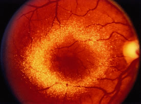
Toxic maculopathy is usually reversible only in its earliest phases. If these drugs have caused skin, eyelid, corneal (hydroxychloroquine), or hair changes, this may be an indicator of possible drug-induced retinopathy. Because the aminoquinolines are concentrated in pigmented tissue, macular changes have been thought to progress long after the drug is stopped. The bull's-eye macula is not diagnostic for aminoquinoline-induced disease because a number of other entities can cause this same clinical picture. Although retinal toxicity occurs in patients taking hydroxychloroquine, the incidence is much lower than with chloroquine.

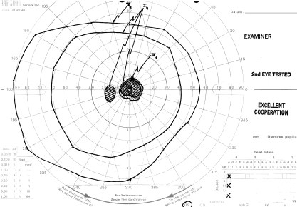
How to detect early toxic changes is still the subject of debate. Easterbrook5–8 has published extensively on chloroquine and hydroxychloroquine retinal toxicity. His current method of examination includes obtaining best corrected vision, examining the cornea with the pupil dilated with retroillumination, and performing an Amsler examination. (He has his patients test themselves every few weeks at home with an Amsler grid.) If the results of the Amsler grid examination are abnormal, a Humphrey field with the 10-2 red and white program is performed. If color vision is deficient on Ishihara testing or if there is any question of a patient's reliability in terms of visual field assessment,fluorescein angiography is done as well. According to Easterbrook, electroretinographic and electro-oculogram studies are either too variable or are abnormal only in late stages of chloroquine retinopathy, so their usefulness is suspect. Color testing is more useful in older patients in whom coincidental age-related macular changes occur. It is also stated that early retinopathy (i.e., small paracentral relative scotomas) (Fig. 2) does not appear to progress, at least in the short term. Patients who present with absolute scotomas and positive fluorescein angiography should be warned that their retinopathy may progress even if the chloroquine therapy is discontinued. There are numerous instances of progression of macular and optic nerve damage, even years after these drugs are discontinued. This may not be as true for hydroxychloroquine because the progression of the maculopathy may be significantly less than with chloroquine once the drug is stopped.

Fig. 2. Paracentral scotoma secondary to chloroquine toxicity.

Some patients wish to continue taking these drugs even with the visual side effects because only these drugs improve their quality of life. If reproducible abnormalities of the Amsler grids occur in this group of patients, kinetic and static perimetry are obtained. If field defects are confirmed, consultation with the rheumatologist concerning discontinuation of the drug is advised. If the patient is reluctant, one may consider halving the usual dosages and following the patient (even those with relative paracentral scotomas with serial fields) every 3 to 4 months. If, however, there is any progression, the recommendation is to stop the drug.

OVERVIEW

Maculopathy must be bilateral and reproducible by Amsler grid and visual field testing. Transient or unilateral defects are not sufficient reasons to implicate the drug and are not an indication to stop therapy. Color vision loss is a late change. The goal is to find early changes, such as relative scotomas substantiated by visual fields. Also suspect are patients with retinal changes, color vision loss, absolute scotoma, and decreased vision, because even if the drug is stopped, two thirds of these patients may continue to lose some vision and/or peripheral fields. Patients with early paracentral relative scotomas do not advance when the drug is discontinued.

GUIDELINES FOR FOLLOWING PATIENTS

Guidelines for follow-up of patients are controversial. Morsman and colleagues,9 Morand and colleagues,2 and Levy10 question the need to screen patients on hydroxychloroquine. Spalton11 suggests every 3 years and Levy10 suggests after 10 years. Blyth and Lane12 suggest ophthalmic examination only after the patient becomes symptomatic. We favor the approach by Easterbrook that is presented here.

  • Initial examination. We prefer baseline complete eye examination. This includes visual acuity, Amsler grids, color vision, and corneal retroillumination. If abnormalities are in the Amsler grid, automated perimetry is indicated.
  • Follow-up examinations. Hydroxychloroquine—Ophthalmic examination, repeating previously mentioned baseline studies every 12 to 18 months if the patients are taking less than 6.5 mg/kg (ideal body weight) with normal kidney and/or liver function. This is in keeping with the American Academy of Ophthalmology's recommendation for biannual eye examination in normal adults. If the patient has been taking the drug for more than 6 years or if the accumulative dose is 200 g, patients should be examined at least annually. Patients should see the ophthalmologist if there is any change in vision or if changes on their “home” Amsler grid testing are noticed. Chloroquine—The previously described tests are performed. The patient should be seen at least annually if dose is less than 3.0 mg/kg (ideal body weight). Patients should be seen every 6 months if dose is greater than 3.0 mg/kg (ideal body weight) or if they are short, are obese, or have renal and/or liver impairment.
  • American Academy of Ophthalmology recommendations are pending and will be similar to what we have described.

TESTS

  • Amsler grids appear to be the most cost-effective method of following patients. Monthly home testing is essential, especially if the patient is on higher doses or long-term therapy or has renal disease.
  • Routine visual fields are not necessary unless Amsler field defects, abnormal color vision, decreased vision, or abnormal retinal findings are detected. A baseline visual field has been advocated by some.
  • Retroillumination of the cornea with the slit lamp with dilated pupils to look for enhanced Hudson-Stähli line, or more commonly, whorl-like superficial corneal deposits; even if faint, these are early indicators of possible retinal toxicity in hydroxychloroquine patients.

CAUTION

To date, there are no data to show hydroxychloroquine toxicity worsening pre-existing macular degeneration. Common sense may make informed consent and explanation of risk-benefit ratios necessary on an individualized basis.

Back to Top
ISORETINOIN
Ocular side effects are dose related and probably the most frequent adverse reactions associated with these drugs. Retinal changes are rare compared with reports of blepharoconjunctivitis, subjective complaints of dry eyes, and transient blurred vision.

However, there are well-documented reports of patients who have decreased ability to see at night after taking these agents. This may occur as early as a few weeks or after taking the drug for 1 to 2 years. It was previously unrecognized that this may be a permanent effect. Retinal dysfunction is probably due to the competition for binding sites between retinoic acid and retinol (vitamin A). The risk of a photosensitizing drug, such as isotretinoin, enhancing the effects of light on the macula and retina is unclear. Although there are cases in the registry of retinal pigmentary changes, to date we consider this just background noise. Excessive amounts of retinoids have been implicated in papilledema secondary to pseudotumor cerebri and may suggest hypervitaminosis A.13 This is confusing, in part because other antibiotics may be used concomitantly (tetracycline, minocycline), which may also cause pseudotumor cerebri.

RECOMMENDATIONS FOR FOLLOWING PATIENTS

  1. If the patient is younger than the age of 40 and has not had an eye examination in a few years or if the patient is older than age 40 and has not had one within 2 years, it may be prudent to have a baseline ocular examination.
  2. Explain risk-benefit ratio to patients with
    1. Retinitis pigmentosa
    2. Chronic blepharoconjunctivitis
    3. Significant tear film abnormalities14

  3. In select patients with anterior segment pathology, consider ultraviolet (UV) blocking lenses because the drug is a photosensitizing agent.
  4. Consider discontinuing or delaying fitting of contact lenses while on this drug.
  5. Patients on long-term isotretinoin should have an annual eye examination.
  6. Suggest that patients see you if any significant ocular signs or symptoms occur.
  7. Question all patients concerning night blindness, keratitis sicca, and decreased color vision. If progressive or persistent, consider discontinuing the drug. Because most cases are transitory, these findings are not necessarily an indication for discontinuing the drug. However, if they persist for a number of weeks, consider closer monitoring and further testing. Informed consent should be considered.
  8. Consider discontinuing the drug if any of the following occurs until a cause is determined:
    1. Pseudotumor cerebri
    2. Optic neuritis
    3. Persistent night blindness
    4. Decreased color vision

Back to Top
SILDENAFIL
Sildenafil citrate, an oral therapy for erectile dysfunction (ED), is one of the largest-selling prescription drugs in the world. This inhibitor of phosphodiesterase type 5 (PDE-5) is a unique class of drugs not previously used in humans. PDE-5 is responsible for the degradation of cyclic guanosine monophosphate (cGMP) in the corpus cavernosum. With increased levels of cGMP, the smooth muscle in the corpus cavernosum is relaxed, allowing inflow of blood.15 Retinal side effects may occur because sildenafil, although selective for PDE-5, has a minor effect on PDE-6, an enzyme involved in light excitation in visual cells. The ocular side effects most commonly associated with sildenafil are a bluish tinge to the visual field, hypersensitivity to light, and hazy vision16 (Fig. 3).

Fig. 3. Sildenafil “blue-tinged” vision.

These reversible side effects may last from a few minutes to hours, depending on drug dose. Visual changes are seen in approximately 3% of men taking the standard 50-mg dose. Eleven percent of men taking 100 mg daily experience visual disturbances, and the incidence rises to 40% at a dose of 200 mg daily. Theoretically, patients with retinitis pigmentosa and patients with a genetic disorder of retinal phosphodiesterase may have increased side effects on sildenafil.17 Although there have been reports of vitreous or retinal hemorrhages, third nerve palsy, and nonarteritic anterior ischemic optic neuropathy (AION), in our opinion, these are not proven as drug related.18 Slight mydriasis may be indirectly due to emotional anticipation, and subconjunctival hemorrhage may be due to vascular dilatation caused by the drug. To date, electroretinogram (ERG) changes are reversible and of no clinical importance.

Back to Top
VIGABATRIN
Vigabatrin is a drug used in more than 50 countries for the treatment of refractory epilepsy. The main ocular side effect is symptomatic or asymptomatic visual field constriction, which is usually bilateral and can progress to tunnel vision.19–25 Unfortunately, more than 80% of these visual field defects appear to be irreversible. The incidence of visual field defects is unknown, but a United Kingdom study suggests that it is three to four cases in 10,000+ patients. Wilton and colleagues26 reported a 2% incidence of visual field abnormalities after 6 months of therapy in a large series of patients. Others suggest that patients on long-term therapy have a 10% to 20% chance of significant ocular problems. There is evidence to suggest that this is dose related; however, the time of onset varies greatly. Some cases occur as soon as 1 month after starting the drug, and others have occurred after more than 6 years of treatment. The mechanism behind the visual field defects could be related to impairment of the highly γ-aminobutyric acid (GABA)-ergic amacrine cells in the retina. Also, histopathologic studies in animals show a microvacuolation in myelin sheaths of white matter when exposed to vigabatrin. How these alterations affect the visual field in patients receiving vigabatrin is not currently known. Retinal changes have been described but disputed by others. Optic nerve changes include a few reports of optic nerve pallor and atrophy. Arndt and colleagues27 believe that visual field changes are enhanced if the patients are also on valproate. Also, outer retinal dysfunction should be present if visual field changes are present. Possibly, electro-oculogram is a more sensitive and specific diagnostic tool than ERG for drug-related retinal effects. Some groups have asked for removal of the drug from the marketplace because of the severity of ocular side effects. Other medications that increase levels of GABA in the central nervous system are being developed and show promise for use in treatment of epilepsy.

Beck28 has reviewed data on 2500 patients in formal clinical trials and 125,000 adverse event reports outside formal trials. He believes that severe visual field constriction resulting from this drug is real but rare. He and others point out that much is yet to be learned in this area.

RECOMMENDATIONS FOR FOLLOWING PATIENTS

The following recommendations are modified from the manufacturer's29 (Aventis Pharma, formerly Hoechst Marion Roussel) recommendations:

  1. Visual field testing every 6 months. If visual fields are abnormal, they must be confirmed by retesting.
  2. Patients should be questioned regularly for visual symptoms.
  3. If any new visual symptoms develop, consider repeating the visual field. If defects are extensive, progressive, and reproducible, then the risk-benefit ratio may need to be revisited. If the drug is discontinued, this must be done over a 2- to 4-week period.
  4. Baseline visual field—if cognitive age is 9 years or older, Goldmann or Humphrey fields should be used. Younger than age 9, there is currently no reliable method available. Possibly, ERG has a role here.
  5. Electroretinography or visual evoked potential is of minimal value to date but may be of benefit as more data are accumulated.

Back to Top
TAMOXIFEN
This antiestrogen is used primarily in the palliative treatment of breast carcinoma, ovarian cancer, pancreatic cancer, and malignant melanoma.

With more than 30 years of clinical use, we are now getting a more complete picture of the adverse retinal effects of this drug. Only recently has the work of Gorin and colleagues30 shown that this agent can cause decreased color vision. The incidence of retinal side effects is dependent on dosage or accumulated dose. More extensive retinal findings were evident when the drugs were given at daily doses of up to 180 mg per day; the standard dosage now is 20 mg per day or less. At lower doses, the severity and incidence of retinal complications are significantly lower. Even more important, the reversibility of these complications appears to be markedly improved at these lower doses. The accumulated dose is important, and major effects are seldom seen at total doses of less than 10 g. Regardless, in very rare individuals, retinal side effects have been seen at total doses of only a few grams. In a prospective study by Pavlidis and colleagues,31 6.3% of patients developed retinopathy from tamoxifen. However, most incidence levels are reported in the 1% to 2% range.

Based on data in the literature and the registry, there appear to be two forms of posterior segment involvement. An acute, debatable form, which is not well defined, may occur after only a few weeks of therapy, with any or all of the following: vision loss, retinal edema, retinal hemorrhage, and optic disc swelling. This may be a result of tamoxifen estrogenic activity, which may cause venous thromboembolism. These findings are reversible with discontinuation of tamoxifen. This may be associated with other systemic changes, and it has been postulated that this has an immune basis or is an idiosyncratic response. Typical crystalline retinopathy reveals striking white-to-yellow perimacular refractile bodies (Fig. 4). This finding has been reported for many medicines including nitrofurantoin, canthaxanthine, and methoxyflurane. These occur most commonly after more than 1 year of therapy with at least 100 g or more of the drug. There are, however, a number of cases in the registry and literature of minimal retinal pigmentary changes occurring after a few months and only a few grams of tamoxifen. This may be associated with cystoid macular edema, punctate macular retinal pigment epithelial changes, parafoveal hemorrhages, and peripheral reticular pigment changes. The refractile bodies are located in the inner retina and histologically may be the products of axonal degeneration. These lesions do not appear to regress if the drug is discontinued. Kalina and Wells32 pointed out that idiopathic juxtafoveal retinal telangiectasis can also be confused with tamoxifen retinopathy. Retinopathy resulting from tamoxifen can be seen without the presence of refractile bodies. Loss of visual acuity in this chronic form is often progressive, dose dependent, and irreversible unless the cystoid macular edema or hemorrhage is the cause of the visual loss.

Fig. 4. Canthaxanthine crystalline retinopathy.

RECOMMENDATIONS FOR FOLLOWING PATIENTS

Overview

Many believe that low-dose tamoxifen (less than 10 g cumulative) causes few to no ocular side effects. They recommend continuing tamoxifen until visual symptoms occur. At that point, even with retinal crystals, the patient should be seen every 3 months and the ophthalmologist should stay in close contact with the oncologist.33 We do not have a good 5-year follow-up on a large number of patients who are now starting this drug prophylactically for breast cancer. Therefore, firm guidelines are difficult.

Guidelines

The following guidelines are modified from Gorin:

  1. Baseline ophthalmic examination within the first year of starting tamoxifen. This should include slit lamp biomicroscopy of the anterior and posterior segments in combination with a handheld indirect or contact lens. Baseline color vision testing is important.
  2. In keeping with the American Academy of Ophthalmology's current recommendations, in normal adults, do a complete eye examination at least every 2 years.
  3. More frequent examinations should be performed if ocular symptoms occur.
  4. The discovery of a limited number of intraretinal crystals in the absence of macular edema or visual impairment does not seem to warrant discontinuation of the drug.
  5. Consultation with the oncologist is essential if significant ocular findings occur.
  6. Presence of age-related maculopathy is not a contraindication to the use of tamoxifen. However, informed consent may be advisable in our litigious society.
  7. Presence of posterior subcapsular cataracts is not an indication to stop the drug because the condition usually progresses even if the drug is discontinued.
  8. Significant color loss may be a valid reason to consider discontinuing the drug. Gorin recommends that stopping the drug for 3 months (in patients on prophylactic therapy) and retesting should be considered. If the color vision returns to normal, restart the drug and retest in 3 months. If, at any time, there is no rebound from stopping the drug, or continued progression, then one may need to consult the oncologist and re-evaluate the risk-benefit ratio.

Back to Top
PHENOTHIAZINES
Phenothiazines are used in the treatment of depressive, involutional, senile, or organic psychoses and various forms of schizophrenia. Some of the phenothiazines are also used as adjuncts to anesthesia, as antiemetics, and in the treatment of tetanus.

The phenothiazines, as a class, are one of the most widely used drugs in the practice of medicine today. The most commonly prescribed drug in this group is chlorpromazine, which has been so thoroughly investigated that over 10,000 publications alone deal with its actions. Even so, these drugs are remarkably safe compared with previously prescribed antipsychotic agents. Their overall rate of side effects is estimated at only 3%.

However, if patients are on phenothiazine therapy for a number of years, a 30% rate of ocular side effects has been reported.34 If therapy continues over 10 years, the rate of ocular side effects increases to nearly 100%. Side effects are dose and drug dependent, with the most significant side effects reported with chlorpromazine and thioridazine therapy, probably because they are the most often prescribed. These drugs in very high doses can cause significant adverse effects within a few days, whereas the same reactions usually would take many years to develop in the normal dose range.

Each phenothiazine has the potential to cause retinal side effects. The basic problem is that pinpointing specific toxic effects to a specific phenothiazine is difficult because most patients have been receiving more than one type. The most common adverse ocular effect with this group of drugs is decreased vision, probably resulting from anticholinergic interference. Retinopathy, optic nerve disease, and blindness are exceedingly rare at the recommended dose levels, and then they are almost only found in patients on long-term therapy.

Retinal pigmentary changes (Fig. 5) are most often found with thioridazine.35 This reaction is dose related and is seldom seen at recommended doses. A review by Marmor36 is the most up-to-date. A phototoxic process has been postulated to be involved in both the increased ocular pigmentary deposits and the retinal degeneration. The group of drugs with piperidine side chains (i.e., thioridazine) has a greater incidence of causing retinal problems than the phenothiazine derivatives with aliphatic side chains (i.e., chlorpromazine), which have relatively few retinal toxicities reported. The phenothiazines combine with ocular and dermal pigment and are only slowly released. This slow release has, in part, been given as the reason for the progression of adverse ocular reactions even after use of the drug is discontinued.

Fig. 5. Thioridazine toxicity.

RECOMMENDATIONS

  1. Photo-induced skin eruptions are well known, especially with chlorpromazine. Pigmentation-induced photosensitivity can be blocked in part by sunglasses that block out UV radiation up to 400 nm.
  2. Sunglasses that block out UV radiation up to 400 nm are helpful for possible lens-induced changes as well.
  3. The patient should avoid bright light when possible.

Back to Top
MISCELLANEOUS AGENTS
The following is a short list of drugs, which have been reported to cause retinal side effects:
  • Ibuprofen has caused visual evoked response alteration.37
  • Indomethacin has been noted to cause retinal pigment epithelium (RPE) disturbances.38–40
  • Desferrioxamine, used in the treatment of systemic iron overload, has been noted to cause retinal epithelial alterations.41
  • Clofazimine, used in the treatment of dapsone-resistant leprosy, has been noted to cause a bull's eye maculopathy in a patient with AIDS.42
  • Methanol ingestion can cause visual field defects, retinal edema, and optic atrophy.43
Back to Top
REFERENCES

1. Fraunfelder FT, Fraunfelder FW: Drug-Induced Ocular Side Effects. Boston: Butterworth Heinemann, 2001

2. Morand EF, McCloud PI, Littlejohn GO: Continuation of long term treatment with hydroxychlorquine in systemic lupus erythematosus and rheumatoid arthritis. Ann Rheum Dis 51:1318, 1992

3. Berstein HN: Ophthalmologic considerations and testing in patients receiving long-term antimalarial therapy. Am J Med 75:25, 1983

4. Johnson MW, Vine AK: Hydroxychloroquine therapy in massive total doses without retinal toxicity. Am J Ophthalmol 104:139, 1987

5. Easterbrook M: Detection and prevention of maculopathy associated with antimalarials. Int Ophthal Clin 39:49, 1999

6. Easterbrook M: An ophthalmological view on the efficacy and safety of chloroquine vs. hydroxychloroquine [Editorial]. J Rheumatol 26:1866, 1999

7. Easterbrook M: Ocular effects and safety of antimalarial agents. Am J Med 85(Suppl 4A):23, 1988

8. Easterbrook M: Is corneal decompensation of antimalarial any indication of retinal toxicity? Can J Ophthalmol 25:249, 1990

9. Morsman CDG, Liversey SJ, Richards IM et al: Screening for hydroxy-chloroquine retinal toxicity: Is it necessary? Eye 4:572, 1990

10. Levy GD: Hydroxychloroquine ocular toxicity. J Rheumatol 25:1030, 1998

11. Spalton DJ: Retinopathy and antimalarial drugs—the British. Lupus S1:570, 1996

12. Blyth C, Lane C: Hydroxychloroquine retinopathy: is screening necessary? BMJ 916:710, 1998

13. Gold JA, Shupack, JL, Nemec MA: Ocular side effects of the retinoids. Int J Dermatol 28:218, 1989

14. Rismondo V, Ubels JL: Isotretinoin in lacrimal gland fluid and tears. Arch Ophthalmol 105:416, 1987

15. Boolell M, Allen MJ, Ballard SA, et al: Sildenafil: An orally active type 5 cyclic GMP-specific phosphodiesterase inhibitor for the treatment of penile erectile dysfunction. Int J Impotence Res 8:47, 1996

16. Fraunfelder FT, Laties A: Visual side effects possibly associated with Viagra®. J Toxicol Cut Ocul Toxicol 19:21, 2000

17. Tsang S.H, Gouras P, Yamashita CK et al: Retinal degeneration in mice lacking the gamma subunit of the rod cGMP phosphodiesterase. Science 272:1026, 1996

18. Donahue SP, Taylor RJ: Pupil-sparing third nerve palsy associated with sildenafil citrate (Viagra). Am J Ophthalmol 126:476, 1998

19. Baulac M, Nordmann JP, Lanoe Y: Severe visual field constriction and side-effects of GABA-mimetic antiepileptic agents [Letter]. Lancet 352:546, 1998

20. Blackwell N, Hayllar J, Kelly G: Severe persistent visual field constriction associated with vigabatrin. Patients taking vigabatrin should have regular visual field testing [Letter, comment]. BMJ 314:180, 1997

21. Eke T et al: Severe persistent visual field constriction associated with vigabatrin. BMJ 314:180, 1997

22. Harding GFA et al: Severe persistent visual field constriction associated with vigabatrin. BMJ 316:232, 1998

23. Mackenzie R, Klistorner A: Severe persistent visual field constriction associated with vigabatrin. Asymptomatic as well as symptomatic defects occur with vigabatrin [Letter, comment]. BMJ 314:233, 1998

24. Ruether K et al: Electrophysiologic evaluation of a patient with peripheral visual field contraction associated with vigabatrin. Arch Ophthalmol 116:817, 1998

25. Wilson EA, Brodie MJ: Severe persistent visual field constriction associated with vigabatrin. Chronic refractory epilepsy may have role in causing these unusual lesions [Letter, comment]. BMJ 314:1693, 1997

26. Wilton LV, Stephens MDB, Mann RD: Interim report of the incidence of visual field defects in patients on long term vigabatrin therapy. Pharmacoepidemiol Drug Safety 8:S9, 1999

27. Arndt CF, Derambure P, Defoort-Dhellemmes S et al: Outer retinal dysfunction in patients treated with vigabatrin. Neurology 52:1205, 1999

28. Beck RW: Vigabatrin-associated retinal cone system dysfunction [Letter]. Neurology 51:1778, 1998

29. Guidelines for Visual Field Screening. Aventis Pharma Ltd, Jan 2000

30. Gorin MB, Day R, Costantino JP et al: Long-term tamoxifen citrate use and potential ocular toxicity. Am J Ophthalmol 125:493, 1998

31. Pavlidis NA et al: Clear evidence that long-term, low-dose tamoxifen treatment can induce ocular toxicity. A prospective study of 63 patients. Cancer 69:2961, 1992

32. Kalina RE, Wells CG: Screening for ocular toxicity in asymptomatic patients treated with tamoxifen. Am J Ophthalmol 119:112, 1995

33. Heier JS, Dragoo RA, Enzenauer RW et al: Screening for ocular toxicity in asymptomatic patients treated with tamoxifen. Am J Ophthalmol 117:772, 1994

34. Ngen CC, Singh P: Long-term phenothiazine administration and the eye in 100 Malaysians. Br J Psychiatry 152:278, 1988

35. Lam RW, Remick RA: Pigmentary retinopathy associated with low-dose thioridazine treatment. Can Med Assoc J 132:737, 1985

36. Marmor HF: Is thioridazine retinopathy progressive? Relationship of pigmentary changes to visual function. Br J Ophthalmol 74:739, 1990

37. Hamburger HA, Beckman H, Thompson R: Visual evoked potentials in ibuprofen (Motrin) toxicity. Ann Ophthalmol 16:328, 1985

38. Burns CA: Indomethacin reduced retinal sensitivity and corneal deposits. Am J Ophthalmol 66:825, 1968

39. Henkes HE, Van Lith GHM, Canta LR: Indomethacin retinopathy. Am J Ophthalmol 73:846, 1972

40. Palimeris G, Koliopoulos J, Velissaropoulos P: Ocular side effects of indomethacin. Ophthalmologica 164:339, 1972

41. Rahi AHS, Hungerford JL, Ahmed AI: Ocular toxicity of desferrioxamine light microscopic histochemical and ultrastructural findings. Br J Ophthalmol 70:373, 1986

42. Craythorn JM, Swartz M, Creel EJ: Clofazimine-induced bull's eye retinopathy. Retina 6:50, 1986

43. Benton CD, Calhoun FP: The ocular effects of methyl alcohol poisoning: Report of a catastrophe involving three hundred and twenty persons. Trans Am Acad Ophthalmol 56:875, 1952

Back to Top