|
| Chapter 32 Retinopathy and Distant Extraocular Trauma ISAAC A. LOOSE and ROBERT P. SCHROEDER Table Of Contents |
|
PURTSCHER'S RETINOPATHY TRAUMATIC ASPHYXIA FAT EMBOLISM RETINOPATHY VALSALVA RETINOPATHY WHIPLASH MACULOPATHY SHAKEN BABY SYNDROME REFERENCES |
| The retinal manifestations of distant trauma may be asymmetric and vary
among patients depending on the type of trauma. Discrepancies in retinal
findings relate to an incompletely understood pathogenesis of the
retina's response to distant trauma, as well as the type of trauma
sustained. For example, retinal changes resulting from long bone fractures
are manifest differently than the retinal changes resulting from
whiplash injuries. The pathophysiologic mechanisms of retinal damage after distant trauma have been debated. Three mechanisms have been proposed to explain the resulting fundus findings: (1) increased intraluminal pressure may damage the retinal vascular endothelial cells; (2) emboli from sources including air, blood products, or fat may also damage the retina, a theory that has been supported in experimental models; and (3) mechanical forces acting at the vitreoretinal interface may damage the retina. In this chapter we describe six clinical entities: Purtscher's retinopathy, traumatic asphyxia, fat embolism retinopathy, Valsalva retinopathy, whiplash maculopathy, and shaken baby syndrome. With the exception of whiplash maculopathy, the five retinopathies have some overlap in either clinical presentation or pathophysiology and the categorization of the retinopathies relates more to the type of trauma than to a unique retinal appearance. |
| PURTSCHER'S RETINOPATHY | |
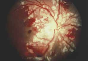
Purtscher's retinopathy is characterized by retinal hemorrhages, exudates, and
decreased vision associated with nonocular trauma (Fig. 1). In 1912, Otmar Purtscher described multiple, white retinal patches
and retinal hemorrhages surrounding a normal-appearing optic
disc in five patients with visual loss after severe head trauma.1 Most commonly, Purtscher's retinopathy develops as a sequela of chest-compressing
trauma. The severity of the traumatic event is
variable, ranging from minimal external trauma to crushing chest wall
injuries. The onset of symptoms usually occurs within 2 days after trauma. Both
eyes are typically involved, but unilateral cases have been
reported.2,3 Patients complain of decreased vision, often from 20/200 (6/60) to
counting fingers. Fundus examination usually reveals numerous
white retinal patches or confluent cotton-wool spots around
the disc, as well as superficial retinal hemorrhages. Other findings
include serous macular detachments, dilated and tortuous vasculature, and
disc edema. The peripheral retina is commonly spared. Fluorescein
angiography can reveal focal areas of arteriolar obstruction, patchy
capillary nonperfusion, disc edema, and dye leakage from retinal arterioles, capillaries, and
venules.3 Purtscher1 originally proposed that the etiology of the white retinal lesions resulted
from lymph extravasated from retinal vessels during a sudden increase
in intracranial pressure. In 1962, Marr and Marr4 wrote that the retinopathy resulted from reflux venous shock waves produced
from intrathoracic chest compression. Several authors have implicated
arteriolar emboli including air and fat as the cause.3,5,6 Other authors have suggested an etiologic role for granulocyte or other
blood product emboli formed after complement activation, arguing that
microembolization is a mechanism common to the varied clinical settings
of Purtscher's retinopathy.7,8 Interestingly, a Purtscher's-like fundus picture may occur
in several nontraumatic settings, such as acute pancreatitis, chronic
renal failure, thrombotic thrombocytopenic purpura lupus erythematosus, and
childbirth.7,9–12 Clinically, the retinal lesions resolve over a period of weeks to a few
months.4 After resolution, the fundus may appear normal, but pigment migration
and optic atrophy can occur.13Although visual acuity can remain reduced, the acuity may return to normal
or near normal.4
|
| TRAUMATIC ASPHYXIA |
| Traumatic asphyxia usually results from severe compression of the thorax and is characterized by a striking, ecchymotic mask, or blue discoloration of the upper chest and the face. External ocular involvement is virtually universal in cases of traumatic asphyxia. Patients have ecchymotic eyelids and hemorrhagic conjunctiva but retinal changes occur less commonly. Trampling, suicide attempts by hanging, seizures, vomiting, and childbirth have caused traumatic asphyxia.14 Visual acuity can be unaffected by traumatic asphyxia but may be reduced to no light perception.4 Fundus examination may reveal intraretinal hemorrhages, as well as cotton-wool spots and disc edema. Often the retina may be ophthalmoscopically normal.4,13 In one case of traumatic asphyxia, fluorescein angiography revealed in one eye blockage of fluorescence by retinal hemorrhage, blurring of the background choroidal pattern associated with cotton-wool spots, and hyperfluorescence with dye leakage associated with hemorrhage at the nasal edge of the disc. The other eye was angiographically normal.15 The fundus changes of this patient improved over a period of weeks, but the visual acuity of the right eye had decreased from 20/30 (6/9) to 20/100 (6/30), presumably as a result of mottling and disruption of the retinal pigment epithelium.15 The pathogenesis of traumatic asphyxia retinopathy and Purtscher's retinopathy may be similar but the ecchymotic appearance associated with traumatic asphyxia separates the two entities. Purtscher's retinopathy usually has no associated external findings.15 Also, the development of Purtscher's retinopathy may be slower than the retinopathy of traumatic asphyxia. Again, patients with traumatic asphyxia are treated supportively. |
| FAT EMBOLISM RETINOPATHY |
| Fat embolism retinopathy is usually secondary to fat embolism syndrome (FES). Retinal findings variably include cotton-wool spots, intraretinal hemorrhages, and visible emboli. FES was first described in 1861 in patients suffering fractures of medullated bones.16 The most likely fractures producing the syndrome are fractures of the lower extremities and the pelvis.17–19 Typically, the manifestations of FES ppear within 24 to 48 hours after the injury, but the syndrome is only recognized in approximately 5% of patients with long bone fractures. Some of these manifestations include petechial rashes, respiratory insufficiency, retinal lesions, and altered mental status.20 In patients with manifest FES, 50% to 60% may have retinal findings.19–21 Chuang et al.22 reported that of 100 consecutive long bone fracture cases, four patients demonstrated retinal pathology resulting from subclinical FES. In this series three patients had normal vision and one patient complained of a visual field defect. Fundus examination classically reveals cotton-wool spots and intraretinal hemorrhages. Additional fundus findings include intravascular fat emboli and central retinal artery occlusion.6,22–24 This retinopathy has occurred after facial autologous fat injection.25,26 Various alterations in lipid homeostasis are probably involved in the pathogenesis of FES. Retinal microinfarcts from fatty emboli have been demonstrated histopathologically, and this may reveal, at least in part, the etiology of the retinal findings.6 The retinal lesions resolve after resolution of the FES. With resolution, most patients are asymptomatic, although permanent scotomas can occur.21 |
| VALSALVA RETINOPATHY | |
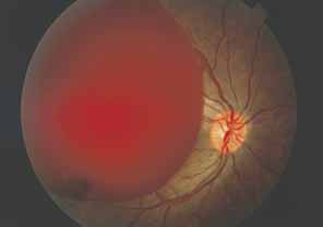
Valsalva retinopathy develops in response to the valsalva maneuver. A rapid
rise in abdominal pressure especially against a closed glottis characterizes
this maneuver (Fig. 2). Some common settings in which the Valsalva maneuver occurs include
heavy lifting, coughing, vomiting, or bowel movement. A history of
such activity is helpful in establishing the diagnosis of Valsalva retinopathy. Patients
may be asymptomatic but usually complain of decreased
vision. Fundus examination most commonly reveals a red, dome-shaped
hemorrhage underneath the internal limiting membrane (ILM).27 A fluid level may be observed in the area of the hemorrhage as the blood
settles inferiorly under the ILM.27 Valsalva retinopathy may cause decreased visual acuity if blood obscures
the macula. Also, extramacular preretinal hemorrhage may diffuse into
the vitreous, which may limit vision.3,21 Usually, the sub-ILM hemorrhage resorbs after several days to weeks. A
serous detachment of the ILM may persist after the blood resolves
but spontaneous reattachment usually occurs, leaving a normal-appearing
fundus.27 Fluorescein angiography typically shows no retinal vascular alterations
except in cases in which Valsalva retinopathy has been associated with
other retinal disorders such as retinal artery macroaneurysm or diabetic
retinopathy.27–29 Duane30 proposed that the pathophysiology might be related to the rapid rise in
intravenous pressure caused by the Valsalva maneuver. With a rise in
intraocular venous pressure, the Valsalva maneuver causes a rupture of
the superficial retinal capillaries.27,30–32 For the usual cases of valsalva retinopathy, the preretinal hemorrhage
clears spontaneously, though neodymium–yttrium-aluminum-garnet
laser disruption of the internal limiting membrane has been
used to allow a more rapid clearing of the preretinal hemorrhage.33
|
| WHIPLASH MACULOPATHY |
| Whiplash maculopathy occurs after flexion–extension head and neck injuries. Patients who have suffered whiplash injuries may complain of a wide range of ophthalmic disturbances. In one study, 26% of patients with whiplash injuries had ophthalmic complications, most commonly a loss of accommodation or convergence.34 Reports of retinal damage attributed to whiplash injury are less common. Whiplash maculopathy symptoms begin with a mild blurring of vision that occurs at the time of the injury, usually in the 20/30 range. The symptoms are bilateral and resolve over a period of a few days. Acutely, the fundus examination may reveal a minimal detachment of the posterior vitreous with a grayish appearance of the fovea. Later, a crater-like depression of less than 100 μmin diameter with slight retinal pigment epithelium disturbances may be found and may remain unchanged over time even though symptoms may resolve.35,36 Some foveal pits may be the result of remote whiplash injury. Small opercula in association with whiplash maculopathy suggest that a true retinal excavation exists and that the pathogenesis of whiplash maculopathy is related to vitreous traction on the macula.35 |
| SHAKEN BABY SYNDROME | |
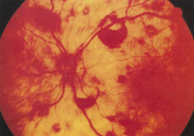
Caffey37 described the first cases of a form of child abuse that he later termed
the whiplash shaken infant syndrome. This syndrome is defined by violent shaking of an infant that commonly
results in intraocular and intracranial hemorrhages (Fig. 3). Ocular complications occur commonly in child abuse cases. Several
studies have found that 35% to 46% of abused children
may have eye injuries.38–40 Importantly, the shaken infant may present with minimal external signs
of trauma. Certainly, numerous cases have occurred in which the head
has shown no signs of visible trauma. The mortality rate (15%) and
the morbidity rate (50%) underscores the
importance of recognizing this form of child abuse.41 Ocular examination usually reveals any combination of subretinal, intraretinal, preretinal, or
vitreous hemorrhage. The presence of intraocular
hemorrhage is a predictor of intracranial hemorrhage, and the severity
of the intraocular hemorrhage correlates with the severity of the
acute neurologic injury.42,43 The usual intracranial manifestation of the shaken baby syndrome is subdural
hematoma. Bridging dural vessels may tear and bleed in response
to repetitive acceleration and deceleration motion of the brain caused
by shaking. Early views held that the pathogenesis of shaken baby retinopathy
related to an acute rise in intracranial pressure.37 More recently, Greenwald and associates44 proposed that the same acceleration and deceleration forces that cause
intracranial hemorrhage act on the vitreous. Vitreous forces perpendicular
to the plane of the retina cause a separation of the ILM or a splitting
of deeper retinal layers. Retinal hemorrhages result from tearing
of small retinal vessels.44,45 The clinical course of shaken baby retinopathy ranges from complete clearing
to severe visual loss secondary to optic atrophy or macular scarring.46 Treatment of the retinal manifestations of shaken baby syndrome is most
commonly supportive, but the ophthalmologist must be wary of the possibility
of significant intracranial trauma in the shaken infant.
|