Chapter 16
Eales' Disease
JORGE H. GUTIÉRREZ and ROBERT P. MURPHY
Main Menu   Table Of Contents

Search

EPIDEMIOLOGY
CLINICAL FEATURES
PROGNOSIS
TREATMENT
PATHOGENESIS
REFERENCES

Although controversy remains about its precise definition, Eales' disease is widely considered to be an idiopathic obliterative vasculopathy that primarily affects the peripheral retina of healthy, young adults. Vascular sheathing and focal occlusion of small peripheral retinal vessels occur early in the course of the disease. With progression, larger areas of nonperfusion develop, extending posteriorly. Neovascularization can occur, usually at the junction of the perfused and nonperfused retina, frequently resulting in vitreal hemorrhages.

The disease is named after Henry Eales, an ophthalmologist who in 1880 described a syndrome of recurrent vitreal hemorrhages in young men with epistaxis and constipation.1 He noted abnormal retinal veins and attributed the hemorrhages to increased venous pressure caused by constipation. He did not observe any new vessels or inflammation preceding or accompanying the hemorrhages. The association with constipation and epistaxis has long since been disproved, and nonperfusion, inflammation, and neovascularization subsequently have been associated with this disease.

Not all have believed that Eales' disease is a specific entity. Duke-Elder believed that Eales' disease represented the clinical manifestation of many diseases.2 Since then, refined diagnostic tests have demonstrated that many of the so-called idiopathic hemorrhages were caused by known etiologies such as sarcoidosis, systemic lupus erythematosus, diabetes mellitus, sickle cell disease, and collagen vascular disease.

However, after elimination of these causes, a group of patients with idiopathic peripheral nonperfusion and perivasculitis of the retina remains. Investigators generally agree that Eales' disease is a distinct entity comprising characteristic funduscopic and fluorescein angiographic features.3 Although this disease has been called “periphlebitis retinae,”4 emphasizing the abnormalities of retinal venules, recent evidence suggests that this disease affects both arterioles and venules.3

Back to Top
EPIDEMIOLOGY
Eales' disease is uncommon in North America; however, this disorder is responsible for widespread visual loss in India, Pakistan, and Afghanistan. The incidence in India has been reported as 1 in 200 to 250 ophthalmic patients,5 and up to 1.35% in an opthalmic referral center.6 It typically affects healthy young adults between their third and fourth decades of life. Patients usually present with symptoms of vitreal hemorrhage such as floaters or decreased vision. Most patients develop bilateral, although often asymmetric, involvement. Most reports, including Henry Eales' original description, indicate a male predominance. However, recent reports suggest an equal prevalence among both men and women.7
Back to Top
CLINICAL FEATURES
Signs of ocular inflammation are commonly encountered in Eales' disease, especially early in its course. Vascular sheathing is seen in most patients (Fig. 1). The degree of sheathing ranges from thin white lines on both sides of the blood column to thick heavy exudative sheathing. Areas of sheathing frequently leak dye with fluorescein angiography (Fig. 2). However, there is not a direct correlation between the regions of sheathing and staining.

Fig. 1. Vascular sheathing. A. Thin white lines surround retinal venule. B. Exudative-type arteriolar sheathing.

Fig. 2. Fluorescein angiogram demonstrating abnormal vascular staining in a patient with Eales' disease. There was venous sheathing in these areas.

In the century since Henry Eales' observation of altered retinal veins, many investigators have described Eales' disease as a primary disease of altered retinal veins. Elliot and Harris suggest the term periphlebitis retinae for this disorder.4 However, recent reports suggest equal involvement of arteriolar and venular sheathing. Because of the evidence of arteriolar involvement (see Fig. 1B), this disease should be considered as a retinal vasculitis or vasculopathy. Others have used the term primary retinal perivasculitis.8 Cystoid macular edema, vitreal cells, keratic precipitates, and cell and flare in the anterior chamber have been observed in patients with Eales' disease.3

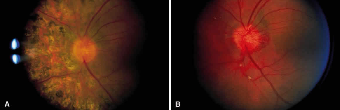
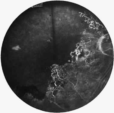
A varying degree of peripheral retinal nonperfusion is present in all patients with this disease. The nonperfusion generally is confluent and sharply demarcated from the posterior perfused retina (Fig. 3). Fine white lines representing the remains of obliterated large vessels (ghost vessels) often are seen in the area of nonperfusion. The temporal retina is most commonly affected.

Fig. 3. Fluorescein angiogram of the peripheral retina demonstrates the junction of normally perfused retinal vessels adjacent to an area of nonperfused retina. Notice the vascular abnormalities at the junction.

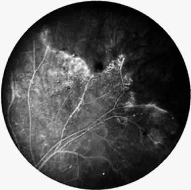
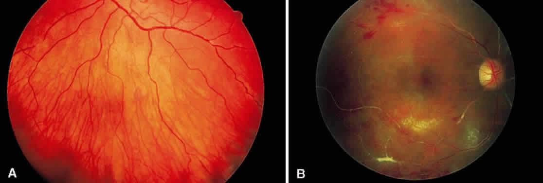
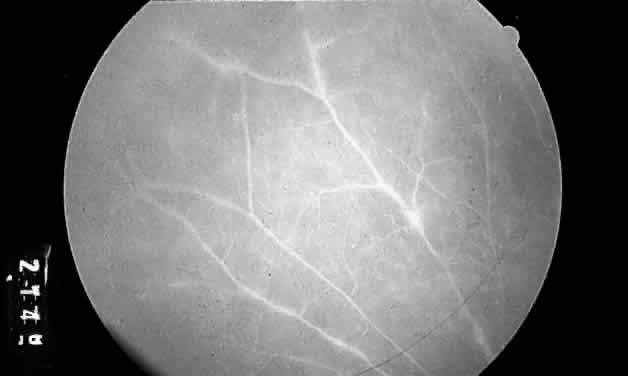
Elliot and Spitnas and colleagues have documented the abnormalities at the junction between the anteroperipheral nonperfused and the posterior perfused retina.9,10 Intraretinal hemorrhages often first appear in the affected area, followed by an increase in vascular tortuosity with frequent collateral formation around occluded vessels (see Fig. 3). Microaneurysms, arteriovenous shunts, and venous beading are commonly seen at the junction (Fig. 4). Fluorescein angiography enhances these abnormalities and often demonstrates staining at the stumps of obliterated vessels.

Fig. 4. Fluorescein angiogram demonstrating severe nonperfusion involving the macula. In this case, the macular nonperfusion was responsible for loss of central vision.

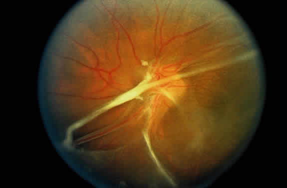
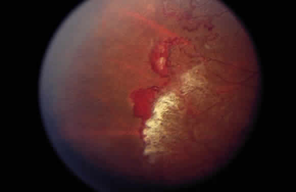
As a result of the retinal nonperfusion, new vessels can form either on the disc (neovascularization of the disc) or, more commonly, neovascularization can occur elsewhere in the retina (Fig. 5). These abnormal blood vessels can hemorrhage and are the major cause of visual loss in this disease. The neovascularization in the peripheral retina usually occurs at the junction of perfused and nonperfused retina, similar to the appearance of neovascularization in the peripheral retina in diabetic retinopathy and the other peripheral proliferative retinopathies. Neovascularization can be associated with extensive fibrovascular proliferation and fibrosis (Fig. 6). The anteroposterior and tangential traction resulting from the fibrovascular proliferation places these eyes at risk for development of retinal detachment. Neovascularization of the iris also has been described.

Fig. 5. Neovascularization of the peripheral retina has developed at the junction of perfused and nonperfused retina.

Fig. 6. Hypovascular fibroproliferation emanating from the disc. Usually, the vascular component of this proliferation is less prominent than that seen in the other proliferative retinopathies. Notice the tractional retinal detachment.

Back to Top
PROGNOSIS
The natural course of this disease is variable. Although the visual acuities of patients with Eales' disease range from normal to no light perception, most eyes retain good acuity. Vitreal hemorrhage is the major cause of visual loss. Usually, the hemorrhage settles to the lower portion of the vitreous and is gradually reabsorbed.

Most patients with Eales' disease retain at least reading acuity in one eye, but a few patients become legally blind. Severe visual loss usually results from complications associated with neovascularization, such as persistent vitreal hemorrhage, retinal detachment, and neovascular glaucoma. Occasionally, loss of vision is caused by cystoid macular edema, macular holes, retinal telangiectasia, or epiretinal membrane. In some patients, relentless nonperfusion progresses across the macula (see Fig. 4); visual acuity in these eyes usually is less than 20/400.

Back to Top
TREATMENT
Henry Eales offered his patients a mixture of laxative, digitalis, and belladonna. Other remedies have included vitamin C, thyroid extract, and high-dose steroids. Because of the previously reported association with tuberculosis (TB), empirical anti-TB therapy is used in India.5 None of these treatments have been conclusively beneficial. Laser photocoagulation is the treatment of choice for the neovascularization of Eales' disease.1112 Numerous investigators have demonstrated regression of neovascularization with light-intensity scatter photocoagulation applied to the nonperfused peripheral retina and to the junction of perfusion and nonperfusion (Fig. 7). Vitrectomy can be used for removing persistent vitreal hemorrhages and fibrosis, often with good results.13 No treatment is known to prevent or reverse the nonperfusion or capillary dropout.

Fig. 7. A. Neovascularization of the disc. Notice the segmental exudative arteriolar sheathing. B. Same patient as in Figure 7A, 2½ years later. The patient had regression of the neovascularization after treatment with scatter photocoagulation of nonperfused retina. Areas of arteriolar sheathing previously observed are no longer present.

Back to Top
PATHOGENESIS
Many investigators emphasize a relation between Eales' disease and TB. A higher than normal incidence of positive reaction to the tuberculin protein has been reported, but there is no direct evidence that the ocular changes are related to active TB. Renie and colleagues noted in their group of 32 patients that 48% had either TB or a history of exposure to TB.7 A recent retrospective study from India involving 1005 patients with active pulmonary and extrapulmonary TB did not detect eyes with Eales' disease in the involved patients.14 Further, in a different study, the same author compared the humoral and cellular immune sensitivity to TB among 56 patients with Eales' disease and 50 healthy controls.6 The only statistically significant difference in the Eales' patients was a higher prevalence of IgM to the A60 mycobacterial antigen compared with controls, whereas the controls had a higher prevalence of IgG against A60. There was no clear relation between immune reactivity to TB antigens and Eales' disease. Further study is needed either to prove or disprove a relation between Eales' disease and TB. Histopathologic studies and biochemical studies suggest an autoimmune mechanism for Eales' disease.15

Bhomma and coworkers implicate cellular damage by lipid peroxidation with a concomitant decrease in antioxidant levels in patients with Eales' disease.16 They suggest that antioxidant therapy may be beneficial for Eales' patients.

Several studies report on concomitant myelopathy secondary to infarction in young patients with Eales' disease.17,18 Although the etiology of this abnormality is unknown, it may have a vascular basis and may indicate that the pathologic consequences of Eales' disease are not confined to the eye.

Eales' disease is a long recognized but poorly understood disorder. Until more is understood about the etiology and pathogenesis of Eales' disease, it will be possible only to continue treating the secondary complications of this disorder.

Back to Top
REFERENCES

1. Eales H: Causes of retinal hemorrhage associated with epistaxis and constipation. Birmingham Med Rev 9:262, 1880

2. Duke-Elder WS: Diseases of the retina. In System of Ophthalmology, vol 10, pp 222–236. St Louis, CV Mosby, 1967

3. Gieser SC, Murphy RP: Eales' disease. In Ryan S (ed): Retina, vol 2, pp 1503–1507. St Louis, CV Mosby, 1994

4. Elliot AJ, Harris GS: The present status of the diagnosis and treatment of periphlebitis retinae (Eales' disease). Can J Ophthalmol 4:117, 1969

5. Das T, Biswas J, Kumar A et al: Eales' disease. Indian J Ophthalmol 42:3, 1994

6. Biswas J, Narain S, Roy S, Madhavan H: Evaluation of lymphocyte proliferation assay to purified protein derivative, enzyme linked immunosorbant assay, and tuberculin hypersensitivity in Eales' disease. Indian J Ophthalmol 45:93, 1997

7. Renie WA, Murphy RP, Anderson KC: The evaluation of patients with Eales' disease. Retina 3:243, 1983

8. Gass JDM: Stereoscopic Atlas of Macular Diseases: Diagnosis and Treatment. St Louis, CV Mosby, 1987

9. Elliot AJ: Thirty year observation of patients with Eales' disease. Am J Ophthalmol 80:404, 1975

10. Spitnas M, Meyer-Schwickerath G, Stephan B: The clinical picture of Eales' disease. Graefes Arch Clin Exp Ophthalmol 194:73, 1975

11. Spitznas M, Meyer-Schwickerath G, Stephen B: Treatment of Eales' disease with photocoagulation. Graefes Arch Clin Exp Ophthalmol 194:193, 1975

12. Meyer-Schwickerath G: Eales' disease: Treatment with light coagulation. Acta XIX Concilium Ophtholmologicurn 2: 862, 1962

13. Smiddy WE, Isernhagen RD, Michels RG, Glaser BM: Vitrectomy for nondiabetic vitreous hemorrhage. Retina 8:88, 1988

14. Biswas J, Badrinath S: Ocular morbidity in patients with active systemic tuberculosis. Int Ophthalmol 19:293, 1996

15. Sen D, Sarin G, Ghosh B et al: Serum alpha-1 acid glycoprotein levels in patients with idiopathic peripheral retinal vasculitis. Acta Ophthalmol 70:515, 1992

16. Bhooma V, Sulochana K, Biswas J, Ramakrishnan S: Eales' diseases: Accumulation of reactive oxygen intermediates and lipid peroxides and decrease of antioxidants causing inflammation, neovascularization and retinal damage. Curr Eye Res 16:91, 1997

17. Atabay C, Erdem E, Kansu T, Eldem B: Eales' disease with internuclear ophthalmoplegia. Ann Ophthalmol 24:267, 1992

18. Katz B, Wheeler D, Weinreb R, Swenson M: Eales' disease with central nervous system infarction. Ann Opthalmol 23:460, 1991

19. Murphy RP, Renie WA, Proctor LR et al: A survey of patients with Eales' disease. In Fines S, Owens S (eds): Management of Retinal Vascular and Macular Disorders, p 29. Baltimore, Williams & Wilkins, 1983

Back to Top