Chapter 2
Molecular Physiology and Pathology of the Retina
STEPHEN H. TSANG and PETER GOURAS
Main Menu   Table Of Contents

Search

FUNCTION OF THE RETINA
PHOTOTRANSDUCTION OF RODS
PHOTOTRANSDUCTION OF CONES
NEUROCIRCUITRY OF THE RETINA
ELECTROPHYSIOLOGIC TESTS
ACQUIRED RETINAL DYSTROPHIES
HEREDITARY RETINAL DYSTROPHIES40,41
GENETIC APPROACHES TO RETINAL DISEASES
RETINAL DEGENERATIONS
STATIONARY NYCTALOPIAS
MACULAR DEGENERATION
OTHER GENETIC RETINAL DISEASES
MOLECULAR PATHOGENESIS
THERAPY
REFERENCES

We have made great progress over the past decade in understanding both the mechanism of phototransduction and the neurocircuitry of the retina. Molecular biology and genetics are revolutionizing the way we study, diagnose, and eventually treat retinal disease by providing important insights into retinal physiology and pathophysiology.
Back to Top
FUNCTION OF THE RETINA
The retina performs two major functions: it transduces light into neural signals, and it transmits these signals in a language usable by our visual brain. Patterns of light and darkness on the photoreceptor mosaic are transformed by retinal circuits that lead to repetitive discharges from a two-dimensional array of ganglion cells. There is no feedback from our brain to the retina, making these ganglion cell responses stereotypical, depending only on the retinal image and their presynaptic circuitry.
Back to Top
PHOTOTRANSDUCTION OF RODS

ACTIVATION

Since 97% of the photoreceptors in cows and frogs consist of rods, research on the physiology and biochemistry of rods has been more approachable technically than has research on cones. With the advent of molecular cloning, however, the phototransduction components of cones are being isolated as well. In rods, phototransduction1,2 starts with the absorption of light by rhodopsin, a seven-loop transmembrane G-protein-coupled receptor containing an 11-cis chromophore covalently linked to a lysine side chain by a protonated Schiff base (Fig. 1). Isomerization by light of the 11-cis retinal to the all-trans retinal form leads to conformational changes in the opsin, producing the activated form metarhodopsin II. Each metarhodopsin II molecule activates hundreds of transducins (heterotrimeric G-proteins [Tαβγ]),3 to exchange guanosine diphosphate (GDP) for guanosine triphosphate (GTP) (Fig. 2). The transducin (Tα)-GTP molecule dissociates from Tβγ and binds to the inhibitory γ-subunits of a cGMP-phosphodiesterase (PDE; a heterotetrameric protein),4,5 thus removing the inhibition that the two γ-subunits exert on the catalytic α- and β-subunits of PDE.3–7

Fig. 1. A schematic view of the rhodopsin molecule with each of its amino acids designated as a small circle; the filled circles indicate amino acids that are identical in different species (i.e., conserved by evolution). The seven segments that are hydrophobic and incorporated into the lipid membranes of the rod discs are also evident, as are the cytoplasmic and intradiscal ends of the molecule. The 11-cis retinal group and its binding to a lysine in the membrane segment of the molecule is also included.

Fig. 2. A flow chart of the phototransduction cascade in which quantal excitation of rhodopsin leads to activation of the phosphodiesterase (PDE), decreasing cyclic guanosine monophosphate (cGMP) levels, closing of Na+ /Ca+ + channels, and hyperpolarization of the rod. Darkness causes the opposite changes, driven by the feedback inactivation mechanism.

It is debatable whether Tα actually binds and removes PDEγ subunits from PDEαβ(γ)27 or whether it binds and remains associated with PDEαβ as a complex.6,8 In frog photoreceptors, Tα activation of PDE results in release of PDEγ from the membrane-bound PDE. In bovine photoreceptors, however, PDEγ remains bound to PDEαβ upon Tα activation.9 The resulting activation of PDEαβ is profound: light-activated PDE is nearly 300 times more active than PDE in the basal level.10 Hydrolysis of cGMP by activated PDE lowers the levels of this cyclic nucleotide,11 which leads to closure of cGMP-gated Na+ /Ca+ + channels on the plasma membrane of the outer segment (see Fig. 2; Fig. 3). This reduces the entry of Na+ and Ca+ + into the cytoplasm, causing the entire rod to hyperpolarize, and thus curtails the release of the rod's transmitter, glutamate, at its synaptic terminal; intracytoplasmic reduction of Ca+ + also reduces transmitter release.

Fig. 3. The phototransduction cascade that results from activation of rhodopsin, subsequent formation of activated Tα, and activation of phosphodiesterase (PDE), which reduces the cGMP in the rod cytoplasm and closes Na+ /Ca+ + channels on the outer plasma membrane of the rod. Cyclase = guanylate cyclase; GCAP = guanylate cyclase-activating protein; P = PDE; Pd = phosducin; R kinase = rhodopsin kinase; T = transducin.

DEACTIVATION

The termination of the photoresponse requires inactivation of the photoexcited rhodopsin and transducin subunit, Tα, as well as reassociation of PDEγ to the PDEαβ catalytic core; it also involves activation of a guanylate cyclase to synthesize more cGMP. Tα possesses intrinsic GTPase activity for its own inactivation, which is accelerated upon binding to PDE.10 Photoactivated rhodopsin is inactivated by phosphorylation of serine and threonine residues at its carboxyl tail by a rhodopsin-specific kinase. Phosphorylated rhodopsin binds arrestin (also known as S-antigen) more efficiently than Tα. Arrestin inhibits the removal of phosphates from rhodopsin by a phosphatase 2A.12 Thus the amount of active (dephosphorylated) rhodopsin available to activate Tα is diminished. In minutes, rhodopsin is regenerated by the replacement with 11-cis retinal, released from arrestin, and dephosphorylated by phosphatase 2A (see Fig. 3).

ADAPTATION

The light-induced drop in Ca+ + influx also leads to adaptation of the photoreceptor to constant light.13 Intracellular Ca+ + inhibits the guanylate cyclases Ret GC-1 and Ret GC-2.14,15 When Ca+ + is reduced, these two enzymes are stimulated to form more cGMP, thus counteracting the reduction in cGMP by the photoresponse (see Fig. 2). In low Ca+ + concentrations, Ret GC-1 and Ret GC-2 are also stimulated by guanylate cyclase-activating proteins (GCAP-1/p20 and GCAP-2/p24) that possess Ca+ + binding sites. Ca+ + also appears to influence the half-life of photoexcited rhodopsin16 as well as its catalytic action. The rate of rhodopsin phosphorylation is inversely proportional to Ca+ + concentration; this Ca+ + sensitivity of rhodopsin kinase phosphorylation is mediated by recoverin. Prolonged light induces a decrease in cytoplasmic Ca+ + concentration as well as a cessation in recoverin inhibition of rhodopsin kinase. In both cases, the light-induced decrease in intracellular Ca+ + concentration leads to a desensitization and speeding up of the response. In addition, Ca+ + influences the cGMP-gated Na+ /Ca+ + channels, which provide the ionic current of the photoresponse.17 The cGMP-gated Na+ /Ca+ + channel is composed of a functional α-subunit and a structurally similar β-subunit. At high levels of Ca+ + , the β-subunit binds to calmodulin, which then increases the channel affinity for cGMP.

Ca+ + may also affect photoreceptor inactivation and recovery through the regulation of Ca+ + -sensitive members of the protein kinase C family, which can phosphorylate rhodopsin, Tα, Tβ, arrestin, and PDEγ. Phosphorylation of PDEγ occurs at threonine 35, and this enhances the inhibitory action of PDE on the catalytic PDEαβ-subunit. When PDEγ remains attached to PDEαβ, the threonine 35 site appears to be less accessible to phosphorylation because free PDEγ is a better substrate for the kinase when separated from PDEαβ(γ)2.18 When light activates the transduction cascade PDEγ complexed with Tα-GTP can dissociate from the catalytic PDEαβ-subunits; after GTP hydrolysis, free PDEγ becomes phosphorylated. The binding of phosphorylated PDEγ to reform the tetrameric PDE holoprotein then results in greater inhibition of the catalytic action. Therefore, light adaptation increases the free pool of PDEγ and consequently the phosphorylated subspecies that can exert greater inhibition of the PDE enzyme.

Back to Top
PHOTOTRANSDUCTION OF CONES
Qualitatively, cone phototransduction is similar to that of rods; however, cones do differ in a number of ways, the most significant of which is their ability to adapt to the brightest ambient lights (e.g., midday sunlight). The rod system is much more sensitive than that of the cones, but rods are virtually incapable of adapting: they saturate at light levels where cones are just beginning to function well.

Light adaptation decreases a photoreceptor's sensitivity, which can be an advantage when there is a plethora of photons, but it also speeds up and increases the temporal accuracy of the photoresponse, which is even more of an advantage. Sensitivity and response speed are inversely coupled in the light-adapting retina, and this is especially apparent for cone vision. The most logical explanation for this behavior is negative feedback exerted biochemically in the cone's photoresponse and neurally by horizontal cell feedback inhibition.

Several phenomena play a more physical role in light adaptation, influencing sensitivity but not response speed. For example, increasing light levels bleach away photopigments in the photoreceptors, making the latter insensitive. There is also decreased sensitivity in the detection of an additional signal in the presence of an increasing level of background light (“noise”). Negative feedback, however, begins only in the biochemistry of the photoresponse. If the speed at which the light-activated molecules (opsins, transducins, PDEs) of the phototransduction cascade are inactivated is progressively increased with stronger ambient lights, then the time scale and temporal accuracy of the photoresponse would increase correspondingly. This is undoubtedly what happens in the cone photoresponse (see Fig. 2). Molecular cloning of cone transduction components shows that cone op sins,19 transducins,20,21 PDEα,22 PDEγ,23 and arrestin are similar to the rod homologues.

Back to Top
NEUROCIRCUITRY OF THE RETINA
The retina consists of a combination of several quasi-independent retinas: (1) a rod retina, which participates neither in high resolution nor in color vision; (2) an S-cone retina, which does not participate in high resolution, but does participate in color vision; and (3) the dominant L-M cone retina, which participates in both high resolution and color vision.

THE ROD SYSTEM

Although the rods outnumber the cones about 20 fold, they are almost vestigial, being seldom used effectively by members of modern society because of artificial illumination. Defects in the rod system are important to detect, however, because they often herald the subsequent or concomitant destruction of cones, which leads to blindness.

The rods transmit their signals to a homogeneous population of rod bipolars called on-bipolars; that is, they are depolarized whenever the rods are hyperpolarized by light (Fig. 4). The rod bipolars synapse on a rod-amacrine interneuron, which subsequently transmits the rod signals to ganglion cells. The ganglion cells are the only retinal structures that rods share directly with cones. The rod system is kept independent of the cones because its signals tend to be out of phase in both space and time with those of cones. It is therefore possible for a person to have major defects in the rod system without there being any significant effects on cone vision (e.g., stationary nyctalopias). Defects that cause rods to degenerate, however, do result in subsequent degeneration of cones.

Fig. 4. Schematic diagram of the rod system's circuitry (upper left). Rods (vertical rectangles) transmit their signals through rod on-bipolars to the unique rod amacrine cells, which transmit these signals to on- and off-ganglion cells. The S-cone system's circuitry (upper right). S-cones (S) transmit their signals through an S-cone on-bipolar to excite ganglion cells when S-cones absorb light; the same ganglion cells receive input from LM cone off-bipolars, which excite the same ganglion cells when LM cones have a decrease in light absorption. The phasic LM cone system's circuitry (lower left). LM cones transmit signals to separate LM cone on- and off-bipolars, which in turn transmit them to separate large on- and off-ganglion cells; specific amacrine cell interneurons are probably responsible for making these responses phasic. The tonic LM cone system's circuitry (lower right). LM cones transmit signals to “midget” cone on- and off-bipolars, which transmit them in turn to separate systems of midget on- and off-ganglion cells.

THE S-CONE SYSTEM

Like the rods, the S-cones may also have only one system of on-bipolars, although this has not been firmly established.24 The S-cone on-bipolars transmit their signals to a unique bistratified ganglion cell,25 which receives excitatory signals from S-cone on-bipolars on its inner dendritic tree and excitatory signals from L-M cone off-bipolars on its outer dendritic tree (see Fig. 4). Short wavelength light (blue, violet, white, or a combination) excites this ganglion cell; minus short wavelength lights (yellow, red, green) excite this ganglion cell when these lights are turned off.

THE L-M CONE SYSTEM

The L-M cones are the most important photoreceptor system, undoubtedly reflecting the earliest evolution of cones, which were probably yellow-green-sensitive cones from which rods and S-cones later evolved. Later in evolution the primeval yellow cones are believed to have split into L and M cones in primates to allow trivariant color vision. The L-M cone system has two distinctly different sets of ganglion cell outputs26: the midget (tonic) system and the larger phasic system (see Fig. 4). Both systems have their own system of on- and off-bipolar cells. The midget system, discovered by Polyak, has a one bipolar to one ganglion cell connection to single foveal cones. The midget system mediates the high spatial resolution of the fovea and also participates in trivariant color vision. The phasic system is a fast, alerting system with poorer spatial resolution and a lack of response to color contrast. It is thought to play a role in luminance and movement detection, perhaps global more than local movement.

HORIZONTAL AND AMACRINE CELLS

The horizontal and amacrine cells tend to be specific for the separate subretinal systems. The horizontal cells provide negative feedback on to the cones. These cells release gamma-aminobutyric acid (GABA), an inhibitory transmitter that hyperpolarizes cones in the dark (i.e., when cones are normally depolarized). With light stimulation, the cones inhibit the horizontal cell, thereby stopping the release of GABA back on themselves. This depolarizes the cones, which counteracts the hyperpolarization of light (negative feedback). This tends to increase spatial and temporal resolution.

The amacrine cells also appear to be coupled to specific bipolar and ganglion cell systems, also providing antagonistic interaction at the inner plexiform layer. The rod amacrine cell is a unique interneuron that brings rod signals into the common ganglion cell stream.

Back to Top
ELECTROPHYSIOLOGIC TESTS
flash electroretinogram.Information about retinal function can be obtained objectively by a number of electrophysiologic techniques, but the flash electroretinogram (ERG) is the oldest and most frequently used (Fig. 5). It reveals a corneal negative a-wave and a positive b-wave, the latter containing oscillations on its rising phase after strong stimulation. The a-wave provides insights into the outer nuclear layer (i.e., photoreceptor function), but it also appears to reflect some signals from bipolar and possibly amacrine cells. The b-wave provides information about the inner nuclear layer, especially bipolar cells. Once the appropriate stimulus conditions are chosen, the flash ERG can selectively examine each of the subretinal systems (rods, S-cones, and L-M cones) (Fig. 6). The flash ERG obtains a distinct signal from each subretinal system:

Fig. 5. The flash electroretinogram, showing a cone response (top) obtained with a ganzfeld white flash on a strong white adapting field, which saturates the rod response; a rod response (middle) obtained with a dim blue light in the absence of any adapting field; and a mixed cone and rod response (bottom) obtained with the same white flash used, above, but in the absence of any adapting field. The ordinate indicates 10 microvolts/division.

Fig. 6. S-cone electroretinogram (ERG) obtained with a blue flash (top) and a quasi-blue pulse (bottom); the pulse is a high-frequency (100-Hz) train of five flashes. These responses are obtained in the presence of a strong white adapting field. The blue light also elicits a quicker LM cone response. The pulse splits the LM cone response into an on-response to the start of the pulse and an off-response to the cessation of the pulse. The S-cone ERG does not split in this way. The ordinate scale indicates 1-microvolt values.

Rod signals are obtained in virtual isolation by giving dim blue flashes to the dark-adapted retina (see Fig. 5). S-cone signals are obtained by giving strong, blue flashes to the light-adapted retina (see Fig. 6); many of these stimuli must be computer-averaged to detect the relatively faint signals of the S-cone response.

L-M cone signals are obtained by giving yellow flashes to the light-adapted retina; these signals are so large compared with those of the S-cones that a white flash also elicits mainly an L-M cone response. A less informative way to isolate the L-M cone ERG is to flicker the stimulus above the fusion frequency of rods.

PULSE ELECTRORETINOGRAM. By using stimuli of longer duration than a flash, one can detect corneal positive off-responses,27 which reflect both the turn-off of the L-M cone receptor response and the excitation of cone off-bipolars.

THE SCOTOPIC THRESHOLD RESPONSE. By exposing dim flashes to the dark-adapted retina, one can detect a negative wave that appears to reflect amacrine or other inner nuclear layer activity.28 There is evidence that the scotopic threshold response is reduced in glaucoma.

FOCAL ELECTRORETINOGRAM. Several new methods are being used to obtain ERGs from local retinal areas, especially the fovea.29,30 One of the most innovative methods uses the simultaneous presentation of random focal stimuli combined with cross-correlation to extract each focal response.31 This greatly shortens the time it usually takes to obtain a record of local retinal function.

PATTERN ELECTRORETINOGRAM. In pattern ERG, the retinal response is generated by a shift in a light/dark pattern, which causes no net change in retinal flux. This reduces the flash ERG enormously and exposes responses to spatial contrast. This response may reflect in part the retinal ganglion cell layer.

ELECTRO-OCULOGRAM. The response to light by the electro-oculogram (EOG) depends on changes induced in the retinal pigment epithelium (RPE) mainly by photoreceptor activity. It requires the subject to track a small light spot while the patient's ocular saccades “chop” the standing potential of the RPE layer across skin electrodes on either side of the eye. The test takes about 1 hour to perform and is in general superseded by the more rapid and usually even more informative flash ERG. A defect in the EOG in the presence of a normal ERG may be pathognomonic of diseases such as vitelliform macular degeneration, pattern retinal dystrophy, and autosomal-dominant vitreoretinal choroidopathy.

EARLY RECEPTOR RESPONSE. This response depends on charge displacement of opsin molecules in the outer segment; it requires an extremely bright flash because it precedes the phototransduction cascade and is therefore not amplified, as is the ERG. It is seldom used because it is prone to artifacts, and equivalent information can usually be obtained by the flash ERG.

MODEL OF PHOTORESPONSE. A mathematic model based on the physical and biochemical parameters of rods and the biochemistry of the transduction cascade provides a quantitative description of the electrical response of rods,32 which can be monitored by the early phase of the ERG a-wave. Several attempts have been made to use this approach to study genetically defined retinal degenerations.33,34 The complexity of the degenerative process, however, impedes easy extrapolations.

Back to Top
ACQUIRED RETINAL DYSTROPHIES

CANCER-ASSOCIATED RETINOPATHY

Recently, Thirkill35 described a syndrome of cancer-associated retinopathy (CAR syndrome), which appears to be due to the formation of autoantibodies induced by the tumor. Recoverin was found to be expressed in the lung tumor of a patient with CAR syndrome, but not in tumors derived from subjects who did not have the syndrome.36 Unauthorized synthesis of recoverin outside the retina may lead to generation of autoantibodies against recoverin in the immune system. The impact on each of the subretinal systems is characterized by striking reductions or changes in the ERG:

Rod/Cone: Retinopathy of both the rods and cones was described first; both rods and cones appear to be directly affected because all components of the ERG are lost. This relatively sudden loss of vision in association with a concomitant cancer is strong evidence for CAR syndrome. In some cases, circulating retinal autoantibodies have been identified.35

Rod: A more specific rod-selective retinopathy has been associated with cutaneous melanomas (melanoma-associated retinopathy).37,38 The insult appears to involve mainly the rod b-wave, indicating that it affects the rod bipolars rather than the rods directly; this hypothesis is supported by immunohistochemistry.38

Cone: A CAR that eliminates the cones but preserves rod function as evidenced by the ERG has been reported by three independent groups.39

Back to Top
HEREDITARY RETINAL DYSTROPHIES40,41
Because there is great genetic and allelic heterogeneity in retinal dystrophies, classifications based on age of onset as well as on psychophysical and electrophysiologic studies have generated some ambiguity. Genetic heterogeneity is defined as different genetic mechanisms producing the identical (or similar) clinical presentation. An example of this is retinitis pigmentosa, which can be caused by mutant phototransduction enzymes (e.g., rhodopsin, PDEβ-subunit) or alternatively by a mutant rod outer segment membrane protein (e.g., RDS/peripherin). Conversely, allelic heterogeneity is defined as different mutant alleles at the same locus producing different phenotypes. For example, different defects in the PDEβ-subunit gene can lead to either retinitis pigmentosa or congenital stationary night blindness. Therefore, the most precise classification will be based on molecular genetic defects. In the future, genetic tests should be used to complement clinical diagnosis.

RETINITIS PIGMENTOSA

Retinitis pigmentosa is a term applied to a group of heterogeneous genetic diseases that feature retinal degeneration. This disease is the most common form of inherited blindness,42 affecting 1 in 3000 persons worldwide.43 Retinitis pigmentosa affects approximately 1.5 million persons in the world44 and between 50,000 and 100,000 persons in the United States alone.45 Retinitis pigmentosa has autosomal-dominant, autosomal-recessive, X-linked, and mitochondrial inheritance patterns.46 In a study conducted in Maine, the frequency of these inheritance patterns is as follows: 19% autosomal dominant, 19% autosomal recessive, 8% X-linked, 46% isolated cases, 8% undetermined, and less than 0.5% mitochondrial.47 The majority of isolated cases probably involve autosomal-recessive inheritance.

At the onset of retinitis pigmentosa, patients present with night blindness due to progressive loss of rod photoreceptor cells. Subsequently, tunnel vision and eventual loss of sight develops as the cones degenerate as well. Light-evoked responses, generated by the massed electrical activity of the retina and detected on ERG, are abnormal in affected persons before the onset of symptoms.48,49 A loss of rod ERG function in persons with intact photoreceptors is found in families with congenital stationary nyctalopia (night blindness); however, normal full-field ERG functions with loss of foveal function are found in hereditary macular dystrophies.

Back to Top
GENETIC APPROACHES TO RETINAL DISEASES
Genetic analyses of rare inherited disorders have shed light into identifying the molecular mechanisms governing retinal function in health and disease. Many initial studies of mammalian genetics have been conducted on the visual system. In 1911, Wilson49a of Columbia University mapped the color-blind locus to the human X-chromosome. This represents the first gene ever mapped. Principles of mammalian genetic linkage were first established based on analysis of the mouse pink-eye dilute and albino loci by Haldane in50 Retinal genetics has been rediscovered recently with the arrival of molecular tools (Fig. 7).

Fig. 7. Different approaches used to define the molecular basis of retinal genetic diseases. The traditional functional cloning approach requires an understanding of the biochemical basis of the disease before cloning of the relevant gene. The positional cloning approach makes no assumption as to the pathologic basis of the disease and systemically isolates the defective gene from its chromosomal location. The positional cloning approach is laborious, but it can be facilitated by a candidate gene at the chromosomal location. DGGE = denaturing gradient gel electrophoresis (also detects mutations); P1 = bacterial phage cloning vector; SSCP = single-strand conformation polymorphism (detects mutations); YAC = yeast artificial chromosome.

POSITIONAL CLONING

Because of the rapid progress in molecular genetic approaches to studying retinal dystrophies, reviews become rapidly outdated. Therefore, we attempt to illustrate some principles of mammalian genetics by studies of specific retinal dystrophies. Knowledge of the chromosomal location of many mutant loci, combined with recombinant DNA technology, has made it possible to elucidate the primary defects of many retinal diseases. Both developments have made it easier to approach the pathophysiology of these defects through a strategy that has become known as “reverse genetics”; the preferred term is now positional cloning. The general scheme of positional cloning is a follows:

  1. Genetic mapping using family linkage analysis
  2. Establishing a physical map of the locus with yeast artificial chromosomes (YACs), bacterial artificial chromosomes (BACs), or P1 phage and cosmids
  3. Isolation of cDNA from YAC or P1 phage by exon trapping or cDNA selection
  4. Screening candidate cDNA for mutations (see Fig. 7).

Presently, retinal dystrophic genes cloned only by positional strategies include those associated with choroideremia and Norrie's disease (Table 1).41,51,52 This approach is labor intensive, particularly in the identification of disease-causing genes from the genomic fragment. Each YAC clone could contain as many as 20 genes, and each of these genes would have to be individually examined as a possible candidate for a given disease.


TABLE 1. Genetic Analysis of Human Retinal Diseases


 Positional CloningCandidate GeneComparative Mapping
Retinal degenerationsChoroideremia (Xq22)ADRP (3q21 - 24: RHO)ARRP: 4p16.3 (rd1 mouse)
Norrie's & EVR1 (Xp11.4)ARRP (4p14 - q13: CNGC)Usher-1:11q13 (Sh1 mouse)
Retinoschisis (Xp22)ARRP (5q31 - q34: PDEA)OCA2:15q11 (p mouse)
Bardet-Biedl (3p11 - 13, 15q13 - 22, 16q21)GA (10q: OAT)ADRP:6p11.2 (Rds mouse)
Leber's amaurosis (17p & others)  
CRD (18q21.1, 19q13.1 - q13.2)  
X-linked RP (Xp11.23, Xp21.1)  
ADRP (7q31 - 35, 7p15.1 - p13, 8p11 - q21, 17p13, 17qq31 - q35, 19q13.4 & others)  
ARRP (1q31 - q32.1, 6p & others)  
Stationary nyctalopiasCSNB1 (Xp11.3)CSNB3 (4p16.3: PDEB)Has not been used
CSNB2 (Xp21.1)Oguchi's (2q37.1: arrestin) 
 RHO (3q21 - 24: RHO) 
Macular degenerationsNorth Carolina (6q16)RDS dystrophies: 6p11.2Has not been used
Best's (11q13)Sorsby's (22q13) 
Stargardt's (1p, 6cen - q14, 13q34)  
DCMD (7p21 - p15)  
Others Kearns-Sayre (Mt) NARP (Mt) 

ADRP = autosomal-dominant RP; ARRP = autosomal-recessive RP; CNGC = cGMP-gated channel α-subunit gene; CRD = cone-rod dystrophy; CSNB = congenital stationary night blindness gene; DCMD = dominant cystoid macular edema; EVR1 = exudative vitreoretinopathy; GA = gyrate atrophy; Mt = mitochondrial; NARP = neurogenic muscle weakness, ataxia, and retinitis pigmentosa; OAT = ornithine-delta-aminotransferase gene; OCA2 = oculocutaneous albinism type 2; PDEA = phosphodiesterase α-subunit gene; PDEB = phosphodiesterase β-subunit gene; RDS = human retinal degeneration slow gene; RHO = rhodopsin gene; RP = retinitis pigmentosa.


CANDIDATE GENES

Another strategy is the candidate gene approach (also known as “position-a-clone approach”) which has been used with great success in retinitis pigmentosa (see Table 1). This involves the co-localization on a chromosomal map of a disease phenotype and a gene for a protein thought to be involved in retinal pathophysiology. This approach allows the testing of hypotheses to examine the cause of a genetic disorder based on a previously described clinical or cellular phenotype. The increasing number of cloned genes in the phototransduction cycle (e.g., rhodopsin, PDE, arrestin) (see Fig. 3) favors a candidate gene approach: a patient and his or her family members can be studied for genetic linkage to the candidate gene(s), or the genes can be sequenced directly from the patient's DNA.

COMPARATIVE MAPPING

This approach takes advantages of the synteny between human and mouse chromosomes (i.e., evolutionary conservation of the gene order across species). Genes associated with a mutant phenotype in mice become candidate genes for the human disorder that maps to the syntenic genomic region.

PRECAUTIONS

Both the positional cloning and the candidate gene approaches require sequencing the candidate genes for pathogenic mutations responsible for the disease. It is often difficult, however, to distinguish between pathogenic mutations and neutral polymorphisms. Polymorphisms are variations fixed in at least 1% of the population. Well-known protein polymorphisms include the G6PD and other isozymes described by Harris as well the blood groups (ABO, Duffy and others). These polymorphisms were used extensively in the 1970s by Bias, McKusick, and others for linkage markers of human genetic diseases. Polymorphisms in DNA, first described by Kan in 1978, are highly informative markers for genetic mapping and have revolutionized mammalian genetic analysis. For example, restriction fragment length polymorphisms are common variations in the DNA sequences that are recognized by restriction endonucleases.

The frequency of a pathogenic mutation should be the same as the frequency of the disease. The pathogenic mutation should never be found in a control population with normal vision. Furthermore, the mutant allele should always segregate with the disease. In other words, the same pathogenic mutation should cause disease in unrelated persons. More important, a pathogenic mutation should disrupt the protein structure that the gene encodes. In most cases, a pathogenic mutation affects an amino acid residue conserved between different species. Therefore, a functional assay is needed to demonstrate how the pathogenic mutation affects the gene's function. The pathogenic phenotypes of the mutation should also be reproduced in cell culture or animal models. One should keep in mind that many so-called mutations described in the literature may have only loosely met these important criteria.

Back to Top
RETINAL DEGENERATIONS

POSITIONAL CLONING

During the past few years, clinical, cytogenetic, and molecular analysis of patients with complex phenotypes have led to the identification of syndromes caused by deletions of adjacent disease genes on a single chromosome. Schmickel called these conditions contiguous gene syndromes, and they are well recognized in human genetics. The DNA isolated from patients with these syndromes is useful for the mapping and cloning of disease genes. The presence of a cytogenetic lesion immediately points to the chromosomal location of the disease gene, as in the cases of choroideremia and Norrie's disease.

X-LINKED RECESSIVE RP3 (XLRP). Twelve years after the initial location of the RP3 gene, accounting for 70% of X-linked RP, Wright and colleagues isolated a candidate exon from direct sequencing of cosmids mapped within the RP3 locus. The transcript encodes a guanine exchange factor and is mutated in all RP3 patients examined. Guanine exchange factors are recruited to interact with G-proteins in plasma membrane upon growth factor stimulation. The level of G-proteins in the GTP-bound state that is available to activate other effectors, is determined by a balance between the activities of guanine exchange factor and GTPase-activating protein (GAP). Loss of the exchange factor may decrease the activity of G-proteins in photoreceptors and causes degeneration.40

CHOROIDEREMIA. Noting the association of cleft lip and palate, neurosensory deafness, mental retardation, and choroideremia, Nussbaum and colleagues,53 Ledbetter, Ropers, and Cremers and associates54,55 probed the presence of a contiguous gene syndrome in a subject with choroideremia. Cytogenetic analysis revealed a deletion in the Xq21 region. The chromosomal localization of choroideremia had been confirmed earlier by linkage analysis.53 The clinical manifestations of choroideremia are similar to those of retinitis pigmentosa, but choroideremia patients present with more severe involvement of the vascular choroid and the RPE layer, which is known to influence the choriocapillaris. Physical mapping studies of DNA from patients affected with the Xq21 contiguous gene deletion syndrome narrowed the search for the choroideremia gene.54 Subtractive cloning strategies similar to those used in identifying the Duchenne muscular dystrophy locus were used to isolate additional probes around the choroideremia locus.

A milestone in the cloning project was the identification of a female choroideremia patient with a t(X;13) translocation. Further studies on other patients with classic presentations of choroideremia identified smaller deletions. Analysis of the translocation breakpoint and the smallest deletion finally localized the choroideremia gene to a 45 kilobase (kb) region. Using conserved DNA within this region as a probe, a candidate transcript, which was found in the choroid and RPE as well as in other nonretinal tissues, was isolated.55 Mutations in this transcript have been found in patients affected with choroideremia.56 The gene codes for RAB geranylgeranyl transferase, subunit A. Lymphoblast cell lines from choroideremia patients have shown deficiency in the activity of component A, but not component B, of RAB geranylgeranyl transferase.57

NORRIE'S DISEASE. Retinopathy, pseudoglioma, iris atrophy and synechiae, cataract, corneal opacities, deafness, microcephaly, mental retardation, and hypogonadism characterize Norrie's disease.41,58 However, only about half of Norrie's disease patients are deaf or mentally retarded. The clinical features of Norrie's disease suggest that the gene product may be involved in neuroectodermal cell fate determination and proliferation. The gene was identified by positional cloning, and its gene product showed similarity to transforming growth factor β with a cystine knot motif being isolated.59,60 Nonsense, missense, insertional, and deletional mutations in this candidate gene have been found in Norrie's disease patients.61 X-linked exudative vitreoretinopathy was later found to be allelic to Norrie's disease.62

EXUDATIVE VITREORETINOPATHY. Autosomal exudative vitreoretinopathy41,63 is characterized by subretinal and intraretinal exudates, peripheral vitreous opacities, retinal traction, congenital retinal folds, and enophthalmos. Interestingly, this disorder maps to the location of autosomal-dominant neovascular inflammatory vitreoretinopathy (11q13)64; however, these disorders are considered nonallelic on the basis of distinct EOG b-wave findings.

RETINOSCHISIS. Retinoschisis, characterized by intraretinal splitting, cystic retinal degeneration, retinal detachment, retinal atrophy, cystic retinal degeneration, choroidal sclerosis, cystic mac-u-lo-pathy, and progressive loss of vision, is mapped to Xp22.2-p22.1.41

BARDET-BIEDL SYNDROME. Bardet-Biedl syndrome41 is an autosomal-recessive disorder that features pigmentary retinopathy, obesity, polydactyly, mental retardation, hypogenitalism, hypertension, renal failure, diabetes mellitus, and hepatic fibrosis. Using homozygosity mapping on a large inbred Bedouin family, Stone and co-workers65 localized one Bardet-Biedl syndrome locus to chromosome 16. Genetic heterogeneity is found in Bardet-Biedl syndrome.

LEBER'S CONGENITAL AMAUROSIS. Leber's congenital amaurosis comprises a group of autosomal-recessive diseases mapped to six different loci. Subjects frequently present with reduced ERG response, pigmentary retinopathy, central visual loss, fundus atrophy, cataract, keratoconus, photophobia, eye poking, sensory hearing loss, mental retardation, growth arrest, hepatomegaly, hyperthreoninemia, and hyperthreoninuria.41

CONE-ROD DYSTROPHY. Cone-rod dystrophy41 is an autosomal-dominant disease characterized by loss of color vision, visual acuity, and peripheral visual field; night blindness; retinal pigmentation; and chorioretinal degeneration. A patient with contiguous gene syndrome of the 18q21.1-qter deletion was found to have cone-rod dystrophy, central postsynaptic hearing loss, hypogonadism, and mental retardation. Thus, a locus for cone-rod dystrophy may be located in this region.66 Linkage analysis was used to map another form of cone-rod dystrophy to 19q13.1.67

OTHER RETINITIS PIGMENTOSA LOCI. Recently, a new autosomal-recessive retinitis pigmentosa (ARRP) gene locus, provided by Jacobson in the laboratories of Gilliam and Ott, was mapped to 6p in a study of a family from the Dominican Republic. This locus is about 20cM telomeric of the RDS/peripherin gene.68 Both the RDS and GCAPI genes have been excluded as candidates for this disease. Efforts are being made to isolate the gene for ARRP by positional cloning strategies. For the progress of positional cloning of other forms of retinitis pigmentosa, see Table 1.

CANDIDATE GENES

autosomal-dominant retinitis pigmentosa. Identification of the gene responsible for one of the autosomal-dominant retinitis pigmentosa (ADRP) forms exemplifies the use of the candidate gene approach. In 1989, Humphries' group mapped an ADRP form in a large Irish pedigree to 3q, which is the locus of the human opsin gene.69 With this hint, Dryja and colleagues70 and Berson's groups discovered the first point mutation, P23H, in the rhodopsin gene in a form of ADRP in 17 of 148 unrelated affected families.70 The P23H mutation was never found in any of the controls tested.

The P23H allele accounts for 10% of all American retinitis pigmentosa cases52 based on haplotype (i.e., pattern of polymorphisms within a gene) analysis, and this mutant allele was found to originate from a single founder. Farrar and associates did not detect this mutation in 91 European pedigrees.71,72

Generally, mutations affecting the intradiscal domain or amino terminal of rhodopsin (see Fig. 1) predict a more favorable clinical outcome than mutations in the cytoplasmic domain or carboxyl terminus. Alteration in the transmembrane region or the middle of the gene generates a phenotype of intermediate severity.73

The demonstration that rhodopsin mutations are responsible for retinitis pigmentosa indicates that it is a genetic disease affecting photoreceptor-specific genes. Previously, investigators were uncertain whether retinitis pigmentosa was a disease caused by defects in photoreceptors or by other cells in the retina (e.g., RPE cells). This finding was a breakthrough in the study of retinitis pigmentosa; it also demonstrated the power of the candidate gene approach in human genetic analysis.

AUTOSOMAL-RECESSIVE RETINITIS PIGMENTOSA. In one study,74 a patient with ARRP was found to be a compound heterozygote for the K139TER allele (results in early truncation of the channel) and the S316F allele. In another study,74 a patient with ARRP carried a homozygous truncation of the last 32 amino acids from the carboxyl terminus of the retinal cGMP-gated channel. The retinal cGMP-gated channel belongs to a family of cyclic nucleotide-activated cationic channels found also in the olfactory epithelium, heart, kidney, and testis.75 The rod channel is 50 times more sensitive to cGMP than to cAMP. The olfactory channel, however, is activated by both cAMP and cGMP. The transmembrane topology of the cyclic nucleotide-gated cationic channel is also related to voltage-gated channels, but it does not undergo desensitization. The channel allows the passage of both monovalent Na+ and divalent Ca+ + . Because of its relatively low affinity for cGMP, the channel closes rapidly in response to the decrease in cGMP level after a flash. The channel is composed of an α-subunit, which is functional as a homo-oligomer. The β-subunit can form a hetero-oligomer with the α-subunit and modulate the channel's sensitivity to Ca+ + .

Removal of the last 32 amino acids from the carboxyl tail or a missense mutation (S316F) prevents the expressed channel from being exported to the membrane, leaving it retained inside the Xenopus oocyte test system.74

GYRATE ATROPHY. Gyrate atrophy is an autosomal-recessive genetic disease with a higher prevalence in the Finnish population. Simell and Takki75a found that patients with gyrate atrophy have a 10- to 20-fold higher systemic level of ornithine in plasma, urine, spinal fluid, and aqueous humor.76 A manifestation of the disease includes night blindness in the early teens. Many also develop subcapsular cataracts in their 20s. The fundus shows delineated circular or scalloped edges of chorioretinal degeneration progressing toward the posterior poles. In some patients, tubular aggregates in type II fibers of skeletal muscle can also be found. In 1977, Valle and others found that subjects with gyrate atrophy exhibit a deficiency of ornithine-delta-aminotransferase (OAT), a pyridoxal-dependent enzyme for ornithine. There are B6-responsive and B6-nonresponsive forms of the disease.

The research groups of Ramesh and co-workers,77 Mitchell and associates,78 and Inana and colleagues79 independently isolated the cDNA and genomic locus for OAT. The OAT locus was assigned to human chromosome 10 and mouse chromosome 7. OAT pseudogenes are found on human Xp21.1-p11.1. The coding sequence of OAT includes 11 exons in a 21-kb genomic sequence. Different OAT mutations have been found in subjects with gyrate atrophy.80,81

Population studies on gyrate atrophy illustrate the principles of genetic drift. Allelic frequency of a population may drift from the parent population because of geographic isolation or immigration. The L402P mutation was found in the homologous form in 78% of unrelated probands of gyrate atrophy in Finnish cases, but never in non-Finnish cases.

Why a systemic rise in ornithine leads to a selective degeneration of the retina is not known. To provide a better understanding of the natural history of gyrate atrophy, however, Valle and colleagues generated a mouse model with a null mutation of the Oat gene.82 Unexpectedly, hypo-ornithinemia and hyperammonemia were found in the newborn OAT-deficient mice, but the lethality of these conditions was able to be counteracted with dietary arginine supplementation. A human infant with OAT deficiency was also later found to have hypo-ornithinemia. This supports the finding that OAT plays different roles in neonates and adult subjects: Once the mutant mice that were “rescued” with arginine supplementation reached about 3 weeks of age, hyperornithinemia developed; when the same mice reached adulthood, retinal degeneration of photoreceptors and RPE developed.

COMPARATIVE MAPPING

The identification of molecular defects in classic mouse mutants immediately provides candidate genes for human genetic disease with similar phenotypic manifestations.

AUTOSOMAL-RECESSIVE RETINITIS PIGMENTOSA. For more than 70 years, mice homozygous for the retinal degeneration (rd1) mutation have been studied as a model for human retinitis pigmentosa. In 1924, Keeler83 first reported that a developmental defect in photoreceptor differentiation led to a rodless retina in r (rodless) mice. However, Keeler's stocks were destroyed at the end of World War II. In the early 1950s, mice with a similar phenotype were also found in Basel.

Recent studies have revealed that r/r mice are genetically identical to rd1/rd1 mice. Pittler and colleagues84 performed polymerase chain reaction (PCR) testing on DNA recovered from a 70-year-old paraffin block of r/r eyes, and identical mutations and intronic polymorphisms were found in both the r/r and rd1/rd1 mice. Sidman and Green85 found that rd1/rd1 is present in many inbred stocks (e.g., Bub/J, ST/bJ, PL/J, NFS/N, BUB/BnJ, WB/ReJ-W, SJL/J, CBA/J, BDP/J, C3H/HeJ, C3H/St, C3HeB/Fe, SWR/J, FVB/N). The rd1 locus was mapped to chromosome 5. The rd1/rd1 phenotype could be detected as early as postnatal day 8, when swelling of the mitochondria and vacuoles in the inner segments was observed. At postnatal day 10, the outer segments became disorganized, and by day 21, only a single row of photoreceptor nuclei was left. The rd1/rd1 retina became largely devoid of photoreceptors when the mice reached adulthood. All other retinal cells, including the RPE cells, survived the disease. Aggregation chimeras between rd1/rd1 and wild-type embryos showed that the rd1 gene defect is intrinsic to photoreceptor cells, rather than RPE cells. The number of granule cells and pyramidal basket cells in the hippocampal dentate gyrus also has been found to be fewer in rd/rd mice than in wild-type mice.86 For unknown reasons, cones also degenerate, but they do so more slowly than rods. The results from the laboratories of Gouras and Zack as well as Simon suggest that rod-specific genes may also be expressed in cone cells. The failure of proper differentiation of rod photoreceptors may signal the removal of rods or cones by programmed cell death.87–89

In the early 1970s, Schmidt and Lolley90 and Farber and Lolley91 found that rising cGMP levels preceded degeneration of photoreceptors and could be correlated with a deficiency of cGMP-PDE activity. Using differential screening between adult rd1/rd1 and normal retinas, Bowes and associates92 isolated a candidate cDNA that mapped to chromosome 5, and sequence analysis revealed that the transcript encoded the PDEβ gene, Pdeb. The PDEβ transcript was depressed before elevation of cGMP levels in the rd1/rd1 retina. Further, segregation analysis showed that no recombination could be detected between Pdeb and rd1 locus.93 Bowes and co-workers92 later found a transcriptionally active MuLV (murine leukemia virus), Xmv28, in reverse orientation in intron 1 of Pdeb. A nonsense mutation in codon 347 that would eliminate the putative catalytic domain was reported by Pittler and Baehr.94 Both mutations are found only in strains of mice that carry rd1/rd1 (i.e., they are absent in all non-rd1 strains). Thus, either the proviral insertion or the nonsense mutation in the Pdeb gene is responsible for the pathology.

Farber and associates95 found that the PDE6B RNA was also defective in Irish setter dogs with rod/cone dysplasia (rcd1), leading to the finding of a nonsense mutation at codon 807 of Pdeb gene.96,97 In a Canadian study in which all 22 exons of PDE6B were scanned with single-strand conformation polymorphism analysis, no mutations were detected in 13 unrelated families with ARRP.98 However, McLaughlin and colleagues99,100 found patients with compound heterozygous mutations in families with ARRP. These mutations affected the catalytic domain and were found to decrease PDE activity and to increase cGMP levels, as in the rd1/rd1 mouse model. In an analysis of 19 Spanish families, Bayes and co-workers101 found a homozygous 71-basepair tandem duplication in exon 1 and a subsequent frame shift of the PDE6B in an affected person in a consanguineous pedigree. PDE6B gene defects were ultimately demonstrated to be the most common identified cause of ARRP, accounting for 4% to 5% of cases. The PDE6B gene was also defective in affected members of a family with autosomal-dominant congenital stationary night blindness.102 Recently, Huang and associates103 also found an association between ARRP and a mutation of the gene encoding PDEα-subunit.

USHER'S SYNDROME TYPE IB. The classic recessive mouse mutant shaker-1 (sh-1/sh-1) manifests some of the features of Usher's syndrome. Vestibular dysfunction and a progressive degeneration of the organ of Corti, the spiral ganglion, and the stria vascularis in the cochlea, and of the saccular macula and the vestibular ganglion in the vestibular labyrinth, are found in sh-1/sh-1 mice. Retinal degeneration has not been reported in these mice. The sh-1 locus maps to chromosome 7, which is tightly linked to olfactory marker protein (Omp). A YAC was isolated from this region, and one of the exon-trapping products was used to screen a mouse inner-ear cDNA library. A cDNA-encoding myosin VIIA was isolated, and the genomic structure was determined. Two missense mutations and a splice acceptor site mutation in the region encoding the myosin head are found in different alleles in the myosin VIIA locus.104 Myosin VIIA belongs to a family of unconventional myosins involved in phagocytosis, endocytosis, vesicular transport, chemotactic movement, and other motilities associated with actin. This 240-kilodalton (kd) protein is found in RPE cells and cochlear hair cells, as well as in the testes, lungs, and kidneys.105

These findings in the sh-1/sh-1 mice prompted Weil and associates106 to undertake a candidate gene approach to studying Usher's syndrome, an autosomal-recessive disorder that exhibits progressive retinal degeneration, congenital deafness, and areflexia. Types I and II differ in terms of the extent of the vestibular involvement: Type II has no vestibular degeneration and a later onset of RP compared with Type I. Type I Usher's syndrome (USH1) maps to chromosome 14 (USH1A), chromosome 11q (USH1B), and chromosome 11p (USH1C). USH1B, which accounts for approximately 75% of USH1 patients, maps to the 11q13.5 region, which is syntenic to mouse chromosome 7. This is why the human homologue of sh-1/myosin VIIA became a strong candidate gene for USH1B.

In one study,105 two different nonsense mutations, a 6-basepair deletion, and two different missense mutations in myosin VIIA caused USH1B in five unrelated families. Other cytoskeletal contractile protein genes in these families may be responsible for other Usher's syndrome types. USH1B is the first form of human retinitis pigmentosa due to an RPE-expressed protein.

OCULOCUTANEOUS ALBINISM TYPE IIA. The mouse pink-eye dilute or pink-eye dilution (p) locus is a classic mutant found around the time mice were first used as pets. The genetic linkage between the p locus and the albino (c) locus was the first mammalian linkage, demonstrated by Haldane in 1915.50

The p gene encodes a 12-transmembrane tyrosine transporter that imports tyrosine into melanocytes in the interior of the melanosomes. The melanosomes of the p/p mice have altered morphology: the biosynthesis of the dark eumelanin is more affected than that of phenomelanin. Thus, the coat of the p/p mice develops as yellow in a wild-type gene background and turns gray in a genetic background in which phenomelanin biosynthesis is affected (e.g., C57BL6). Both eye and coat pigmentations are affected. Numerous alleles exist for the p locus, and the p unstable locus (pun) has a high reversion rate to the wild-type phenotype, both somatically and in the germline.

The presence of the pun allele allowed application of the genome scanning technique to access the pun locus.107 A 75-kb DNA fragment was found to be duplicated in the pun locus, but this fragment was not found in the revertant. Deletion of the entire paternal P locus, the last coding exon, and the poly A-tail of the maternal P gene was found in a patient with tyrosinase-positive oculocutaneous albinism type 2 (OCA2) and Prader-Willi syndrome.108 Mutations of the P gene have been detected in OCA2 patients with nonpigmented fundi, nystagmus, strabismus, and poor visual acuity.109 In the overall population in the United States, OCA2 occurs in 1 in 36,000; in African-Americans, it occurs in 1 in 10,000. In South Africa, OCA2 occurs in 1 in 3900 blacks.

AUTOSOMAL-DOMINANT RETINITIS PIGMENTOSA. Heterozygous mice at the retinal degeneration slow (RDS) locus display abnormal outer segments and a slow rate of photoreceptor degeneration. Homozygosity at this locus leads to retinal degeneration of early onset and slow progression. In the Rds/Rds retina, the specialized outer segment membranes that contain the light-detecting visual pigment and associated neural signaling molecules are never properly assembled. Slowly, the inner segments, nuclei, and synaptic terminals degenerate. Photoreceptors begin to degenerate slowly by 2 weeks of age. The time course of the degeneration takes approximately 1 year, with greater severity in the peripheral compared to the central part of the retina.

A null mutation arising from a retrotransposable element is inserted into the Rds gene110 encoding Rds/peripherin. Based on size (approximately 10 kb) and the copy number of the inserted sequences in the mouse genome (10 to 30 copies), this insert represents a retrotransposable element homologous to the t haplotype-specific element in the H-2 complex. The transcript of the mutant locus includes this transposable element.111 No protein is made because the aberrant transcript does not leave the nucleus.111 The research groups of Travis and colleagues,110,112 McInnes, and Molday independently showed that the Rds gene product is a 39-kd integral membrane glycoprotein located on the rim of both rod and cone outer segments.112

Travis and associates113 isolated the human cDNA and showed that there are two transcripts that correspond to 3 and 5.5 kb. The human RDS protein shares about 92% homology with murine rds/peripherin. Using somatic cell hybrids and in situ hybridization, Travis and co-workers mapped RDS to human chromosome 6. Given the phenotype of the rds/rds mice, RDS became a strong candidate gene for human retinal dystrophies. Farrar and associates114 and Kajiwara and co-workers115 independently demonstrated that mutations of the RDS gene caused ADRP.

DIGENIC RETINITIS PIGMENTOSA. Using differential screening, McInnes's and Inana's laboratories independently isolated a 1.4-kb human retinal-specific cDNA.116 The transcript encodes a 33-kd transmembrane protein found in the rim of the rod photoreceptor disc. The ROM gene product forms a heterodimer with the RDS gene product. With the use of in situ hybridization, ROM was mapped to 11q13.

Recently, Kajiwara and colleagues117 reported on retinitis pigmentosa patients who had double heterozygosity for ROM1 and RDS mutations. The three ROM1 alleles are likely to be null mutations because a different 1-basepair insertion occurred at the 5' end of the gene and created a frame shift downstream. The other RDS allele in these patients bears the P185L. Retinitis pigmentosa does not develop in heterozygous carriers of either ROM1 or RDS. Interaction between distinct mutations at two unlinked genetic loci causes human retinitis pigmentosa of nonmendelian inheritance, known as digenic inheritance. Thus, nonallelic noncomplementation occurs between these two loci.

HETEROGENEITY AT THE RDS LOCUS. Great allelic heterogeneity is found at the RDS locus. Mutations in RDS can lead to either central or peripheral dystrophies affecting primarily rods or cones, respectively. RDS gene mutations account for approximately 3% to 5% of ADRP as well as cases of retinitis punctata albescens, macular dystrophy, vitelliform macular dystrophy, butterfly dystrophy, retinitis pigmentosa with bull's eye maculopathy, or adult-onset foveomacular dystrophy with choroidal neovascularization.45,52

Noncovalent linkage occurs between RDS and rod ROM1. A rod ROM1 homologue may also exist in cone disc membranes. If the RDS mutation were to affect the interaction domain with rod ROM1, peripheral rod dystrophies would be a consequence; however, if the mutation were to affect the cone ROM1 homologue interaction domain, central macular dystrophies might be generated.51 The clinical heterogeneity at the RDS locus can also be due to differences in individual response to the same primary defect.52

ANIRIDIA. Although aniridia is considered an anterior segment disorder, the story of the cloning of aniridia illustrates the power of the comparative gene mapping approach. The human-mouse synteny maps have been exploited to clone the Pax3 gene responsible for Waardenburg's syndrome in humans. Similar strategies have been used to clone the murine small eye (Sey) locus after the identification of the molecular defect of human aniridia. The aniridia and Sey genes are flanked by the same genes in homologous regions of human chromosome 11 and mouse chromosome 2.118 Aniridia is a congenital absence of an iris that exhibits an autosomal-dominant mode of inheritance. Aniridia can also occur as part of a contiguous gene deletion syndrome, the WAGR complex: Wilms' tumor, aniridia, genitourinary malformations, and mental retardation. Two unrelated persons with familial aniridia were reported to have conceived a stillborn fetus that lacked eyes, a nose, and the adrenal glands.119 Therefore, homozygosity at the aniridia locus is lethal in early development. Similar to aniridia, the original Sey allele is semidominant and characterized in the heterozygote by malformed lenses and congenital cataracts, in addition to smaller eyes. Homozygous mice are characterized by complete failure of eye and nose development.

By molecular cytogenetic analysis of a patient with a contiguous gene syndrome of the 11p13 region and another patient with an intragenic deletion of the aniridia gene, Ton and associates120 were able to isolate a candidate cDNA that is completely or partially deleted in two patients with aniridia. The cDNA encodes the aniridia (PAX6) gene. PAX6 mutations are also found in other anterior segment disorders, such as Peters' anomaly.121 Features of Peters' anomaly also develop in some of the heterozygous small eye mice. A different allele of the PAX6 mutation causes autosomal-dominant keratitis, a disease featuring corneal opacification and foveal hypoplasia.122 Ectopic expression of the Drosophila fly Pax6 gene can induce eye formation in the wings and legs of transgenic Drosophila flies.

Back to Top
STATIONARY NYCTALOPIAS
There are diseases that eliminate or greatly reduce rod vision but have virtually no effect on cone vision. Presumably the rods or rod bipolars do not degenerate but only lose their function. Hypothetically, the rod amacrine cell could be the site of such a defect, but there is no evidence for this so far; it would be detectable by nyctalopia in the presence of a normal flash ERG.

POSITIONAL CLONING

CONGENITAL STATIONARY NIGHT BLINDNESS (X-LINKED). X-linked congenital stationary night blindness (CSNB1), mapped to Xp11.3, is characterized by night blindness, myopia, and hemeralopia.41

CONGENITAL STATIONARY NIGHT BLINDNESS (X-LINKED). X-linked congenital stationary night blindness (CSNB2), mapped to Xp21.1, is characterized by night blindness and decreased visual acuity. It is tightly linked to the X-linked RP3 locus.123

CANDIDATE GENES

congenital stationary night blindness (dominant). A mutation H258D in the PDEβ-subunit has been found in autosomal-dominant congenital stationary night blindness (CSNB3) in a Danish kindred.102 H258D lies close to the putative PDEβ binding site of PDEγ. The loss of one of the two PDEγ binding sites in the catalytic core may contribute to constitutive PDE hydrolytic activity and a low cGMP level in the dark. Thus, the cGMP-gated channels would always remain closed, and the rod photoreceptor would be constantly hyperpolarized. Failure of the photoreceptor to return to the depolarized state could result in night blindness. The lack of retinal degeneration in this Danish family suggests that only high (not low) levels of cGMP are associated with cell death.

CONGENITAL STATIONARY NIGHT BLINDNESS (RHODOPSIN-RELATED). Night blindness was documented at 18 and 34 years of age, as evidenced by loss of rod ERG a- and b-waves, in a 34-year-old man.124 Normal cone ERG b-wave amplitudes and implicit times were recorded in both visits. No change in fundus appearance was noted between the first and second visits, indicating a retinal dystrophy of nonprogressive nature. DNA analysis of this genetically isolated case revealed a heterozygous missense mutation, A292E. In human embryonic kidney cell culture systems, transfected A292E rhodopsin can activate transducin without a chromophore. Based on these observations, the authors proposed that constitutive activation of the photoexcitation cascade generated constantly light-adapted rods in this patient.

Sieving and colleagues125 reported cosegregation of G90D as an autosomal trait in affected persons in a large family with mildly progressive congenital night blindness. Defects of rod vision in dim but not in bright light suggested that the affected rod photoreceptor was partially light-adapted. In vitro, G90D is able to activate Tα without light or a chromophore.126

OGUCHI'S DISEASE. Oguchi's disease is a nonprogressive autosomal-recessive congenital stationary night blindness that maps to chromosome 2. A candidate gene in that region is the arrestin gene (also known as S-antigen). Examination of five of six unrelated Japanese patients with Oguchi's disease revealed an identical deletion of nucleotide 1147 in codon 309 of the arrestin gene.127 This 1-basepair deletion created a frame shift and produced early truncation of arrestin. Genetic heterogeneity was determined to exist in Oguchi's disease because the deletion could not be detected in the sixth patient.

Oguchi's disease patients have problems with light adaptation. Single rod cell recordings of Chen's mice carrying a targeted disruption of the arrestin gene showed that light adaptation is impaired in these mice (Chen J, personal communication, 1996). Interestingly, severe light-dependent photoreceptor degeneration developed in flies lacking arrestin, whereas mice lacking arrestin showed only a slow light-dependent degeneration of photoreceptors.76 The rod ERG in Oguchi's disease becomes normal after prolonged dark adaptation, but recovery from a single flash is greatly delayed; however, only one case has been studied so far. The cone ERG is entirely normal, although no S-cone ERG has yet been reported.

AUTOSOMAL-DOMINANT CONGENITAL STATIONARY NIGHT BLINDNESS. The earliest and most well-documented autosomal-dominant pedigree in human genetics affected the decendents of Nougaret (1637 to 1719), a Southern French butcher with CSNB.40 Using the candidate gene approach the research groups of Maumenee-Hussels, Arnaud, and Dryla, found segregation of G38D in affected family members.40 This mutation is thought to lock transducin in the GTP bound state and become constitutively active. Ablation of the GTPase activity in transducin like G-protein, H-ras, conferred its oncogenic potential with growth factor independent cell proliferation. Constitutively active transduction cascade in affected individuals in the Nougaret kindred may increase the threshold of light activation leading to night blindness.

Recently, rod transducin was also found to mediate bitter taste transduction. Affected individuals in the Nougaret kindred may also be defective in taste transduction tolerating bitter substances more than normal individuals.

Back to Top
MACULAR DEGENERATION
Macular degenerations are localized to the macular region and produce their main deficit by destroying foveal vision. They are distinguished from global retinal, and especially cone degenerations, by the presence of normal full-field ERGs, color vision, and peripheral visual fields.

POSITIONAL CLONING

north carolina macular dystrophy. North Carolina macular dystrophy (MCDR1) is also known variously as “dominant macular degeneration and aminoaciduria,” “dominant progressive foveal dystrophy,” “central pigment epithelial dystrophy (CAPED),” or “pigment epithelial and choroidal degeneration.” Patients with MCDR1 show progressive foveal dystrophy, macular pigmentary changes, and drusen. A founder effect was observed in a large North Carolina pedigree of more than 5000 descendants of three Irish brothers who became tobacco farmers. Two of these three brothers are affected.128 Unrelated families, however, also have been found in Belize, Spain, France, England, Canada, and Mexico. In 1992, Small and associates mapped MCDR1 to 6q14-q16.2.129

POLYMORPHIC VITELLIFORM MACULAR DYSTROPHY (BEST'S DISEASE). Best's disease, also known as vitelliform macular dystrophy, is a true autosomal-dominant disease in which homozygotes manifest the same features as the heterozygotes. The EOG has been useful in preclinical detection. In Best's disease patients, an egg yolk-appearing mass develops in the macular area from birth. Later the lesion has the appearance of a scrambled egg, becoming irregularly pigmented. The RPE cells accumulate excessive lipofuscin and present as a previtelliform lesion. In 1992, Stone's and Sheffield's groups mapped the Best's disease gene to 11q13, closely linked to the INT2 (FGF3) gene, using a five-generation family with 29 affected persons. A candidate gene in the region is the ROM1 gene; however, Stone's, McInnes', and Sheffield's team were not able to detect any mutation in all the coding exons.130

ATYPICAL VITELLIFORM MACULAR DYSTROPHY. Atypical vitelliform macular dystrophy maps to 8q24.41

STARGARDT'S DISEASE. Stargardt's disease is characterized by bilateral maculopathy with RPE degeneration consisting of one or more of the following: atrophies, dispersion, and granulation. Patients usually present at 6 to 20 years of age with reduced visual acuity. There are currently three known loci for Stargardt's disease: an autosomal-recessive form on 1p, an autosomal-dominant form on 6cen-q14, and another autosomal-dominant form on 13q34.41

CYSTOID MACULAR EDEMA. Cystoid macular edema is an autosomal-dominant disease that features normal ERG, but low visual acuity, hyperopia, strabismus, whitish punctate vitreous deposits, and pericentral retinitis pigmentosa.131,132 Dominant cystoid macular edema (DCMD) was mapped to 7p21-p15 and was found to be closely linked to an RP locus.133

CANDIDATE GENES

sorsby's fundus dystrophy. Sorsby's fundus dystrophy is characterized by macular edema, hemorrhage, exudate, and diffuse choroidal atrophy. A diffuse, yellow subretinal material also can be observed in the peripheral retina. Linkage analysis of a large pedigree placed SFD gene on chromosome 22q12.1-qter.134 A strong candidate gene in that region is the gene for tissue inhibitor of metalloproteinase-3 (TIMP3). The TIMP gene families are best known to limit trophoblast invasiveness to the uterine wall during implantation. The TIMPs also play active roles in tissue remodeling in organogenesis. The pseudoinflammatory response of the choroid in Sorsby's fundus dystrophy could be due to a defect in excess proteinase activity. Weber and co-workers135 examined the TIMP3 gene in a family with Sorsby's fundus dystrophy and found a S181C mutation that segregated with all 12 affected persons in the pedigree. Further demonstration of a Y168C mutation in this gene in a different family confirmed that TIMP3 is the gene for Sorsby's fundus dystrophy. Vitamin A therapy appears beneficial in reversing night blindness in the early stages of the disease.136

RDS-RELATED CENTRAL DYSTROPHIES. The RDS gene has been implicated in both macular and generalized retinal degenerations.41,128 Table 2 includes examples of both types where there is evidence for the genetic mutation.137–143


TABLE 2. RDS-Related Central Dystrophies


DiseaseMutationPhenotype
Macular Dystrophies
Macular dystrophyHeterozygous A172Q137Macular degeneration with normal peripheral fundus
 Heterozygous R172W137 
Vitelliform macular dystrophyHeterozygous Y258ter137Yellow deposits at RPE level in the foveal region
Butterfly dystrophy of RPEHeterozygous G167D138 2-bp deletion at codon 299139Yellow, white, or black butterfly-shaped pigment deposits in the foveal region
Adult-onset foveomacular dystrophy with choroidal neovascularizationHeterozygous P210R140Foveomacular dystrophy with bilateral, subfoveal, and vitelliform lesions
Pattern dystrophyHeterozygous 4-bp-insertion at codon 140141 
Generalized Retinal Dystrophies
Retinitis pigmentosa with bull's-eye maculopathyHeterozygous N244K142Diffuse pigmentary degeneration and bull's-eye maculopathy; loss of ERG response
Retinitis punctata albescens2-bp deletion at codon 25 truncated at 42 amino acid residue143Retinal degeneration involving the macula; subretinal flecks; loss of ERG response

ERG = electroretinographic; RDS = retinal degeneration slow gene; RPE = retinal pigment epithelium.


Back to Top
OTHER GENETIC RETINAL DISEASES
CONGENITAL HYPERTROPHY OF THE RETINAL PIGMENT EPITHELIUM. Congenital hypertrophy of the RPE is found to be associated with mutations in the adenomatous polyposis coli gene (APC), which causes the formation of hundreds of polyps in the colon and rectum that eventually progress to carcinomas. The APC gene, isolated by the laboratories of Vogelstein and White, was found to bind to β catenin, a calcium dependent adherens junction protein. In persons with APC mutations located after exon 9, congenital hypertrophy of the RPE develops.144

DUCHENNE'S MUSCULAR DYSTROPHY. Many boys with Duchenne's muscular dystrophy (DMD) have normal ERG a-waves but reduced-amplitude rod-isolated b-waves. A functional retinal or on-bipolar DMD product, dystrophin, may be missing in boys with defective ERG findings; however, no abnormality in night vision has been reported.41

MITOCHONDRIAL PIGMENTARY RETINOPATHIES

If there is an error in mitochondrial DNA, then mitochondrial pigmentary retinopathies are maternally inherited. If mutations are found in the nuclear DNA affecting the structure, importation, or assembly of nuclear proteins into the mitochondria, however, then mitochondrial pigmentary retinopathies may have a mendelian inheritance pattern.

The expressivity of the phenotype is highly variable because of the phenomenon of heteroplasmy: coexistence of mutant and normal mitochondrial DNA in the same cell. Mitochondria have about five genomes per organelle, and there are approximately thousands of mitochondria per cell. Homoplasmy occurs if all the genomes are identical, whereas heteroplasmy occurs when there is a diversity of mitochondrial genomes. Mitochondrial replication is not in phase with mitosis or cellular division. After a mitochondrion divides, it may not carry along its replicated DNA. Thus, the number of mutant mitochondrial genomes can change over time.

The phenotypic expression of a mitochondrial mutation further depends on the oxidative activity of the tissue (threshold effect): retina, brain, and muscle, which have much higher requirements for oxidative energy, have a lower threshold than kidney or liver. In the retina, 80% of mitochondrial mutant genomes will be sufficient to cause a pathologic defect in oxidative metabolism, whereas in kidney or liver, 80% is below threshold. Therefore, mitochondrial disorders are not tissue specific genotypically, but can be phenotypically.

KEARNS-SAYRE SYNDROME. In patients with Kearns-Sayre syndrome,145 retinal degeneration, ptosis, ophthalmoplegia (paralysis of extraocular muscles), heart block, and occasionally corneal dystrophy develops. Ragged red fibers are seen when the muscle is stained with Gomori trichrome. The mitochondria are abnormally large and creatine phosphokinase deposits as paracrystalline inclusions in the muscle. Early in oogenesis or embryogenesis, clonal expansion of deleted mitochondrial DNA occurs. At the cellular level, the deletion may not have many phenotypic consequences; however, at the organismic level, such deletions are deleterious.

NEUROGENIC MUSCLE WEAKNESS, ATAXIA, AND RETINITIS PIGMENTOSA. The syndrome of neurogenic muscle weakness, ataxia, and retinitis pigmentosa (NARP syndrome) is associated with a missense mutation in the ATPase 6 gene in mitochondrial DNA.146

LEBER'S HEREDITARY OPTIC NEUROPATHY. Wallace and co-workers147 first showed that a pathogenic point mutation in subunit 4 of NADH dehydrogenase (complex I) caused Leber's hereditary optic neuropathy. Patients with this disease typically become blind at about 20 years of age and are more commonly male than female. The blindness may be reversible later in life.148

Back to Top
MOLECULAR PATHOGENESIS

DOMINANT ALLELES

Persons with dominant diseases have a normal allele on one chromosome and a mutant allele on the other chromosome. Phenotypic consequences of dominant disorders can be attributed either to interruption or gain of function by the mutant allele (dominant negative) or to insufficient levels of the gene product due to inactivation of the mutant allele (haploinsufficiency). The majority of dominant disorders seem to exhibit features of dominant negative function at the cellular level. In contrast to normal rhodopsin, P23H mutant rhodopsin accumulates in the rough endoplasmic reticulum and leads to photoreceptor degeneration. The P23H allele is a gain-of-function mutation because it acquires the new property of being retained in the endoplasmic reticulum. The wild-type allele has no effect on altering cell fate determined by the P23H allele. Therefore, the treatment of dominant diseases requires pharmacologic or genetic disabling of the dominant gain-of-function allele. Simple replacement of the protein product may not have any beneficial effects.

RECESSIVE ALLELES

In contrast, many recessive alleles are loss-of-function mutations. The protein product translated from a missense or nonsense allele is generally unstable and degraded rapidly. Consequently, the cellular concentration of the protein is decreased and function subsequently lost. The level of protein product of a heterozygous carrier is usually half that of a normal person. Most enzymes are naturally made in vast excess, and therefore most heterozygous carriers do not have any detectable phenotype. In principle, replacement of the wild-type protein product at a sufficient level and at the appropriate time should cure the disease.

MOLECULAR CELLULAR MECHANISMS

Genetic analysis of Drosophila mutants has revealed that photoreceptor degeneration can be classified into stimulus-independent and stimulus-dependent degenerations. Light-independent degenerations occur in mutants with defects in maintaining a normal level of rhodopsin or other structural components of the photoreceptors. On the contrary, light-dependent degenerations occur in mutants with defects in the regulation of phototransduction.

Human genetics in combination with somatic cell genetic analyses has yielded insights into the molecular pathogenesis of rhodopsin mutants. Different rhodopsins carrying different human mutations were transfected into culture cells by Sung and co-workers (see Table 3). The mutants generally can be divided into three major categories:


TABLE 3. Transgenic Mouse Models of Rhodopsin Mutants


ResearchersConstructExpressionPhotoreceptorsTime CoursePathophysiology
Olson et al158Normal humanHigh (6X) (one founder)Degeneration<20 daysRegulated expression of rhodopsin is essential for photoreceptor survival
Roof et al159P23H humanSame as endogenous (one founder)NormalNo degeneration reported 
  High (two founders)Degeneration<20 daysP23H-dominant RP mutation is a gain-of-function mutation
     Centrally located photoreceptors are the first to degenerate
     P23H rhodopsin, transducin, and PDE are found in the outer plexiform layer
     P23H rhodopsin is misrouted (transducin and PDE accompany this misrouted P23H rhodopsin)
  Low (one founder)Degeneration>20 daysERG and histologic findings resemble RP patients
Naash et al157P23H/V20G/P27L mouseNot reportedDegeneration>200 daysERG and histologic findings resemble RP patients
Sung et al160Mouse Q344terNot reportedDegenerationFirst few monthsTransport to outer segment (OS) inefficient; Q344ter rhodopsin is found only in plasma membrane of photoreceptor cell body Slowing of response kinetics by 15%
Chen et al156Mouse S334terHighDegenerationEarlyTransgenic rhodopsin cannot be phosphorylated
  Low (one founder)Degeneration3 monthsShort OS and 20% loss of photoreceptor layer Rhodopsin transported to the OS and can activate transducin Termination of photoresponse prolonged

ERG = electroretinographic; PDE = phosphodiesterase; RP = retinitis pigmentosa.


  Class I: Mutants affecting signaling
  Class II: Mutants affecting folding
  Class III: Mutants affecting localization.51

In class I, deletion of amino acids 68 to 71 leads to failure of binding to 11-cis retinal. Transducin activation is affected in T58R, R137L, and R135W alleles.149 In class II, many rhodopsin defects affect protein folding and subsequent glycosylation. These abnormally processed rhodopsins, such as P23H, accumulate in the rough endoplasmic reticulum and are deleterious to the cell. In class III, the Q344TER mutant fails to be localized to the outer segment, even though protein folding is apparently normal.

DEFECTS IN TRANSDUCTION: EQUIVALENT LIGHT HYPOTHESIS

Constant exposure to room light is known to produce photoreceptor degeneration in rodents.150 Fain and Lisman151 proposed that photoreceptor degeneration in vitamin A deficiency and some forms of retinitis pigmentosa is a consequence of the uninterrupted photoexcitation cascade. There has been controversy, however, about whether a continuous activation of the phototransduction cascade leads to cell death. Genetic analysis in Drosophila clearly demonstrated that continuous photoexcitation leads to photoreceptor degeneration. Arrestin is necessary for the termination of the light response. A genetic screen of Drosophila mutants based on the loss of arrestin immunoreactivity, yielded several mutations of this gene (arr1 and arr2) mutants. Functional defects in arr2 lead to a rapid light-dependent degeneration.76 The inability to inactivate metarhodopsin suggests that sustained activation of the phototransduction cascade may cause a calcium-dependent excitotoxicity.

There is little experimental evidence that such a constitutive activation of phototransduction leads to vertebrate photoreceptor degeneration in vivo. There have been attempts to develop transgenic mouse strains with a continuously active photoexcitation. The K296E rhodopsin mutant becomes phosphorylated and permanently bound to arrestin when it is expressed in the mouse retina.152 In vitro, however, the K296E mutant can constitutively activate transducin and is not phosphorylated by rhodopsin kinase.153,154 Another attempt to lock the mouse photoreceptor in a light-adapted state with expression of a GTPase-deficient cone transducin resulted in downregulation of PDE and guanyl cyclase, but no degeneration.155 Recently, deletion of the carboxyl-terminal 15 amino acids of rhodopsin resulted in a receptor that could exhibit photoexcitation but was resistant to inactivation in the mouse photoreceptors.156 This leads to a slow, progressive loss of photoreceptors in these mice within a year.156 Interestingly, mice lacking arrestin do seem to show slow, progressive light-dependent retinal degeneration (Chen J, personal communication, 1996).

TRANSGENIC MODELS

Most of the models of diseases of retinal degeneration have been provided by naturally occurring mutations in humans or animals, mostly mice. The further understanding of the roles of defective genes in these diseases will depend on our ability to manipulate the structure of the genes and examine the effects of these manipulations in animal models. One experimental approach to this problem has been to introduce dominant-acting transgenes into normal mice and to determine whether their expression produces retinal degeneration.157,158 There are two drawbacks to this approach: (1) it requires dominantly expressed alleles; and (2) the effect of the transgene is superimposed on the normal gene complement of the transgenic animal, including the endogenous wild-type allele. Table 3 shows what has been uncovered to date by this approach.156–160

Recent advances161,162 in the ability to target exogenous DNA sequences into specific chromosomal locations in mammalian cells via homologous recombination together with manipulation of murine embryos and embryonic stem (ES) cell lines make possible the generation of mouse strains with specific genes inactivated. Genetically altered ES cells can be introduced into preimplantation mouse embryos, where they take part in the formation of all tissues, including the germline, thus generating transgenic offspring.

Gene replacement techniques enable the study of the effects of either recessive or dominant transgenes in isolation. Null alleles of retinal genes have now been generated by targeted gene inactivation with mouse ES cell technology. The null mutant provides a perfect genetic background to study the structure and function of retinal proteins or any other proteins in vivo. Animal models with precise human genetic defects are essential not only for understanding the variable expressivity of the disease, but also for designing pharmacologic or genetic therapies.

APOPTOSIS

Different models of hereditary or induced photoreceptor degenerations all display features of apoptotic photoreceptor death,88,89 characterized by an absence of inflammatory response, chromatin condensation, cytoplasmic blebbing, and nucleosomal DNA fragmentation. The molecular basis of apoptosis has been elucidated through studies of developmental mutants and human cancer genetics. In Caenorhabditis elegans, ced-3 and ced-4 promote but ced-9 prevents apoptosis. Ced-9 is a homologue of the human BCL2 (B-cell lymphoma) gene. Croce's group discovered the BCL2 gene on human chromosome 18 and the immunoglobulin heavy-chain enhancer on chromosome 14 in the t(14;18) translocation. In transgenic mice carrying the BCL2 gene driven by the immunoglobulin heavy-chain enhancer, lymphoid hyperplasia and high-grade malignant lymphomas develop.

Transfection of BCL2 into cell lines is able to block growth factor withdrawal, glucocorticoid-induced, or γ-irradiation-induced apoptosis. During embryogenesis, BCL2 is expressed in proliferative zones. BCL2 may prevent the formation of apoptosis by forming heterodimers with BAX. Apoptotic photoreceptor cells in two mouse models (mutant rhodopsin transgenic mice and the rd1/rd1 mice) can be temporarily and partially halted when crossed with transgenic mice that expressed BCL2 under the control of the mouse 4.4-kb opsin promoter.163 The genetic studies of apoptosis may generate new molecular therapeutic strategies to deter retinal degenerations.

Back to Top
THERAPY
Isolation of novel genes with the positional cloning approach and examination of known genes with the candidate gene approach should shed light into the mechanisms of retinal dysfunction and degeneration. Further, elucidation of the specific gene defect and the cell system in which these mutants are expressed can suggest strategies for rational therapy, which will evolve with greater rapidity as more and more of the necessary genetic information is obtained.
Back to Top
REFERENCES

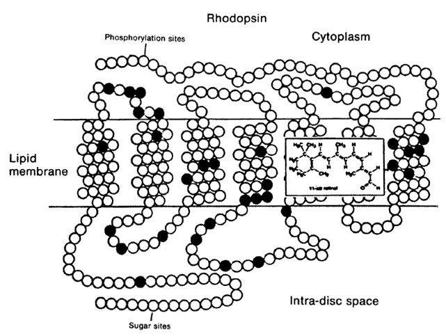
1. Stryer L: Visual excitation and recovery. J Biol Chem 266:10711, 1991

2. Yarfitz S, Hurley JB: Transduction mechanisms of vertebrate and invertebrate photoreceptors. J Biol Chem 269:14329, 1994

3. Fung BK-K, Hurley JB, Stryer L: Flow of information in the light-triggered cyclic nucleotide cascade of vision. Proc Natl Acad Sci USA 78:152, 1981

4. Baehr W, Devlin MJ, Applebury ML: Isolation of bovine ROS phosphodiesterase. J Biol Chem 254:11669, 1979

5. Fung BKK, Young JH, Yamane HK, Griswold-Prenner I: Subunit stoichiometry of retinal rod cGMP phosphodiesterase. Biochemistry 29:2657, 1990

6. Wensel TG, Stryer L: Reciprocal control of retinal rod cyclic GMP phosphodiesterase by its <gt> subunit and transducin. Proteins Struct Funct Genet 1:90, 1986

7. Deterre P, Bigay J, Forquet F et al: cGMP phosphodiesterase of retinal rods is regulated by two inhibitory subunits. Proc Natl Acad Sci USA 85:2424, 1988

8. Clerc A, Bennett N: Activation of cGMP phosphodiesterase of retinal rods: A complex with transducin alpha subunit. J Biol Chem 267:6620, 1992

9. Sitaramayya A, Harkness J, Parkes JH et al: Kinetic studies suggest that light-activated cyclic GMP phosphodiesterase is a complex with G-protein subunits. Biochemistry 25:651, 1986

10. Arshavsky VY, Bownds DM: Regulation of deactivation of photoreceptor G protein by its target enzyme and cGMP. Nature 357:416, 1992

11. Miki N, Baraban JM, Keins JJ et al: Purification and properties of the light-activated cyclic nucleotide phosphodiesterase of rod outer segments. J Biol Chem 260:6320, 1975

12. Palczewski K, McDowell JH, Jakes S et al: Regulation of rhodopsin dephosphorylation by arrestin. J Biol Chem 264:15770, 1989

13. Lagnado L, Baylor D: Signal flow in visual transduction. Neuron 8:995, 1992

14. Koutalos Y, Nakatani K, Yau K-W: Guanylate cyclase studied in truncated rod outer segments. Biophys J 61:428, 1992

15. Torre V, Matthews HR, Lamb TD: Role of calcium in regulating the cyclic GMP cascade of phototransduction in retinal rods. Proc Natl Acad Sci USA 83:7109, 1986

16. Kawamura S: Rhodopsin phosphorylation as a mechanism of cyclic GMP phosphodiesterase regulation by S-modulin. Nature 362:855, 1993

17. Yau K-W: Phototransduction mechanism in retinal rods and cones. Invest Ophthalmol Vis Sci 35:9, 1994

18. Udovichenko IP, Cunnick J, Gonzalez K, Takemoto DJ: Functional effect of phosphorylation of the photoreceptor phosphodiesterase inhibitory subunit by protein kinase C. J Biol Chem 269:9850, 1994

19. Nathans J, Thomas D, Hogness DS: Molecular genetics of human color vision: The genes encoding blue, green, and red pigments. Science 232:193, 1986

20. Lerea CL, Somers DE, Hurley JB et al: Isolation of specific transducin α subunits in retinal rod and cone photoreceptors. Science 243:77, 1986

21. Lerea CL, Bunt-Milam AH, Hurley JB: α Transducin is present in blue-, green-, and red-sensitive cone photoreceptors in the human retina. Neuron 3:367, 1989

22. Gillespie PE, Beavo JA: Characterization of a bovine cone photoreceptor phosphodiesterase purified by cyclic GMP-sepharose chromatography. J Biol Chem 263:6122, 1988

23. Hamilton SE, Prusti RK, Bentley JK et al: Affinities of bovine photoreceptor cGMP phosphodiesterases for rod and cone inhibitory subunits. FEBS Lett 318:157, 1993

24. Gouras P: Retinal circuitry and its relevance to diagnostic psychophysics and electrophysiology. Curr Opin Ophthalmol 3:803, 1992

25. Dacey DM: Physiology, morphology and spatial densities of identified ganglion cells in primate retina. Ciba Found Symp 184:12, 1994

26. Kolb H: The architecture of functional neural circuits in the vertebrate retina. Invest Ophthalmol Vis Sci 35:2385, 1994

27. Bush RA, Sieving PA: A proximal retinal component in the primate photopic ERG a-wave. Invest Ophthalmol Vis Sci 35:635, 1994

28. Sieving PA, Fishman LJ, Steinberg RH: Scotopic threshold response of proximal retina in cat. J Neurophysiol 56:1049, 1986

29. Yamamoto S, Gouras P, Lopez R: The focal cone electroretinogram. Vision Res 35:1641, 1995

30. Sandberg MA, Ariel M: A hand-held two channel stimulator-ophthalmoscope. Arch Ophthalmol 101:232, 1978

31. Sutter EE, Tran D: The field topography of ERG components in man—I. Vision Res 32:443, 1992

32. Pugh ENJ, Lamb TD: Amplification and kinetics of the activation steps in phototransduction. Biochem Biophys Acta 1141:111, 1993

33. Breton ME, Schueller AW, Lamb ED, Pugh JEN: Analysis of ERG a-wave amplification and kinetics in terms of the G-protein cascade of phototransduction. Invest Ophthalmol Vis Sci 35:295, 1994

34. Hood DC, Birch DG: A quantitative measure of the electrical activity of human rod photoreceptors using electroretinography. Vis Neurosci 5:379, 1990

35. Thirkill CE: Cancer associated retinopathy: The CAR syndrome. Neuro Ophthalmol 13:297, 1994

36. Polans AS et al: Recoverin, a photoreceptor-specific calcium-binding protein, is expressed by the tumor of a patient with cancer-associated retinopathy. Proc Natl Acad Sci USA 92:9176, 1995

37. Berson EL, Lessell S: Paraneoplastic night blindness with malignant melanoma. Am J Ophthalmol 106:307, 1988

38. Milan AH, Saari JC, Jacobson SG et al: Autoantibodies against retinal bipolar cells in cutaneous melanomaassociated retinopathy. Invest Ophthalmol Vis Sci 34:91, 1933

39. Jacobson DM, Thirkill CE: Paraneoplastic cone dysfunction: An unusual visual remote effect of cancer. Arch Ophthalmol 113:1580, 1995

40. Pearson P et al: The status of Online Mendelian Inheritance in Man (OMIM) medio 1994. Nucleic Acids Res 22:3470, 1994

41. McKusick VA, Francomano CA, Antonarakis SE, Pearson PL: Mendelian Inheritance in Man. Baltimore, The Johns Hopkins University Press, 1994

42. Heckenlively JR: Retinitis Pigmentosa. Philadelphia, JB Lippincott, 1988

43. Humphries P, Kenna P, Farrar J: On the molecular genetics of retinitis pigmentosa. Science 256:804, 1992

44. Berson EL: Retinitis pigmentosa: The Friedenwald Lecture. Invest Ophthalmol Vis Sci 34:1655, 1993

45. Rosenfeld PJ, Dryja TP: In Wiggs J (ed): Molecular Genetics of Ocular Diseases, p 99. New York, Wiley-Liss, 1995

46. Dryja TP, Berson EL: Retinitis pigmentosa and allied diseases: Implications of genetic heterogeneity. Invest Ophthalmol Vis Sci 36:1197, 1995

47. Bunker CH, Berson EL, Bromley WC et al: Prevalence of retinitis pigmentosa in Maine. Am J Ophthalmol 97:357, 1984

48. Berson EL, Gouras P, Gunkel RD: Rod responses in retinitis pigmentosa, dominantly inherited. Arch Ophthalmol 80:355, 1968

49. Berson EL, Gouras P, Hoff M: Temporal aspects of the electroretinogram. Arch Ophthalmol 81:207, 1969

49a. Wilson EB: The sex chromosomes. Arch Mikrosk Anat 77:249, 1911

50. Haldane JBS, Sprunt AD, Haldane NM: Reduplication in mice. J Genet 5:133, 1915

51. Bird AC: Retinal photoreceptor dystrophies: The LI Edward Jackson Memorial Lecture. Am J Ophthalmol 119:543, 1995

52. Dryja TP, Li T: Molecular genetics of retinitis pigmentosa. Hum Mol Genet 4:1739, 1995

53. Nussbaum RL, Lesko JG, Lewis RA et al: Isolation of anomalous DNA sequences from within a submicroscopic X chromosomal deletion in a patient with choroideremia, deafness, and mental retardation. Proc Natl Acad Sci USA 84:6521, 1987

54. Cremers FPM et al: Chromosomal jumping from the DXS165 locus allows molecular characterization of four microdeletions and a de novo chromosome X/13 translocation with choroideremia. Proc Natl Acad Sci USA 86:7510, 1989

55. Cremers FPM, van de Pol DJR, van Kerkoff LPM et al: Cloning of a gene that is rearranged in patients with choroideraemia. Nature 347:674, 1990

56. Sankila E-M, Tolvanen R, van den Hurk JAJM et al: Aberrant splicing of the CHM gene is a significant cause of choroideremia. Nature Genet 1:109, 1992

57. Seabra MC, Brown MS, Goldstein JL: Retinal degeneration in choroideremia: Deficiency of Rab geranylgeranyltransferase. Science 259:377, 1993

58. Zhu D et al: Microdeletion in the X-chromosome and prenatal diagnosis in a family with Norrie disease. Am J Med Genet 33:485, 1989

59. Berger W et al: Isolation of a candidate gene for Norrie disease by positional cloning. Nature Genet 1:199, 1992

60. Chen Z-Y et al: Isolation and characterization of a candidate gene for Norrie disease. Nature Genet 1:204, 1992

61. Wong F, Goldberg MF, Hao Y: Identification of a nonsense mutation at codon 128 of the Norrie's disease gene in a male infant. Arch Ophthalmol 111:1553, 1993

62. Chen Z-Y et al: A mutation in the Norrie disease gene (NDP) associated with X-linked familial exudative vitreoretinopathy. Nature Genet 5:180, 1993

63. Criswick VG, Schepens CL: Familial exudative vitreoretinopathy. Am J Ophthalmol 68:578, 1969

64. Stone EM et al: Genetic linkage of autosomal dominant neovascular inflammatory vitreoretinopathy to chromosome 11q13. Hum Mol Genet 1:685, 1992

65. Kwitek-Black AE et al: Linkage of Bardet-Biedl syndrome to chromosome 16q and evidence for non-allelic genetic heterogeneity. Nature Genet 5:392, 1993

66. Warburg M, Sjo O, Tranebjaerg L, Fledelius HC: Deletion mapping for a retinal cone-rod dystrophy: Assignment to 18q21.1. Am J Med Genet 39:288, 1991

67. Evans K et al: Genetic linkage of cone-rod retinal dystrophy to chromosome 19q and evidence for segregation distortion. Nature Genet 6:210, 1994

68. Knowles JA et al: Identification of a locus, distinct from RDS-peripherin, for autosomal recessive retinitis pigmentosa on chromosome 6p. Hum Mol Genet 3:14401, 1994

69. McWilliam P et al: Autosomal dominant retinitis pigmentosa (ADRP): Localization of an ADRP gene to the long arm of chromosome 3. Genomics 5:619, 1989

70. Dryja TP et al: A point mutation of the rhodopsin gene in one form of retinitis pigmentosa. Nature 343:364, 1990

71. Farrar GJ et al: Autosomal dominant retinitis pigmentosa: A novel mutation in the rhodopsin gene in the original 3q family. Hum Mol Genet 1:769, 1992

72. Farrar GJ et al: Autosomal dominant retinitis pigmentosa: Absence of the rhodopsin proline-to-histidine substitution (codon 23) in pedigrees from Europe. Am J Hum Genet 47:941, 1990

73. Sandberg MA, Weigel-Di Franco C, Dryja TP, Berson EL: Clinical expression correlates with location of rhodopsin mutation in dominant retinitis pigmentosa. Invest Ophthalmol Vis Sci 36:1934, 1995

74. Dryja TP et al: Mutations in the gene encoding the alpha subunit of the rod cGMP-gated channel in autosomal recessive retinitis pigmentosa. Proc Natl Acad Sci USA 92:10177, 1995

75. Yau K-W: Cyclic nucleotide-gated channels: An expanding new family of ion channels. Proc Natl Acad Sci USA 91:3481, 1994

75a. Simell O, Takki K: Raised plasma ornithine and gyrate atrophy of the choroid and retina. Lancet 1:1031, 1973

76. Dolph PJ et al: Arrestin function in inactivation of G protein-coupled receptor rhodopsin in vivo. Science 260:1910, 1993

77. Ramesh V et al: The ornithine aminotransferase (OAT) locus: Analysis of RFLP in gyrate atrophy. Am J Hum Genet 42:365, 1988

78. Mitchell GA et al: Human ornithine-delta-aminotransferase: cDNA cloning and analysis of the structural gene. J Biol Chem 263:14288, 1988

79. Inana G et al: Molecular cloning of human ornithine aminotransferase mRNA. Proc Natl Acad Sci USA 83:1203, 1986

80. Akaki Y et al: A deletion in the ornithine aminotransferase gene in gyrate atrophy. J Biol Chem 267:12950, 1992

81. Brody LC et al: Ornithine delta-aminotransferase mutations in gyrate atrophy: Allelic heterogeneity and functional consequences. J Biol Chem 267:3302, 1992

82. Wang T et al: Mice lacking ornithine-delta-amino-transferase have paradoxical neonatal hypoornithinaemia and retinal degeneration. Nature Genet 11:185, 1995

83. Keeler CE: The inheritance of a retinal abnormality in white mice. Proc Natl Acad Sci USA 10:329, 1924

84. Pittler SJ, Keeler CE, Sidman RL, Baehr W: PCR analysis of DNA from 70-year-old sections of rodless retina demonstrates identity with the mouse rd defect. Proc Natl Acad Sci USA 90:9616, 1993

85. Sidman RL, Green MC: Retinal degeneration in the mouse: Location of the rd locus in linkage group XVII. J Hered 56:23, 1965

86. Wimer RE, Wimer CC, Alameddine L, Cohen AJ: The mouse gene retinal degeneration (rd) may reduce the number of neurons present in the adult hippocampal dentate gyrus. Brain Res 547:279, 1991

87. Lolley RN, Rong H-M, Craft CM: Linkage of photoreceptor degeneration by apoptosis with inherited defect in phototransduction. Invest Ophthalmol Vis Sci 35:358, 1994

88. Chang C-Q, Hao Y, Wong F: Apoptosis: Final common pathway of photoreceptor death in rd, rds, and rhodopsin mutant mice. Neuron 11:595, 1993

89. Poetera-Cailliau C, Sung CH, Nathans J, Adler R: Apoptotic photoreceptor cell death in mouse models of retinitis pigmentosa. Proc Natl Acad Sci USA 91:974, 1994

90. Schmidt SY, Lolley RN: Cyclic-nucleotide phosphodiesterase: An early defect in inherited retinal degeneration of C3H mice. J Cell Biol 57:117, 1973

91. Farber DB, Lolley RN: Cyclic guanosine monophosphate: Elevations in degenerating photoreceptor cells of the C3H mouse retina. Science 186:449, 1974

92. Bowes C et al: Retinal degeneration in the rd mouse is caused by a defect in the β subunit of rod cGMP phosphodiesterase. Nature 347:677, 1990

93. Danciger M, Bowes C, Kozak CA et al: Fine mapping of a putative rd cDNA and its cosegregation with rd expression. Invest Ophthalmol Vis Sci 31:1427, 1990

94. Pittler SJ, Baehr W: Identification of a nonsense mutation in the rod photoreceptor cGMP phosphodiesterase beta-subunit gene of the rd mouse. Proc Natl Acad Sci USA 88:8322, 1991

95. Farber DB, Danciger J, Aguirre G: The beta subunit of cyclic GMP phosphodiesterase mRNA is deficient in canine rod-cone dysplasia. Neuron 9:349, 1992

96. Suber ML et al: Irish setter dogs affected with rod/cone dysplasia contain a nonsense mutation in the rod cGMP phosphodiesterase beta-subunit gene. Proc Natl Acad Sci USA 90:3968, 1993

97. Ray K, Baldwin VJ, Acland GM et al: Cosegregation of codon 807 mutation of the canine rod cGMP phosphodiesterase beta gene and rcd1. Invest Ophthalmol Vis Sci 35:4291, 1994

98. Riess O, Noerremoelle A, Weber B et al: The search for mutations in the gene for the beta subunit of the cGMP phosphodiesterase (PDEB) in patients with autosomal recessive retinitis pigmentosa. Am J Hum Genet 51:755, 1992

99. McLaughlin ME, Sandberg MA, Berson EL, Dryja TP: Recessive mutations in the gene encoding the beta-subunit of rod phosphodiesterase in patients with retinitis pigmentosa. Nature Genet 4:130, 1993

100. McLaughlin ME, Ehrhart TL, Berson EL, Dryja TP: Mutation spectrum of the gene encoding the beta subunit of rod phosphodiesterase among patients with autosomal recessive retinitis pigmentosa. Proc Natl Acad Sci USA 92:3249, 1995

101. Bayes M et al: Homozygous tandem duplication within the gene encoding the beta-subunit of rod phosphodiesterase as a cause for autosomal recessive retinitis pigmentosa. Hum Mutat 5:228, 1995

102. Gal A, Orth U, Baehr W et al: Heterozygous missense mutation in the rod cGMP phosphodiesterase beta-subunit in autosomal dominant stationary night blindness. Nature Genet 7:64, 1994

103. Huang SH et al: A mutation in the gene encoding the alpha-subunit of rod cGMP phosphodiesterase (PDEA) in retinitis pigmentosa. Invest Ophthalmol Vis Sci 36(suppl):S825, 1995

104. Gibson F et al: A type VII myosin encoded by the mouse deafness gene shaker-1. Nature 374:62, 1995

105. Hasson T, Heintzelman MB, Santos-Sacchi J et al: Expression in cochlea and retina of myosin VIIa, the gene product defective in Usher syndrome type 1B. Proc Natl Acad Sci USA 92:9815, 1995

106. Weil D et al: Defective myosin VIIA gene responsible for Usher syndrome type 1B. Nature 374:60, 1995

107. Gardner JM et al: The mouse pink-eye dilution gene: Association with human Prader-Willi and Angelman syndromes. Science 257:1121, 1992

108. Rinchik EM et al: A gene for the mouse pink-eye dilution locus and for human type II oculocutaneous albinism. Nature 361:72, 1993

109. Lee S-T et al: Mutations of the P gene in oculocutaneous albinism, ocular albinism and Prader-Willi syndrome plus albinism. N Engl J Med 330:529, 1994

110. Travis GH, Brennan MB, Danielson PE et al: Identification of a photoreceptor-specific mRNA encoded by the gene responsible for retinal degeneration slow (rds). Nature 338:70, 1989

111. Ma J et al: Retinal degeneration slow (rds) in mouse results from simple insertion of a t haplotype-specific element into protein-coding exon II. Genomics 28:212, 1995

112. Travis GH, Sutcliffe JG, Bok D: The retinal degeneration slow (rds) gene product is a photoreceptor disc membrane-associated glycoprotein. Neuron 6:61, 1991

113. Travis GH et al: The human retinal degeneration slow (RDS) gene: Chromosomal assignment and structure of the mRNA. Genomics 10:733, 1991

114. Farrar GJ et al: A three-base-pair deletion in the peripherin-RDS gene in one form of retinitis pigmentosa. Nature 354:478, 1991

115. Kajiwara K et al: Mutations in the human retinal degeneration slow gene in autosomal retinitis pigmentosa. Nature 354:480, 1991

116. Bascom RA et al: Localization of the photoreceptor gene ROM1 to human chromosome 11 and mouse chromosome 19: Sublocalization of human 11q13 between PGA and PYGM. Am J Hum Genet 51:1028, 1992

117. Kajiwara K, Berson EL, Dryja TP: Digenic retinitis pigmentosa due to mutations at the unlinked peripherin/RDS and ROM1 loci. Science 264:1604, 1994

118. Hill RE et al: Mouse small eye results from mutations in a paired-liked homeobox-containing gene. Nature 354:522, 1991

119. Glaser T et al: PAX gene dosage effect in a family with congenital cataracts, aniridia, anophthalmia, and central nervous system defects. Nature Genet 7:463, 1994

120. Ton CCT et al: Positional cloning and characterization of a paired box- and homeobox-containing gene from the aniridia region. Cell 67:1059, 1991

121. Hanson IM et al: Mutations at the PAX6 locus are found in heterogeneous anterior segment malformations including Peters' anomaly. Nature Genet 6:168, 1994

122. Mirzayans F, Pearce WG, MacDonald IM, Walter MA: Mutation of the PAX6 gene in patients with autosomal dominant keratitis. Am J Hum Genet 57:539, 1995

123. Bergen AAB, ten Brink JB, Riemslang F et al: Localization of a novel X-linked congenital stationary night blindness locus: Close linkage to the RP3 type retinitis pigmentosa gene region. Hum Mol Genet 4:931, 1995

124. Dryja TP, Berson EL, Rao VR, Oprian DD: Heterozygous missense mutation in the rhodopsin gene as a cause of congenital stationary night blindness. Nature Genet 4:280, 1993

125. Sieving PA et al: Dark-light: Model for nightblindness from the human rhodopsin gly90-to-asp mutation. Proc Natl Acad Sci USA 92:880, 1995

126. Rao V, Cohen GB, Oprian DB: Rhodopsin mutation G90D and a molecular mechanism for congenital night blindness. Nature 367:639, 1994

127. Fuchs S et al: A homozygous 1-base pair deletion in the arrestin gene is a frequent cause of Oguchi disease in Japanese. Nature Genet 10:360, 1995

128. Small KW: In Wiggs J (ed): Molecular Genetics of Ocular Diseases, p 127. New York, Wiley-Liss, 1995

129. Small KW et al: North Carolina macular dystrophy is assigned to chromosome 6. Genomics 13:681, 1992

130. Nichols BE et al: Refining the locus for Best vitelliform macular dystrophy and mutation analysis of the candidate gene ROM1. Am J Hum Genet 54:95, 1994

131. Deutman AF, Pickers AJL, Aan de Kerk AL: Dominantly inherited cystoid macular edema. Am J Ophthalmol 82:540, 1976

132. Fishman GA, Goldberg MF, Trautmann JC: Dominantly inherited cystoid macular edema. Ann Ophthalmol 11:21, 1979

133. Kremer H et al: Localization of the gene for dominant cystoid macular dystrophy on chromosome 7p. Hum Mol Genet 3:299, 1994

134. Weber BHF, Vogt G, Wolz W et al: Sorsby's fundus dystrophy is genetically linked to chromosome 22q13-ter. Nature Genet 7:158, 1994

135. Weber BHF, Vogt G, Pruett RC et al: Mutations in the tissue inhibitor metalloproteinases-3 (TIMP3) in patients with Sorsby's fundus dystrophy. Nature Genet 8:352, 1994

136. Jacobson SG et al: Night blindness in Sorsby's fundus dystrophy reversed by vitamin A. Nature Genet 11:27, 1995

137. Wells J et al: Mutations in the human retinal degeneration slow (RDS) gene can cause either retinitis pigmentosa or macular dystrophy. Nature Genet 3:213, 1993

138. Nichols BE et al: Butterfly-shaped pigment dystrophy of the fovea caused by a point mutation in codon 167 of the RDS gene. Nature Genet 3:202, 1993

139. Nichols BE et al: A 2 base pair deletion in the RDS gene associated with butterfly-shaped pigment dystrophy of the fovea. Hum Mol Genet 2:601, 1993

140. Feist RM, White MF Jr, Skalka H, Stone EM: Choroidal neovascularization in a patient with adult foveomacular dystrophy and a mutation in the retinal degeneration slow gene (pro210-to-arg). Am J Ophthalmol 118:259, 1994

141. Kim RY et al: Autosomal dominant pattern dystrophy of the retina associated with a 4-base pair insertion at codon 140 in the peripherin/RDS gene. Arch Ophthalmol 113:451, 1995

142. Kikawa E, Nakazawa M, Chida Y et al: A novel mutation (asn244-to-lys) in peripherin/RDS gene causing autosomal dominant retinitis pigmentosa associated with bull's-eye maculopathy detected by nonradioisotopic SSCP. Genomics 20:137, 1994

143. Kajiwara K, Sandberg MA, Berson EL, Dryja TP: A mutation in the human peripherin/RDS gene in a family with autosomal dominant retinitis punctata albescens. Nature Genet 3:208, 1993

144. Olschwang S et al: Restriction of ocular fundus lesions to a specific subgroup of APC mutations in adenomatous polyposis coli patients. Cell 75:959, 1993

145. Moraes CT et al: Mitochondrial DNA deletions in progressive external ophthalmoplegia and Kearns-Sayer syndrome. N Engl J Med 320:1293, 1989

146. Holt IJ, Harding AE, Petty RKH, Morgan-Hughes JA: A new mitochondrial disease associated with mitochondrial DNA heteroplasmy. Am J Hum Genet 46:428, 1990

147. Wallace DC et al: Mitochondrial DNA mutation associated with Leber hereditary optic neuropathy. Science 242:1427, 1988

148. Stone EM et al: Visual recovery in patients with Lever's hereditary optic neuropathy and the 11778 mutation. J Clin Neuro Ophthalmol 12:10, 1992

149. Min KC, Zvyaga TA, Cypess AM, Sakmar TP: Characterization of mutant rhodopsins responsible for autosomal dominant retinitis pigmentosa: Mutations on the cytoplasmic surface affect transducin activation. J Biol Chem 1993:9400, 1993

150. Noell WK: Possible mechanisms of photoreceptor damage by light in mammalian eyes. Vision Res 20:1163, 1980

151. Fain GL, Lisman JE: Photoreceptor degeneration in vitamin A deprivation and retinitis pigmentosa: The equivalent light hypothesis. Exp Eye Res 57:335, 1993

152. Li T, Franson WK, Gordon JW et al: Constitutive activation of phototransduction by K296E opsin is not a cause of photoreceptor degeneration. Proc Natl Acad Sci USA 92:3551, 1995

153. Robinson PR, Cohen GB, Zhukovsky EA, Oprian DD: Constitutively active mutants of rhodopsin. Neuron 9:719, 1992

154. Robinson PR, Buczylko J, Ohguro H, Palczewski K: Opsins with mutations at the site of chromophore attachment constitutively activate transducin but are not phosphorylated by rhodopsin kinase. Proc Natl Acad Sci USA 91:5411, 1994

155. Raport CJ et al: Downregulation of cGMP phosphodiesterase induced by expression of GTPase-deficient cone transducin in mouse rod photoreceptors. Invest Ophthalmol Vis Sci 35:2932, 1994

156. Chen J, Makino CL, Peachey NS et al: Mechanisms of rhodopsin inactivation in vivo as revealed by a COOH-terminal truncation mutant. Science 267:374, 1995

157. Naash MI, Hollyfield JG, al-Ubaidi MR, Baehr W: Simulation of human autosomal dominant retinitis pigmentosa in transgenic mice expressing a mutated murine opsin gene. Proc Natl Acad Sci USA 90:5499, 1993

158. Olson JE et al: Transgenic mice with a rhodopsin mutation (Pro23His): A mouse model of autosomal dominant retinitis pigmentosa. Neuron 9:815, 1992

159. Roof DJ, Adamian M, Hayes A: Rhodopsin accumulation at abnormal sites in retinas of mice with a human P23H rhodopsin transgene. Invest Ophthalmol Vis Sci 35:4049, 1994

160. Sung CH, Makino C, Baylor D, Nathans J: A rhodopsin gene mutation responsible for autosomal dominant retinitis pigmentosa results in a protein that is defective in localization to the photoreceptor outer segment. J Neurosci 14:5818, 1994

161. Capecchi M: The new mouse genetics: Altering the genome by gene targeting. Trends Genet 5:70, 1989

162. Hogan B, Beddington R, Costantini F, Lacy E: Manipulating the Mouse Embryo: A Laboratory Manual. Cold Spring Harbor, NY, Cold Spring Harbor Laboratory Press, 1994

163. Flannery JG, Chen J, Xu J, Simon MI: Overexpression of Bcl-2 can interfere with apoptosis in retinal degeneration. Invest Ophthalmol Vis Sci 36(suppl 4):S615, 1995

Back to Top