Chapter 31
Cystic Tumors
R. PATRICK YEATTS
Main Menu   Table Of Contents

Search

DERMOID AND EPIDERMOID CYSTS
TERATOMAS
CEPHALOCELES
MICROPHTHALMOS WITH CYST AND CONGENITAL CYSTIC EYE
MUCOCELE
CYSTS OF THE OPTIC NERVE SHEATH
DACRYOCELE
HEMATIC CYSTS
PARACYTIC CYSTS
SIMPLE CYSTS
MISCELLANEOUS CYSTIC DISEASE
SUMMARY
REFERENCES

A true orbital cyst is any closed cavity or sac within the bony orbital confines that is lined with epithelium and contains a liquid or semisolid material.1 Many orbital cysts are developmental and present at birth, but some may not be manifest for many years; these cysts include dermoid and epidermoid cysts, cystic teratomas, encephaloceles, congenital cystic eyes, and perioptic hygromas. Other orbital cysts are acquired, arising from neighboring sinuses or adjacent adnexal structures; these cysts include mucoceles or dacryoceles. Epithelial implantations secondary to trauma or surgery also can give rise to cyst formation. Finally, vascular neoplasms, chiefly those in the lymphangioma–venous varix group, may develop cystic cavities or bleed spontaneously, forming blood-filled or “chocolate” cysts.

Often, the term cyst is used loosely in its clinical application to orbital disease. Parasitic encystment is more often an inflammatory granuloma than a cyst, although cyst is commonly used in reference to the manifestation of these infestations. Hematic cysts are not lined with epithelium but are a fibrous encapsulation of blood or blood products. Malignant epithelial neoplasms that secondarily invade the orbit may develop central necrosis and a cyst-like character.

True orbital cysts are discussed in this chapter; those arising from vascular neoplasms are discussed elsewhere in these volumes. Paracytic cysts and hematic cysts, although not epithelially lined, are discussed because of their importance in the differential diagnosis of true orbital cysts.

Back to Top
DERMOID AND EPIDERMOID CYSTS
Dermoid and epidermoid cysts are among the most common periorbital and orbital tumors presenting in childhood.2–5 Although their incidence is reportedly low, many of them are excluded from orbital tumor series because they commonly are found anterior to the orbital septum along the upper margin of the orbital rim. Most are first seen during the preschool years, although a dermoid cyst may be seen at any age.

Dermoid and epidermoid cysts are choristomas arising from subcutaneous epidermal rests or epidermal tissues trapped along bony suture lines or within the diploe of the bone during embryonic development. The epidermal anlage develops into a cyst lined with stratified squamous epithelium, which is usually intimately associated with or firmly attached to the frontozygomatic suture superotemporally or to the maxillofrontal suture superonasally. Rarely, these cysts involve the suture confluence of the greater with of the sphenoid, zygoma, and frontal bones or the intradiploic space of the lateral orbital rim.6 If the cyst wall contains skin appendages, such as hair follicles, sweat glands or sebaceous glands, the cyst is termed a dermoid cyst. If skin appendages are absent, the cyst is termed an epidermoid cyst.7

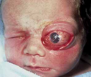
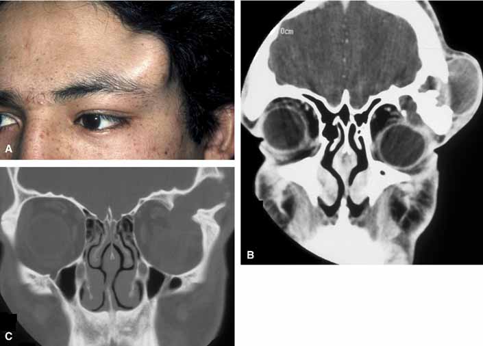
Typically, a dermoid cyst presents in a child as a mass protruding forward from beneath the superior orbital rim anterior to the orbital septum (Fig. 1). Dermoid cysts are found most commonly in the superotemporal quadrant and less frequently superonasally, but these cysts may occur elsewhere in the periobital region both anterior and posterior to the orbital septum and in the temporal fossa.

Fig. 1 A. Dermoid cyst in an adult who had a clinically apparent lesion in the superior temporal quadrant of the right eye since early childhood. Note the contour deformity of the eyelid without globe displacement of proptosis. A computed tomogram shows two cysts of different radiodensity in both axial (B) and coronal (C) sections. The two dermoid cysts show differing radiodensities. The small, dark, or radiolucent, lesion belies its high fat content; the larger, radiopaque lesion contains keratin debris. D. Morphologically, the cysts are smooth-walled and vary in content from a tan, oily liquid (small cyst) to a yellow-white, cheesy material (large cyst) as shown in cross section (E). F. Histology of a dermoid cyst shows a stratified squamous epithelium with keratin debris filling the lumen and a pilosebaceous apparatus in the cyst wall (magnification × 100).

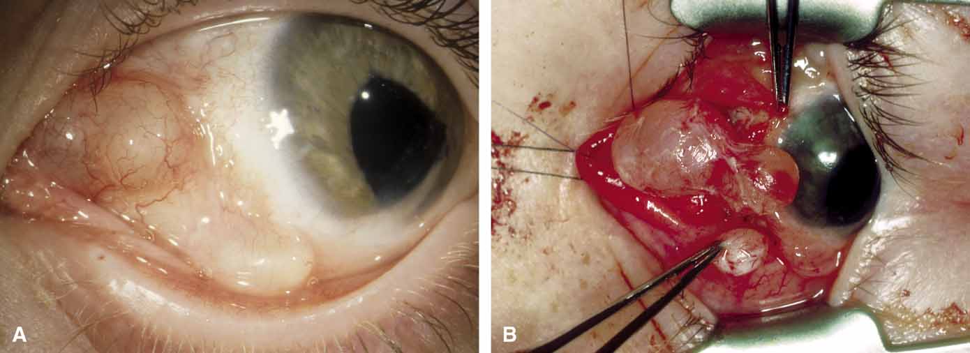
The usual cyst is a painless, smooth, ovoid-to-round, firm, rubbery mass. It may be mobile or immobile, being relatively free or firmly attached to the underlying bone (periosteum), but it is not attached to the overlying skin, distinguishing them from implantation cysts. Although contour abnormalities of the eyelid are common, there is little or no displacement of the globe with dermoid cysts located along the orbital rim. In an adult presentation, a dermoid cyst may frequently have a more posterior location.8 These posteriorly located cysts more typically present in adulthood, are more difficult to palpate, and proptosis and globe displacement are more common.

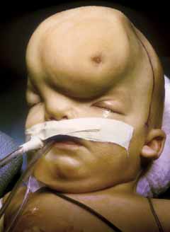
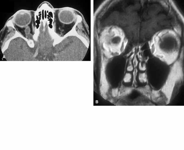
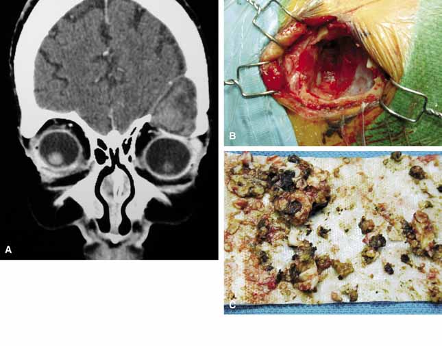
Rarely, an orbital cyst may pass through bony suture lines to extend intracranially or to the temporal fossa (Fig. 2).5,6 Pressure placed on the extracranial portion of a bilobed cyst may be transmitted through the bony dehiscence into the orbit, and is a cause for the mastication proptosis reported by Bullock and Bartley.9

Fig. 2 A large dermoid cyst is seen in the left temple of a young man (A) that shows both an intraorbital and extraorbital component with associated bone change seen on computed tomography (B). A coronal view of the orbit with a setting to highlight bone detail shows a confluence of the intraorbital and extraorbital components through a bone defect in the superior lateral orbital wall (C).

The nature of an orbital dermoid cyst can be demonstrated well by computed tomography (CT): the cyst has a low-density lumen and its relationship to the underlying bone is often manifested by smooth remodeling of the bone secondary to cyst expansion; the high content of fatty material within the cyst makes it radiolucent.

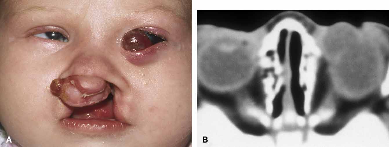
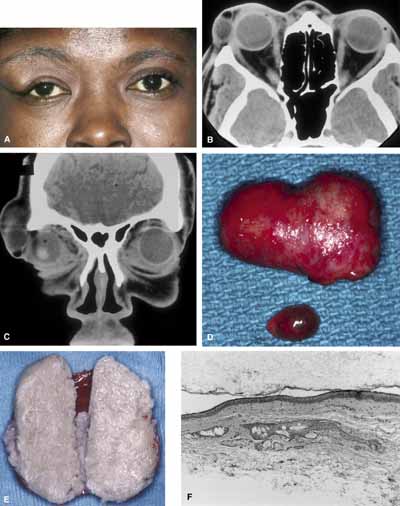
Because of the variable presentation of dermoid cysts, Shields et al.,5 have suggested a classification of orbital dermoid cysts by their association (or lack of association) with suture lines of the skull and assist the clinician in appropriate management. Cysts are classified as juxtasutural, sutural, or soft-tissue dermoid cysts. Those cysts adjacent to the bony suture line but not firmly attached are juxtasutural. A sutural dermoid cyst is firmly attached to bony sutures causing bone erosion, tunnels or an hourglass configuration. Soft tissue dermoid cysts may be strictly confined to soft tissues without any connection to a bone structure. Intradiploic epidermoid cysts are distinctly uncommon and were not included in Shields' classification (Fig. 3).

Fig. 3 In a coronal image of a computed tomogram, an intradiploic dermoid cyst is shown to involve the superior orbital rim and roof of the left orbit displacing the globe downward (A). The intraoperative cavity (B) of the cyst is observed with the evacuated keratin debris shown (C).

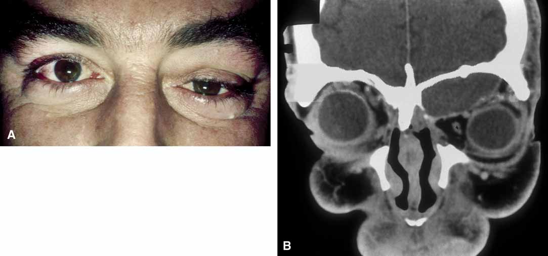
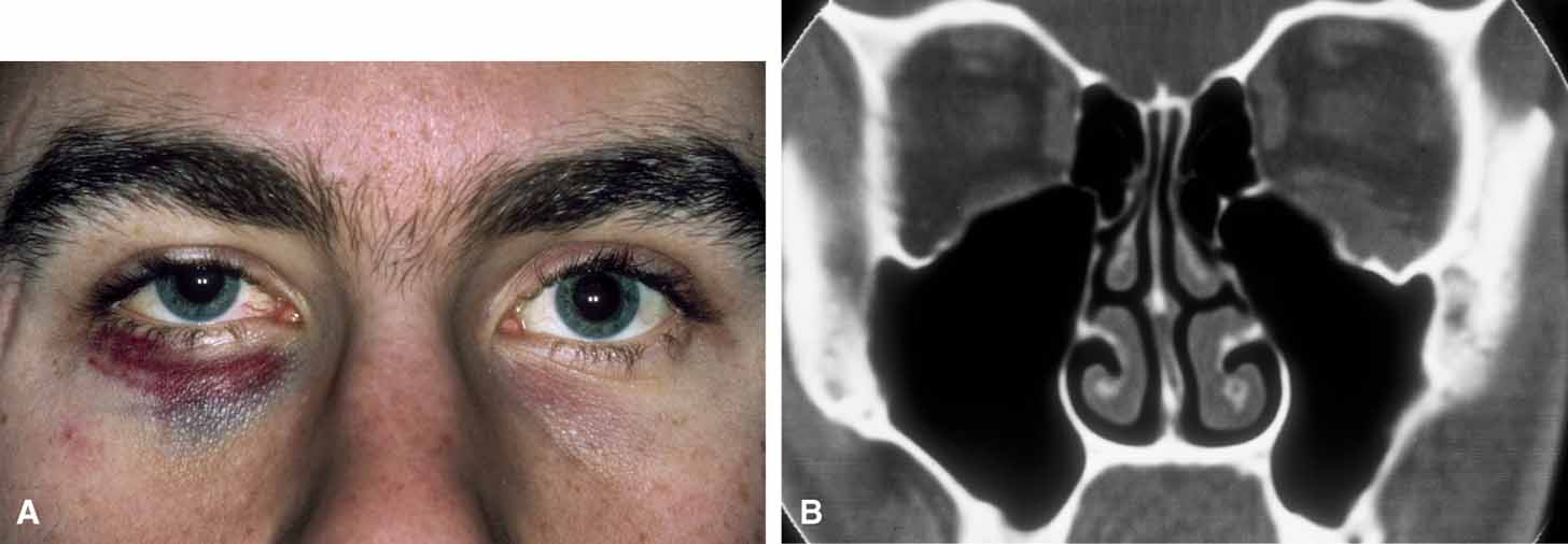
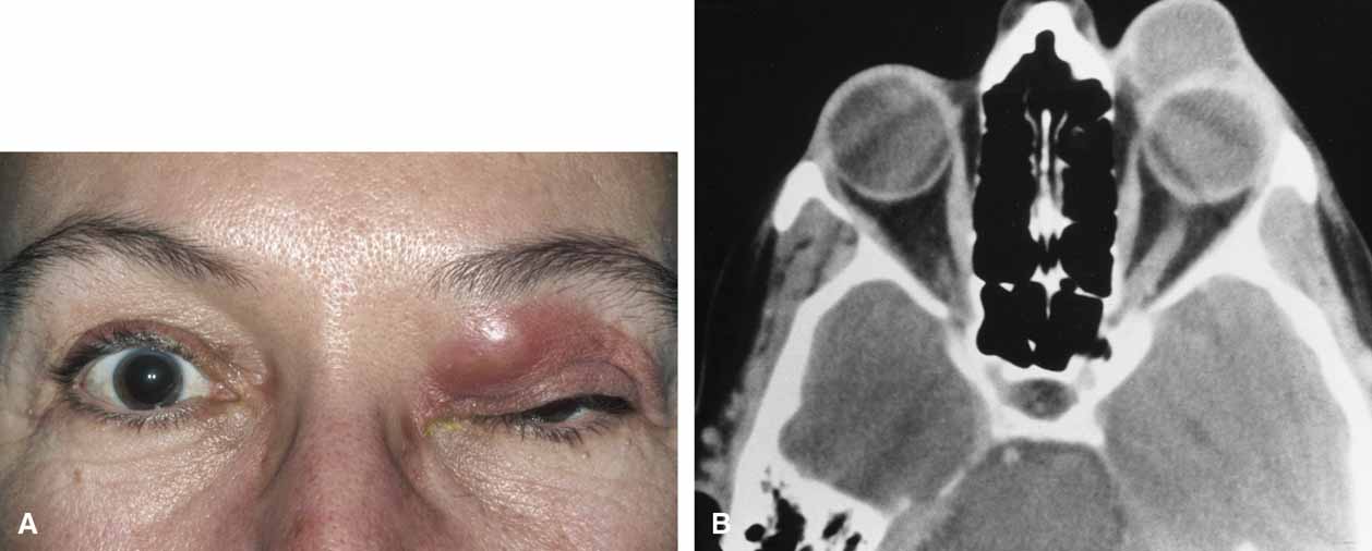
It is often cosmetic considerations that prompt the parents of an affected child to seek treatment. These cysts enlarge as the child grows and the possibility of accidental traumatic rupture is ever present. In adults, mass effect or periorbital inflammation due to leakage of cyst contents may prompt medical attention and surgical removal (Fig. 4).5,10

Fig. 4 A. Neglected dermoid cyst in a 47-year-old woman showing recurrent inflammation manifested by lid edema and erythema. B. Computed tomogram shows large cyst attaining a size nearly equal to that of the globe.

Surgical removal is the treatment of choice. The surgical approach is dictated by the location of the cyst and its association with boney suture lines. In the majority of juxtasutural dermoid cysts, the lesion can be reached through an incision placed directly over it. Because many of these cysts are located along the superior orbital rim, a brow incision may be placed directly over the cyst along the superior orbital rim. Because of a potentially visible scar, however, use of an upper eyelid crease incision has been advocated.11 A posteriorly located cyst (soft-tissue dermoid) or a bilobed cyst with transmission through the orbital rim (sutural dermoid), in contrast, requires more careful planning for an approach through an anterior and/or a lateral orbital route. Large intradiploic cysts and cysts located along the orbital roof and temporal fossa may require multidisciplines and approach transcranially or a temporal skull base approach.12,13

Surgical extirpation should be complete. Intraoperative rupture of the cyst with release of its contents into the orbit may incite a mild but smoldering granulomatous inflammation. The contents of these cysts may vary from an oily, tan liquid to a cheesy, yellow-white material. When inadvertent rupture occurs, the operating surgeon must flood the wound with irrigating solution to be sure that all this material has been washed away. Complete removal of the cyst wall is curative; incomplete removal may be followed by recurrence. Although marsupialization of deep and extensive dermoid cysts has been advocated by some practitioners,14 this technique is not recommended.8

Histologic study of all these cysts is recommended, because rare cases of epidermoid cysts undergoing malignant transformation into squamous cell carcinoma have been reported in adult patients.15 Although granulomatous inflammation may be seen histologically in as many as two-thirds of dermoid cysts removed in one large retrospective series, the clinical signs of inflammation are observed in the minority of patients.5,10

Back to Top
TERATOMAS
Similar to dermoid cysts, teratomas are congenital tumors and choristomas.7,16,17 Although dermoid and epidermoid cysts are derived from only one germinal layer, a true teratoma has all three germinal anlagen: ectoderm, mesoderm, and endoderm.

A teratoma may become manifest at any age, but the majority are present at or shortly after birth. The tumor is rare,17–19 often only single cases being reported in large series of orbital tumors. Rarely is the presentation of other orbital tumors as dramatic as that of a teratoma, the eye seeming to be a mere appendage to the massive orbital component of the tumor (Fig. 5). CT imaging reveals the multicystic nature of these lesions and the often-encountered intracranial extensions.

Fig. 5 Cystic teratoma in a newborn. (Photograph courtesy of Thomas J. Leisegang, MD, Mayo Clinic, Jacksonville, FL)

The histology of teratomas is complex because of the presence of all three germinal layers. The presence of multiple epidermoid cysts and embryonic forms of mucin-secreting gastrointestinal mucosa within these lesions contributes to their cystic nature. Most teratomas are benign, although some seen in newborns may be malignant.

Tumor growth and unacceptable cosmetic appearance dictate the surgical removal of an orbital teratoma. Although the globe and optic nerve are not intrinsic parts of the tumor, surgical efforts to salvage vision and ocular function have been disappointing. One should preserve the globe whenever possible16,20; however, incomplete removal of the cyst leads to recurrence. Exenteration, although not recommended,18 may be necessary to effect a complete cure.

Back to Top
CEPHALOCELES
A congenital dehiscence in the bony cranium may enable the meningeal tissue to herniate into the orbit forming a cystic structure filled with cerebrospinal fluid: an orbital meningocele. If brain protrudes inside the meningeal sac, the term encephalocele or meningoencephalocele applies.

These cephaloceles are rare. Their diagnosis most often is made in infancy, although a cephalocele presenting deep in the orbit may not be recognized until later in life.21 Cephaloceles form during embryonic development and their formation is either the result of the failure of the neuroectoderm to separate from the surface ectoderm or to defects in osseous development when fetal fissures fail to close. In the former instance, brain tissue is pinched off by the developing bony cranium, the result being a cephalocele attached to the brain by a tissue stalk that extends through sutures between the frontal, ethmoid, lacrimal, and/or maxillary bones. In the latter instance, defects in osseous development result in true herniation of intracranial contents.22

An encephalocele most commonly presents as an anterior protrusion between the nose and eyebrow or as a lateral protrusion into the orbit (Fig. 6). Painless and elastic, the encephalocele may enlarge slowly, and pulsations may be present. The larger the defect, the greater the degree of pulsation and often, the more rapid the increase in size. A Valsalva maneuver or crying can cause a mild increase in size. Orbital signs are limited to the mass effect. Globe displacement or proptosis occurs and is related to the location and size of the cephalocele.23,24 When protruding from the nose into the lower lid, a cephalocele may resemble, and be confused with, a mucocele of the nasolacrimal sac or with a dacryocele.

Fig. 6 Large frontal encephalocele.

The diagnosis of an encephalocele should be considered in any child with a midline or paramedian cystic tumor. Facial anomalies such as hypertelorism, broad nasal root, or increased bitemporal diameter should alert the clinician to the possibility of an encephalocoele.

Radiographic analysis by CT or magnetic resonance imaging (MRI) is critical, both to the diagnosis and to the management of cephaloceles. Recognition of these cysts permits appropriate management rather than an accidental surgical encounter. Surgical treatment is indicated, consisting of exposure, ligation, and excision of a small encephalocele, and of transfrontal craniotomy with excision of a larger encephalocele followed by dural patching, and possibly by bone grafting if the bone defect is large.

Back to Top
MICROPHTHALMOS WITH CYST AND CONGENITAL CYSTIC EYE
Failure of the fetal fissure to close at 4 weeks of development results in proliferation of neuroectoderm through the opening, leading to formation of an orbital cyst. In almost all cases, the resulting eye is defective, smaller than normal, and has an attached cystic mass at birth.25,26 The cyst may be smaller or larger than the eye; its appearance is dependent on the size of the ocular defect and its rate of expansion; microphthalmos with a cyst may be bilateral.27–29 The cyst is slate blue and translucent, and may displace the globe (Fig. 7).

Fig. 7 A. Microphthalmos with cyst in a newborn with congenital facial cleft abnormality. B. Computed tomogram shows globe communication with the cyst cavity; normal anterior segment structures are absent.

In contrast, congenital cystic eye results from failure of the primary optic vesicle to invaginate.25 The eye is filled with both solid and cystic forms of dysplastic neuroglial tissue. The cyst is connected to the brain by an astrocyte-filled stalk, but it does not communicate with the anterior ventricle.

Both microphthalmos with cyst and congenital cystic eye should be distinguished from cystic teratoma and cephalocele. Management of microphthalmos with cyst and congenital cystic eye involves excision of the cyst and/or globe along with the abnormal neuroepithelial structures. Each lesion is self-contained, and often an orbital implant can be placed and a prosthesis fitted to obtain a satisfactory cosmetic effect.

Back to Top
MUCOCELE
Destruction of sinus ostia because of recurrent inflammation, trauma, or intrinsic mucosal disease results in a mucus-filled sinus, or mucocele, which can expand slowly to involve the orbital cavity. If the sinus is inflamed and the cyst contains pus or pus and mucus, the terms pyocele and mucopyocele, respectively, apply. The prevalence of mucoceles among orbital tumors is reportedly low (1.5% to 15%),2 the variability in the prevalence depending on the reporting medical specialty. Because this disease belongs in the purview of otolaryngologists, many believe that the prevalence of mucoceles affecting the orbit is underreported. Henderson and Farrow,2 reviewing data collected at the Mayo Clinic, reported a prevalence of 8.5% of all orbital tumors. Although mucoceles may occur at any age, most occur between the ages of 40 and 70 years. Mucocele occurring in a child should alert the clinician to the possibility of cystic fibrosis.31,32 In the adult, it is common to elicit a history of recurring sinusitis, trauma, or previous sinus surgery.

Mucoceles of the frontal and ethmoid sinuses are most common; and their clinical features are usually related to a mass effect and inflammation.33–34 The enlargement of the mucocele may be insidious, with proptosis and displacement of the globe being manifest in association with a palpable, smooth-walled mass in the upper inner quadrant of the orbit (Fig. 8).

Fig. 8 Frontal sinus mucocele in a man with a history of sinus disease. A. The left eye is displaced inferotemporally, indicating the superior medial location. B. Coronal section of a computed tomogram shows expansion of the opacified left frontal sinus with loss of the bony anterior and inferior walls of the frontal sinus.

Mucoceles of the posterior orbit may present more insidiously, with the patient having vague complaints of headache and orbital discomfort. These mucoceles arise in the sphenoid sinus and posterior ethmoid air cells and may mimic retrobulbar neuritis or cause ophthalmoplegia and ptosis by affecting branches of cranial nerve III. Axial proptosis is observed, in contrast to the global displacement in the horizontal or vertical plane associated with anterior mucocele of the frontal sinus.

Radiographic analysis, including CT, serves to delineate the origin and extent of the mucocele. Thinning of the overlying bone because of progressive expansion of the mucocele is apparent. Intracranial extension can be observed in untreated mucocele.

The treatment of mucoceles is surgical. Frontal and frontoethmoidal mucoceles are often approached subperiosteally with elevation of an osteoplastic flap and eradication and extirpation of all diseased mucosa. Successful sinus obliteration and/or reestablishing sinus drainage intranasally protect against recurrence. Sphenoid sinus mucoceles are approached intranasally. Neurosurgical consultation is indicated for those mucoceles with intracranial extension.

Back to Top
CYSTS OF THE OPTIC NERVE SHEATH
Dilatation and expansion of the optic nerve sheath has been observed in a variety of neoplastic and nonneoplastic conditions. Such a cystic change in an optic nerve sheath filled with cerebrospinal fluid has been called perioptic hygroma, arachnoid cyst of the optic nerve sheath, patulous subarachnoid space, optic hydrops, and meningocele.2,36–38

Most perioptic hygromas present in patients between the ages of 30 and 60 years, whose complaints are headache and visual disturbance. A variety of optic nerve signs may be present, including decreased vision, deafferented pupil, optic disc edema, optic atrophy, and visual field defects. Choroidal folds and progressive hyperopia in the absence of overt proptosis suggest this disorder.

The relationship of the intracranial pressure and the finding of a patulous optic nerve sheath is unclear. Perioptic hygroma may be observed in children, although it is often found in association with neurofibromatosis type I and with optic nerve glioma.

MRI is the radiographic imaging technique of choice, emphasizing high spatial resolution, fat-suppression techniques, and gadolinium contrast (Fig. 9).38 These techniques should assist in differentiating the patulous optic nerve sheath from optic nerve sheath meningioma in adults and from optic nerve glioma in children, which can be mimicked by cysts of the optic nerve sheath. Visual prognosis in these cases is guarded and the role of surgical treatment (optic nerve fenestration) is uncertain.

Fig. 9 Axial (A) computed tomography scans show enlarged right optic nerve sheath. The dilated optic nerve sheath is seen in a coronal plane (B) and is filled with cerebrospinal fluid in a T1-weighted magnetic resonance scan.

Back to Top
DACRYOCELE
Distention of the nasolacrimal sac by captured or entrapped mucoid material results in the formation of a dacryocele.39,40 The retention of mucus results from an obstruction in the distal nasolacrimal duct together with a proximal obstruction at the junction of the common canaliculus and the sac (valve of Rosenmüller). Often, the proximal obstruction is functional, permitting reflux of lacrimal sac material on digital pressure over the lacrimal sac, or permitting passage of a lacrimal probe to the lacrimal sac.

A congenital dacryocele, variably referred to as a congenital dacryocystocele, a mucocele, a lacrimal sac cyst, or an amniocele, is manifest at birth as a firm bluish swelling in the medial canthal area at or below the medial canthal tendon. The lacrimal sac is tense and is filled with mucus. In a large multicenter retrospective review of congenital dacryoceles, female patients were more often affected (male to female ratio, 1:3); there was equal involvement of the right and left sides, with 13% of cases occurring bilaterally; and dacryocystitis with or without cellulitis was a frequent presentation, occurring in 75% of cases.34 Nasal cysts, which have been observed by others,41 were present in 6 of 54 patients, with 4 patients requiring marsupialization of the cysts to effect a cure. Surgical intervention is indicated in cases of dacryocystitis, cellulitis, recurrent dacryocele, difficulty in breathing from large nasal cysts, and failure to respond to conservative measures such as digital massage.39,42 One study of patients undergoing conservative (nonsurgical) management reported spontaneous resolution in 80% of patients, although the remaining 20% developed dacryocystitis.39

The differential diagnosis of congenital dacryocele includes cephaloceles,43 mucoceles,35 dermoid cysts,4 capillary hemangiomas, and sudoriferous cysts.44 Transillumination, ultrasound, and radiographic studies (CT, MRI, and dacryocystography) should assist in differentiating these entities.

Dacryoceles also may occur as an acquired disorder in adults.45 Although the nature of the disorder is rarely in question, one still should consider the possibility of a primary tumor of the nasolacrimal sac whenever decompression of the sac is not possible with manipulation or whenever the mass extends above the medial canthal tendon.46 Radiographic imaging is recommended in such cases. Dacryocystorhinostomy generally is curative.

Back to Top
HEMATIC CYSTS
The term hematic cyst refers to the accumulation of hematogenous debris within a cavity lined with fibrous tissue but not epithelial or endothelial tissue.47,48 Hematic cysts may be acute or chronic, can occur at any age, and often, but not always, are associated with antecedent trauma. Hematic cysts should be distinguished from endothelial-lined, blood-containing cysts, such as the “chocolate” cysts associated with lymphangiomas, venous varices, and hemangiomas.

Hematic cysts are uncommon, and the clinical history and ophthalmic findings rarely are diagnostic.48–50 Mass effect with proptosis, globe displacement, and motility disturbance are common with chronic hematic cysts. Spontaneous eyelid ecchymosis and edema may suggest this diagnosis (Fig. 10). In acute hematic cysts, optic nerve signs may be present, including decreased vision, afferent pupillary defect, and choroidal folds.51

Fig. 10 A. Spontaneous periorbital ecchymosis and diplopia in a man who had undergone a left orbital blowout fracture 5 years previously suggest the presence of a hematic cyst. B. Computed tomogram shows the left globe and a previously placed Silastic plate displaced superiorly by the expanding hematic cyst in the floor of the left orbit.

The pathogenesis of hematic cysts is unclear. Some believe they are caused by trauma and an incompletely reabsorbed orbital hemorrhage, wherein the presence of old blood and blood-breakdown products elicits a granulomatous inflammation and fibrous encapsulation.2 Others believe that the cysts arise from bleeding within the bony diplöe, which breaks out into the peripheral orbital space.50

Radiographic imaging is invaluable in diagnosing a hematic cyst.52,53 Plain films can show bony erosion, especially when the cyst arises in the superior orbit and involves the orbital plate of the frontal bone. CT shows a well-defined, nonenhancing mass having the same density as the brain. Within a blood-containing tumor, the evolution from acute to chronic hematoma is manifested by characteristic, highly diagnostic changes in the MRI signal.52

Histologically, both acute and chronic hematic cysts contain blood or blood-breakdown products. A brown fluid often is evacuated from these thin-walled cysts. Variable stages of organization are present, with chronic cysts showing cholesterol clefts,54 pigment-laden macrophages, hemosiderin depositions, and granulomatous inflammation.

Hematic cysts should be treated surgically with evacuation of cyst contents, removal of the fibrous wall lining, and establishment of meticulous hemostasis to prevent recurrence. Removal of one wall of the cyst is often all that is necessary, and the wall that is attached to the orbital tissues can be left intact. Cure can be achieved with minimal risk of postoperative complications.

Back to Top
PARACYTIC CYSTS
Cysticercosis and echinococcosis are parasitic diseases whose orbital manifestation is often a cyst with associated mass effect or inflammation accompanying cyst rupture or parasite death.8,55–57 These parasitic infestations have a specific geographic prevalence with the highest incidence seen in the rural areas of developing countries of the world.55 The prevalence of these diseases may vary greatly even within the same country because of the differences in personal hygiene and habits of the local people. While these diseases are rare in the United States, they may be found among immigrant populations.

Cysticercosis is caused by the cestode, Taenia solium. The pig is the main host for the larval stage (intermediate host) of T. solium. Humans may serve as both the definitive or intermediate host; however, human cysticercosis occurs when the human acts as the intermediate host. Ingestion of the T. solium ovum may permit embryos to survive that escape the gastric or intestinal mucosa to the hepatic portal system. From there, they are dispersed passively throughout the tissues of the body by the bloodstream. A mature larvae, called Cysticercus cellulosae, forms over 3 months and once fully grow, the cysticercus cyst may lie dormant, without producing symptoms for 30 years or more.

The clinical manifestations of orbital cysticercosis are dependent on the location, size, and relation to adjacent structures of the cysticerci as well as the viability of the parasite. A live cyst produces mass effect, whereas the dying parasite releases toxins that lead to secondary inflammation. Although any tissue in the orbit may be involved, there is a predilection for involvement of the anterior orbit with involvement of the extraocular muscles, subconjunctival space and eyelid.55

Diagnosis of orbital cysticercosis can be made based on history laboratory investigation, orbital imaging and histologic findings. Important historical information to elicit from a patient include: (1) travel to or being from an endemic area; (2) history of exposure to infected pork or ingestion of raw pork; (3) history of tapeworm infection in the patient or family member. Mild eosinophilia may be present and stool tests may reveal ova or tapeworm structures. Immunologic tests (enzyme-linked immunosorbent assay [ELISA]) are helpful if positive but do not exclude the possibility of disease when negative. High-resolution B-scan ultrasonography and CT may show what has been described as a “pea in a pod” or hanging drop sign, representing the larval head (scolex) attached to the cyst wall. Histologically, the cysticercus cysts have a typical appearance and if degenerating or dying, may be surrounded by varying degrees of inflammatory cells.

Treatment has improved because of the encouraging results of medical therapy. Although surgical removal of the scolex is curative with subsequent degeneration of the cyst wall, oral albendazole or praziquantel has been associated with successful treatment.55 Concomitant use of systemic corticosteroid may be required to control associated orbital inflammation.

Similar to cysticercosis, echinococcosis in humans is the result of parasitic larvae. Echinococcus granulosus infests the intestine of dogs, sheep, pigs, cows, and other animals. Ingested parasite ova develop and find there way to the hepatic portal system and are distributed similar to cysticercosis. Orbital cysts are said to occur in 1% of cases of echinococcosis.8

The onset of echinococcosis is insidious with symptoms of mass effect most prominent. Although a cyst may occur anywhere in the orbit, there is a predilection for the superior and posterior orbit. Cyst rupture either from trauma or surgical manipulation can be associated with sudden and intense orbital inflammation.

Diagnosis is made based on the presence of a cyst and history suggesting parasitic infestation. Peripheral eosinophilia may be observed but is usually mild and seen in the minority of cases. Results from the Casoni intradermal antigen test and the compliment-fixating and hemagglutinating antibody testing are often unreliable. The extent of the cyst can be defined by CT or MRI study.

Treatment is surgical with complete removal of an intact cyst. A surgical field contaminated by the contents of the cysts results in a fulminate inflammatory response. If complete excision is not possible, cyst drainage via syringe has been advocated followed sterilization of the cyst with alcohol or formalin and subsequently rinsed with saline. If an inflammatory response ensues, control with systemic corticosteroids in indicated.

Back to Top
SIMPLE CYSTS
Henderson and Farrow,2 in their discussion of orbital tumors seen at the Mayo Clinic, suggested that the term simple cyst be applied to three types of cysts that differ little from one another, except in terminology. These three—serous, retention, and implantation cysts—have certain shared features. They are rare in the orbit and are only slightly more common in the eyelids and conjunctivae, their source of origin; orbital involvement is secondary rather than primary. These simple cysts are lined with simple epithelium, which reflects their origin; and they are of little functional significance, although they may cause cosmetic embarrassment.

Serous cysts arise in the small delicate bursae between the tendon sheaths of the ocular muscles; they are lined with a cuboidal epithelium. Retention cysts originate in the glandular appendages of the conjunctiva and adnexal structures. Obstruction of the orifices of the lacrimal gland or accessory lacrimal glands (glands of Krause or Wolfring) from trauma or cicatrix may result in a thin-walled epithelium-lined cyst in the superior fornix, which can cause a mass effect. Implantation cysts arise from misplacement of surface epithelium into the orbit as a result of trauma or surgery.

Of these simple cysts, the implantation cysts are the most difficult to eradicate. This difficulty perhaps is because of their variable depth, in that they commonly arise from penetrating wounds or a surgical procedure that penetrates the orbit. An implantation cyst may follow enucleation,58,59 dermal-fat grafting to the orbit, retinal detachment repair, or strabismus surgery (Fig. 11). Removal of the entire cyst or electrodesiccation of the epithelial lining affects a cure. In cases of retention cysts involving the lacrimal ductules (lacrimal ductule cyst), marsupialization may be preferable to compete excision, because attempts to excise the cyst may unnecessarily damage the remaining lacrimal ductules.8 Similarly, cysts whose origins are the accessory lacrimal glands may be associated closely with the levator aponeurosis. Thus, one should be cautious in the management of these simple cysts by excision lest one cause a needless complication.

Fig. 11 A. Conjunctival implantation cysts following retinal detachment and strabismus repair. B. Multiple cysts are observed and are intimately associated with the medial rectus muscle at its insertion and the encircling element (buckle).

Back to Top
MISCELLANEOUS CYSTIC DISEASE
Although true eye cysts are lined by an epithelium, many diseases will appear morphologically to be a cystic structure but do not represent true cyst histologically. These entities are important in the differential diagnosis of true orbital cysts. Cutaneous malignancies with orbital involvement,60 aneurysmal bone cysts,61,62 and hematic cysts47–53 previously discussed are a few examples.

Aggressive basal cell or squamous cell carcinomas with orbital involvement will rarely present as a cystic mass.60 A fast-growing malignancy can outgrow its blood supply with central tumor necrosis and liquidification suggesting an orbital cyst morphologically and radiographically. Aneurysmal bone cysts, although a common entity affecting the long bones of the young, rarely affects the cranial bones occurring in 2.5% to 6% of cases and even more rarely the bones of the orbit.61 An aneurysmal bone cyst appears as painless, multiloculated, expansile bony lesion with thinning of the cortical bone but without destruction. Leveling of hemorrhagic fluid within the honeycomb-like spaces of the lesion gives a radiographic appearance of a cyst within the diploë of the bone. Surgical removal is often curative. Surgical curettage has been used for lesions of the skull base not amenable to resection albeit with a higher recurrence rate.

Back to Top
SUMMARY
Orbital cysts may be developmental (dermoid and epidermoid cysts, cystic teratomas, cephaloceles, microphthalmos, and congenital cystic eye) or acquired (mucocele, perioptic hygroma, parasitic cysts hematic cysts, and simple cysts). Some may arise from developmental abnormalities of the globe (microphthalmos with cyst, congenital cystic eye), ocular adnexal structures (dacryoceles, lacrimal ductule cysts), or adjacent structures (mucoceles, encephaloceles). Some entities do not represent true epithelially lined cysts, but morphologically or radiographically resemble a true cyst (hematic cyst, aneurismal bone cyst, cystic epithelial malignancy). The most common clinical feature of orbital cysts is mass effect. The age at onset, the location, and any history of trauma, antecedent surgery, or sinus disease assists in making the correct diagnosis. Radiographic imaging and analysis are invaluable in characterization of the cyst and in planning the therapeutic approach. Most orbital cysts are approached surgically, with cure being affected by successful elimination of the cyst's contents and extirpation of its epithelial lining.
Back to Top
REFERENCES

1. Dorland's Illustrated Medical Dictionary. Philadelphia: WB Saunders, 1974:398

2. Henderson JW, Farrow GM: Orbital Tumors, 2nd ed. New York: Brian C. Decker, 1980

3. Stefanyszyn MA, Handler SD, Wright JE: Pediatric orbital tumors. Otolaryngol Clin North Am 21:103, 1988

4. Pfeiffer RL, Nicholl RJ: Dermoid and epidermoid tumors of the orbit. Arch Ophthalmol 40:639, 1948

5. Shield JA, Kaden IH, Eagle RC, et al: Orbital dermoid cysts: clinicopathologic correlations, classification, and management, the 1997 Josephine E. Schueler Lecture. Ophthalmic Plast Reconstr Surg 13: 265, 1997

6. Meyer DR, Lesser AM, Yeatts RP, et al: Primary temporal fossa dermoid cysts: characterization and surgical management. Ophthalmology 106:342, 1999

7. Yanoff M, Fine BS: Ocular Pathology: A Text and Atlas, 3rd ed. Philadelphia: JB Lippincott, 1989

8. Rootman J: Diseases of the Orbit: A Multidisciplinary Approach. Philadelphia: JB Lippincott, 1988

9. Bullock JD, Bartley GB: Dynamic proptosis. Am J Ophthalmol 102:104, 1986

10. Abou-Rayyah Y, Rose GE, Konrad H, et al: Clinical, radiological and pathological examination of periocular dermoid cysts: evidence of inflammation from an early age. Eye 16:507, 2002

11. Kronish JW, Dortzbach RK: Upper eyelid crease: surgical approach to dermoid and epidermoid cysts in children. Arch Ophthalmol 106:1625, 1988

12. Eijpe AA, Koornneeff, L, Verbeeten B, et al: Intradiploic epidermoid cysts of the bony orbit. Ophthalmology 98:1737, 1991

13. Blanco G, Esteban R, Galarretts D, et al: Orbital intradiploic giant epidermoid cyst. Arch Ophthalmol 119:771, 2001

14. Kennedy RE: Marsupialization of inoperable orbital dermoids. Trans Am Ophthalmol Soc 68:146, 1970

15. Wright J, Morgan G: Squamous cell carcinoma developing in an orbital cyst. Arch Ophthalmol 95:635, 1977

16. Duke-Elder S: System of Ophthalmology, Vol XIII, Normal and Abnormal Development: Part II. Congenital Deformation. St. Louis: CV Mosby, 1963

17. Levin ML, Leone CRJr , Kincaid MC: Congenital orbital teratomas. Am J Ophthalmol 102:476, 1986

18. Mamalis N, Garland PE, Argyle JC, et al: Congenital orbital teratoma: a review and report of two cases. Surv Ophthalmol 30:41, 1985

19. Sinniah D, Prathap K, Somasundram K: Teratoma in infancy and childhood: a ten-year review at the University Hospital, Kuala Lumpar. Cancer 46:630, 1980

20. Chang DF, Dallow RL, Walton DS: Congenital orbital teratoma: Report of a case with visual preservation. J Pediatr Ophthalmol Strabismus 17:88, 1980

21. Leone CRJr , Marlowe JF: Orbital presentation of an ethmoidal encephalocele: report of a case of a 62-year-old woman. Arch Ophthalmol 83:445, 1970

22. Pollock JA, Newton TH, Hoyt WF: Transsphenoidal and transethmoidal encephaloceles: a review of clinical and roentgen features in 8 cases. Radiology 90:442, 1968

23. Mortada A: Pulsating frontocele and exophthalmos. Am J Ophthalmol 65:425, 1968

24. Strandberg B: Cephalocele of posterior part of orbit. Arch Ophthalmol 42:254, 1949

25. Waring GOIII , Roth AM, Rodrigues MM: Clinicopathologic correlation of microphthalmos with cyst. Am J Ophthalmol 82:714, 1976

26. Lieb W, Rochels R, Gronemeyer U: Microphthalmos with colobomatous orbital cyst: clinical, histological, immunohistological, and electronmicroscopic findings. Br J Ophthalmol 74:59, 1990

27. Slamovits XL, Kimball GP, Friberg TR, et al: Bilateral optic disc colobomas with orbital cysts and hypoplastic optic nerves and chiasm. J Clin Neuro Ophthalmol 9:172, 1989

28. Magni R, Pierro L, Brancato R: Microphthalmos with colobomatous orbital cyst in trisomy 13. Ophthalmic Paediatr Genet 12:39, 1991

29. Goldberg SH, Farber MG, Bullock JD, et al: Bilateral congenital ocular cysts. Ophthalmic Paediatr Genet 12:31, 1991

30. Helveston EM, Malone EJr , Lashmet MH: Congenital cystic eye. Arch Ophthalmol 84:622, 1970

31. Robertson DM, Henderson JW: Unilateral proptosis secondary to orbital mucocele in infancy. Am J Ophthalmol 68:845, 1969

32. Stool S, Kertesz E, Sibinga M, et al: Exophthalmos due to pyocele of the sinus in children with cystic fibrosis. Trans Am Acad Ophthalmol Otolaryngol 70:811, 1966

33. Alberti PWRM, Marshall HF, Munro Black JI: Frontoethmoidal mucocoele as a cause of unilateral proptosis. Br J Ophthalmol 52:833, 1968

34. Reese AB: Expanding lesions of the orbit. Trans Ophthalmol Soc UK 91:85, 1971

35. Iliff CE: Mucoceles in the orbit. Arch Ophthalmol 89:392, 1973

36. Harris GJ, Sacks JG, Weinberg PE, et al: Cyst of the intraorbital optic nerve sheaths. Am J Ophthalmol 81:656, 1976

37. Miller NR, Green WR: Arachnoid cysts involving a portion of the intraorbital optic nerve. Arch Ophthalmol 93:1117, 1975

38. Garrity JA, Trautmann JC, Bartley GB, et al: Optic nerve sheath meningoceles: clinical and radiographic features in 13 cases with a review of the literature. Ophthalmology 97:1519, 1990

39. Mansour AM, Cheng KP, Mumma JV, et al: Congenital dacryocele: a collaborative review. Ophthalmology 98:1744, 1991

40. Harris GJ, DiClementi D: Congenital dacryocystocele. Arch Ophthalmol 100:1766, 1982

41. Berkowitz RG, Grundfast KM, Fitz C: Nasal obstruction of the newborn revisited: clinical and subclinical manifestations of congenital nasolacrimal duct obstruction presenting as a nasal mass. Otolaryngol Head Neck Surg 103:468, 1990

42. Levy NS: Conservative management of congenital amniotocele of the nasolacrimal sac. J Pediatr Ophthalmol Strabismus 16:254, 1979

43. Rashid ER, Bergstrom TJ, Evans RM, et al: Anterior encephalocele presenting as nasolacrimal obstruction. Ann Ophthalmol 18:132, 1986

44. Saunders JF: Congenital sudoriferous cyst of the orbit. Arch Ophthalmol 89:205, 1973

45. Traquair HM: Chronic dacryocystitis: Its causation and treatment. Arch Ophthalmol 26:165, 1941

46. Hornblass A, Jakobiec FA, Bosniak S, et al: The diagnosis and management of epithelial tumors of the lacrimal sac. Ophthalmology 87:476, 1980

47. Krohel GB, Wright JE: Orbital hemorrhage. Am J Ophthalmol 88:254, 1979

48. Pearson PA, Rakes SM, Bullock JD: Clinicopathologic study of hematic cysts of the orbit [letter]. Am J Ophthalmol 102:804, 1986

49. Bergin DJ, McCord CD, Dutton JJ, et al: Chronic hematic cyst of the orbit. Ophthalmic Plast Reconstr Surg 4:31, 1988

50. Cameron JD, Letson RD, Summers CO: Clinical significance of hematic cyst of the orbit. Ophthalmic Plast Reconstr Surg 4:95, 1988

51. Amrith S, Baratham G, Khoo CY, et al: Spontaneous hematic cysts of the orbit presenting with acute proptosis. A report of three cases. Ophthalmic Plast Reconst Surg 6:273, 1990

52. Kersten RC, Kersten JL, Bloom HR, et al: Chronic hematic cyst of the orbit: role of magnetic resonance imaging in diagnosis. Ophthalmology 95:1549, 1988

53. Loeffler M, Hornblass A: Hematic cyst of the orbit. Arch Ophthalmol 108:886, 1990

54. McNab AA, Wright JE: Orbitofrontal cholesterol granuloma. Ophthalmology 97:28, 1990

55. Pushker N, Bajaj MS, Betharia SM: Orbital and adnexal cysticercosis. Clin Exp Ophthalmol 30:322, 2002

56. Awad MA, Ray CJ, Karciolu ZA: Echinococcus cysts of the orbit and substernum. Am J Ophthalmol 118:676, 1994

57. Ergun R, Okten AI, Yaksel M, et al: Orbital hydatid cysts: report of four cases. Neurosurg Rev 20:33, 1997

58. Smit TJ, Koornneef L, Zonneveld FW: Conjunctival cysts in anophthalmic orbits. Br J Ophthalmol 75:342, 1991

59. McCarthy RW, Beyer CK, Dallow RL, et al: Conjunctival cysts of the orbit following enucleation. Ophthalmology 88:30, 1981

60. Karcioglu ZA, Al-Hussain H, Svedberg AH: Cystic basal cell carcinoma of the orbit and eyelids. Ophthalmic Plast Reconstr Surg 14:134, 1998

61. Cakirer S, Cakier D, Kabukcuoglu F: Aneurysmal bone cyst of the orbit: a case of a rare location and rview of the literature. J Clin Imaging 26:386, 2002

62. Patel BC, Sabir DI, Flaharty PM, et al: Aneurysmal bone cyst of the orbit and ethmoid sinus. Arch Ophthalmol 111:586, 1993

Back to Top