Chapter 30
Congenital and Developmental Anomalies of the Orbit
MICHAEL T. YEN, PATRICK M. FLAHARTY and RICHARD L. ANDERSON
Main Menu   Table Of Contents

Search

LOCALIZED ORBITAL ANOMALIES
CRANIOSYNOSTOSIS
FACIAL CLEFTS
OTHER ANOMALIES OF THE ORBIT
REFERENCES

The various patterns of congenital facial and orbital anomalies have been recognized for many years. However, recent advances in gene mapping and surgical technique have dramatically changed our understanding as well as our approach to the management of these problems. Many congenital anomalies of the orbit are associated with more extensive defects that involve other structures of the face and skull. Although the orbital surgeon is well equipped to handle many localized problems of the orbit, more extensive anomalies require a multidisciplinary team, including craniofacial surgeons, neurosurgeons, pediatricians, orthodontists, ocularists, and others.

Congenital anomalies can affect the orbit in two ways. First, there can be a primary defect in the structural architecture of the bony orbit. This type includes defects of the anterior cranial base and facial skeleton. Alternatively, defects in the development of the globe and orbital soft tissues can induce secondary changes in the bony orbit. Moss and Salentijn's theory of the functional matrix proposes an ongoing interdependence between the growth and development of orbital soft tissues and the surrounding bone.1

Most congenital and developmental anomalies of the orbit can be classified in one of three categories: localized anomalies of the orbit; craniosynostosis, or deformities of premature cranial suture closure; and facial clefting syndromes. Several other congenital anomalies, which do not fit into any of these categories, are discussed separately.

Back to Top
LOCALIZED ORBITAL ANOMALIES
Localized anomalies of the orbit and periorbital adnexa are the most common congenital defects seen in the routine practice of ophthalmology. These defects may affect other facial and intracranial structures, and it is extremely important for the ophthalmologist to identify the full extent of what may initially appear to be an isolated problem prior to any surgical intervention.

MICROPHTHALMOS AND ANOPHTHALMOS

True anophthalmos is a rare condition that results from failure of development or complete regression of the optic vesicle. This condition may be clinically indistinguishable from severe microphthalmos, which results from incomplete invagination of the optic vesicle or closure of the embryonic fissure. The term clinical anophthalmos has been used to describe patients who have no clinical or radiographic evidence of any ocular remnant, although true anophthalmos can only be verified after careful histologic sectioning of the orbital tissues.

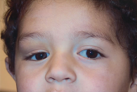
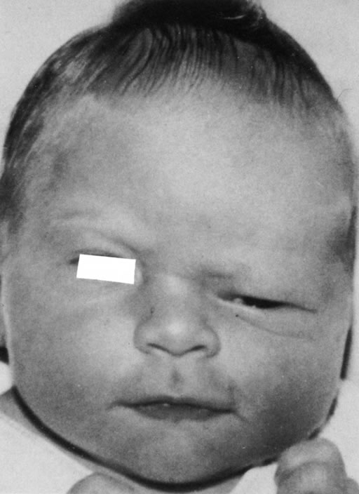
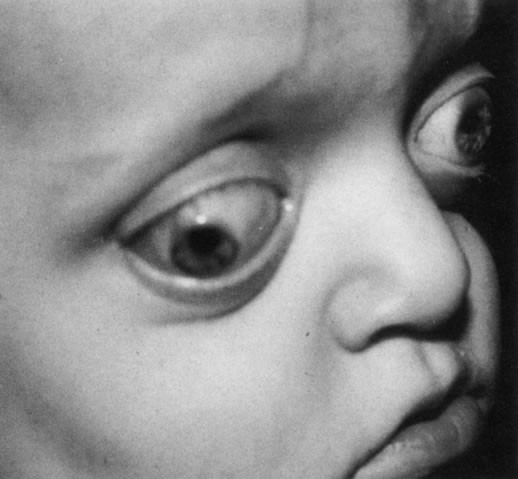
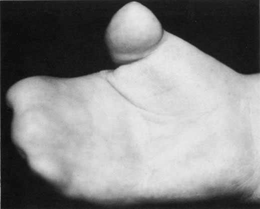
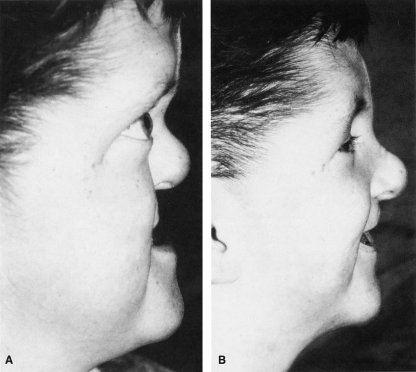
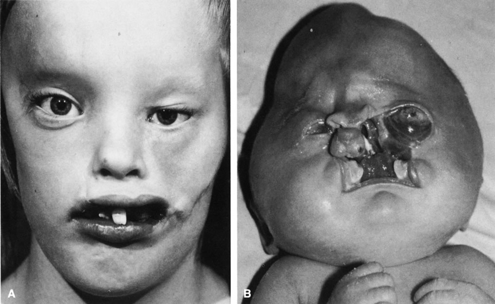
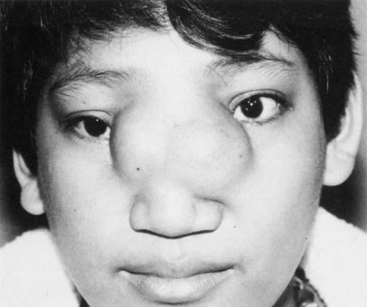
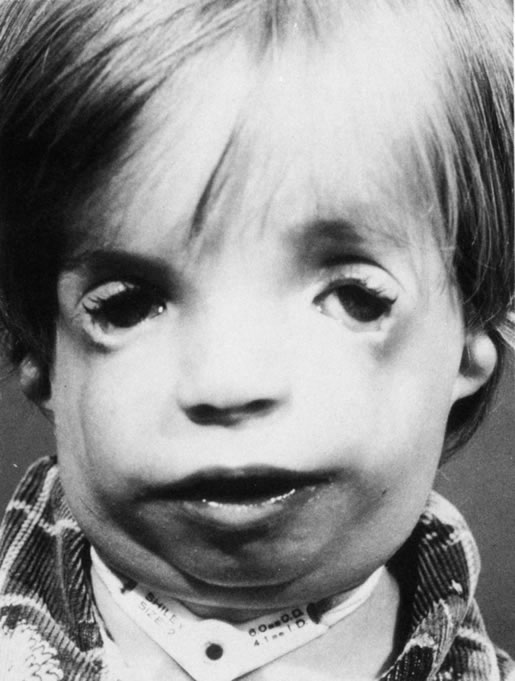
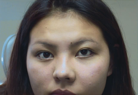
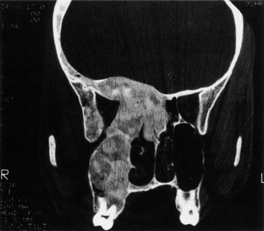
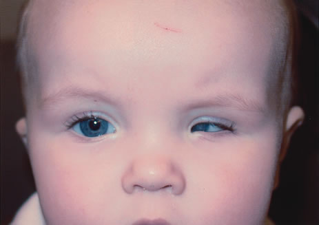
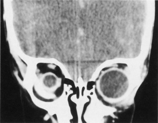
Anophthalmos and microphthalmos are usually unilateral and may be associated with a variety of craniofacial and systemic anomalies, including orbital hypoplasia, facial clefts, basal encephalocele, hemifacial microsomia, mandibulofacial dysostosis, cardiac anomalies, polydactyly, and mental retardation. When they occur unilaterally, they also can be associated with anomalies of the contralateral “normal” eye, including cataract, cornea1 opacities, microphthalmos, coloboma, epibulbar dermoids, and nystagmus. Anophthalmos and severe microphthalmos frequently are associated with contracted conjunctival fornices, phimotic eyelids, and generalized hypoplasia of the periocular soft tissues (Fig. 1). When soft tissue contractures occur, the early use of conformers is essential to expand these tissues.2 This treatment should be instituted in the first month of life, with progressive enlargement of the conformer over time to achieve maximum expansion of the conjunctival fornix. Unfortunately, this treatment usually does not stimulate adequate orbital bone growth, and unilateral microphthalmos and anophthalmos may be associated with secondary orbital hypoplasia (Fig. 2). Serial implantation of progressively larger orbital implants or placement of expansile orbital implants has been advocated to stimulate bony orbital development.3,4

Fig. 1. Microphthalmos of the left eye has resulted in secondary soft tissue contractures, including shortened fomices and small, phimotic eyelids.

Fig. 2. Abnormal globe development for any reason may result in secondary orbital hypoplasia. Here, microphthalmos of the right globe is associated with relative orbital hypoplasia.

CYSTIC ANOMALIES OF THE ORBIT

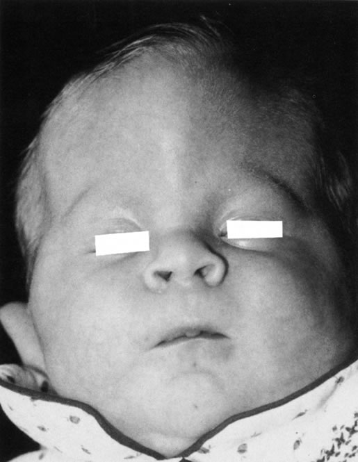
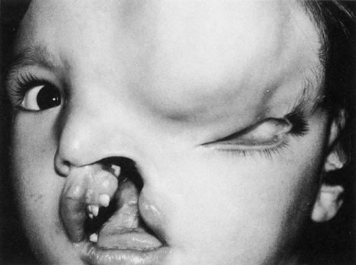
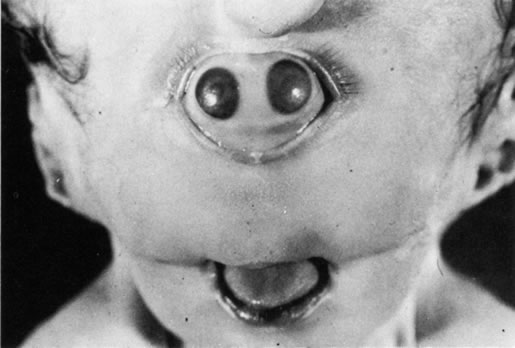
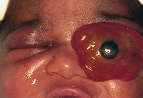
Many congenital cystic structures may arise from or involve the orbit. Some cystic structures, such as meningoencephaloceles or mucoceles, result from defects in the bony sutures of the cranial skeleton, allowing herniation of adjacent structures into the orbit. Other cystic structures, such as dermoid cysts, teratomas, and epithelial cysts, result from developmental anomalies of the orbital soft tissues. Most isolated orbital cysts have a subtle clinical presentation at birth, although some may present with extreme proptosis (Fig. 3). Ultrasonography can aid in the prenatal detection and monitoring of large orbital cysts.5

Fig. 3. Extreme proptosis at birth caused by a congenital orbital cyst.

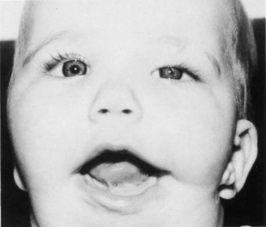
Dermoid cysts are the most common congenital orbital anomalies and represent developmental choristomas that are believed to arise from ectodermal nests pinched off by the fusion of bony sutures around the orbit. These cysts often originate from the frontozygomatic suture temporally but can also be seen nasally, arising from the frontonasal and frontolacrimal sutures; they rarely occur deep in the orbit.6 They commonly present during the first decade of life as a well-circumscribed, firm, rubbery subcutaneous mass just below the temporal eyebrow. Deeper dermoids can remain asymptomatic for many years, often presenting later in life as a slowly expanding orbital mass. Complete excision of these encapsulated lesions is the preferred treatment. Rupture of the cyst from trauma or during surgery may result in severe orbital inflammation (Fig. 4).

Fig. 4. A ruptured orbital dermoid cyst on the right side has caused swelling of the brow and upper eyelid as well as inferomedial displacement of the globe.

Back to Top
CRANIOSYNOSTOSIS
Craniosynostosis implies premature fusion of the bony sutures of the skull. These craniofacial anomalies usually display a sporadic inheritance pattern, although several well-recognized syndromes have distinct inheritance patterns, such as the autosomal dominant pattern of Crouzon's disease. Although premature suture fusion was believed to be the primary pathologic process, genetic mapping and molecular studies suggest that this early fusion may be a result of altered cytokine and extracellular matrix component expression.7

The phenotypic pattern of deformity is a direct result of the sutures involved (Fig. 5). Scaphocephaly is an elongated, narrow cranium associated with premature fusion of the sagittal suture. Brachycephaly is a short, wide cranial vault associated with bilateral coronal synostosis. Plagiocephaly results from premature closure of one coronal suture, leading to prominent orbital asymmetry (Fig. 6). A flattened, recessed forehead occurs on the affected side, and persistent growth of the contralateral side results in frontal bossing, inferolateral orbital dystopia, and a prominent occiput. This deformity is reminiscent of hemifacial microsomia. Trigonocephaly is a triangular deformity of the anterior cranial fossa that results in medial displacement of the orbits (hypotelorism) (Fig. 7). Acrocephaly results from multiple suture closure, including bicoronal synostosis. Typically, there is excessive skull height and a pointed head.

Fig. 5. The skull deformity in cranlosynostosis is a direct result of the sutures involved.

Fig. 6. Unilateral coronal synostosis results in severe orbital asymmetry (ptogiocephaly). The uninvolved side (left) shows dramatic growth relative to the prematurely fused side (right)).

Fig. 7. Trigonocephaly or triangular deformity of the anterior cranial fossa, with associated hypotelorism.

Back to Top
CROUZON'S DISEASE
In 1912, Crouzon described the froglike facies characteristic of this distinct anomaly, and his name has been associated with this syndrome ever since (Fig. 8). Most cases of this autosomal dominant disease have been mapped to the fibroblast growth factor receptor 2 gene located on chromosome 10.8 Ocular findings include exophthalmos, hypertelorism, strabismus, extraocular muscle agenesis or anomaly, nystagmus, papilledema, and optic atrophy. A variable pattern of suture closure is seen, including coronal, sagittal, and lambdoid sutures. The deformities of the orbit and cranial vault are in part a result of the compensatory expansion of the cranium from increased intracranial pressure. Forward displacement of the greater wing of the sphenoid bone results in a shortening of the lateral orbital wall and a dramatic reduction in orbital volume. To compound the problem, there is also inferior displacement of the orbital roof from anterior cranial fossa expansion and shortening of the orbital floor from maxillary hypoplasia. Tessier estimated that these defects account for a 6-cc reduction in orbital volume, or approximately 20% to 25% of the total volume of the orbit.9

Fig. 8. The froglike facies of Crouzon's disease results from maxillary hypoplasia, shallow orbits, and prominent exophthalmos.

Back to Top
APERT'S SYNDROME
Apert's syndrome is similar to Crouzon's disease, although the craniofacial deformities are usually more severe. The genetic defect has also been localized to the fibroblast growth factor receptor 2 gene.7 Ocular findings may include brachycephaly, exophthalmos, hypertelorism, strabismus, extraocular muscle agenesis or anomaly, and maxillary hypoplasia. The distinguishing feature of Apert's syndrome is syndactyly of the hands and feet (Fig. 9). Ptosis, an antimongoloid slant to the intrapalpebral fissure, and oculomotor palsies also can be seen in Apert's syndrome.9

Fig. 9. The main differentiating feature between Crouzon's disease and Apert's disease is the associated syndacryly in the latter.

Back to Top
TREATMENT
Early intervention in craniosynostosis is necessary not only to reduce the intracranial pressure and permit normal visual and mental development, but also to achieve a satisfactory cosmetic result. The techniques employed include a variety of frontal and facial bone advancements designed to expand the intracranial volume and improve the cosmesis of the facial skeleton. Traditionally, Le Fort III osteotomies and bone grafting have been used to achieve midface advancement to address the maxillary hypoplasia and shallow orbits that develop in craniosynostosis (Fig. 10). More recently, internal distraction devices have allowed for a more gradual but safer and less morbid technique for midface advancement.10 If detected in the neonatal period (before 6 months of age), many cases of craniosynostosis can be successfully managed with small-incision endoscopic craniectomies and external skull molding devices.11

Fig. 10. Preoperative (left)) and postoperative (right)) views of a patient with Apert's syndrome who underwent LeFort III osteotomies and midface advancement.

Back to Top
FACIAL CLEFTS
The confusing array of skeletal and soft tissue deformities that constitute the facial clefting syndromes were brought to order in 1976, when Tessier developed an anatomic classification for this diverse group of congenital anomalies.12 The Tessier classification numbers the clefts based on their location in relation to the orbit (Fig. 11). Facial clefts range from small, isolated soft tissue defects to severe, disfiguring craniofacial deformities (Fig. 12).13

Fig. 11. The Tessier classification system for cranial and facial clefts is based on their location in relation to the orbit.

Fig. 12. A. Mild facial distortion is evident in this patient with a subtle Tessier cleft 4 on the right and a more pronounced Tessier cleft 7 on the left, B. The grotesque deformity in this patient is a result of severe, bilateral Tessier cleft 3, or naso-orn-ocular cleft.

Tessier cleft 0-14, a true median cleft, is associated with orbital hypertelorism and meningoencephalocele (Fig. 13). Clefts 1 and 2 are associated with telecanthus from involvement of the soft tissues of the medial canthus but spare the lacrimal system and eyelids. Clefts 3 and 4 involve the inferomedial orbit and lower eyelid medial to the punctum. Disruption of the nasolacrimal system is associated with bony defects between the orbit, maxillary sinus, and nasal cavities, with inferior displacement of the globe.14 Cleft 5 is associated with a defect in the inferolateral orbital rim and floor, a lateral lower eyelid cleft, and frequently microphthalmia. Features of clefts 6, 7, and 8 are seen in Treacher Collins syndrome, Goldenhar's syndrome, and hemifacial microsomia. Ocular abnormalities may include a cleft or antimongoloid slant of the lateral lower eyelid, hypoplasia of the maxilla with downward slanting of the lateral orbital floor, and hypoplasia or absence of the lateral orbital wall. Cleft 9 is characterized by defects in the superolateral orbital rim and the lateral one-third of the upper eyelid, and distortion of the lateral canthus. A central cleft of the eyebrow, upper eyelid, supraorbital rim, and orbital roof characterize cleft 10. Fronto-orbital meningoencephaloceles are common (Fig. 14). Cleft 11 is characterized by defects in the medial aspect of the upper eyelid and brow, but no bony defect in the supraorbital rim. Cleft 12 is associated with telecanthus, hypertelorism, and a defect at the medial root of the eyebrow. Clefts 13 and 14 are characterized by hypertelorism with sparing of the orbital soft tissues.

Fig. 13. Tessier cleft 0=N14 with a prominent midline meningoencephalocele.

Fig. 14. This Tessier cleft 3=N10 is associated with a large frontal meningoencephalocele.

Back to Top
HYPERTELORISM
Orbital hypertelorism is defined as an abnormally wide distance between the orbits. It is not a syndrome but a physical finding that is found in a variety of craniofacial anomalies, including facial clefts. Hypertelorism implies an increased interpupillary distance. This condition should not be confused with telecanthus, which is defined as an increased distance between the two medial canthal angles with a normal interpupillary distance. Telecanthus is classically seen with traumatic disinsertion of the medial canthal tendons. In Waardenburg's syndrome, it is associated with heterochromia iridis, a white forelock, and congenital deafness.

Hypertelorism is often associated with a variety of facial clefts, craniosynostosis, and meningoencephaloceles. The normal distance between the orbits is roughly 16 mm at birth and increases to 25 to 28 mm in adults.15 A widening of the anterior ethmoid air cells is believed to be the main anatomic defect responsible for primary orbital hypertelorism, resulting in an increase in soft tissue, bone, and cartilage between the medial canthi.16 The posterior ethmoid air cells and the sphenoid bone are usually normal, and as a result, the optic foramina are usually normal as well. The cribriform plate is not widened but can be depressed 10 mm below its usual level, making the extracranial approach to the correction of this defect hazardous. The angle between the central axes of the orbits is normally 45°. In orbital hypertelorism, the axes of the orbits are more divergent, measuring up to 60° in severe cases.

Surgical correction of hypertelorism usually entails a combined intracranial and extracranial approach. All four walls of each orbit are osteotomized to free them from the frontal, zygomatic, maxillary, nasal, and sphenoid bones. The excessive intervening tissues are removed, and the orbits are brought closer together in the midline. The resultant bone gaps are filled with bone grafts.

Back to Top
ORBITAL DYSTOPIA
Orbital dystopia is defined as a vertical misalignment of the globes. Like hypertelorism, orbital dystopia is a physical finding with a variety of etiologies, not a distinct clinical syndrome. Congenital anomalies of orbital development are the most common cause of orbital dystopia. These anomalies include craniosynostosis, hemifacial microsomia, and orbitofacial clefts. Acquired orbital dystopia may occur as a result of facial and orbital fractures or mass lesions that arise from the orbit, periorbital sinuses, and adjacent structures. Progressive orbital dystopia of unknown etiology requires a thorough evaluation to exclude the presence of mass lesions encroaching on the ocular structures.
Back to Top
MENINGOENCEPHALOCELE
Congenital defects in the bony sutures of the cranial skeleton may result in a herniation of brain and meninges into the orbit, known as meningoencephalocele (see Fig. 13). This defect is frequently associated with a variety of facial clefts and usually occurs medially between the sutures of the frontal, ethmoidal, lacrimal, or nasal bones. A soft, pulsatile mass that bulges with coughing and Valsalva maneuvers appears in the upper medial canthal area during the first years of life. Rarely, a congenital dehiscence in the greater wing of the sphenoid bone may also result in a slowly progressive pulsatile exophthalmos that presents later in life.
Back to Top
HEMIFACIAL MICROSOMIA AND OCULOAURICULAR DYSPLASIA
Hemifacial microsomia is a complex disorder of unknown etiology that is characterized by facial asymmetry with ipsilateral abnormalities of the middle ear, mandibular ramus, and condyle. Dystopia may result from hypoplasia of the orbital bones. Associated systemic defects can involve the heart, kidneys, and limbs.

Oculoauriculovertebral dysplasia (Goldenhar's syndrome) is a variant of hemifacial microsomia characterized by unilateral malformations of the eye, ear, and malar and vertebral structures (Fig. 15). Soft tissue findings include epibulbar dermoids, orbital lipodermoids, eyelid colobomas, preauricular appendages, and aural fistulas. Marked facial asymmetry can occur due to unilateral hypoplasia of the zygoma, mandible, and chin.

Fig. 15. Note the limbal dermoid, eyelid coloboma, and preauricular skin lags in this patient with Goldenhar's syndrome.

Back to Top
MANDIBULOFACIAL DYSOSTOSIS
Mandibulofacial dysostosis (Treacher Collins syndrome, Franceschetti syndrome) is a bilateral, autosomal dominant condition that results from abnormal development of structures derived from the first and second branchial arches (Fig. 16). The genetic defect for Treacher Collins syndrome, TCOF1, has been isolated to chromosome 5, and the gene product, treacle, has been identified as a nucleolar protein.17,18 The exact function of treacle, however, has not yet been determined. The craniofacial anomalies include hypoplasia of the maxilla, mandible, and zygoma associated with a variety of soft tissue malformations. These malformations include underdevelopment of the midfacial musculature, lower eyelid colobomas, inferior displacement of the lateral canthi (antimongoloid slant), inferior punctal agenesis, and blepharoptosis. Additional ocular anomalies may include high myopia, dermolipoma, lens subluxation, and secondary glaucoma.19

Fig. 16. Note the maxillary and mandibular hypoplasia, inferior displaced lateral canthi, lower eyelid colobomas, and blepharoptosis in this patient with Treacher Collins syndrome (Tessier clefts 6, 7, and 8)).

Back to Top
PROGRESSIVE HEMIFACIAL ATROPHY
Progressive hemifacial atrophy (Romberg's syndrome) is a rare disorder characterized by progressive atrophy of the skin, subcutaneous tissue, muscle, cartilage, and bone, usually involving only one side of the face. The genetic basis of this disorder has not been elucidated. This condition has been associated with a variety of ocular findings, including progressive enophthalmos, heterochromia, uveitis, restrictive strabismus, papillitis, retinal vasculitis, oculomotor nerve palsy, Horner's syndrome, Fuchs's syndrome, and Duane's retraction syndrome.20 Surgical intervention has been aimed at masking the atrophy with the use of various flaps, grafts, and implants.21,22
Back to Top
PIERRE ROBIN SEQUENCE
In 1923, Pierre Robin described a condition characterized by micrognathia, glossoptosis, cleft palate, and respiratory distress from airway obstruction at the level of the tongue. Although the exact genetic defect for this disorder has not yet been identified, recent studies have localized a candidate locus to the long arm of chromosome 2.23 Associated ocular findings may include microphthalmia, congenital glaucoma, and high myopia with associated retinal detachments.
Back to Top
OTHER ANOMALIES OF THE ORBIT

FIBROUS DYSPLASIA

Fibrous dysplasia is a benign disorder characterized by an arrest of bone maturation that results in immature bone and osteoid in a cellular fibrous matrix. This congenital disease usually becomes clinically apparent in children and young adults. Fibrous dysplasia can extensively involve the facial bones and skull.24 The maxillary, frontal, and sphenoid bones are most commonly involved. Maxillary bone involvement can cause nasolacrimal duct obstruction. Involvement of the frontal and sphenoid bones may result in orbital asymmetry from contour deformities, vertical dystopia, and exophthalmos (Fig. 17). Radiographically, fibrous dysplasia appears as an expansile bone lesion with a characteristic ground-glass appearance (Fig. 18). Additional ocular complications include compressive optic neuropathy, oculomotor nerve palsy, and trigeminal neuralgia.25 Involvement of the sphenoid bone may result in narrowing of the optic canal, with secondary compressive optic neuropathy. Definitive treatment entails unroofing the optic canal by way of a transcranial approach, although high-dose steroids can be used as a temporizing measure.

Fig. 17. Fibrous dysplasia of the left maxilla has resulted in marked hypertrophy of the maxilla and cheek, lower eyelid ectropion, and hyperglobus of the left globe.

Fig. 18. Fibrous dysplasia that involves the frontal, sphenoid, ethmoid, and zygomatic bones illustrates the characteristic ground-glass appearance of this disorder.

Fibrous dysplasia is not a true neoplasm; however, there is a small incidence of malignant degeneration, usually into osteogenic sarcoma. Treatment options range from careful observation to aggressive debulking of the diseased bone, with subsequent reconstruction. Recent advances in craniofacial surgery, including cranial bone grafting with mini- and microplate fixation, have made the latter approach more appealing.

ANENCEPHALY

Anencephaly, either partial or total absence of the brain, is a severe congenital birth defect incompatible with life. This dramatic deformity results from a failure of forebrain development. The vault of the skull is absent, and the forebrain consists of a degenerated mass of glial tissue. The orbits are shallow and tilted upward. The eyes are fairly well developed, but the optic nerves, when present, taper down to a loose mass of glial tissue at the optic canal.

CYCLOPIA/SYNOPHTHALMOS

True cyclopia is a rare congenital anomaly characterized by a single eye situated in a single median orbit. Synophthalmos, which is also rare but much more common than true cyclopia, occurs when paired ocular structures are found in a single median orbit (Fig. 19). These disorders result from a failure of lateralization of the midline facial structures.

Fig. 19. Synophthalmos.

Back to Top
REFERENCES

1. Moss ML, Salentijn L: The primary role of functional matrices in facial growth. Am J Orthodontia 38:38, 1968

2. Dootz GL: The ocularists' management of congenital microphthalmos and anophthalmos. Adv Ophthalmic Plast Reconstr Surg 9:41, 1992

3. Krastinova D, Kelly MB, Mihaylova M: Surgical management of the anophthalmic orbit, part 1: Congenital. Plast Reconstr Surg 108:817, 2001

4. Weise KG, Vogel M, Guthoff R, Gundlach KK: Treatment of congenital anophthalmos with self-inflating polymer expanders: A new method. J Craniomaxillofac Surg 27:72, 1999

5. Yen MT, Tse DT: Congenital orbital cyst detected and monitored by prenatal ultrasonography. Ophthal Plast Reconstr Surg 17:443, 2001

6. Chawda SJ, Moseley IF: Computed tomography of orbital dermoids: A 20-year review. Clin Radiol 54:821, 1999

7. Carinci P, Becchetti E, Bodo M: Role of the extracellular matrix and growth factors in skull morphogenesis and in the pathogenesis of craniosynostosis. Int J Dev Biol 44:715, 2000

8. Reardon W, Winter RM, Rutland P, et al: Mutations in the fibroblast growth factor receptor 2 gene cause Crouzon syndrome. Nat Genet 8:98, 1994

9. Tessier P: The definitive plastic surgical treatment of the severe facial deformities of craniofacial dysostosis, Crouzon's and Apert's disease. Plast Reconstr Surg 48:419, 1971

10. Holmes AD, Wright GW, Meara JG, et al: LeFort III internal distraction in syndromic craniosynostosis. J Craniofac Surg 13:262, 2002

11. Jimenez DF, Barone CM, Cartwright CC, Baker L: Early management of craniosynostosis using endoscopic-assisted strip craniectomies and cranial orthotic molding therapy. Pediatrics 110:97, 2002

12. Tessier P: Anatomical classification of facial, craniofacial, and latero-facial clefts. J Maxillofac Surg 4:69, 1976

13. David DJ, Moore MH, Cooter RD: Tessier clefts revisited with a third dimension. Cleft Palate 26:163, 1989

14. Stretch JR, Poole MD: Nasolacrimal abnormalities in oblique facial clefts. Br J Plast Surg 43:463, 1990

15. Cohen MMJr , Richieri-Costa A, Guion-Almeida ML, Saavedra D: Hypertelorism: Interorbital growth, measurements, and pathogenetic considerations. Int J Oral Maxillofac Surg 24:387, 1995

16. Converse JM, Ransohoff J, Mathews ES, et al: Ocular hypertelorism and pseudohypertelorism: Advances in surgical treatment. Plast Reconstr Surg 45:1, 1970

17. The Treacher Collins Syndrome Collaborative Group: Positional cloning of a gene involved in the pathogenesis of Treacher Collins syndrome. Nat Genet 12:130, 1996

18. Winokur ST, Shiang R: The Treacher Collins syndrome (TCOF1)) gene product, treacle, is targeted to the nucleolus by signals in its C-terminus. Hum Mol Genet 7:1947, 1998

19. Posnick JC, Ruiz RL: Treacher Collins syndrome: Current evaluation, treatment, and future directions. Cleft Palate Craniofac J 37:434, 2000

20. Miller MT, Spencer MA: Progressive hemifacial atrophy. A natural history study. Trans Am Ophthalmol Soc 93:203, 1995

21. Abyholm FE, Skolleborg KC: Aesthetic treatment of progressive hemifacial atrophy (Romberg's disease)): Use of a pedicled platysma flap. Plast Reconstr Surg 95:71, 1995

22. Rigotti G, Cristofoli C, Marchi A, et al: Treatment of Romberg's disease with parascapular free flap and polyethylene porous implants. Facial Plast Surg 15:317, 1999

23. Houdayer C, Portnoi MF, Vialard F, et al: Pierre Robin sequence and interstitial deletion 2q32.3-q33.2. Am J Med Genet 102:219, 2001

24. Camilleri AE: Craniofacial fibrous dysplasia. J Laryngol Otol 105:662, 1991

25. Liakos GM, Walker CB, Carruth JA: Ocular complications in craniofacial fibrous dysplasia. Br J Ophthalmol 68:611, 1979

Back to Top