Chapter 22
Introduction to Orbital Disease
JOHN T. HARVEY
Main Menu   Table Of Contents

Search

DEMOGRAPHICS OF ORBITAL DISEASE
HISTORY TAKING
PRESENTATION
QUESTIONS
ORBITAL EXAMINATION
SPECIAL INVESTIGATIONS IN ORBITAL DISEASE
CONCLUSION
REFERENCES

This chapter provides the reader with a guide to history taking and examination in the patient presenting with orbital disease. In addition the demographics of orbital disorders are presented to give an overview of the territory. Specific disease entities are not covered in any great detail because these are discussed in subsequent chapters. Similarly, specific orbital investigative techniques and imaging modalities are not dealt with in an extensive manner because these are covered in more detail in separate chapters. It is assumed that the reader has a good grasp of orbital anatomy and is familiar with basic history taking and general physical examination techniques.
Back to Top
DEMOGRAPHICS OF ORBITAL DISEASE
All the major reports on the incidence of orbital disease have been consolidated into two tables: Table 1 covers orbital disease as a whole and Table 2 deals with pediatric orbital disorders. Together they include the major studies of the past 40 years.


TABLE 1. Review of 4635 Orbital Cases (1960-2001)


 Shields 1Reese 2Moss 3Henderson 4Silva 5Kennedy 6Rootman 7Harvey 20Total
LesionNo.%No.%No.%No.%No.%No.%No.%No.%No.%
Inflammatory8713%9118%5725%409%4916%18122%80857%5521%136830%
Cystic19330%469%136%255%5017%8911%836%3313%53211%
Secondary/metastatic8613%337%83%13629%6622%7910%614%104%47910%
Vasculogenic406%11222%4821%5712%196%729%967%2811%47210%
Lymphoid/leukemic619%4810%2210%398%217%10212%544%2811%3758%
Lacrimal8313%275%177%296%145%455%141%218%2505%
Optic nerve/meningeal102%296%198%4610%196%111%504%187%2024%
Peripheral nerve142%235%73%215%134%536%242%21%1573%
Osseous, fibrous, cartilagenous203%112%21%266%289%334%262%125%1583%
Rhabdomyosarcoma81%377%52%102%52%81%50%10%792%
Melanocytic10%245%00%225%103%30%121%31%752%
Adipose234%41%00%51%00%00%81%52%451%
Histiocytic10%00%21%72%00%40%81%31%251%
Other183%194%3013%20%62%14017%16011%4316%4189%
 TOTAL645 504 230 465 300 820 1409 262 4635 

The “Inflammatory” row in the reports of Moss, Silva, and Kennedy & Rootman include Graves' disease.
All percentages are rounded to the nearest whole number.


TABLE 2. Review of 1370 Cases of Orbital Tumors in Children (1953-1987)


 Ingalls8Porterfield9MacCarty and Brown10Yousefi11Templeton12Eldrup-Jorgenson and Fledelius13Iliff and Green14Crawford15Shields16et al.Bullock17et al.Total 
LesionNo.%No.%No.%No.%No.%No.%No.%No.%No.%No.%No.%
Cystic1529251242315000384860346413052594236827
Vasculogenic142728133619915126827162416177231618514
Optic nerve/meningeal363918512723009111481711627514811
Rhabdomyosarcoma4856260023121013159149104321158
Inflammatory241051792391511951394116541098
Lymphoid/leukemic3612611003660452123156254927
Secondary/metastatic363111671112231061499443645
Peripheral nerve61263630012341161494296604
Osseous, fibrous, cartilaginous00212111127121142643175524
Adipose0031112300345300177118423
Histiocytic0021000000001120131032272
Lacrimal00422112002353116232242
Melanocytic0000210000000000000020
Other1224113418584711116000021826
 TOTAL51 214 186 62 60 80 174 152 250 141 1370 

All percentages are rounded to the nearest whole number.
(Modified from Bullock JD, Goldberg SH, Rakes SM: Orbital tumours in children. Ophthalmic Plast Reconstr Surg 5:13–16, 1989)


There are several difficulties inherent in this type of data collation. Not all authors use the same categories for orbital disease, which means that the data in these papers must be made to fit into the classification that I have chosen to use, which is a modified Shields classification.1 The studies often have very different biases. Some studies are based on large pathologic series, whereas others use clinical material. Some studies insist on biopsied proof of the diagnosis and other studies do not. Some of the older studies were carried out in the pre-computed tomographic scanning era. Some represent data accumulated at large referral centers, whereas others represent data collected in smaller practices. Some authors include Graves' disease in the inflammatory category, whereas others do not. Despite these limitations, I believe that both tables fairly represent the incidence of orbital disorders that an ophthalmologist could reasonably expect to encounter in practice.

Table 1 shows that the two leading categories (inflammatory and cystic) account for approximately 41% of all orbital lesions. If one adds vascular lesions, which account for 10%, and all the other nonmalignant categories, one finds that well over half of all orbital lesions are nonmalignant.

Bullock nicely summarized the pediatric data and this has been reproduced with slight modification in Table 2. Cystic and vascular lesions make up 41% of the orbital masses seen in the pediatric age group. Rhabdomyosarcoma, which is rare in adults but relatively frequent in children, occurs fourth in frequency and accounts for approximately 8% of pediatric orbital tumors. The percent of cases in the lymphoid/leukemic category is approximately the same in both children and adults.

Back to Top
HISTORY TAKING
Obtaining the medical history is important in all aspects of ophthalmology, especially in the case of the patient presenting with orbital disease. Ophthalmologists are accustomed to dealing with ocular disorders that can be readily visualized using either biomicroscopy or funduscopy. Because of this reliance on visual clues for diagnosis, ophthalmologists frequently take an abbreviated history and rely mainly on the ocular examination to arrive at a diagnosis. Orbital disorders are more akin to abdominal disease, in which the pathologic entity is not visible to the naked eye. Consequently, a thorough history is important for reaching a correct diagnosis.
Back to Top
PRESENTATION
Orbital disorders typically present in one of five ways, and the type of presentation is helpful in steering the examiner to the correct anatomic location and, hence, the correct differential diagnosis.

PROPTOSIS

Proptosis implies an axial protrusion of the globe from the bony socket. It is helpful to determine if the globe is displaced in another direction, because a lesion presenting in one quadrant of the orbit will frequently displace the globe into the opposite quadrant, as well as produce a degree of proptosis. For example, lacrimal gland tumors frequently displace the eye infranasally in addition to creating proptosis.

Pseudoproptosis can occur if the pathologic side is enophthalmic. Occasionally this may cause confusion. The most frequent cause of enophthalmos is blowout fracture, but metastatic breast carcinoma may cause enophthalmos.18

PAIN

Patients with orbital disorders frequently complain of pain or discomfort on the affected side. The pain may range from very mild to very severe. The quality of the pain is an important characteristic. Burning, scratching, and irritation are frequently described in patients presenting with Graves' disease and are often related to the desiccation of the cornea and conjunctiva. Patients with inflammatory lesions, whether benign or malignant, frequently complain of an aching, throbbing, or boring pain that resides behind the eye and may radiate into the forehead, cheek, or temporal areas. Dysesthesias, in the distribution of the supraorbital nerve or the infraorbital nerve, are encountered following orbital trauma, especially during the recovery period as the initial posttraumatic hypoesthesia is resolving.

DIPLOPIA

Diplopia is a common symptom in orbital disorders, related to a paralysis of the extraocular muscles or a restriction of ocular movement. Typically, lesions residing in the cavernous sinus or posterior orbit are responsible for paralytic abnormalities of ocular movement. More anterior orbital lesions typically produce diplopia by means of a mechanical effect. This mechanical restriction of function in the extraocular muscles may be caused by lesions that are immediately adjacent to the extraocular muscles or diseases that involve the muscle tissue, such as myositis and Graves' disease.

DECREASE IN VISUAL ACUITY

Loss of visual acuity usually implies involvement of the optic nerve, either by compression, infiltration, vascular compromise, or inflammation. Signs of optic nerve dysfunction, such as relative afferent pupillary defect, loss of color vision, visual field cuts, and increased latency in the visually evoked responses, may be identified.

LID MALPOSITION

Lid malpositions are less common but may be associated with orbital masses in the anterior extraconal space. For example, lateral ptosis of the upper eyelid is often associated with lacrimal gland masses. Superior orbital tumors may produce upper eyelid ptosis because of interference with the normal function of the levator muscle. Lid retraction of the upper and lower eyelids is one hallmark of Graves' orbital disease.

Back to Top
QUESTIONS

WHAT IS THE PROBLEM?

It is important to ask “What is the problem?” directly of the patient and not rely too heavily on the description provided in the referring letter or in the history taken by ophthalmic assistants because patients will often reveal important information as they recount their history. The examiner must remember not to be misled by the garrulous or vague patient who may attribute the orbital condition to an event that is unrelated but coincidental.

WHEN DID IT START?

The time of onset of the orbital condition is important. In many cases the patient is unable to give this information because the onset of the proptosis has been very gradual. Asking the patient to bring old pictures can be very helpful in this situation. In the case of pain or diplopia, patients are usually accurate and specific about the time of onset. The practitioner should try to get a sense of whether the orbital symptom has been progressive or stable since its onset. In addition some long-standing orbital conditions may present with a sudden worsening, an example is sudden bleeding into an orbital lymphangioma.

WHAT MAKES IT BETTER?

Asking the patient presenting with pain about what relieves pain will sometimes give valuable information. A patient requiring narcotics for relief of pain is in most cases having more severe pain than the patient who requires simple aspirin, although varying pain thresholds and occasional drug abusers make this less than absolute.

WHAT MAKES IT WORSE?

Asking about what makes the pain worse is especially useful in patients presenting with double vision, because diplopia is often worse in specific fields of gaze. The proptosis of vascular lesions sometimes worsens with head position or the Valsalva maneuver (see following discussion). Lymphangiomas typically enlarge during upper respiratory tract infections.

ASK ABOUT THE EFFECT OF HEAD POSITION AND A VALSALVA MANEUVER

Asking about head position and the Valsalva maneuver is important in patients who have a vascular lesion. An increase in proptosis is often reported by a patient with orbital varices who places his or her head in a dependent position or who performs a Valsalva maneuver. Frequently patients with inflammatory conditions of the orbit will describe worsening of their pain when their head is placed in a dependent position. Presumably this increase in pain is caused by the engorgement of orbital vessels when the head is dependent.

ASK ABOUT SYSTEMIC DISORDERS

The value of inquiring about systemic disorders cannot be overestimated, because it is obvious that many systemic disorders are associated with orbital disease or influence the choice of treatment. Obvious examples include hyperthyroidism and a history of previous malignancy. The question of a previous malignancy must be approached with delicacy because patients may be reluctant to mention it because of denial or ignorance of previous diagnoses. Often questioning a family member, the family physician, or the referring doctor is helpful.

Back to Top
ORBITAL EXAMINATION
The orbital examination consists of the routine ophthalmic examination plus some specialized evaluations that are pertinent to orbital disease.

ROUTINE

Vision

The vision is an integral part of any orbital examination. Visual acuity provides an indicator of the extent of orbital disease, and decreased vision suggests involvement of the optic nerve or globe. Vision gives a baseline from which to measure progression of disease and is a medical-legal necessity. This is even more important in patients presenting with orbital trauma. It is vital to perform a visual acuity examination before taking trauma patients to surgery even if this requires using a Desmarres retractor to open the swollen eyelids. Nonophthalmic surgeons who are called on to operate for trauma in the orbital area frequently neglect this vital part of the orbital examination.

Pupils

Examination of the pupils is very important and may provide information about the underlying orbital condition. The small pupil of a Horner's syndrome may be the clue that one is dealing with a pseudoenophthalmos. The large pupil of a third cranial nerve palsy indicates that the associated diplopia is due to a paralytic abnormality as opposed to a restrictive phenomenon. The presence of a relative afferent pupillary defect is confirmation that the optic nerve is compromised by orbital disease, even though many patients have an optic nerve head that appears normal on funduscopic examination.

Extraocular Movements

The cross cover test is invaluable for assessing ocular position in primary position, upgaze, downgaze, right gaze, and left gaze. In most orbital disorders the deviation will vary in different positions of gaze. The nonconcomitance of strabismus associated with orbital disease helps to distinguish it from other common forms of strabismus. It is important to note abnormal ductions because a slight limitation in one eye may be the only evidence of an abnormality and may be very useful in localizing the orbital lesion.

Slit-Lamp Examination

Slit-lamp examination is most useful for assessing the status of the cornea that may demonstrate exposure keratopathy caused by proptosis or lid retraction. Many orbital disorders cause dilatation of conjunctival vessels and conjunctival edema. These findings are relatively nonspecific, but large tortuous conjunctival veins may be a sign of carotid cavernous fistula.

Intraocular Pressure

Intraocular pressure may be elevated in patients with orbital disease, through a variety of mechanisms. Carotid cavernous fistulas cause an increase in episcleral venous pressure and, therefore, impede aqueous outflow. Mass lesions may press directly against the globe, especially when a retrobulbar hemorrhage causes a sudden expansion of the mass. Restriction of the extraocular muscles, such as may occur with blowout fracture or thyroid eye disease, produces a factitious elevation of intraocular pressure when the eye attempts to rotate against the restricted muscle. In cases of mild thyroid eye disease, the progressive increase in intraocular pressure with elevation of the eye may help to confirm the diagnosis.

Fundus Examination

Fundus examination is important to assess the optic nerve. Tumors may compress the optic nerve, causing disc edema or optic atrophy. Optic nerve meningiomas may produce shunt vessels on the disc. Choroidal striae may be noted if a mass is indenting the globe. On occasion intraocular tumors may extend through the sclera into the orbit, and the correct diagnosis is made when the intraocular tumor is identified.

Color Vision

Assessment of color vision is an important test for optic nerve dysfunction, although one must be careful not to attribute optic nerve dysfunction to patients who have a congenital abnormality of color vision. Sophisticated tests for color vision measurement, such as the Farnsworth-Munsell test, are not generally required for clinical assessment of optic nerve dysfunction. The Ishihara color plates are satisfactory for clinical use.

SPECIALIZED ORBITAL EXAMINATION

Inspection

Inspection of the face and more specifically the periocular adnexa is the simplest of all examinations and yet this inspection is frequently overlooked by neophyte examiners. It is important to look at the entire face to get a sense of the facial proportion and symmetry.

Globe Displacement

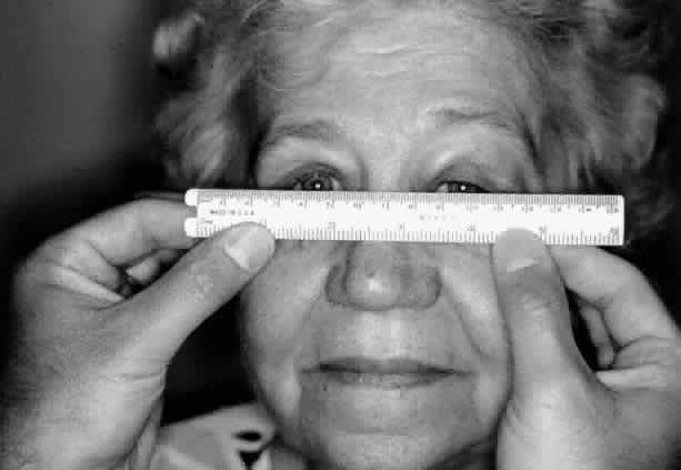
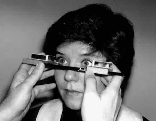
Globe displacement (horizontal or vertical) does not always result in diplopia. Even so, patients may present with the complaint that the eye does not “look right.” It is important to determine whether the apparent displacement of the globe is real or illusory. Posttraumatic telecanthus or large congenital epicanthal folds may create the false impression that eyes are displaced medially. Tripod malar fractures may produce a displacement of the lateral canthal complex and the illusion that the eye is abnormally positioned, when the true anatomic abnormality involves the zygoma and eyelids. Vertical displacement of the globe is frequently seen after blowout fractures or orbital decompression. Orbital roof fractures or tumor masses present in the superior orbit may cause the globe to be displaced inferiorly. This displacement may or may not be associated with ipsilateral ptosis and/or decreased supraduction. The horizontal position of the globes is measured from the center of the bridge of the nose using a ruler to determine the distance from the midline to the center of the pupil (Fig. 1). Previous trauma or surgery to the nose may make this measurement unreliable. Vertical displacement of the globe is evaluated by placing a ruler in a horizontal position across the bridge of the nose. The relative positions of the globes may be assessed and the vertical displacement can be either measured or estimated in millimeters. Once again significant trauma to the midface renders this measurement inaccurate.

Fig. 1. The ruler is held horizontally across the bridge of the nose. The distance from the midline to the pupil is recorded on each side.

Color

The color of the eyelids may provide a clue to the underlying orbital pathology. In many cases there will be no discoloration of the eyelids. However, inflammatory lesions often cause an erythema of the eyelids. This redness may be associated with edema and tenderness. Bruising of the eyelids is most frequently seen following trauma but may be associated with hemorrhage into an underlying orbital lesion. Spontaneous hemorrhage most frequently occurs with lymphangioma and hemangioma but may be seen with other tumors, such as childhood neuroblastoma. Lymphangiomas and deep seated hemangiomas have a bluish tint. More superficial vascular lesions are red or maroon.

Pulsations

Pulsation of the eyelids and orbital contents is an infrequent finding and may be easier to appreciate with palpation rather than visual inspection. Pulsation is most commonly seen in neurofibromatosis, in which the absence of sphenoid bone allows brain pulsations to be transmitted to the orbital contents. Extremely vascular tumors may occasionally pulsate because of the high blood flow in these lesions.

Palpation

Palpation of the orbit is a simple maneuver that often provides important information. The upper and lower eyelids should be palpated gently. It is possible to insert the tip of a small finger between the globe and the orbital rims on all four sides so that the anterior orbital contents may be palpated. In this way abnormalities of the lacrimal gland or the lacrimal sac can be identified, and abnormalities of the orbital rim are easily appreciated. This technique is especially useful for trauma patients who may have a step deformity of the orbital rim.

Retropulsion of the globes is a test that has received considerable attention. This test is performed by placing the fingers over both eyes and gently pushing them in a posterior direction. A firm lesion present behind the globe will resist retropulsion and this sensation is palpable. Unfortunately, this test is nonspecific and has little diagnostic value.

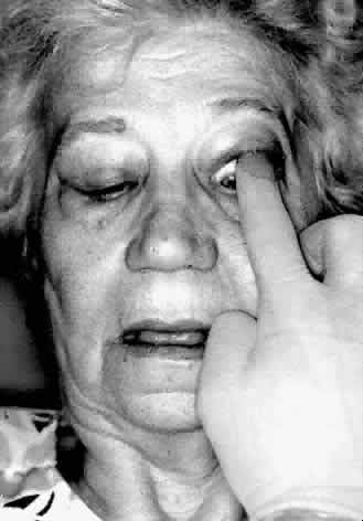
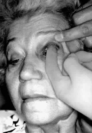
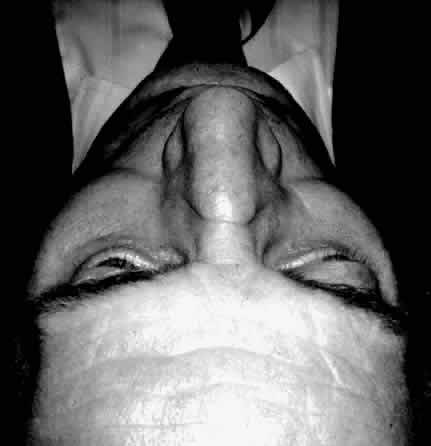
Digital examination of the superior fornix is a particularly useful technique for evaluating superior orbital lesions. To perform this examination the conjunctival sac is anesthetized with a topical agent. A gloved finger is gently inserted behind the upper eyelid into the superior fornix (Fig. 2). The finger is moved medially and laterally and anteriorly and posteriorly to palpate any lesions present in the superior part of the orbit. Bimanual palpation is sometimes helpful (Fig. 3). It is useful to have the patient look down during the examination because this position is more comfortable. In addition, downward rotation of the globe will sometimes bring an orbital mass more anteriorly so that it may be more readily palpated. Small orbital rim fractures may also be identified in this way. Lacrimal gland abnormalities may be easily palpated. This technique is particularly useful if the upper eyelid is swollen, making palpation of the orbital mass through the eyelid difficult. The same technique may be used in the inferior fornix.

Fig. 2. The conjunctival sac is anesthetized. A gloved finger is inserted into the superior fornix. The patient is more comfortable in downgaze. Palpation of the superior orbital mass is gently performed.

Fig. 3. Bimanual palpation is sometimes helpful in more anterior lesions.

Sensation

The first and second divisions of the trigeminal nerve provide sensation to the face in the orbital area. The ophthalmic branch provides sensation in the forehead, upper lid, and globe, whereas the maxillary division via the infraorbital nerve gives sensation to the cheek and upper lip. Sensation is usually tested by touching these areas with a wisp of cotton and asking if the patient is able to feel the touch. It is useful to compare one side to the other because the sensory deficit is incomplete in many cases of orbital disease. Hypoesthesia of the cheek and lip is a typical finding in patients with blowout fractures because of injury to the infraorbital nerve as it travels through its bony canal in the orbital floor.
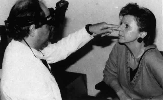
Exophthalmometry

Exophthalmometry is an accurate technique for the measurement of proptosis. This measurement may be accomplished in several ways, but in each case the reference points are the lateral orbital rim and the apex of the cornea. The generally accepted normal value is less than 22 mm. However, there are some patients without orbital disease who have exophthalmometry readings greater than 22 mm. The absolute value of the exophthalmometry reading is less important than the difference between the two eyes. The left and right measurements should not differ by more than 2 mm.

Exophthalmometry may be performed with a Hertel type of instrument. This instrument rests against the lateral orbital rim and the position of the corneal apex is measured by means of mirrors (Fig. 4). Frueh has shown that inexperienced examiners give less reliable measurements than experienced examiners.19

Fig. 4. The Hertel exophthalmometer rests against the lateral orbital rims. By means of mirrors, the distance from the lateral orbital rim to the apex of the cornea is measured for each side.

Although the Hertel exophthalmometer is the most widely used instrument, exophthalmometry readings may be obtained by simply placing a transparent ruler on the lateral orbital rim and measuring the position of the cornea. Inspection from behind the patient looking over the forehead down onto the proptosed globes is also a way to estimate proptosis (Fig. 5).
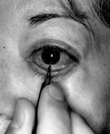
Fig. 5. The examiner stands behind the patient and looks down over the forehead. The amount of proptosis is estimated.

Forced Duction Test

The forced duction test is a useful test for distinguishing paralytic strabismus from restrictive strabismus. The forced duction test is used to detect an inelastic extraocular muscle that prevents the globe from rotating. For example, a tight inferior rectus muscle will not allow the globe to rotate superiorly. The test is carried out by placing topical anesthetic into the conjunctival sac. The globe is grasped with a pair of toothed forceps at either the limbal area (where Tenon's capsule is tightly adherent to the globe) or the insertion of the extraocular muscle, and an attempt is made to “force” the globe to rotate in the desired direction (Fig. 6). Restriction may be felt when the globe reaches the end of its “tether.” (For some patients, especially in the pediatric age group, application of the forceps to the globe is too frightening or painful. In these situations the forceps may be replaced with a cotton swab and the globe gently pushed.) The forced duction test is particularly useful for differentiating between a medial wall blowout fracture with medial rectus entrapment and a sixth cranial nerve palsy.

Fig. 6. The conjunctiva is anesthetized and grasped with a toothed forceps at the limbus. The globe is rotated (in this case upward) until it reaches the end of its “tether.” This restriction of movement can be felt and constitutes a positive forced duction test.

Auscultation

Auscultation of the orbit will detect bruits. Orbital bruits are generally due to high flow, high-pressure carotid cavernous fistulas, or in some cases high-flow arteriovenous malformations. The examiner must be careful to ensure that the fellow eye remains in primary position during auscultation because ocular movements behind the closed eyelid cause noise that can be readily heard through the stethoscope.

Nasal Examination

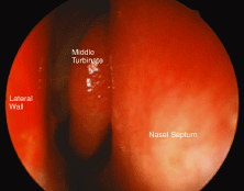
Nasal examination is a useful technique for the orbital surgeon because nasal or sinus inflammation and tumors sometimes have an orbital presentation. The nasal mucosa is anesthetized and decongested with 5% cocaine spray (or something similar). When the nasal speculum is inserted into the nose it is helpful to hold the external naris with the index finger to stabilize the speculum. Nasal visualization is carried out by means of the time-honored center-hole head mirror. However, the indirect ophthalmoscope is an excellent tool for looking into the nose and gives the examiner the added advantage of binocularity (Fig. 7).

Fig. 7. The nose is anesthetized and decongested with 5% cocaine spray. The nasal speculum is inserted into the nose and steadied with the index finger, which is placed on the external naris. The indirect ophthalmoscope provides the examiner with good illumination and binocular viewing.

Nasal endoscopy is another excellent way of examining the nose. The nose is similarly prepared and the endoscope is placed into the nostril. Visualization is much easier using a camera and monitor than looking directly though the endoscope. In addition, photographic documentation of nasal abnormalities is possible (Fig. 8).

Fig. 8. A normal right nose (cocainized) as visualized through a nasal endoscope.

Back to Top
SPECIAL INVESTIGATIONS IN ORBITAL DISEASE
The diagnosis of an orbital disease is often not clear, even after a thorough clinical examination. In an earlier era, this dilemma would have necessitated an exploratory orbitotomy to determine the diagnosis. Currently, there is no role for the exploratory orbitotomy.

Modern orbital investigations such as orbital ultrasound, computed tomography (CT), magnetic resonance (MR) imaging, and angiography have allowed the orbital surgeon to define a limited differential diagnosis before embarking on orbital surgery.

These specialized orbital investigations are discussed in depth elsewhere in this volume. A brief overview of each modality follows.

X-RAY EXAMINATION

Plain x-ray films of the orbits and sinuses, which have been the standard form of orbital investigation for many years, are now rarely requested. The main limitation of this investigation is that, although the bony structures of the orbit can be identified relatively well, the soft tissues cannot. However, plain x-ray studies still serve a useful function in certain situations. Most orbital blowout fractures can be identified on plain films when the Waters and Caldwell views are examined. Orbital expansion associated with orbital tumors or failure of orbital development in anophthalmic patients can be readily appreciated with the posteroanterior view. The main advantage of plain x-ray studies is that they are universally available and are quickly obtained in an emergency situation.

ORBITAL ULTRASONOGRAPHY

Orbital ultrasonography is especially useful for intraocular diagnosis and is useful for anterior orbital lesions. The internal acoustic properties of the lesion often allow the ultrasonographer to differentiate the mass into a solid, cystic, or vascular category. Precise measurements of extraocular muscles may be obtained, especially when A mode is used. The main disadvantage of orbital ultrasonography is that the posterior regions of the orbit are poorly evaluated. Orbital ultrasonography requires an experienced and well-trained examiner.

COMPUTED TOMOGRAPHY

Thin-section orbital CT scanning allows detailed visualization of orbital structures in both the coronal and axial planes. It is especially useful for showing bony abnormalities, and in this area it is better than MR imaging. The orbital fat provides good contrast with the extraocular muscles and optic nerve, and in may cases injection of contrast material is not required.

MAGNETIC RESONANCE IMAGING

MR imaging is an excellent tool because of the detailed soft tissue images that this scanning method produces. Tissue diagnoses may be made, in some cases, with MR imaging techniques. Surface coils can provide exquisite tissue detail. The main drawback of MR imaging is that orbital bones are not well delineated.

VISUAL EVOKED RESPONSE

The visual evoked response is generated by presenting a standardized visual stimulus to the patient, usually a reversing grid pattern. Cutaneous electrodes are placed over the occipital region to record cortical electrical activity. The typical visual evoked response shows an initial deflection known as the P100 wave at about 100 milliseconds after the stimulus. A second deflection in the opposite direction follows at about 120 milliseconds and is known as the N120 wave. Abnormalities in the waveform provide the basis for interpretation. Delay in the formation of the wave typically indicates prolonged conduction in the visual pathways and may indicate a disorder of the retina, optic nerve, or optic radiations. Orbital conditions such as Graves' disease, optic nerve sheath meningioma, optic nerve glioma, and space-occupying lesions that compress the optic nerve may also produce abnormal results.

VISUAL FIELDS

Visual field testing is useful if there is a clinical suspicion that the optic nerve is diseased. It is also important in suspected cavernous sinus or pituitary lesions in which field cuts may provide useful information about the location and severity of the disorder. Sequential visual field testing is also a useful way to follow these lesions to note improvement or deterioration.

ANGIOGRAPHY

Arterial angiography is used infrequently in orbital lesions because other less invasive diagnostic modalities usually provide a diagnosis. However, arteriography is useful in cases of carotid cavernous fistula and some arteriovenous malformations to delineate the extent of the lesion and also to provide an arteriographic map. The interventional radiologist can treat these lesions using intra-arterial embolism.

In the past, venography was used extensively in the evaluation of orbital disease but now is rarely indicated. Venography is performed by catheterizing the supraorbital vein and filling the orbital venous system with intravenous contrast material. The main current indication for this test is the presence of an orbital varix. Other less invasive diagnostic modalities give the same information in most cases.

Back to Top
CONCLUSION
The diagnosis and management of orbital disorders remains a challenging subspecialty within ophthalmology and overlaps with neurosurgery, otolaryngology, maxillofacial surgery, and plastic surgery. A complete history and thorough orbital examination should allow the examiner, in most cases, to arrive at a relatively short list of differential diagnoses for the patient with orbital disease. Sophisticated orbital imaging techniques permit the orbital surgeon to accurately diagnose the orbital condition in the preoperative period and to precisely plan the approach to orbital surgery.
Back to Top
REFERENCES

1. Shields JA, Bakewell B, Augsburger JJ et al: Classification and incidence of space-occupying lesion of the orbit: A survey of 645 biopsies. Arch Ophthalmol 102:1606, 1984

2. Reese AB: Bowman lecture: Expanding lesions of the orbit. Trans Ophthalmol Soc U K 91:85, 1971

3. Moss HM: Expanding lesion of the orbit. Am J Ophthalmol 54:761, 1962

4. Henderson JW: Orbital Tumors. Philadelphia: WB Saunders, 1973:78–79

5. Silva D: Orbital tumors. Am J Ophthalmol 65:318, 1968

6. Kennedy RE: An evaluation of 820 orbital cases. Trans Am Ophthalmol Soc 82:134, 1984

7. Rootman J: Diseases of the Orbit: A Multidisciplinary Approach. Philadelphia: JB Lippincott, 1988:121–124

8. Ingalls RG: Tumors of the Orbit and Allied Pseudo Tumors. Springfield, IL: Charles C Thomas, 1953

9. Porterfield JF: Orbital tumors in children: A report on 214 cases. Int Ophthalmol Clin 2:319, 1962

10. MacCarty CS, Brown DN: Orbital tumors in children. Clin Neurosurg 11:76, 1964

11. Yousefi B: Orbital tumors in children: A clinical study of 62 cases. J Pediatr Ophthalmol Strabismus 6:177, 1969

12. Templeton AC: Orbital tumors in African children. Br J Ophthalmol 55:254, 1971

13. Eldrup-Jorgensen P, Fledelius H: Orbital tumors in infancy: An analysis of Danish cases from 1943-1962. Acta Ophthalmol Copenh 53:887, 1975

14. Iliff WJ, Green WR: Orbital tumors in children. In Jakobiec FA (ed): Ocular and Adnexal Tumors. Birmingham, AL: Aesculapius, 1978:669–684

15. Crawford JS: Diseases of the orbit. In Crawford JS, Morin JD (eds): The Eye in Childhood. New York: Grune & Strutton, 1983:361

16. Shields JA, Bakewell B, Augsburger JJ et al: Space occupying orbital masses in children: A review of 250 consecutive biopsies. Ophthalmology 93:379, 1986

17. Bullock JD, Goldberg SH, Rakes SM: Orbital tumors in children. Ophthalmic Plast Reconstr Surg 5:13, 1989

18. Manor RS: Enophthalmos caused by orbital metastatic breast carcinoma. Acta Ophthalmol 52:881, 1974

19. Musch DC, Frueh BR, Landis JR: The reliability of Hertel exophthalmometry: Observer variation between physician and lay readers. Ophthalmology 92:1177, 1985

20. Harvey JT: Unpublished data, orbital cases collected 1995 to 2001

Back to Top