Chapter 19
Ischemic Stroke
JOSE G. ROMANO
Main Menu   Table Of Contents

Search

EPIDEMIOLOGY
STROKE SIGNS AND SYMPTOMS
ACUTE ISCHEMIC STROKE: EVALUATION AND TREATMENT
EVALUATION OF THE CAUSES OF CEREBRAL ISCHEMIA AND PREVENTION OF RECURRENT STROKE
REFERENCES

EPIDEMIOLOGY
In spite of decreasing rates of stroke mortality in the last decades, with 700,000 new strokes each year and 160,000 annual deaths, stroke is the third cause of death and leading cause of disability in the United States.1 The cost of stroke to the health care system is over $40 billion,2 but the cost in human suffering is difficult to estimate. Seventy percent of the estimated 4 million stroke survivors are vocationally disabled,1 approximately 30% fulfill criteria for dementia,3,4 and a similar number have major depression.5,6 The elderly are particularly vulnerable, because age is the most important risk factor for stroke. Stroke incidence triples with each decade after age 35, and seven of eight stroke deaths occur in those age 65 years or older. The magnitude of the problem can be put in perspective by the estimation that the population older than 65 years will increase by 50% from 1995 to 2025.7 Stroke fatality varies according to stroke subtype, but on average 20% to 25% of those with ischemic strokes and 50% of those with hemorrhagic strokes will die within 30 days of the event.8 However, new understandings of stroke mechanisms and acute revascularization techniques have created optimism to counter these ominous figures.

Stroke is not one disease but a group of conditions that result in sudden brain injury caused by alterations of cerebral blood flow. This review focuses on ischemic stroke, which accounts for 80% of all strokes, and emphasizes those areas of particular interest to ophthalmologists.

Back to Top
STROKE SIGNS AND SYMPTOMS

LARGE VESSEL MANIFESTATIONS

The carotid arteries carry 80% of cerebral blood flow, irrigate the territories of the middle cerebral, anterior cerebral, and anterior choroidal arteries, and provide blood flow to the eye through the ophthalmic artery. The symptoms of carotid embolism vary according to the recipient artery. Occlusion of the ophthalmic artery or its branches results in monocular visual loss (amaurosis fugax, central or branch retinal artery occlusion), often described as blurring, graying or curtaining of vision.9 In addition, in those with chronic flow deprivation to the retinal circulation, transient and brief monocular visual loss in response to bright light can be seen.10 Middle cerebral artery syndrome often includes contralateral weakness affecting the face and arm more than the leg, contralateral sensory loss, and aphasia if the dominant hemisphere is affected, or hemi-inattention/neglect with nondominant lesions. In addition, there may be gaze deviation toward the affected hemisphere.11 Anterior cerebral artery strokes affect the contralateral leg and shoulder more than face and arm, and can have associated abulia and incontinence.12 The anterior choroidal stroke affects the basal ganglia and internal capsule, with contralateral hemiplegia, hemisensory loss, and hourglass-shaped homonymous visual field defects.13 Anterior choroidal infarcts may be difficult to differentiate from lacunar strokes because of occlusion of small penetrators.

Clues for carotid disease on examination include an audible bruit in the anterior neck, evidence of a cholesterol retinal embolus (Hollenhorst plaque), and a history of various spells of ischemia in the same distribution. In addition, careful craniovascular examination may reveal asymmetric preauricular and orbital pulses, which are stronger on the side of the stenosis, representing external carotid artery collateral flow. On occasions, dilatation of the extracranial vasculature may be noted over the forehead.

The posterior circulation accounts for 20% of embolic strokes. Vertebral syndromes may affect the medulla or cerebellum as these structures are fed by branches of the vertebral arteries, or may cause distal embolization into the basilar and posterior cerebral arteries.14 The most common medullary manifestation is the posterolateral medullary syndrome or Wallenberg syndrome, caused by occlusion of the vertebral artery or the posterior inferior cerebellar artery (PICA). It is characterized by vertigo, nausea, vomiting, miosis and ptosis, ipsilateral face and contralateral hemibody numbness, hoarseness, and dysphagia; hiccups may also be present. When the PICA is involved, there are also cerebellar manifestations, with consequent hemidysmetria and gait ataxia. The medial medullary syndrome caused by distal vertebral occlusion, results in hemiparesis and tongue deviation. When the basilar artery occludes, there is usually the combination of extraocular movement abnormalities, descending motor fiber involvement with hemi or quadriparesis, and alteration of the state of consciousness. A comatose state is common in basilar artery occlusion, but large hemispheric strokes may impair consciousness through increased intracranial pressure, as do large cerebellar strokes that press on the brainstem and result in hydrocephalus. Occlusion of the posterior cerebral artery causes visual field loss by virtue of occipital injury. The proximal posterior cerebral artery also feeds the thalamus and the medial temporal lobes, and therefore there may also be sensory, memory, and behavioral manifestations.15

On general physical examination it is important to record the blood pressure in each arm and compare both radial pulses. Differential blood pressure or radial pulse, as well as a bruit in the supraclavicular area, constitute indirect evidence of subclavian and possibly vertebral origin disease.

Border zone infarcts resulting from hemodynamic failure as a consequence of large vessel flow obstruction may affect the anterior frontal as well as the corona radiata in a longitudinal fashion (border zones between the anterior and middle cerebral arteries) or the parietooccipital area (border zone between the middle and posterior cerebral arteries).16 The classic description of bilateral border zone infarcts that may be seen after significant hypotension is the “man in the barrel” syndrome, where proximal upper and lower limb weakness is noted.17 More commonly, the border zone is unilateral and the manifestations are often similar to those seen in embolic strokes.18

Headache as a symptom of cerebral ischemia occurs as an initial manifestation of carotid or vertebral artery dissection, on occasions accompanies the acute occlusion of a large artery such as the vertebral and basilar artery, is common in stroke caused by giant cell arteritis, and may develop later in the course of ischemia as a result of increased intracranial pressure.19

SMALL VESSEL MANIFESTATIONS

The manifestations of small penetrating artery occlusion depend on the location of injury. The lacunar syndromes usually result from single small perforating artery damage, but larger strokes, such as a striatocapsular stroke caused by proximal middle cerebral artery occlusion with infarct in the distribution of multiple penetrating arteries, may have similar clinical findings. The classic lacunar syndromes include pure hemiparesis (internal capsule), pure hemihypoesthesia (thalamus), hemiparesis with hemihypoesthesia (thalamocapsular), ataxic hemiparesis (corona radiata and pons), dysarthria with a clumsy hand (corona radiata and pons), but many other less common syndromes have been described.20 When small basilar penetrator arteries occlude, there may be diplopia, hemiparesis, perioral numbness, ataxia, and dysarthria. The accumulation of multiple small subcortical infarcts results in a range of cognitive impairments, including dementia.21 Dementia from cerebrovascular disease may also be caused by single strategic strokes (such as caudate or thalamic strokes) or multiple cortical strokes.22 In addition, right parietal strokes and dominant hemisphere temporal strokes (with fluent aphasia) may be confused with psychiatric conditions.

VENOUS STROKES

Thrombosis of cerebral veins, or the larger cerebral sinuses, causes symptoms both by its location and by increased intracranial pressure. These infarcts commonly have a hemorrhagic component because venous outflow obstruction results in extravasation from the capillary bed. Blood products are quite irritative and therefore seizures frequently accompany venous infarcts. When large cerebral sinuses are occluded, particularly the superior sagittal sinus, increased intracranial pressure may result. This syndrome is characterized by headaches, papilledema, and occasionally abduscens nerve palsy, and may be confused with benign intracranial hypertension or pseudotumor cerebri. When the cavernous sinus is affected, ipsilateral proptosis, oculomotor palsy and trigeminal involvement (of its V1 and sometimes V2 segments) is noted.23

TRANSIENT ISCHEMIC ATTACKS

An episode of sudden neurologic dysfunction because of cerebral ischemia that is transient and leaves no observable sequelae is denominated a transient ischemic attack (TIA). Although the duration of such spells has traditionally been defined as less than 24 hours, this concept is currently being reconsidered to events of 1 hour or less.24 The average duration of a TIA is 14 minutes for the anterior circulation and 8 minutes for the posterior circulation.25 In those whose symptoms last 1 hour or more, only 14% have complete recovery by 24 hours,26 and magnetic resonance imaging (MRI) studies have revealed small strokes in half of those with transient neurologic symptoms.27

When cerebral blood flow decreases below approximately 20 mL per 100 g of brain tissue per minute, neuronal function will cease and symptoms will become manifest.28 If blood flow is restored promptly, usually because of the body's endogenous thrombolytic ability, then full function will be restored. Therefore, transience of symptoms depends in large part on the size and composition of the responsible embolic particles, as well as the robustness of collateral flow.

Cardioembolic strokes commonly debut with strokes, as the emboli produced in the cardiac chambers are usually large: the diameter of the middle cerebral, vertebrals, and basilar arteries is approximately 3 mm, and it is not unusual for cardiac emboli to be larger than this. In comparison, the platelet-rich emboli formed in high-shear rate areas such as stenotic arteries are much smaller and therefore TIA is a frequent predecessor to aortic, carotid, and vertebral strokes. This phenomenon of continuous or repetitive embolization is illustrated by monitoring the cerebral arteries with transcranial Doppler ultrasonography, where the detection of small microembolic signals distal to carotid stenosis is common.29,30 Presumably, many of these signals represent small emboli that are washed into the venous circulation,31 but their presence predicts plaque instability and risk of stroke. Embolism is not the only cause of TIA, which may occur in the territory distal to a fixed arterial stenosis or occlusion in the setting of hypotension or hypovolemia, with resolution of symptoms with increased flow. Recurrent TIAs with similar manifestations are suggestive of a hemodynamic mechanism.32 In addition, TIAs may occur in small vessel disease; whether this represents a small thrombus in the presumed preocclusive state of a small penetrating artery is unclear.

The occurrence of a TIA is a concerning event as it may precede a stroke. In one report of over 1,700 TIAs evaluated in an emergency room, 10% went on to have a stroke within 90 days, and one-half developed within 48 hours.33 This underscores the importance of emergent evaluation and treatment of TIAs.

Back to Top
ACUTE ISCHEMIC STROKE: EVALUATION AND TREATMENT
It is now possible to treat selected acute strokes to reduce or abort the cerebral insult. In order to do this it is important to remember that brain tissue requires approximately 50 mL of blood flow per 100 g of tissue per minute to meet its metabolic demands. Areas with flow greater than 20 mL per 100 g/min are able to function normally, but tissue with less than 10 mL per 100 g/min promptly develops irreversible damage. Cerebral tissue with blood flow between 10 and 20 mL per 100 g/min is salvageable, if reperfused within a certain amount of time; this area is denominated the ischemic penumbra. Even without further decreases in regional blood flow, irreversible changes in the penumbra will ensue within approximately 6 hours, mainly because of excitotoxic insults arising from the core of the stroke. This damage is mediated by various neurotransmitters, mainly glutamate. This substance is normally present in the brain, but nonfunctional astrocytes are unable to recapture it, leading to high extracellular concentrations that through a number of cell-membrane interactions result in increasing intracellular calcium levels that result in cell death.34,35

The treatment of acute ischemic stroke is directed at saving the penumbra, which initially represents the major portion of the tissue at risk; over time (hours), irreversible injury develops. The three main tenets of intervention include reperfusion or opening the occluded vessel, increasing collateral flow to the ischemic penumbra, and blocking excitotoxic influnces.

REPERFUSION

Since its approval by the Food and Drug Administration (FDA), the administration of the recombinant tissue plasminogen activator (rt-PA) for intravenous (IV) thrombolytic therapy in acute ischemic stroke has become the standard of care in appropriate patients.36 The National Institute of Neurological Disorders and Stroke (NINDS) study37 showed that individuals treated with intravenous rt-PA (0.9 mg/kg of weight, not to exceed 90 mg total dose, 10% given as a bolus and the remainder over 1 hour) had one-third greater chance of being asymptomatic or with minimal sequelae than those treated with placebo. With a 6% incidence of hemorrhagic complications, it remains a potentially dangerous drug, and certain precautions should be observed to avoid a higher complication rate. Establishing the precise onset of symptoms is extremely important as r-tPA is approved for use within 3 hours of stroke onset. Administration after this period results in decreased efficacy, and possibly an elevated rate of symptomatic cerebral hemorrhage. Noncontrast brain computed tomography (CT) needs to be done urgently because the presence of cerebral hemorrhage, which cannot be reliably excluded on clinical grounds alone, precludes the use of thrombolytic medications. Patients presenting within 3 hours of onset of symptoms may have a normal brain CT, or one with early ischemic changes, such as loss of gray-white matter distinction, effacement of the insular ribbon, or loss of basal ganglia definition. A dense middle cerebral artery represents thrombus within this vessel. Further evidence of ischemia, particularly a well-established hypodensity, represents cytotoxic edema in infracted tissue, and should preclude the use of intravenous rt-PA. The brain parenchyma can also be studied anatomically by MRI. The latter is much more sensitive, particularly when diffusion sequences are obtained.38,39 Restriction of the normal diffusion of water across membranes in response to energetic failure becomes apparent within minutes and remains positive for up to 2 weeks after insult.40 Quantification of neurologic deficits is helpful in estimating the risk of administering thrombolytics and in assessing response to treatment; the National Institutes of Health Stroke Scale (NIHSS)41 is the most widely used for this purpose. An NIHSS grade greater than 24 (a higher grade indicates more deficits) is associated with more intracerebral hemorrhagic complications after therapy, but does not represent an absolute contraindication because these patients have a poor prognosis if left untreated. Conversely, minimal deficits, such as isolated sensory symptoms or an NIHSS less than 4 does not warrant the risk of thrombolytic therapy. Factors that may preclude thrombolytic use such as recent surgery, cerebral aneurysms, cranial vascular malformations or tumors, or a potential source of systemic bleeding (i.e., active gastric ulcer) should be sought. Laboratory screening for coagulopathic conditions or significant thrombocytopenia should be obtained.

Unfortunately, the 3-hour window for intravenous thrombolysis limits the number of patients treated, currently fewer than 5% of all strokes in this country. Other intravenous rt-PA studies have not shown efficacy with this agent beyond 3 hours. This has led to the exploration of intraarterial administration of thrombolytic agents, with the expectation that it may prove to be a safer and more effective way of opening occluded cerebral vessels, and may provide a longer window of opportunity for intervention than intravenous application. In PROACT II,42 prourokinase (9 mg) was administered to individuals with large strokes from a middle cerebral artery occlusion within 6 hours of onset of symptoms. There was a 15% absolute difference in improvement to complete resolution or minimal sequelae between patients treated with prourokinase (40%) versus those in the placebo group (25%). In spite of an excess in symptomatic cerebral hemorrhages in the treated group (10.2 vs. 1.8%), this did not translate into greater mortality.

It is possible that the posterior circulation has a longer window of viability than the anterior circulation.43 Small series have reported benefit from intra-arterial thrombolysis in basilar artery occlusion up to 48 hours, a condition with a mortality of up to 90% if left untreated. Brandt and colleagues43 and the AUST Study Group44 found recanalization rates of 51% to 69% with a reduction in mortality of over 50%. As the intraarterial thrombolytic process requires specialized centers and the setup of the angiographic suite is time consuming, some have attempted to initiate intravenous rt-PA early on at a reduced dose, to be followed by intraarterial thrombolysis if needed. With this approach, greater recanalization rates, compared to intravenous rt-PA, have been reported.

As diffusion-perfusion MRI and other techniques to quantify blood flow, such as perfusion CT and xenon-CT, become more widely available, clinicians will be better equipped to estimate the relative benefits and risks of thrombolysis, based more on physiologic data than rigid time constraints. Patients with large areas of penumbra tissue would benefit most, while those with large tissue volume with blood flow under 10 mL per 100 g/min would not benefit and may be at increased risk for hemorrhage. A recent report utilizing diffusion-perfusion MRI to guide thrombolysis with desmoteplase allowed the safe and apparently efficacious administration of this thrombolytic agent up to 9 hours after the onset of ischemia.45 Figure 1 exemplifies the use of diffusion-perfusion MRI in acute stroke treatment. Ideally, multimodal testing of the parenchyma to exclude hemorrhage, a viability study to determine the amount of salvageable brain, and a vascular study to assess vessel patency would provide the clinician with the information to make rational decisions regarding the need to treat. Magnetic resonance (MRI, diffusion-perfusion imaging, and magnetic resonance angiography [MRA]) and CT technology (with CT perfusion and CT angiography) have the potential to guide this multimodal evaluation.

Fig. 1. An 81-year-old man with atrial fibrillation developed acute onset of left-sided weakness and presented to hospital 3 hours and 40 minutes after onset of symptoms. A: The diffusion-sequence magnetic resonance imaging (MRI), with infracted tissue in the right basal ganglia in white. B: Decreased perfusion in the right middle cerebral artery distribution (white area). Given the relatively small diffusion abnormality and the large perfusion defect, it was decided to proceed with thrombolysis despite the time elapsed.

However, it should be noted that at the time of this writing, only intravenous rt-PA is approved by the FDA in the United States for treatment of acute ischemic stroke.

INCREASING COLLATERAL FLOW TO THE ISCHEMIC PENUMBRA

The most important single measure to reach this goal is to avoid hypotension. Because the hypertensive response after acute cerebral ischemia may be a normal compensatory response, it is recommended not to treat systolic blood pressure under 220 mm Hg or diastolic pressures below 120 mm Hg in the first 24 hours after onset of the event. If thrombolytics are used, then the systolic blood pressure should be kept under 180 mm Hg, and the diastolic BP under 110.46 In the presence of overt cardiac failure, coronary ischemia or aortic dissection, blood pressure needs to be reduced more aggressively. When blood pressure reduction is required, β-blockers such as labetalol are the preferred initial therapy. Vasodilators, particularly nifedipine, should be avoided because they can produce a precipitous drop in blood pressure resulting in reduction of cerebral blood flow to the ischemic penumbra. Patients with low blood pressures may need colloids and occasionally vasopressors to ensure adequate perfusion to the ischemic penumbra. The use of isotonic intravenous fluids is recommended unless there is a cardiovascular contraindication such as congestive heart failure.

BLOCKING EXCITOTOXIC INFLUENCES

The reduction of excitotoxic influences should preserve penumbra neurons. Multiple agents have been tried in a variety of studies with disappointing results, but the search continues.47 If a safe agent is developed, it may be administered in the prehospital setting by paramedics. Nevertheless, there are two very effective interventions with clear neuroprotective effects that should currently be used.46 The first is avoidance of hyperglycemia because it worsens ischemic injury. The second is treatment of hyperthermia: even small increases of temperature adversely affect the reversibility of ischemic penumbra damage. Fever should be treated aggressively with antipyretics and even cold blankets if needed. Hypothermia has been shown to have beneficial effects on stroke outcome in animal studies; human studies are currently underway.

ANTITHROMBOTIC THERAPY IN ACUTE ISCHEMIC STROKE

The use of antithrombotic agents is based mainly on the risk of early recurrence of ischemia, and to a lesser degree on prevention of progression of symptoms. Although earlier reports suggested a high risk of early recurrent stroke,48 most recent large studies suggest a low risk of early recurrence. In the placebo arm of early anticoagulation trials, the risk of recurrence was 1.1%49 to 4.4%50 in the first weeks after a stroke, and even in cardioembolism early recurrence is rare.51,52 In addition, full anticoagulation carries a certain risk of cerebral hemorrhage.50 As to early deterioration, there is a theoretical concern that an intravascular thrombus may extend and cause further ischemic injury. However, the causes of early progression of deficits are multiple and include cerebral edema, hemorrhagic transformation and infections, amongst others. A number of trials of very early anticoagulation with unfractionated heparin, low-molecular-weight heparins, and heparinoids have not shown any benefit of this practice in improving outcomes.46 Therefore, full anticoagulation is not indicated in the acute phase after an ischemic stroke. For most patients, antiplatelet therapy should be administered.53 However, subcutaneous anticoagulants for prevention of deep vein thrombosis are usually indicated in those with impaired mobility. After the acute period, the appropriate antithrombotic therapy is instituted based on the mechanism of ischemia.

Back to Top
EVALUATION OF THE CAUSES OF CEREBRAL ISCHEMIA AND PREVENTION OF RECURRENT STROKE
Once the acute stroke has been treated (or when confronted by a TIA, evidence of an old stroke, or asymptomatic arterial disease) then evaluation of the risk factors and mechanisms of ischemia is warranted in order to establish rational and effective preventive interventions. In the evaluation of stroke, it is important to evaluate the brain parenchyma, potential embolic sources (cardiac and arterial), and vascular risk factors.

The most common mechanisms of ischemia include embolism, occlusion of small cerebral arteries, hemodynamic events, and less commonly cerebral artery in situ thrombosis (caused by arteritis or a hypercoagulable condition) and cerebral venous thrombosis.

Embolism is probably the most common cause of ischemic stroke,54 and it consists of the occlusion of a cerebral vessel by particle formed upstream in the vascular tree. The common manifestations of cerebral embolism are a sudden onset of neurologic dysfunction, with deficits maximum at onset, and that commonly have associated cortical signs and symptoms. The embolic material will depend on the source of embolism; for example, thrombus formed in the heart chambers or deep veins is rich in erythrocytes and fibrin (red clot), while the material that arises from the large vessels, and that is often associated with atherosclerosis may be platelet-rich (white clot), and may also contain atherosclerotic plaque components such as cholesterol crystals, calcium and other debris. Determination of the embolic source and the probable composition of the embolic material are important to institute effective and rational therapy.

CARDIOEMBOLISM

Cardioembolism accounts for 15% to 30% of all embolic strokes.55,56 Thrombus formation is extraordinarily rare in a healthy heart, but structural changes, such as valvular diseases (particularly of the mitral valve), ventricular wall dysfunction (ventricular akinesis or aneurysm) after myocardial ischemia, and septal abnormalities such as a patent foramen ovale and atrial septal aneurysm, constitute a favorable milieu for generation of emboli.

Cardiac rhythm abnormality, particularly atrial fibrillation, is the predominant source of cardioembolism. The noncontracting and often dilated left atria and atrial appendage are nidus for thrombi, which can then be expelled into the cerebrovascular circulation. The incidence of atrial fibrillation duplicates with each decade of life, reaching a frequency of 39 per 1,000 in those aged 80 to 89.57 The risk of cerebral ischemia is much greater if there is associated valvular disease.58 When it is not, the presence of other factors such as hypertension, diabetes mellitus, heart failure, coronary artery disease, and certain echocardiographic findings such as dilated atria and “smoke” in the left atria appears to predict embolic risk.59–61 The risk, however, is similar for chronic and paroxysmal atrial fibrillation.62

The patent foramen ovale (PFO) is increasingly recognized as a common cause of cerebral embolism. It is formed as a channel between the septum primum and septum secundum that divide the atria. Although required during in utero life, this right-to-left atrial communication usually closes at birth with the recruitment of the pulmonary circulation. However, in up to one in four it remains patent,63 either as a constant anatomic channel or as a potential conduit that opens up with increases in right heart pressures. There are many possible mechanisms through which the PFO may contribute to cerebral embolism. The first is as a mere channel through which thrombi generated in the deep veins of the extremities or pelvis can bypass the lung filter and reach the brain. These thrombi may develop in the presence of a hypercoagulable condition such as immobility or stasis, dehydration, postoperative state, or in the presence of a congenital or acquired procoagulant factor. Although a PFO is a common finding, paradoxical embolization is relatively rare. Factors that increase the risk of stroke in the presence of a PFO include a large PFO, an associated hypercoagulable state, right atrial developmental variants such as a Chiari network and a Eustachian valve that may act as nidus for thrombi formation, and, more importantly, the presence of a floppy interatrial septum also denominated atrial septal aneurysm (ASA). The coexistence of a PFO with an ASA appears to be the most dangerous anatomic condition in predicting stroke recurrence. Mas and colleagues64 found a threefold risk of recurrence with a PFO plus an ASA compared to a PFO alone. Therefore, some have suggested that thrombosis may occur within a complex PFO itself. Finally, there appears to be some increased risk of developing atrial fibrillation in the presence of a PFO. Other conditions that may result in embolic cerebral damage and that have been associated with the PFO include the fat embolism syndrome (fat globules)65 and decompression illness (gaseous particles or bubbles).66

When cardioembolism is suspected, echocardiography is an invaluable tool in the evaluation of this condition. Transthoracic echocardiography is excellent in the evaluation of the ventricles, while transesophageal echocardiography (TEE) is superior in the visualization of the atria and interatrial septum. Therefore, when paradoxical embolization across a patent foramen ovale is considered, TEE is the preferred study.67 It not only allows the detection of the shunt, usually by detecting intravenously administered microbubbles in the left atrium, but can characterize the septum itself and determine the presence of an associated ASA. When atrial fibrillation is suspected and the electrocardiogram cannot detect it, then prolonged monitoring with a Holter or with event recorders is appropriate.

For the great majority of cardioembolic conditions, warfarin is the appropriate antithrombotic prophylactic therapy. A number of clinical trials have shown this in atrial fibrillation, with an estimated relative reduction in embolic events of 68% for warfarin compared to placebo in the intention-to-treat analysis of one meta-analysis, which was twice as effective as aspirin.68 After a left ventricular myocardial infarction, the risk of embolic stroke is approximately 5% at 30 days, because the akinetic myocardium may form thrombus. With time, this thrombus may become endothelized, with a decreased risk of stroke. Therefore, anticoagulation is usually recommended only for a few months.69 The appropriate treatment of a PFO is uncertain at this time and is a subject of controversy. The risks of any therapeutic modality should be weighed against the risk of no therapy, particularly in the presence a common finding as a PFO. Most would agree that an incidentally detected PFO needs no particular therapy, although there may be a benefit of aspirin. Although a recent study showed no benefit of warfarin over aspirin in preventing recurrent stroke in patients with cryptogenic stroke and a PFO,70 certain concomitant factors such as the presence of a hypercoagulable condition or an atrial septal aneurysm may warrant the use of anticoagulants. Surgical or endovascular closure of a PFO is feasible and relatively safe. Ongoing randomized trials will eventually determine if such an intervention is indicated.

LARGE VESSEL DISEASE

The large vessels are a common source of cerebral ischemia. Although carotid disease is the most recognized cause of large vessel disease, the vertebral arteries, which carry 20% of cerebral flow, are also a common cause of posterior circulation ischemia. Since the advent of transesophageal echocardiography, the aortic arch is also increasingly recognized as an embolic donor. Finally, the large intracranial vessels such as the intracranial internal carotid artery, the middle and anterior cerebral artery, and the intracranial basilar and vertebral arteries may be affected. Large vessel atherostenotic disease is also a marker for coronary atherosclerosis and is associated with cardiovascular death: population studies have revealed that the presence of a carotid bruit doubles the mortality rate. Even in the absence of a defined carotid atherosclerotic plaque, the mere increase in the intima and media thickness is a predictor for greater risk of stroke and myocardial infarction: the Cardiovascular Health Study showed an increase in stroke and myocardial infarction from 5% to 25% at 7 years when comparing the lower and higher quintiles of intima-media thickness. In those with defined carotid plaques, the risk may even be higher.71

Atherosclerosis, the formation of plaques in the arterial wall, constitutes the main pathology found in large vessels. In this condition, in response to common risk factors such as hypertension, diabetes, hyperlipidemia, tobacco use, and hyperhomocystenemia, accumulation of lipids, smooth muscle cells, fibroblasts, and calcium occurs within the arterial wall. A fibrous cap usually covers these plaques.72 At this stage, plaques are unlikely to cause symptoms. However, rupture of this covering can lead to an ulcer on the endothelial surface, or to a plaque accident by which blood enters the plaque. Once blood is exposed to the thrombogenic influences of the plaque contents, thrombus formation on the plaque surface can occur. The thrombus, and occasionally plaque contents (i.e., cholesterol crystals) can embolize to the intracranial vessels, and result in cerebral ischemia. Indeed, most strokes and transient ischemic attacks related to large vessel disease are caused by embolism, while hemodynamic ischemia is less common. Large vessel related cerebral ischemia is caused most frequently by artery-to-artery embolism. If the embolus is promptly lysed, either therapeutically or spontaneously, symptoms resolve without deficits, and this episode will be characterized as a TIA. Hemodynamic symptoms related to poor flow through a narrowed carotid are associated with hypotensive or hypovolemic states. The border zone between the major cerebral branches is commonly affected with low flow states. Another mechanism of ischemia is seen in the intracranial large vessels, where in addition to artery-to-artery embolism, the atherosclerotic plaque accident may result in plaque expansion that occludes perforators off these intracranial vessels.

CAROTID DISEASE

Patients who have had symptoms of carotid disease (stroke, TIA, or amaurosis fugax) are at a much higher risk of recurrent stroke. The seminal North American Symptomatic Carotid Endarterectomy Trial73 defined a 26% risk of ipsilateral stroke at 2 years in the presence of stenosis of 70% or more for those treated medically, with increasing risk at greater degree of stenosis. The risk of stroke after purely ocular symptoms (amaurosis fugax) is less than in those with cerebral symptoms. For stenosis of 50% to 69% in symptomatic patients, the risk of stroke was less impressive: at 5 years, the risk of ipsilateral stroke on antiplatelet therapy was 22.2%.74 Stenosis under 50% is unlikely to result in stroke. The risk of stroke with asymptomatic carotid stenosis is clearly less than the risk following a TIA or stroke. The Asymptomatic Carotid Atherosclerosis Study75 studied carotid stenosis of 60% or more; in the medically treated group, the risk of ipsilateral stroke was estimated at 11% at 5 years, suggesting an annual risk of 2.2%. It should be recalled that these studies were conducted before certain agents such as the statins were available. Their common use may have a significant beneficial effect and their impact on the risk of arterial atherostenosis is probably significant.

Although embolism is the most common cause of carotid-related strokes,76 hemodynamic factors clearly play a role.77 Therefore, it is important to evaluate collateral flow to the brain distal to a significant carotid stenosis. This usually arises from the external carotid artery through retrograde ophthalmic flow into the distal intracranial carotid artery; through the posterior communicating artery; or across the anterior communicating artery from the contralateral carotid. In addition, there are a number of leptomeningeal collaterals that may be recruited. The functionally most important source of collateral flow is the anterior communicating artery.78

In the evaluation of extracranial large vessel disease, mainly carotid disease, ultrasound is the initial indicated study. It is commonly available, relatively inexpensive, and accurate. In addition, compared to other vascular imaging methods, it has the ability to directly image and characterize the plaque. Certain plaque characteristics may predict an increased risk of embolization. For example, the presence of an ulcer at the plaque surface may promote the formation of thrombus that can secondarily embolize, while a hypoechoic plaque, representing either intraplaque hemorrhage or a lipid-rich plaque, is associated with the potential for plaque destabilization.79,80 Carotid artery ultrasound has a sensitivity of 90% to 95% and specificity of 85% compared to catheter angiography.81 However, ultrasound may be unable to detect high-grade stenosis and mistakenly diagnose a carotid occlusion in 5% of cases. It is important to make this distinction as a patent vessel may be amenable to a revascularization procedure. Although power Doppler and echo-contrast agents may improve the odds of a correct diagnosis, for now it is standard practice to corroborate carotid occlusions with another imaging modality such as MRA, computed tomographic angiography (CTA), or conventional catheter angiography. MRA is an evolving technology comparable in accuracy to ultrasound, with the advantage of being able to image the distal carotid. In the past, there was a risk of overestimation of carotid stenosis by MRA, and beyond 70% stenosis, the absence of signal (flow gap) impeded adequate quantification of narrowing.82 Evolution in MRA technology and the use of contrast agents has improved the yield of this diagnostic method. Catheter angiography is the gold standard for carotid disease, but has inherent risks, with approximately 1% risk of stroke, although probably less in experienced centers. Figure 2 demonstrates the use of imaging technology in carotid disease.

Fig. 2. This 48-year-old diabetic man suddenly developed confusion and clumsiness of the right hand. On examination, he had inability to identify objects by touch and difficulty identifying the right from the left. A. Fluid attenuated inversion recovery (FLAIR) MRI shows a left parietal cortical infarct. Two potential embolic sources were noted: a left ventricular apical clot was seen on echocardiography from a presumed recent silent myocardial infarction, and ultrasound and subsequent angiography confirmed a significant left internal carotid stenosis. B: A 70% left internal carotid artery origin stenosis is shown on angiography. Monitoring both middle cerebral arteries with transcranial Doppler showed microembolic signals only over the left, suggesting that the carotid atherostenotic plaque was active or destabilized. C: An interruption (arrow) of the normal Doppler flow pattern of the middle cerebral artery is shown, representing a microembolic signal. The artery was stented rather than revascularized by endarterectomy because of the recent myocardial infarction. D: A patent revascularized carotid is revealed by ultrasound; note the struts of the stent.

The choice of treatment should be guided by knowledge of the natural history of carotid stenosis, concomitant diseases, and life expectancy. Patients who have had symptoms of carotid disease (stroke, TIA or amaurosis fugax) are at a much higher risk of recurrent stroke. The seminal NASCET73 trial in symptomatic patients defined the risk of ipsilateral stroke at 2 years in the presence of stenosis of 70% or more as 26% for those treated medically versus 9% for those treated with carotid endarterectomy. For stenosis of 50% to 69%, the risk of stroke and the benefits of surgery were less impressive: at 5 years, the risk of ipsilateral stroke was 22.2% for those treated medically and 15.7% for those submitted to endarterectomy.74

The risk of stroke with asymptomatic carotid stenosis is clearly less than after a TIA or stroke. The ACAS trial75 studied asymptomatic individuals with carotid stenosis of 60% or more; in the medically treated group, the risk of ipsilateral stroke was estimated at 11% at 5 years; carotid endarterectomy reduced this risk to 5%. Therefore, endarterectomy decreases the risk by over 50%, but this risk is small to begin with (approximately 2% per year). In addition, certain subgroups (women, patients with diabetes) did not have a significant advantage with surgery over medical treatment. Table 1 outlines the benefits of endarterectomy in carotid disease.

TABLE 1. Benefits of Carotid endarterectomy in symptomatic and asymptomatic patients

Degree of stenosis Medical arma Endarterectomyb Absolute risk reduction NNTc
Symptomatic 50%–69%d22.2%15.7%6.5% at 5 years20
Symptomatic 70%–99%d26%9%17% at 2 years8
Asymptomatic 60%–99%d11%5.1%5.9% at 5 years83

aRisk of ipsilateral stroke.
bNumbers needed to treat to prevent one stroke at 2 years.
cFrom NASCET Collaborators. Beneficial effect of carotid endarterectomy in symptomatic patients with high-grade stenosis. N Engl J Med 325:445, 1991; and NASCET Collaborators. Beneficial effect of carotid endarterectomy in patients with symptomatic moderate or severe stenosis. N Engl J Med 339:1415,1998
dFrom Executive Committee for the Asymptomatic Carotid Atherosclerosis Study. Endarterectomy for asymptomatic carotid ertery stenosis. JAMA 273:1421, 1995


Carotid angioplasty and stenting is a novel endovascular approach to revascularizing the carotid artery. As a technique, it is in its infancy, and catheter technology and distal protection devices to prevent periprocedural strokes are constantly evolving. To date, no randomized study has shown superiority of this technique over surgical endarterectomy in the average patient,83,84 although direct comparisons between endarterectomy, and angioplasty and stenting are currently underway.85 However, high-risk patients appear to do better with endovascular approaches.86 In the author's opinion, for now angioplasty and stenting should be reserved for symptomatic patients who cannot undergo surgery or generalized anesthesia because of cardiac or pulmonary conditions. Certain situations, such as radiation-induced arteriopathy, contralateral occlusion or high-grade stenosis, or contralateral vocal cord paralysis, are relative contraindications to endarterectomy; in their presence angioplasty and stenting is an appropriate therapeutic choice. An example of carotid stenting is shown in Figure 2.

Vertebral artery atherosclerotic disease is a common source of stroke. It should be remembered that 20% of cerebral blood flow comes through the vertebral, and the frequency of vertebral-related strokes is approximately one-fifth of carotid strokes. Even so, imaging of the vertebral arteries is often neglected. Atherosclerosis of the extracranial vertebral artery occurs at its origin from the subclavian. Detection of vertebral origin stenosis can be achieved through ultrasound, MRA (usually of the great vessels and not of the neck), or angiography. Medical treatment is usually indicated, but endovascular revascularization with angioplasty and stenting is also feasible.

Intracranial atherostenosis, namely the intracranial carotid, middle cerebral, vertebral, and basilar arteries, account for approximately 10% of all strokes.87 African Americans and Asians have a greater relative incidence of intracranial disease. The gold standard for detecting intracranial stenosis is catheter angiography, but transcranial Doppler and MRA, particularly in combination, have good accuracy.

In sickle cell disease, the altered hemoglobin structure results in abnormally formed erythrocytes. These in turn may damage the endothelium and cause a non-atherosclerotic vasculopathy that typically manifests with intracranial stenosis of the terminal internal carotid artery and proximal middle cerebral and anterior cerebral arteries. These are usually found in the young. In response to the stenosis, there is formation of small and fragile collateral vessels that are reminiscent of those seen in Moya-Moya disease, another vasculopathy of unknown cause seen in Asians. Strokes affect 1% to 10% of sickle cell victims, depending on the state of the intracranial vessels. This can be determined by transcranial Doppler.88 In those with elevated flow velocities, suggesting significant intracranial stenosis, transfusion therapy to decrease the amount of abnormal hemoglobin is effective in decreasing the risk of stroke.89 It is recommended that those with sickle cell disease be screened every 6 months to establish their stroke risk.90

AORTIC ARCH ATHEROSCLEROTIC DISEASE

The availability of transesophageal echocardiography has allowed the recognition of the aortic arch as a common source of embolic stroke. Those plaques that protrude more than 4 mm into the aortic lumen are associated with stroke, and the presence of a mobile element, usually representing thrombus on the plaque, is particularly dangerous.91 The choice of antithrombotic agents for this condition is not well established; many stroke experts would agree that the presence of a mobile aortic arch element, probably representing a thrombus on a plaque, warrants anticoagulation, while nonmobile plaques should be treated with antiplatelet agents.

Arterial dissection is a relatively common condition that is often misdiagnosed. Injury to the arterial wall, usually in response to trauma, may lead to cerebral ischemia through various mechanisms. A tear of the endothelial surface allows blood to enter the arterial wall. If there is recanalization into the lumen, thrombotic material may reenter the circulation. When there is a large amount of blood in the arterial wall, there may be occlusion of the lumen. Distal to the occlusion, the intravascular blood stagnates and clots, usually up to the first branch of the artery (the ophthalmic in the case of carotid dissections). Thrombus may detach from the distal end of this clot and embolize to the cerebral circulation. Finally, when the dissection penetrates into the media, blood will accumulate between the medial and adventitial tunics, resulting in a dissecting aneurysm. These may act as a nidus for thrombus formation. There is often well-defined trauma preceding the dissection: contact sports and neck chiropractic maneuvers are well recognized associations. Occasionally, more subtle trauma may be responsible, such as sexual intercourse and extreme head turning. This minimal trauma, as well as the so-called spontaneous dissections in which no major injury is identified, often affects diseased arteries. The arteriopathies associated with spontaneous dissection include Marfan's disease, Ehler-Danlos IV, and fibromuscular dyplasia. There is now an increasing recognition of more subtle collagenopathies that may be better defined by future genetic studies.92

The carotid artery usually dissects in the distal extracranial carotid artery at the level of C2, where it may be injured in flexion by compression between the angle of the jaw and the vertebrae, and in extension by puncture from the styloid process or from injury by the vertebral body itself. The vertebral artery tends to be damaged at its V3 segment, where it may become trapped between the vertebral body and the base of skull, or in its interforaminal V2 portion. In the vertebral artery, the dissection may extend intracranially. The intracranial vertebral lacks an external elastic portion, and aneurysmal dilatation and rupture may occur; therefore, the presentation may be that of subarachnoid hemorrhage. The extracranial carotid dissection almost never extends intracranially. Dissections that arise in the intracranial vessels are rare.

Arterial dissections are usually accompanied by headache. In the carotid artery, miosis and ptosis may develop from interruption of the internal carotid sympathetic fibers; vertebral dissections very often have visual disturbance in the form of diplopia, oscillopsia, visual field defect, and rarely upside down vision.93 Involvement of the lower cranial nerves from ischemic neuropathy or even compression from a dissecting aneurysm can manifest with tongue deviation and dysgeusia.94 Most subocclusive dissections will heal within a few weeks. In occlusions from dissection, approximately half will recanalize. The treatment is anticoagulation for a few weeks, followed usually by antiplatelet therapy. When there is recurrent embolism from a dissecting aneurysm, the mouth of the sac may be stented. Recurrent dissection is usually seen in arteriopathic conditions. Counseling regarding avoidance of injury is recommended.95

SMALL VESSEL DISEASE

The presence of a small subcortical stroke and the absence of a clear embolic source make the diagnosis of small vessel disease more likely.96 These infarcts result from occlusion of small penetrating arteries that arise from the large intracranial arteries. The most commonly affected structures are the basal ganglia and internal capsule, thalamus, pons, and cerebellum. Lipohyalinosis as a consequence of arterial hypertension is the most common cause of small vessel disease.97 However, microatheromata at the mouth of the penetrating arteries, or even embolism may cause small subcortical infarcts. Small vessel disease not only results in small discreet infarcts, but there may be more diffuse involvement of the periventricular white matter in a confluent manner. This is thought to result from chronic hypoperfusion of the white matter through small vessels that have lost adequate vasoreactivity in response to a multiplicity of insults, but mainly from hypertension. The progression of white matter disease is associated with cognitive decline.

In addition, certain genetic conditions preferentially affect the small vessels. In amyloid angiopathy, the small vessels accumulate amyloid in their wall.98 The lumen may occlude, producing a small infarct, or alternatively rupture, thus causing lobar hemorrhages. In addition, TIA may be present; whether these are the result of ischemia or actually represent seizures from microhemorrhages is unclear. The presence of old blood products on MRI, best seen on gradient echo sequences99 and in an older individual, raises the suspicion for this condition. Cerebral autosomal dominant arteriopathy with subcortical infarcts and leukoencephalopathy (CADASIL) is another disease that, contrary to amyloid angiopathy, usually affects young individuals. Here, eosinophilic material accumulates in the arterial wall. A family history, common association with migraine, and extensive white matter involvement suggests this diagnosis. Genetic testing for Notch 3 mutation on chromosome 19 is diagnostic.100

ANTITHROMBOTIC THERAPY

Two categories of antithrombotic agents are available for ischemic stroke prophylaxis: anticoagulants and antiplatelet agents (Table 2). Anticoagulation with warfarin is indicated for cardioembolic sources, in cerebral sinus and vein thrombosis, in arterial dissections and in hypercoagulable states. Ximelagatran, a direct thrombin inhibitor, promises to be an equally efficacious and probably safer alternative to warfarin that will not require laboratory monitoring.101

TABLE 2. Antithrombotic therapy in stroke prevention


Condition Agent Comments
Cardioembolism  
 Atrial fibrillationWarfarin INR 2–3 
 Valvular heart diseaseWarfarin INR 3–4 For metallic valves
 Left ventricular thrombus WarfarinINR 2–3 For 6 months
Aortoembolism Antiplatelet agentWarfarin if mobile element
Arterial dissection Warfarin INR 2–3 For 6–12 weeks
Carotid disease  
 Asymptomatic Antiplatelet agentRevascularization in select cases
 Symptomatic 50%–69%Antiplatelet agentRevascularization in select cases
 Symptomatic 70%–99%Antiplatelet agentRevascularization
Intracranial stenosisAntiplatelet agent 
Small vessel disease Antiplatelet agent 
Hypercoagulable stateWarfarin 
Cerebral sinus and vein thrombosisWarfarin 

INR, International normalized ratio.


Large or small vessel arterial-related strokes require antiplatelet therapy.102 With a track record of over 100 years, aspirin is an effective, safe, and inexpensive agent. Meta-analyses have shown a 13% to 18% relative risk reduction in the risk of stroke recurrence with the use of aspirin.69,103 It is not clear that a particular dose of aspirin is more effective.69 Newer agents such as the thienopyridines, ticlopidine, and clopidogrel, or the combination of aspirin and dipyridamol, may be superior but are more expensive. Ticlopidine is now rarely utilized mainly because of its side effect profile that includes neutropenia, thrombotic thrombocytopenic purpura, and diarrhea.104 Clopidogrel was studied in a large cohort of vasculopaths with cerebral, coronary, and peripheral ischemic syndromes, and was found to have an 8.7% relative risk reduction over aspirin in the combined end point of stroke, myocardial infarction, and cardiovascular death.105 The combination of aspirin and extended release dipyridamole resulted in a 23% relative risk reduction in stroke compared to aspirin alone in one trial.106 The combination of aspirin and clopidogrel appears to carry a higher risk of hemorrhage,107 but is commonly used when a stent is placed within an artery.

VASCULAR RISK FACTORS

In addition to surgical or endovascular treatment of large vessel disease and antithrombotic therapy, treatment of the underlying vascular risk factors is extremely important. The next paragraphs address only the eminently treatable risk factors.

The most important modifiable vascular risk factor for cerebral vascular disease is arterial hypertension (HTN). It is estimated that half of all strokes are attributable to HTN108 and that its presence increases the risk of stroke by a factor of 3.109,110 It has become increasingly clear that there is no “J” effect in the treatment of HTN,111 and recent guidelines have suggested lower goals for blood pressure control.112 In the past, more emphasis was placed on diastolic HTN than systolic hypertension.113,114 It is now also clear that even isolated systolic hypertension increases the risk of stroke and that its treatment is effective; a recent meta-analysis reported a 30% decrease in stroke in those with treated isolated systolic hypertension.115 There has been considerable interest on angiotensin-converting enzyme inhibitors as endothelial protecting agents, particularly as two recent trials showed a reduction in stroke in pateients treated with these drugs, even in nonhypertensive agents.116,117 Others have suggested that blood pressure reduction with any agent is effective in stroke prevention, and that the goals for blood pressure reduction should be more aggressive than in the past.118

Hypercholesterolemia, and in particular the increase in the low-density lipoprotein (LDL), is now a well-established risk factor for ischemic stroke, although not for hemorrhagic stroke.119,120 A number of intervention trials have demonstrated a reduction of approximately 30% in the risk of stroke recurrence in those treated with statins for hypercholesterolemia.121 In addition to the effects on lipids, this class of drugs also has beneficial effects on the endothelium; however, the clinical significance of this action is not clearly understood.122 The recommended goal LDL for prevention of recurrence of vascular events is 100 mg/dL or less,123 although ongoing studies may modify this recommendation.124

Diabetes mellitus causes a significant increase in the risk of cerebrovascular events.125 In addition, it is associated with worse outcomes in those that suffer a stroke,126 because hyperglycemia may exert adverse effects on the ischemic penumbra and increase the chance of developing poststroke medical complications. The control of glycemia prevents not only the microvascular complications of diabetes such as retinopathy, nephropathy, and neuropathy, but also large vessel atherosclerosis. One study reported a 12% decrement in the risk of stroke with each 1% reduction in glycosilated hemoglobin.127

Hyperhomocysteinemia is a newly recognized risk factor for both atherosclerotic stroke and stroke related to small vessel disease.128 The mechanism through which high homocysteine levels result in an increased risk of stroke are not completely understood, but it may directly injure the endothelium, favor smooth muscle proliferation, play a role in LDL peroxidation, and have effects on platelet aggregability.129 Hyperhomocystenemia can be treated by high doses of folic acid and B vitamin supplementation. However, the clinical impact on stroke risk of this intervention is still unclear.130,131

Obesity, particularly central or abdominal obesity,132,133 and a sedentary lifestyle,134 are independent risk factors for stroke. Habits such as tobacco and excessive alcohol intake also increase the risk of stroke. Contrary to the persistent effects on the development of malignancy, tobacco cessation eliminates the risk of stroke in 2 to 5 years.135 Excessive alcohol intake increases the risk of stroke, because binging may cause severe HTN and induce atrial fibrillation.136–138 On the other hand, modest to moderate intake may have beneficial effects on lipids and may decrease the risk of stroke.139,140 Finally, it has now become clear that hormonal replacement therapy does not protect against stroke and cardiovascular disease in general, and may actually be harmful. Although observational studies in the past suggested a beneficial effect,141 studies in primary prevention,142,143 secondary prevention after stroke144 and myocardial infarction145 have convincingly recommended against these agents for prevention of vascular events.

In conclusion, ischemic stroke is a significant health care problem. However, it is now possible to effectively prevent strokes by identifying the mechanisms of ischemia and the vascular risk factors. In addition, early identification and therapy of acute strokes leads to improved outcomes.

Back to Top
REFERENCES

1. American Heart Association: 2004 Heart and Stroke Statistics—2004 Update. Dallas, TX (http://www.americanheart.org)

2. Taylor TN, Davis PH, Torner JC, et al: Lifetime cost of stroke in the united States. Stroke 27:1459, 1996

3. Barba R, Martínez-Espinosas S, Rodriguez-García E, et al: Post-stroke dementia: clinical features and risk factors. Stroke 31:1494, 2000

4. Prencipe M, Ferretti C, Casini AR, et al: Stroke, disability, and dementia: results of a population survey. Stroke 28:531, 1997

5. Astrom M, Adolfsson R, Asplund K. Major depression in stroke patients: a 3 year longitudinal study. Stroke 24:976, 1993

6. Wilkinson PR, Wolfe CDA, Warburton FG, et al: A long-term follow-up of stroke patients. Stroke 28:507, 1997

7. US Bureau of the Census. Statistical abstract for the United States: 1995. 115th ed. Washington, DC: US Bureau of the Census, 1995

8. Bonita R. Epidemiology of stroke. Lancet 339:342, 1992

9. Wray SH. Visual symptoms (eye). In Bogousslavsky J, Caplan LR (eds): Stroke Syndromes. New York: Cambridge University Press, 1995

10. Furlan AJ, Whisnant JP, Kearns TP. Unilateral visual loss in bright light: an unusual symptom of carotid artery occlusive disease. Arch Neurol 36:675, 1979

11. Saver JL, Biller J. Superficial middle cerebral artery. In Bogousslavsky J, Caplan LR (eds): Stroke Syndromes. New York: Cambridge University Press, 1995

12. Bogousslavsky J, Regli F. Anterior cerebral artery territory infarction in the Lausanne Stroke Registry: clinical and etiological patterns. Arch Neurol 47:144, 1990

13. Helgason C, Caplan LR, Goodwin J, et al: Anterior choroidal artery territory infarction. Arch Neurol 43:681, 1986

14. Caplan LR, Amarenco P, Rosengart A, et al: Embolism from vertebral artery origin disease. Neurology 45:1505, 1992

15. Caplan LR. Top of the basilar syndrome. Neurology 30:72, 1980

16. Bogousslavsky J, Moulin T. Border-zone infarcts. In Bogousslavsky J, Caplan LR (eds): Stroke Syndromes. New York: Cambridge University Press, 995:358–365

17. Sage JI. “Man-in-the-barrel syndrome” after cerebral hypoperfusion: clinical description, incidence and prognosis. Ann Neurol 14:131, 1983

18. Graeber MC, Jordan JE, Mishra SK, et al: Watershed infarction on computed tomographic scan: an unreliable sign of hemodynamic stroke. Arch Neurol 49:311, 1992

19. Mitsias P. Head pain and stroke. In Welch KMA, Caplan LR, Reis DJ, Siesjo BK, Weir B (eds): Primer on Cerebrovascular Diseases. New York: Academic Press, 1997

20. Fisher CM. Lacunar strokes and infarcts. A review. Neurology 32:871, 1982

21. Fisher CM. Binswanger's encephalopathy: a review. J Neurol 236:65, 1989

22. Tatemichi TK. Dementia. In Bogousslavsky J, Caplan LR (eds): Stroke Syndromes. New York: Cambridge University Press, 1995:161–181

23. Ameri A, Bousser MG. Cerebral venous thrombosis. Neurol Clin 10:87, 1992

24. Easton JD, Albers GW, Caplan LR, et al: TIA Working Group. Reconsideration of TIA terminology and definitions. Neurology 62(8 Suppl 6):S29, 2004

25. Dyken ML, Conneally M, Haerer AF, et al: Cooperative study of hospital frequency and character of transient ischemic attacks. I. Background, organization, and clinical survey. JAMA 237: 882, 1977

26. Levy DE. How transient are transient ischemic attacks? Neurology 38:674, 1988

27. Ay H, Oliveira-Filho J, Buonanno FS, et al: ‘Footprints’ of transient ischemic attacks: a diffusion-weighted MRI study. Cerebrovasc Dis 14:177, 2002

28. Jones T, Morawetz R, Crowell RM, et al: Threshold of focal cerebral ischemia in awake monkeys. J Neurosurg 54:773, 1981

29. Babikian VL, Wijman C, Hyde C, et al: Cerebral microembolism and early recurrent cerebral or retinal ischemic events. Stroke 28:1314, 1997

30. Siebler M, Kleinschmidt A, Sitzer M, et al: Cerebral microembolism in symptomatic and asymptomatic high grade internal carotid artery stenosis. Neurology 44:615, 1994

31. Russel D. The detection of cerebral emboli using transcranial Doppler. Theoretical, experimental and clinical aspects. In Newell DW, Aaslid R (eds): Transcranial Doppler. New York: Raven Press, Ltd., 1992:207–214.

32. Fisher CM. Concerning recurrent transient cerebral ischemic attacks. Can Med Assoc J 86:1091, 1962

33. Johnston SC, Gress DR, Browner WS, et al: Short-term prognosis after emergency department diagnosis of TIA. JAMA 284: 290, 2000

34. Rothman SM, Olney JW. Glutamate and the pathophysiology of hypoxic-ischemic brain damage. Ann Neurol 19:105, 1986

35. Castillo J, Dávalos A, Noya M. Progression of ischemic stroke and excitotoxic amino acids. Lancet 349:79, 1997

36. Adams HP, Brott TG, Furlan AJ, et al: Guidelines for thrombolytic therapy for acute stroke: a supplement to the guidelines for the management of patients with acute ischemic stroke. Circulation 94:1167, 1996

37. The National Institute of Neurological Disorders and Stroke rt-Pa Stroke Study Group. Tissue plasminogen activator for acute ischemic stroke. N Engl J Med 333:1581, 1995

38. van Everdingen KJ, van der Grond J, Kappelle LJ, et al: Diffusion-weighted magnetic resonance imaging in acute stroke. Stroke 29:1783, 1998

39. Lansberg MG, Albers GW, Beaulieu C, et al: Comparison of diffusion-weighted MRI and CT in acute stroke. Neurology 54:1557, 2000

40. Warach S, Chien D, Li W, et al: Fast magnetic resonance diffusion-weighted imaging of acute human stroke. Neurology 42:1717, 1992

41. Brott T, Adams HP, Olinger CP, et al: Measurements of acute cerebral infarction: a clinical examination scale. Stroke 20:864, 1989

42. Furlan A, Higashida R, Wechsler L, et al: Intra-arterial prourokinase for acute ischemic stroke. The PROACT II study: a randomized controlled trial. Prolyse in Acute Cerebral Thromboembolism. JAMA 282:2003, 1999

43. Brandt T, von Kummer R, Muller-Kuppers M, et al: Thrombolytic therapy of acute basilar artery occlusion: variables affecting recanalization and outcome. Stroke 27:875, 1996

44. Australian Urokinase Stroke Trial (AUST). Stroke 29:550, 1998

45. Warach S. Desmoteplase in Acute Ischemic Stroke. 29th International Stroke Conference, 2004

46. Adams HP, Adams RJ, Brott T, et al: Guidelines for the early management of patients with ischemic stroke. Stroke 34:1056, 2003

47. Martinez-Vila E, Sieira PI. Current status and perspectives of neuroprotection in ischemic stroke treatment. Cerebrovasc Dis 11:60, 2001

48. Cardiogenic Brain Embolism: The Second Report of the Cerebral Embolism Task Force. Arch Neurol 46:727, 1989

49. The Publications Committee for the Trial of Org 10172 in acute stroke treatment (TOAST) Investigators. Low molecular weight heparinoid ORG 10172 (danaparoid), and outcome after acute ischemic stroke: a randomized controlled trial. JAMA 279:1265, 1998

50. International Stroke Trial Collaborative Group. The International Stroke Trial (IST): a randomised trial of aspirin, subcutaneous heparin, both, or neither among 19435 patients with ischaemic stroke. Lancet 349:1569, 1997

51. Berge E, Abdelnoor M, Nakstad PH, et al: Low molecular weight heparin versus aspirin in patients with acute ischemic stroke and atrial fibrillation: a double-blind randomized study. Lancet 355:1205, 2000

52. Saxena R, Lewis S, Berge E, et al: Risk of early death and recurrent stroke and effect of hepain in 3169 with acute ischemic stroke and atrial fibrillation in the International Stroke Trial. Stroke 32:2333, 2001

53. CAST (Chinese Acute Stroke Trial) Collaborative Group. CAST: a randomised placebo-controlled trial of early aspirin use in 20,000 patients with acute ischaemic stroke. Lancet 349:1641, 1997

54. Bock RW, Lusby RJ. Lesions, dynamics, and pathogenetic mechanisms responsible for ischemic events in the brain. In Moore WS (ed): Surgery for Cerebrovascular Disease. Philadelphia: WB Saunders, 1996:48–71

55. Sacco RL, Foulkes MA, Mohr JP, et al: Determinants of early recurrence of cerebral infarction: Stroke Data Bank. Stroke 20:983, 1989

56. Wolf PA, D'Agostino RB, O'Neal A, et al: Secular trends in stroke incidence and mortality: The Framingham Study. Stroke 23:1551, 1992

57. Wolf PA, Abbott RD, Kannel WB. Atrial fibrillation: a major contributor to stroke in the elderly. The Framingham Study. Arch Intern Med 147:1561, 1987

58. Wolf PA, Dawber TR, Thomas HE, et al: Epidemiologic assessment of chronic atrial fibrillation and risk of stroke: The Framingham Study. Neurology 28:973, 1978

59. The Stroke Prevention in Atrial Fibrillation investigators. Predictors of thromboembolism in atrial fibrillation. I. Clinical features of patients at risk. Ann Intern Med 116:1, 1992

60. The Stroke Prevention in Atrial Fibrillation Investigators. Predictors of thromboembolism in atrial fibrillation. II. Echocardiographic features of patients at risk. Ann Intern Med 116:6, 1992

61. Benjamin EJ, Levy D, Vaziri SM, et al: Independent risk factors for atrial fibrillation in a population-based cohort: The Framingham Heart Study. JAMA 271:840, 1994

62. Connolly SJ, Paupacis A, Gent M, et al: Canadian Atrial Fibrillation Anticoagulation (CAFA) Study. J Am Coll Cardiol 18:349, 1991

63. Lechat P, Mas JL, Lascault G, et al: Prevalence of patent foramen ovale in patients with stroke. N Engl J Med 318:1148, 1988

64. Mas JL, Zuber M, for the French Study Group on Patent Foramen Ovale and Atrial Septal Aneurysm. Recurrent cerebrovascular events in patients with patent foramen ovale, atrial septal aneursym, or both and cryptogenic stroke or transient ischemic attack. Am Heart J 130:1083, 1995

65. Forteza AM, Koch S, Romano JG, et al: Transcranial Doppler detection of fat emboli. Stroke 30:2687, 1999

66. Knauth M, Ries S, Pohimann S, et al: Cohort study of multiple brain lesions in sport divers: role of a patent foramen ovale. BMJ 314:701, 1997

67. DeRock FA, Comess KA, Albers G, et al: Transesophageal echocardiography in the evaluation of stroke. Ann Intern Med 117:992, 1992

68. The Atrial Fibrillation Investigators. Risk factors for stroke and efficacy of antithrombotic therapy in atrial fibrillation: analysis of pooled data from five randomized controlled trials. Arch Intern Med 154:1449, 1994

69. Albers GW, Easton JD, Sacco RL, et al: Antithrombotic and thrombolytic therapy for ischemic stroke. Chest 114:683s, 1998

70. Homma S, Sacco RL, Di Tullio MR, et al: PFO in Cryptogenic Stroke Study (PICSS) Investigators. Effect of medical treatment in stroke patients with patent foramen ovale: patent foramen ovale in Cryptogenic Stroke Study. Circulation 105:2625, 2002

71. O'Leary DH, Polak JF, Kronmal RA, et al: Carotid artery intima and media thickness as a risk factor for myocardial infarction and stroke in older adults. N Engl J Med 340:14, 1999

72. Stary HC, Chandler AB, Dinsmore RE, et al: A definition of advanced types of atherosclerotic lesions and a histological classification of atherosclerosis. A report from the Committee on 
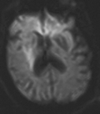
Vascular Lesions of the Council on Arteriosclerosis, American Heart Association. Circulation 92:1355, 1995

73. The North American Symptomatic Carotid Endarterectomy Trial Collaborators. Beneficial effect of carotid endarterectomy in symptomatic patients with high-grade stenosis. N Engl J Med 325:445, 1991

74. The North American Symptomatic Carotid Endarterectomy Trial Collaborators. Beneficial effect of carotid endarterectomy in patients with symptomatic moderate or severe stenosis. N Engl J Med 339:1415, 1998

75. Executive Committee for the Asymptomatic Carotid Atherosclerosis Study. Endarterectomy for asymptomatic carotid artery stenosis. JAMA 273:1421, 1995

76. Ringelstein EB, Zeumer H, Angelou D. The pathogenesis of strokes from internal carotid artery occlusion: diagnostic and therapeutical implications. Stroke 14:867, 1983

77. Klijn CJ, Kappelle LJ, van Huffelen AC, et al: Recurrent ischemia in symptomatic carotid occlusion: Prognostic value of hemodynamic factors. Neurology 55:1806, 2000

78. Bisschops RHC, Klijn CJM, Kappelle LJ, et al: Collateral flow and ischemic brain lesions in patients with unilateral carotid artery occlusion. Neurology 60:1435, 2003

79. Gomez CR. Carotid plaque morphology and risk for stroke. Stroke 21:148, 1990

80. Polak JF, Shemanski L, O'Leary D, et al: Hypoechoic plaque at US of the carotid artery: an independent risk factor for incident stroke in adults aged 65 or older. Radiology 208:649, 1998

81. Blakeley DD, Oddone EZ, Hasselblad V, et al: Non-invasive carotid artery testing: a meta-analytic review. Ann Intern Med 122:360, 1995

82. Heiserman JE, Zabramski JM, Drayer BP, et al: Clinical significance of the flow gap in carotid magnetic resonance angiography. J Neurosurg 85:384, 1996

83. Forteza AM, Rabinstein A. Angioplastía y Stenting de la estenosis carotídea. Alternativa terapéutica o posibilidad técnica? Rev Neurol 32:270, 2001

84. CAVATAS Investigators. Endovascular versus surgical treatment in patients with carotid stenosis in the Carotid and Vertebral Artery Transluminal Angioplasty Study (CAVATAS): a randomised trial. Lancet 357:1729, 2001

85. Howard VJ, Sheffet AJ, Brott TG, et al: Crest. Progress Report of the Carotid Revascularization Endarterectomy vs. Stenting Trial (CREST). 29th International Stroke Conference, 2004

86. SAPPHIRE Investigators. Stenting and Angioplasty with Protection in Patients and High Risk for Endarterectomy. 28th International Stroke Conference, 2003

87. Chimowitz MI, Kokkinos J, Strong J, et al: for the Warfarin-Aspirin Symptomatic Intracranial Disease Study Group. The Warfarin-Aspirin Symptomatic Intracranial Disease Study. Neurology 45:1488, 1995

88. Adams RJ, McKie VC, Carl EM, et al: Long-term stroke risk in children with sickle cell disease screened with transcranial Doppler. Ann Neurol 42:699, 1997

89. Adams RJ, McKie VC, Hsu L, et al: Prevention of a first stroke by transfusions in children with sickle cell anemia and abnormal results on transcranial Doppler ultrasonography. N Engl J Med 339:5, 1998

90. Goldstein LB, Adams R, Becker K, et al: Primary prevention of ischemic stroke: a statement for healthcare professionals from the Stroke Council of the American Heart Association. Stroke 32:280, 2001

91. Amarenco P, Cohen A, Tzourio G, et al: Atherosclerotic disease of the aortic arch and the risk factor of ischemic stroke. N Engl J Med 331:1474, 1994

92. Schievink WI. Spontaneous dissections of the carotid and vertebral arteries. N Engl J Med 344:898, 2001

93. Hicks PA, Leavitt JA, Mokri B. Ophthalmic manifestations of vertebral artery dissection: patients seen at the Mayo Clinic from 1976 to 1992. Ophthalmology 101:1786, 1994

94. Mokri B, Schievink WI, Olsen KD, et al: Spontaneous dissection of the cervical internal carotid artery: presentation with lower cranial nerve palsies. Arch Otolaryngol Head Neck Surg 118:431, 1992

95. Scievink WI. The treatment of spontaneous carotid and vertebral artery dissections. Curr Opin Cardiol 15:316, 2000

96. Fisher CM. Lacunes: small deep cerebral infarcts. Neurology 15:774, 1965

97. Fisher CM. The arterial lesions underlying lacunes. Acta Neuropathol 12:1, 1991

98. Okazaki H, Reagan TJ, Campbell RJ. Clinicopathological studies of primary cerebral amyloid angiopathy. Mayo Clin Proc 54:22, 1979

99. Greenberg SM, O'Donnell HC, Schaefer PW, et al: MRI detection of new hemorrhages: potential marker of progression in cerebral amyloid angiopathy. Neurology 53:1135, 1999

100. Tournier-Lasserve E, Joutel A, Melki J, et al: Cerebral autosomal dominant arteriopathy with subcortical infarcts and leukoencephalopathy maps to chromosome 19q12. Nat Genet 3:256, 1993

101. Elkind MSV, Sacco RL. Direct thrombin inhibition: a novel approach to stroke prevention in patients with atrial fibrillation. Stroke 35:1519, 2004

102. Mohr JP, Thompson JL, Lazar RM, et al: Warfarin-Aspirin Recurrent Stroke Study Group. A comparison of warfarin and aspirin for the prevention of recurrent ischemic stroke. N Engl J Med 345:1444, 2001

103. Algra A, van Gijn J. Aspirin at any dose above 30 mg offers only modest protection after cerebral ischaemia. J Neurol Neurosurg Psychiatry 60:197, 1996

104. Hass WK, Easton JD, Adams HP Jr, et al: A randomized trial comparing ticlopidine hydrochloride with aspirin for the prevention of stroke in high-risk patients. Ticlopidine Aspirin Stroke Study Group. N Engl J Med 321:501, 1989

105. CAPRIE Steering Committee. A randomised, blinded, trial of clopidogrel versus aspirin in patients at risk of ischaemic events (CAPRIE). Lancet 339:1329, 1996

106. Diener HC, Cunha L, Forbes C, et al: European Stroke Prevention Study. 2. Dipyridamole and acetylsalicylic acid in the secondary prevention of stroke. J Neurol Sci 143:1, 1996

107. Diener HC, for the MATCH Steering Committee. Management of atherothrombosis with clopidogrel in high-risk patients with recent TIA or ischemic stroke. Presented at the 13th European Stroke Conference, 2004

108. McMahon S, Rodgers A. The epidemiological asscociation between blood pressure and stroke: implications for primary and secondary prevention. Hypertens Res 17:s23, 1994

109. Wolf PA, Agostino RB, Belanger AJ, et al: Probability of stroke: a risk profile from the Framingham Study. Stroke 22:312, 1991

110. Elkind MS, Sacco RL. Stroke risk factors and stroke prevention. Semin Neurol 18:429, 1998

111. Hansson L, Zanchetti A, Carruthers SG, et al. for the HOT Study Group. Effects of intensive blood pressure lowering and low-dose aspirin in patients with hypertension: principal results of the Hypertension Optimal Treatment (HOT) randomised trial. Lancet 351:1755, 1998

112. Chobanian AV, Bakris GL, Black HR, et al: Seventh Report of the Joint National Committee on Prevention, Detection, Evaluation, and Treatment of High Blood Pressure. Hypertension 42:1206, 2003

113. MacMahon S, Peto R, Cutler J, et al: Blood pressure, stroke and coronary heart disease: part 1) Prolonged differences in blood pressure: prospective observational studies corrected for the regression dilutional bias. Lancet 335:756, 1990

114. Collins R, Peto R, MacMahon S, et al: Blood pressure, stroke and coronary heart disease: part 2) Short term reductions in blood pressure: overview of randomized trials, their epidemiological context. Lancet 335:827, 1990

115. Staessen JA, Gasowski J, Wang JG, et al: Risks of untreated and treated isolated systolic hypertension in the elderly: a meta-analysis of outcome trials. Lancet 355:865, 2000

116. Heart Outcomes Prevention Evaluation (HOPE) Study Investigators. Effects of an angiotensin-converting-enzyme inhibitor, ramipril, on cardiovascular events in high-risk patients. N Engl J Med 342:145, 2000

117. PROGRESS Collaborative Group. Randomised trial of a perindopril-based blood-pressure-lowering regimen among 6105 individuals with previous stroke or transient ischaemic attack. Lancet 358:1033, 2001

118. The ALLHAT Officers and Coordinators for the ALLHAT Collaborative Research Group. Major Outcomes in High-Risk Hypertensive Patients Randomized to Angiotensin-Converting Enzyme Inhibitor or Calcium Channel Blocker vs. Diuretic: the Antihypertensive and Lipid-Lowering Treatment to Prevent Heart Attack Trial (ALLHAT). JAMA 288:2981, 2002

119. Iso H, Jacobs DR Jr., Wentworth D, et al: Serum cholesterol levels and six-year mortality from stroke in 350,977 men screened for the Multiple Risk Factor Intervention Trial. N Eng J Med 320:904, 1989

120. Yano K, Reed DM, MacLean CJ. Serum cholersterol and hemorrhagic stroke in the Honolulu Heart Program. Stroke 20:1460, 1989

121. Hebert PR, Gaziano JM, Chan KS, et al: Cholesterol lowering with statin drugs, risk of stroke, and total mortality: an overview of randomized trials. JAMA 278:313, 1997

122. Hess DC, Demchuk AM, Brass LM, et al: HMG-CoA reductase inhibitors (statins): a promising approach to stroke prevention. Neurology 54:790, 2000

123. Albers GW, Hart RG, Lutsep HL, et al: Supplement to the guidelines for the management of transient ischemic attacks. Stroke 30:2502, 1999

124. Sillesen H, for the SPARCL Investigators. Design of the Stroke Prevention by Aggressive Reduction in Cholesterol Levels (SPARCL) Study. Ongoing Clinical Trials Session, 29th International Stroke Conference, 2004

125. Barrett-Connor E, Khaw K. Diabetes mellitus: an independent risk factor for stroke. Am J Epidemiol 128:116, 1988

126. Bruno A, Biller J, Adams HP, et al: Acute blood glucose level and outcome from ischemic stroke. Trial of ORG 10172 in Acute Stroke Treatment (TOAST) Investigators. Neurology 52:280, 1999

127. Stratton IM, Adler AI, Neil HAW, et al: Association of glycaemia with macrovascular and microvascular complications of type 2 diabetes (UKPDS 35): prospective observational study. BMJ 321:405, 2000

128. Boushey CJ, Beresford AA, Omenn GS, et al: A quantitative assessment of plasma homocysteine as arik factor for vascular disease. JAMA 274:1049, 1995

129. Fridman O. Hyperhomocysteinemia: atherothrombosis and neurotoxicity. Acta Physiol Ther Latinoam 49:21, 1999

130. Toole JF, Malinow MR, Chambless LE, et al: Lowering homocysteine in patients with ischemic stroke to prevent recurrent stroke, myocardial infarction, and death: the Vitamin Intervention for Stroke Prevention (VISP) randomized controlled trial. JAMA 291:565, 2004

131. Hankey GJ, for the VITATOPS Trial Group. The VITATOPS (VITAmins TO Prevent Stroke) Trial: Rationale, Design and Progress of an International, Large, Simple, Randomised Trial of Homocysteine-Lowering Multivitamin Therapy in Patients with Recent Transient Ischaemic Attack or Stroke. 29th International Stroke Conference, 2004

132. Abbott RD, Behrens GR, Sharp DS, et al: Body mass index and thromboembolic stroke in non-smoking men in older middle age: The Honolulu Heart Program. Stroke 25:2370, 1994

133. Walker SP, Rimm EB, Ascherio A, et al: Body size and fat distribution as predictor of stroke among US men. Am J Epidemiol 144:1143, 1996

134. Kiely DK, Wolf PA, Cupples LA, et al: Physical activity and stroke risk: The Framingham Study. Am J Epidemiol 140:608, 1994

135. Kawachi I, Colditz GA, Stampfer MJ, et al: Smoking cessation and decreased risk of stroke in women. JAMA 269:232, 1993

136. Hillbom M, Kaste M. Ethanol intoxication: a risk factor for ischemic brain infarction in adolescents and young adults. Stroke 12:422, 1981

137. Klatsky AL. Alcohol and hypertension. Clin Chem Acta 246:91, 1996

138. Thornton JR. Atrial fibrillation in healthy non-alcoholic people after an alcoholic binge. Lancet 2:1013, 1984

139. Sacco RL, Elkind M, Boden-Albala B, et al: The protective effect of moderate alcohol drinking on ischemic stroke. JAMA 281:53, 1999

140. Berger K, Ajani UA, Kase C, et al: Light-to-moderate alcohol consumption and the risk of stroke among US male physicians. N Engl J Med 341:1557, 1999

141. Finucane FF, Madans JH, Bush TL, et al: Decreased risk of stroke among postmenopausal hormone users: results from a national cohort. Arch Intern Med 153:73, 1993

142. Writing Group for the Women's Health Initiative Investigators. Risks and Benefits of Estrogen Plus Progestin in Healthy Postmenopausal Women: Principal Results From the Women's Health Initiative Randomized Controlled Trial. JAMA 288:321, 2002

143. The Women's Health Initiative Steering Committee. Effects of Conjugated Equine Estrogen in Postmenopausal Women With Hysterectomy: The Women's Health Initiative Randomized Controlled Trial. JAMA 291:1701, 2004

144. Viscoli CM, Brass LM, Kernan WN, et al: A clinical trial of estrogen-replacement therapy after ischemic stroke. N Engl J Med 345:1243, 2001

145. Hulley S, Gradt D, Bush T, et al: Randomized trial of estrogen plus progestin for secondary prevention of coronary heart disease in postmenopausal women. JAMA 280:605, 1998

Back to Top