Chapter 18
The Dizzy Patient: Disturbances of the Vestibular System
RONALD J. TUSA
Main Menu   Table Of Contents

Search

VESTIBULAR SYSTEM: AN OVERVIEW
CLINICAL ASSESSMENT
ABNORMALITIES AND THEIR MANAGEMENT
REFERENCES

There can be few physicians so dedicated to their art that they do not experience a slight decline in spirits on learning that their patient's complaint is of dizziness (giddiness). This frequently means that after exhaustive inquiry it will not be entirely clear what it is that the patient feels wrong or even less so why he feels it.

W.B. Matthews in Practical Neurology

Back to Top
VESTIBULAR SYSTEM: AN OVERVIEW
The vestibular system serves to stabilize eye position and movements during changes in head rotation and is an essential mechanism for clear vision. In fact, patients with bilateral vestibular defects must interrupt walking in order to see, and while reading a book they must even brace the head against the wall to prevent the smallest head movements transmitted from each heart beat. A classic article written by a physician describes the problems he experienced from ototoxicity.1 This chapter describes the pathophysiology of vestibular function, elaborates the clinically relevant symptoms, and reviews treatment and management options.

NORMAL FUNCTION: THE VESTIBULAR REFLEXES

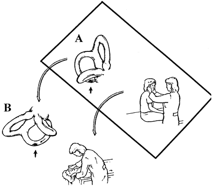
Vestibular reflexes are triggered by head movements. Imagine a bird watcher standing in a rocking boat traveling downstream (Fig. 1A). To identify a bird roosting on a tree, the image of the bird must be kept stable on the retina by three separate vestibular reflexes (see Fig. 1B). As the boat pitches up and down the eyes must move in the opposite direction and synchronously with the angular motion of the head to keep the eyes stable in space. This is accomplished by the angular vestibulo-ocular reflex (VOR), which is sensed by the semicircular canals in the inner ear (see Fig. 1B). As the boat moves down the river toward the bird, the eyes must move horizontally in the opposite direction synchronously with the linear motion of the boat. This is accomplished by the linear VOR, which is sensed by the otoliths in the inner ear (see Fig. 1C). Figure 1D shows the most complicated reflex as the boat tilts left and right. To keep the head and eyes level during tilt to the right, the right eye is elevated in the orbit and the left eye is depressed (skew deviation). Both eyes undergo torsion to the left within the orbit (counter-roll deviation) and the head is tilted to the left on the body (head tilt). This is accomplished by the ocular tilt reflex, which is sensed by the otoliths. Without these vestibular reflexes, visual acuity would be degraded and diplopia would occur. Table 1 lists the three vestibular reflexes, the sensory organs involved, and the motor output for each reflex.

Fig. 1. Illustration of vestibulo-ocular reflex (VOR) and ocular tilt reflex (OTR). A. Bird-watcher standing in a rocking boat traveling downstream. B. As the boat pitches up and down, the angular VOR moves the eyes in the opposite direction, synchronous with the angular motion of the head. C. As the boat moves down the river toward the bird, the linear VOR moves the eyes horizontally in the opposite direction, synchronous with the linear motion of the boat. D. As the boat tilts to the left and right, the OTR tilts the eyes and head in the opposite direction to maintain an earth-horizontal plane. The figure shows what happens with tilt to the right. The right eye is elevated in the orbit and the left eye is depressed (skew deviation); both eyes undergo torsion to the left within the orbit (counter-roll deviation) and the head is tilted to the left on the body.


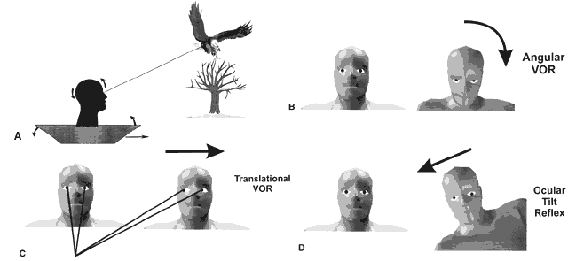
TABLE 18-1. Vestibular Reflexes


Vestibular ReflexSensory OrganMotor Output
Angular VORSemicircular canals (SCCs) Horizontal Posterior AnteriorEyes move opposite to angular movement (rotation) of the head. Shaking the head up and down (yes) is termed pitch and is sensed by the anterior and posterior SCCs. Shaking the head horizontally (no) is termed yaw and is sensed by the horizontal SCCs.
Linear VOROtoliths Saccule UtricleEyes move opposite to linear movement of the head. Linear movement up and down (riding in an elevator) is sensed by the saccule. Linear movement horizontally (riding in a train on a straight track) is sensed by the utricle.
Ocular tilt reflexOtolith UtricleEyes and head move opposite to tilt of the head. Tilt left causes elevation of the left eye, depression of the right eye, torsion of both eyes to the right, and rightward tilt of the head.

VOR, vestibulo-ocular reflex.


PATHOPHYSIOLOGY

Peripheral Structures

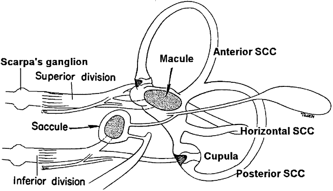
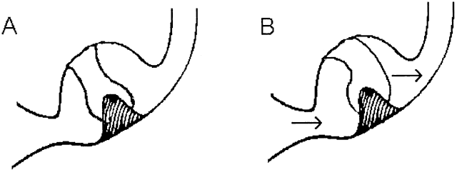
The vestibular sensory organs lie within the membranous labyrinth of the inner ear, protected in the petrous portion of the temporal bone. The membranous labyrinth consists of three semicircular canals (SCCs)—the anterior, posterior, and horizontal—that lie 90 degrees (orthogonal) to each other, and two otoliths, the utricle (horizontally aligned) and the saccule (vertically aligned) (Fig. 2). The labyrinth is innervated by the vestibular nerve, which is part of the VIIIth nerve. The vestibular nerve contains two fascicles, the superior and inferior divisions. The cell bodies of each axon of the VIIIth nerve lie in Scarpa's ganglion, located in the internal auditory canal. The superior division innervates the utricle and the anterior and horizontal SCCs. The inferior division innervates the saccule and posterior SCCs. Within each semicircular canal is an area of hair cells that protrude theirprocesses into a gelatinous matrix called the cupula. Angular head acceleration imposes inertial forces on the endolymph fluid within the canal, which causes relative fluid flow through the canal in the direction opposite to that of head acceleration. This flow deflects the cupula and bends the hair cells (Fig. 3). During head acceleration, these hair cells bend in proportion to head acceleration and change the neuronal firing rate in the VIIIth nerve. Because each Scarpa's ganglion is spontaneously “firing” at 100 spike/sec, head motion (acceleration or deceleration) modulates this firing rate. The firing rate is increased for ipsilateral angular head movements and decreased for contralateral angular head movements. The otoliths also contain a local region of hair cells. The hair cells protrude their processes into a gelatinous matrix called the macula, which is covered by a surface of calcium carbonate crystals, the otoconia. The otolith organs respond to linear acceleration and sustained head tilt relative to gravity. Linear acceleration (including tilt of the head) causes these crystals to move, which bends the hair cells and modulates the firing rate in the VIIIth nerve. The firing rate is increased for ipsilateral linear head movement or tilt, and is decreased for contralateral linear head movement and tilt.

Fig. 2. Labyrinth. See text for details.

Fig. 3. A. Enlarged view of cupula of the horizontal semicircular canal when the head is still. B. The arrows indicate direction of endolymph flow during head rotation to the left.

Semicircular Canals

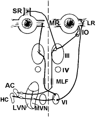
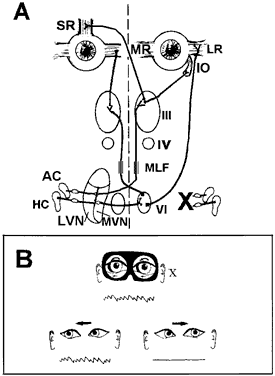
Each SCC innervates two eye muscles by means of a three-neuron arc. The central connections of the horizontal and anterior SCCs on one side are shown in Figure 4. The projections of these two SCCs are shown together because usually they are both involved in vestibular neuritis, the most common cause of severe vertigo. The pathophysiology of vestibular neuritis is diagrammed in Figure 5. This disorder disrupts the superior division of the vestibular nerve.2 Because the Scarpa's ganglion on each side normally is firing at 100 spikes/sec, any loss of activity on one side results in relative excessive excitation from the intact side. This large bias in neural activity causes nystagmus. The direction of nystagmus is determined according to the quick phase, but the vestibular deficit is actually driving the slow phase of the nystagmus. Vestibular neuritis results in a mixed horizontal and torsional nystagmus. This pattern of nystagmus is caused by the innervation pattern of the superior division of the VIIIth nerve on the intact side (recall that the superior division innervates the horizontal and anterior semicircular canals; see Fig. 2). Relative excitation of the horizontal SCC causes horizontal nystagmus with the slow phase toward the side of the lesion. Figure 5 depicts a left-sided lesion, which would cause a right-beating nystagmus. Relative excitation of the anterior SCC causes torsional nystagmus (counterclockwise nystagmus for a left-sided lesion). Because of the confusion over whether to label torsional nystagmus from the perspective of the observer or the patient, the current trend is to assess torsional nystagmus according to the direction of the quick phases toward which the superior poles of both eyes are beating. Thus, a left-sided lesion results in right beating and right torsional nystagmus (see Fig. 5B). There are two other key features of vestibular nystagmus. The intensity of nystagmus is increased when fixation is blocked (Fig. 5B depicts increased nystagmus when the subject looks through Frenzel or 20-diopter lenses), and the intensity of nystagmus also increases when the patient looks in the direction of the quick phases.

Fig. 4. Central connections of the horizontal and anterior semicircular canals mediating angular vestibulo-ocular reflex. Primary afferents of the horizontal semicircular canal (HC) project to the medial vestibular nucleus (MVN). Neurons in the medial vestibular nucleus project across the midline to terminate in the VIth nerve nucleus. Two types of neurons are in the VIth nerve nucleus, abducens neurons that project to the lateral rectus (LR) muscle, and interneurons that cross the midline and travel up the medial longitudinal fasciculus (MLF) to innervate the medial rectus subnuclei of the IIIrd nerve nucleus. Primary afferents of the anterior semicircular canal (AC) project to the lateral vestibular nucleus (LVN) and MVN, which in turn project across the midline and travel up the MLF to terminate in the superior rectus and inferior oblique subnuclei of the IIIrd nerve nucleus.

Fig. 5. Acute unilateral loss of semicircular canal function causing vestibular nystagmus. A. Disruption of the left superior division of the VIIIth nerve from vestibular neuritis. Loss of spontaneous neural activity from the left side causes slow phases to the left from relative excitation of the right horizontal semicircular canal (HC). B. Features of nystagmus. By convention, right eye position is up and left eye position is down. As the eyes move across the orbit, a quick phase resets the eyes back to the right. Quick phases are generated by burst cells, which are not part of the vestibular system. Nystagmus is labeled according to the direction of the quick phases. Consequently, this would be called a right-beating nystagmus. The lesion also disrupts spontaneous neural activity from the left anterior semicircular canal (AC), which results in right torsional nystagmus (the superior pole of each eye beats right). The intensity of nystagmus increases when the patient looks in the direction of the quick phases (B, bottom). If fixation is blocked by Frenzel glasses, nystagmus also increases and is seen clearly even in primary gaze (B, top).

Otoliths

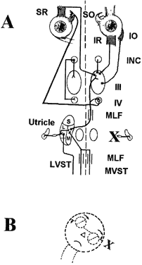
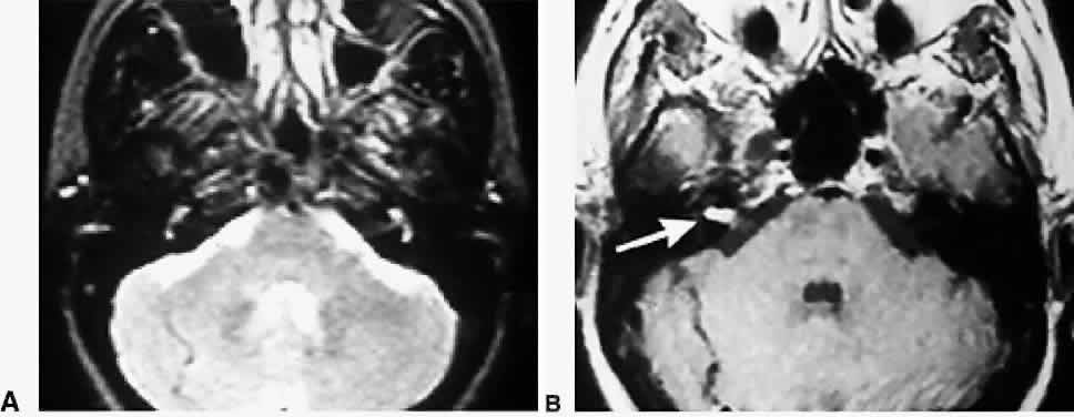
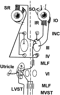
Each otolith innervates four eye muscles by means of a three-neuron arc. The central connections of the utricle on one side are shown in Figure 6.3 The projection to the vertical muscles causes vertical eye deviation and torsion during head tilt. The projections in the lateral and medial vestibulospinal tracts mediate the head tilt during the ocular tilt reflex. Acute loss of function of the utricle on one side from the VIIIth nerve section or vestibular neuritis causes a pathologic ocular tilt response because of the unopposed excitation of the intact utricle. Figure 7 depicts the results of a left-sided lesion.4 Excitation of the right superior rectus and oblique muscles causes elevation and intorsion of that eye. Excitation of the left inferior rectus and oblique muscles causes depression and extorsion of that eye. Excitation of the neck muscles innervated by the intact vestibulospinal tracts causes a left head tilt.

Fig. 6. Central connections of the utricle mediating the ocular tilt reflex. This figure illustrates the projections from the right utricle. Primary afferents of the utricle project to the lateral (L) and medial (M) vestibular nuclei. Neurons in the lateral vestibular nucleus cross the midline and travel up the medial longitudinal fasiculus (MLF) to project to the IVth and IIIrd nerve nuclei, which innervate eye muscles involved with vertical and torsional eye movements. The IIIrd nerve nucleus also innervates the interstitial nucleus of Cajal (INC). The INC in turn projects back to the IIIrd and IVth nerve nuclei. Neurons in the L and M vestibular nucleus also project to the spinal cord through the lateral and medial vestibular spinal tracts (LVST and MVST).

Fig. 7. Unilateral loss of the utricle, causing pathologic ocular tilt reflex. A. Disruption of the left utricular division of the VIIIth nerve from vestibular neuritis. B. The pathologic ocular tilt reflex from this lesion. Relative excitation of the right medial vestibular nucleus (M) causes elevation and intorsion of the right eye (mediated by the right superior rectus and right superior oblique muscles), and causes depression and extorsion of the left eye (mediated by the left inferior rectus and left inferior oblique muscles). Excitation of the neck muscles innervated by the intact vestibulospinal tract causes a left head tilt.

Back to Top
CLINICAL ASSESSMENT

HISTORY

Determining a patient's history is by far the most important part of a clinical evaluation. Unfortunately, taking a good history can be tedious because complaints by patients with dizziness are often vague and frequently confounded with anxiety-provoked symptoms. For these reasons a preliminary questionnaire is helpful. Three key items of the history are most helpful in determining the cause of dizziness.

Tempo

It must be determined whether the patient is suffering from an acute attack of dizziness (3 days or less), chronic dizziness (more than 3 days), or episodes of dizziness. An acute attack frequently is caused by vestibular neuritis, labyrinthitis, Meniere's disease, or brainstem infarct. Chronic dizziness often is caused by an uncompensated unilateral vestibular defect, bilateral vestibular hypofunction (ototoxicity, sequential vestibular neuritis), or disequilibrium from a variety of different causes, or psychological problems. If the patient suffers from episodic symptoms, the average duration of the dizziness spells should be determined. Spells that last for seconds are usually caused by benign paroxysmal positional vertigo (BPPV) or orthostatic hypotension; spells that last for minutes are usually caused by transient ischemic attacks (TIAs) or migraine; and spells that last for hours are usually from migraine or Meniere's disease.

Symptoms

What the patient means by “dizziness” should be expanded on. Dizziness is an imprecise term used to describe a variety of symptoms including vertigo, disequilibrium, light-headedness, floating, and rocking, each of which has a different pathophysiologic mechanism and significance (Table 2).


TABLE 18-2. Symptoms


TermSymptomsMechanism
VertigoRotation, linear movement, or tiltSudden imbalance of tonic neural activity to vestibular nucleus
DysequilibriumImbalance or unsteadiness while standing or walkingLoss of vestibulospinal, proprioception, visual, and motor function; joint pain or instability; and psychological factors
Light-headednessPresyncopeDecreased blood flow to the brain
Psychologically inducedFloating, swimming, rocking, and spinning inside of headAnxiety, depression, and somatiform disorders
OscillopsiaIllusion of visual motionSpontaneous: acquired nystagmus Head-induced: severe, bilateral loss of the vestibulo-ocular reflex


VERTIGO. Vertigo is the illusion of movement (rotation, linear movement, or tilt) and is caused by a sudden imbalance of tonic neural activity in the vestibular nucleus. It may be caused by either normal head movements or disease in the labyrinth, VIIIth nerve, vestibular nucleus, vestibular cerebellum (nodulus and flocculus), or central otolith pathways (from the medial longitudinal fasciculus to the interstitial nucleus of Cajal). The causes of vertigo may be simply divided into those lesions that cause a mechanical problem to the inner ear (e.g., BPPV), and those that cause loss of function (ablation) of the inner ear or central pathways (e.g., vestibular neuritis).

DISEQUILIBRIUM. This is an imbalance or unsteadiness while standing or walking. It is caused by a variety of factors including diminished or double vision, loss of vestibular function, defects in proprioception from peripheral neuropathy or spinal cord lesions, defects in motor function from central nervous or peripheral nervous system abnormalities, joint pain or instability from arthritis or weakness, and psychological factors.

LIGHT-HEADEDNESS. Also called presyncope, light-headedness usually is related to a momentary decrease in blood flow to the brain.

FLOATING, SWIMMING, ROCKING, AND SPINNING INSIDE OF HEAD. These frequently are symptoms of psychological disorders, which include anxiety (panic attacks, agoraphobia, obsessive-compulsive disorder), somatiform disorders (including conversion), and depression.

OSCILLOPSIA. This is the subjective illusion of visual motion. Patients with acquired nystagmus report apparent motion of the visual scene caused by movement of the retina (retinal slip). Patients occasionally interpret oscillopsia as dizziness, although it differs from true vertigo in that it only occurs with the eyes open. Patients with congenital nystagmus usually do not report oscillopsia because of compensatory neural mechanisms, including the foveation period, during which the eye is relatively stable, and the feedback of involuntary eye movement to the central nervous system (efference copy). Another form of oscillopsia occurs in patients with severe, bilateral loss of the VOR, which is frequently caused by ototoxicity from aminoglycosides. This form of oscillopsia occurs only during head movements and is caused by the lack of stabilizing features of the VOR; it is sometimes referred to as head-induced oscillopsia to differentiate it from oscillopsia caused by spontaneous nystagmus.

Circumstance

The circumstances under which dizziness occurs should be well defined. When dizziness occurs without provocation (spontaneously) and is vestibular in origin, it frequently is exacerbated by head movements. Dizziness may be provoked only by certain movements, such as standing up after reclining for at least 10 minutes (suggesting orthostatic hypotension), or may occur when the head is moved into certain vertical or oblique head positions (lying down or sitting up), suggesting BPPV.

Medications

Several medications may cause subjective symptoms of dizziness, especially in patients older than 65 years of age.5,6 Table 3 lists common medications and their primary effects. Certain drugs cause disequilibrium and light-headedness. These include anticonvulsants, antidepressants, antihypertensives, antiinflammatory agents, hypnotics, muscle relaxants, tranquilizers, and (when used chronically) vestibular suppressants. Sensitization to meclizine and scopolamine may occur after a few days of continuous use, and withdrawal symptoms occur when the medication is discontinued; this may be misinterpreted as a recurrence of the disorder itself, so physicians should be cautious about restarting these medications. It is suggested that meclizine, scopolamine, and other vestibular suppressants be used for only a few days during acute vestibular hypofunction caused by vestibular neuritis or labyrinthitis. These drugs should then be discontinued because they interfere with central compensation within the denervated vestibular nucleus. Patients with brainstem medullary lesions may have nausea lasting for weeks and may require medication for a longer time. Certain drugs may cause vestibular ototoxicity and spare hearing, yet lead to disequilibrium. These include certain aminoglycosides (streptomycin, gentamicin, tobramycin), furosemide, and ethacrynic acid.


TABLE 18-3. Drug-Induced Dizziness


 Drugs That Can Cause DizzinessDrugs that Interfere With Vestibular CompensationOtotoxic Drugs (Vestibular)
Antiarrythmics   
Amiodarone  X (synergistic)
Quinine  X (synergistic)
Anticonvulsants   
BarbituratesX  
CarbamazepineX  
PhenytoinX  
EthosuximideX  
Antidepressants   
AmitriptylineX  
ImipramineX  
Antihypertensive agents   
Diuretics   
 HydrochlorthiazideX  
 Furosemide  X (synergistic)
 Ethacrynic acid  X (synergistic)
α1-Blockers   
 ProzosineX  
 TerosineX  
β-Blockers   
 AtenololX  
 PropranalolX  
Calcium antagonists   
 NifedipineX  
 VerapamilX  
Antiinflammatory drugs   
Ibuprofen   
IndomethacinX  
Acetylsalicylic acid  X (reversible)
Antibiotics   
Streptomycin  X
Gentamicin  X
Tobramycin  X
Chemotherapeutics   
Cisplatin  X
Hypnotics   
FlurazepamX  
TriazolamX  
Muscle Relaxants   
CyclobenzaprineX  
OrphenidrineX  
MethocarbamolX  
Tranquilizers   
ChlordiazepoxideX  
MeprobamateX  
Vestibular Suppressants   
MeclizineXX 
ScopalamineXX 
ChlordiazepoxideXX 
DiazepamXX 


OFFICE EXAMINATION

Eye Movements

VESTIBULO-OCULAR REFLEX. There are three bedside tests of the VOR (Table 4). The vestibular dynamic visual acuity test compares static acuity with the head still to dynamic acuity with the head moving. First, distance visual acuity is measured using a Snellen chart. The patient is then asked to read the smallest possible line on the chart while the examiner manually oscillates the patient's head horizontally at 2 Hz, with the face moving about 1 to 2 inches in each direction; this is greater than the frequency at which smooth eye movements pursuit can track a target. If the VOR is normal, the patient's eyes move smoothly in the opposite direction of the head such that ocular fixation is always maintained. The patient should be able to read the same line that was readable when his or her head was still, or the adjacent line with larger letters. If the patient can read only letters more than three lines above his or her initial static visual acuity, the patient most likely has a vestibular defect. Second, VOR is examined using head thrust. Have the patient fixate on a target and observe the eyes after passive horizontal and vertical head thrusts. After a head thrust, an observed refixation saccade indicates decreased VOR.7 Third, determine whether head-shaking nystagmus is present. Have the patient close his or her eyes, pitch the head down 30 degrees, and then oscillate the head 20 times horizontally. Elicitation of nystagmus during this procedure indicates a vestibular imbalance.8 This sign may persist indefinitely after a peripheral or central unilateral vestibular lesion.


TABLE 18-4. Bedside Tests of the Vestibulo-Ocular Reflexes


TestProcedureResult
Vestibular dynamic visual acuityStatic, distant visual acuity is determined with the head still. Dynamic visual acuity is then determined while the patient's head is oscillated manually at 2 Hz.A dynamic visual acuity of 3 or more lines above static visual acuity indicates a vestibular defect.
Head thrustThe patient fixates a distant visual target, and eye position is observed immediately after a small thrust of the head to the left and right.A refixation saccade after the head thrust indicates decreased vestibulo-ocular reflex.
Head-shaking nystagmusPatient's head is pitched down 30º and the head is oscillated horizontally 20 times.Elicitation of jerk nystagmus after this procedure indicates a vestibular imbalance.


In very young children and babies, VOR can be assessed by picking up the child and rotating him or her in outstretched arms while observing the eyes for nystagmus. During this test, visual fixation should be blocked by having the patient wear Frenzel glasses (20- to 30-diopter lenses) to prevent the optokinetic response.

SMOOTH PURSUIT AND VESTIBULO-OCULAR REFLEX CANCELLATION. The patient is asked to track a slowly moving target, both horizontally and vertically, with the head still (smooth pursuit) and with the head moving synchronously with the target (VOR cancellation). Lesions in the parieto-occipital cortex, pons, and cerebellum cause deficits in smooth pursuit (catch-up saccades are observed) and VOR cancellation (inappropriate VOR) for targets moving toward the side of the lesion.

NYSTAGMUS. Selective lesions in the peripheral and central vestibular pathways result in spontaneous nystagmus because of the unopposed higher spontaneous neural activity in the intact vestibular pathways (Table 5). For example, vestibular neuritis on one side results in peripheral vestibular nystagmus because of the unopposed activity of the lateral and posterior SCCs on the intact side. These two SCCs project to the medial vestibular nucleus. Dorsolateral medullary lesions (Wallenberg syndrome) result in torsional nystagmus caused by involvement of the anterior and posterior SCC pathways centrally on one side.9 The presence of spontaneous nystagmus should be assessed with and without fixation because peripheral causes of nystagmus usually can be suppressed with fixation, whereas central causes cannot be suppressed. An easy way to test this is during the ophthalmoscopic examination, with the other eye fixating on a target.10 During this procedure, the fixating eye can be covered with the hand. Torsional nystagmus is the only form of central vestibular nystagmus that is associated with vertigo. This may be because the lesions that cause other types of nystagmus are located between the vestibular nuclei and oculomotor nuclei. In contrast, lesions that cause vertigo lie within the vestibulothalamocortical system.


TABLE 18-5. Spontaneous vestibular nystagmus due to peripheral and central lesions


NystagmusPathologyPossible Mechanism
Peripheral vestibular nystagmusLabyrinth, vestibular nerve or root entry zone lesionDecreased tonic neural activity to the MVN from horizontal and anterior SCC pathways on one side
Torsional nystagmusDorsolateral medulla lesion9Decreased tonic neural activity to the INC from central anterior and posterior SCC pathways on one side
Downbeat nystagmusCerebellular flocculus lesion or floor of fourth ventricle lesion11Decreased tonic neural activity to the INC from central posterior SCC pathways on both sides.
Upbeat nystagmusBrachium conjunctivum lesion Dorsal upper medulla lesion12,13Decreased tonic neural activity to INC from central anterior SCC pathways on both sides
Seesaw nystagmusUnilateral lesion of INC14Unilateral inactivation of INC on one side
Periodic alternating nystagmusCerebellar nodulus lesions15Unstable (high gain) neural activity in the MVN
Latent nystagmusLoss of cortical binocular visual input to the NOT usually from congenital esotropia16Decreased tonic neural activity to MVN from the NOT on one side when one eye is covered (NOT provides all of the visual input into the MVN)

MVN, medial vesbibular nucleus; SCCs, semicircular canals; INC, interstitial nucleus of Cajal; NOT, nucleus optic tract.


Certain maneuvers that may evoke nystagmus also should be performed. Results of the Hallpike-Dix test are positive in patients with BPPV. During this test the patient is seated on a flat table with the head rotated 45 degrees to one side. The patient is then quickly moved backward into a supine position with the head still deviated and hanging over the side of the table. Nystagmus from BPPV should begin within 30 seconds and lasts less than 30 seconds. If nystagmus persists while the patient is in this position, and is not present while he or she is sitting, it is likely caused by a central disorder (central positional vertigo), although there are exceptions.17 Nystagmus or a drift of the eyes also should be assessed after positive and negative pressure directed to the external auditory canal (Hennebert sign), valsalva, or loud noise (Tullio's phenomenon).18,19 A positive response is found in patients with perilymphatic fistula or hypermobile stapes, and occasionally in patients with posttraumatic endolymphatic hydrops.

Stance and Gait

The Romberg, “sharpened” Romberg (heel-to-toe tandem stance), Fukuda stepping test, normal gait, and tandem gait should be examined. In the Romberg tests, the patient is asked to stand with feet slightly apart, first with eyes open, and then with eyes closed. The patient is asked to fold his or her arms across the chest for 30 seconds with eyes open and then 30 seconds with eyes closed. A positive Romberg is one in which the patient is stable with eyes open but looses balance with eyes closed. A positive Romberg may be found in patients with acute vestibular defects or proprioceptive defects from a peripheral neuropathy. A positive sharpened Romberg also is found in these circumstances, as well as in patients with chronic vestibular defects, and in some normal individuals over the age of 65 years. For the Fukuda stepping test, the patient steps in place for 50 steps with arms extended and eyes closed.20 Progressive turning of more than 30 degrees toward one side is abnormal. A positive Fukuda stepping test frequently is found in patients with a unilateral vestibular defect, but it also is found in patients with a leg-length discrepancy or other structural abnormalities of the legs.

The Romberg test also is useful in identifying a functional component (nonorganic), which is suggested when patients rock backward on their heels yet remarkably do not fall. Other features of stance and gait help identify a functional component, including knee-buckling without fall, small-amplitude steps, uneconomical posture and movement, exaggerated sway during the Romberg test without fall, excessive slowness in gait, and fluctuations in levels of impairment.21,22

SPECIAL APPARATUSES

Electronystagmography

Electronystagmography consists of eye movement recordings during visual tracking and during vestibular testing (rotary chair or caloric stimulation). It is objective and quantitative and ideally should be performed on all patients who exhibit a vestibular defect during the clinical examination.

ROTARY CHAIR. This is the most sensitive test for assessing vestibular function and can be performed in children of any age, although calibrations usually are obtainable only in infants 6 months or older (by using a “happy face” or similar stimulus). Children less than 4 years of age can sit in a parent's lap during the test. Eye movements are measured by electro-oculography. After calibration, peak slow phase eye velocity is measured during either sinusoidal chair rotations or constant-velocity step rotations in the dark to assess VOR gain (eye velocity/chair velocity). VOR gain normally is near 1.0 at birth and decreases to about 0.7 in adults. Values less than 0.4 are abnormal and indicate vestibular hypofunction.

CALORIC TEST. Compared with rotary chair, the caloric test is a less sensitive but more specific test of peripheral vestibular function. This is the best method for determining whether a vestibular defect is peripheral or central, and also for indicating the side of the defect. This test usually is not well tolerated in children less than 8 years of age. Before the caloric test is performed, the internal auditory canal is examined with an otoscope, and any wax or debris blocking the canal is removed. If there is a tympanic membrane perforation, different temperatures of air instead of water should be used as the stimulus. The caloric test uses a nonphysiologic stimulus (water) to induce endolymphatic flow in the semicircular canals by creating a temperature gradient within each canal. Each ear canal is irrigated for 40 seconds, with a constant flow rate of water at two temperatures (30° and 44°C). Eye movements are recorded for 2 minutes after each irrigation. At the end of this period, the ear is emptied of any remaining water and sufficient time is allowed for the nystagmus to stop before proceeding with the next irrigation (usually 5 minutes).

Posturography

Postural sway is quantified with dynamic posturography that measures sway in conditions in which visual and somatosensory cues are absent or altered. Automatic postural responses also can be measured in response to perturbations of the support platform. Deficits in a variety of different neural systems can be identified, including the cerebral cortex, anterior cerebellum, and spinal cord. The test is not specific for vestibular disorders, although patients with uncompensated or severe vestibular deficits typically have difficulty maintaining their balance when both visual and somatosensory cues are altered. This test is also useful in demonstrating objective signs of a functional component.23,24

Audiometry

Audiometry should include both pure-tone and speech audiometry, and test acoustic reflexes and middle ear function. Audiometry should be performed on all individuals who complain of hearing loss. Acoustic neuromas usually present with unilateral hearing loss or tinnitus. Nonpulsatile and constant tinnitus without documented hearing loss is extremely rare. A nonorganic loss of hearing can be determined by the inconsistency of the audiogram (more than a 10-dB change in threshold on successive trials), which occurs in up to 10% of head-injured patients.25

Brainstem Auditory-Evoked Response

The brainstem auditory-evoked response (BAER) is recorded with scalp electrodes and represents the averaged surface-recorded activity of the auditory neural generators of the peripheral and central auditory pathways in the pons and midbrain. It can be used to determine auditory threshold when standard audiography cannot be performed. It also is an excellent screening test for abnormalities involving VIIIth nerve and central auditory brainstem pathways. The BAER has been reported to be abnormal in patients with postconcussive syndrome even when all other studies are normal.26

Neuroimaging

COMPUTED TOMOGRAPHY. High-resolution computed tomography (CT) of the temporal bone is highly sensitive for evaluating petrous bone and ossicular chain abnormalities, including petrous bone fractures, cholesteatoma, and congenital defects.27 The cause of conductive hearing loss and peripheral sensorineural hearing loss usually can be identified by the type of temporal bone fracture indicated by CT.28 The location of cerebrospinal fluid leaks also is best determined with a high-resolution CT scan of the temporal bone after intrathecal injection of water-soluble contrast material.

MAGNETIC RESONANCE IMAGING. Magnetic resonance imaging (MRI) with gadolinium-enhanced sections through the VIIIth nerve clearly defines the internal auditory canal, cerebellopontine angle, and brainstem.29 Enhancement on MRI has been reported in patients with inflammation of the labyrinth,30 but the significance of these findings is not yet clear.

Back to Top
ABNORMALITIES AND THEIR MANAGEMENT

ACOUSTIC NEUROMA

Acoustic neuroma is a benign tumor of the myelin sheaths of the vestibular nerve (VIII) and usually is characterized by unilateral hearing loss or tinnitus. Rarely does it cause vertigo or disequilibrium. Audiography shows features consistent with a retrocochlear process, which consists of poor speech discrimination relative to the degree of tone loss and recruitment in which increments in tone intensity cause a higher than expected increase in perceived tone loudness. The diagnostic procedure of choice for acoustic neuroma is gadolinium-enhanced MRI sections of the posterior fossa that include the VIIIth nerve and other cerebellopontine angle structures (Fig. 8). Surgery is the treatment of choice, either by middle cranial fossa approaches (for tumors confined to the internal auditory canal, with usable hearing), by suboccipital craniotomy (for large tumors or those adherent to the brainstem, with usable hearing), or by a translabyrinthine approach (for small tumors, with no usable hearing).31 With questionable or small intracanalicular tumors, especially in elderly patients or in those in poor medical condition, watchful waiting is prudent, with neuroimaging repeated at 6- to 12-month intervals. Focal radiosurgery may be ideal for recurrent tumors and for patients in poor medical condition who cannot tolerate posterior fossa surgery.31 Tumor control is achieved in 95% of patients, but approximately 25% to 30% develop 5th and 7th cranial nerve loss and hearing loss caused by involvement of the vascular supply.31,32

Fig. 8. Small acoustic neuroma revealed in an enhanced MRI of the head. Left. Axial T2 MRI through the cerebellar pontine angle. Right. Same section in a gadolinium-enhanced T1 MRI. The intracanalicular portion of the VIIIth nerve on the right side is enhanced (arrow) consistent with an acoustic neuroma.

ACUTE VESTIBULAR NEURITIS AND LABYRINTHITIS

Acute vestibular neuritis and labyrinthitis are associated with intense vertigo, nausea, and disequilibrium that persist for days. It is caused by a viral infection of the superior portion of the vestibular nerve (neuritis) or of the endolymph of the labyrinth (labyrinthitis). A viral etiology has been postulated, and recent data suggest a reactivation of dormant herpes infection in Scarpa's ganglia.33,34 Diagnosis of these entities is based primarily on clinical presentations. If associated with significant hearing loss (frequently with tinnitus), the disorder is labeled labyrinthitis; otherwise it is labeled vestibular neuritis. Viral serologic studies are superfluous and their results do not alter treatment. Differential diagnoses include infarcts of the labyrinth and a first attack of Meniere's disease. Audiography should be obtained when patients complain of hearing loss. After several days, caloric testing may document the extent of vestibular defects. In appropriate circumstances, serum fluorescent treponemal antibody absorption testing and erythrocyte sedimentation rate are used are performed to rule out otic syphilis and giant cell arteritis.

Admission to a hospital may be required for extreme dehydration from vomiting or when a central disorder is suspected. During the first few days, vestibular suppressants should be used, such as intramuscular promethazine (Phenergan; 25 to 50 mg) in the office and promethazine suppositories at home. Ondansetron (Zofran) may also be appropriate for patients with severe vertigo and nausea, but currently it is approved only for chemotherapy-induced nausea.35 Keeping patients on vestibular suppressants too long is a common error because these drugs delay vestibular adaptation. Consequently, these medications should be discontinued and vestibular exercises started as soon as possible. The patient should be reassessed after a few days to make certain that symptoms are resolving. After an acute insult, the imbalance of spontaneous neural activity in the vestibular nucleus usually corrects itself within several days, possibly through commissural pathways between the vestibular nuclei. What remains is a low gain in the VOR from which patients perceive vertigo or unsteadiness during head movements. This defect can be treated with vestibular rehabilitation (see Disequilibrium).

BENIGN PAROXYSMAL POSITIONAL VERTIGO

Benign paroxysmal positional vertigo is characterized by vertigo that lasts less than 1 minute. It usually occurs in the morning upon arising or even when turning over in bed. Symptoms also may be caused by reclining or extending the head backward. After a severe attack, patients frequently complain of disequilibrium that lasts for several hours. BPPV usually is idiopathic but also may occur after head trauma, labyrinthitis, or ischemia in the distribution of the anterior inferior cerebellar artery. The pathophysiologic mechanism of BPPV is related to portions of otoconia from the utricle that are displaced and free floating (canalithiasis) in the posterior SCC. Occasionally it is caused by otoconia attached to the cupula of this canal (cupulolithiasis). Both these conditions cause inappropriate neural afferent discharge from the posterior SCC after the head stops moving backward. The diagnosis is secured by eliciting a torsional-upbeat nystagmus associated with vertigo during the Hallpike-Dix test when the affected ear is inferior. In this test, the patient is seated on an examination table with the head rotated 45 degrees to one side; he or she is then quickly moved backward into a lying position with the head still deviated and hanging over the side of the table (Fig. 9). BPPV also may occur from debris in the anterior or horizontal SCC, but the nystagmus induced by the Hallpike-Dix test is downbeat and horizontal, respectively. The nystagmus found in BPPV usually has a latency of 3 to 20 seconds, fatigues within 1 minute, and decreases with repeat testing.

Fig. 9. Hallpike-Dix maneuver for benign paroxysmal positional vertigo. This figure shows the maneuver of the head and body during the test, along with the labyrinth (enlarged). A. The patient sits on the examination table with the head turned 45 degrees horizontally. B. The head and trunk are quickly brought straight back en bloc so that the head is hanging over the edge of the examination table by 20 degrees. The patient is assessed for nystagmus and is asked whether he or she has vertigo. Although not shown in the figure, the patient is then brought up slowly to a sitting position, with the head still turned 45 degrees, and nystagmus is sought again. This test is then repeated with the head turned 45 degrees in the other direction. This figure also shows movement of free-floating otoconia in the right posterior semicircular canal (large black arrows) during the Hallpike-Dix test. In this example, the patient has nystagmus and vertigo when the test is performed on the right side but not when the test is performed on the left side.

BPPV is best treated by a maneuver called the canalith repositioning maneuver,36 which moves the otoconia out of the posterior SCC and back into the utricle, where it is reabsorbed into the calcium matrix. Total remission or significant improvement from BPPV occurs in 90% of patients using theis maneuver,37 and complications are rare.38 During the canalith repositioning maneuver, the patient is seated with the head rotated toward the side that elicited nystagmus during the Hallpike-Dix maneuver. The patient is then moved backward into a lying position with the head hanging over the side of the table and kept is there until the vertigo and nystagmus stop. The head is then rotated toward the unaffected side, and the patient is rolled over onto this side until the face is pointed down. The patient is kept in this position for 1 minute. With the head deviated toward the unaffected side, the patient then slowly sits up. To make certain that the debris does not move back toward the cupula, the patient is fitted with a soft collar and told not to bend over or look up or down for 1 or 2 days. In addition, the patient is told to sleep sitting up during this period. For the subsequent 5 days the patient is allowed to lie down, but only on the unaffected side. After 7 days the patient is reevaluated. If the initial treatment does not work, then the patient is retreated. In patients that cannot tolerate sleeping upright for 1 or 2 days, a different maneuver is used.39 In this maneuver, the patient sits on a table sideways, rotates the head 45 degrees horizontally, and then rapidly lies on his or her side in the opposite direction and waits until the vertigo has resolved or for 10 seconds if the vertigo does not resolve. The patient then rapidly sits up and waits for the same amount of time. He or she next performs the movement in the opposite direction. This can be repeated 5 to 10 times, 1 or 2 times each day. Unlike the single treatments described earlier, this latter treatment usually takes 1 to 2 weeks before symptoms resolve. The maneuver works either by habituation or by dislodging debris from the cupula of the posterior SCC. Vestibular suppressant drugs do not have a role in the treatment of BPPV unless excessive vertigo and nausea prevent the patient from doing the maneuvers.

DIZZINESS IN CHILDREN

Dizziness in children is uncommon but alarming to parents. The most common cause is periodic dizziness (ataxia) of childhood, which is a migraine aura.40 Meniere's disease and BPPV usually do not present in childhood. The clinical evaluation of the dizzy child is the same as that of the adult.41 A neurologic examination is performed to rule out a CNS defect, and reassurance and follow-up visits usually are sufficient.

DISEQUILIBRIUM

Disequilibrium is an imbalance or unsteadiness while standing or walking. It may be caused by a number of problems, including loss of normal vestibular function, peripheral neuropathies, motor weakness, poor vision, disabling arthritis, or fear of falling. Normal balance is maintained through a complex integration of sensory input (vestibular, somatosensory, and visual) and appropriate automatic postural responses involving the frontal lobe, basal ganglia, cerebellum, spinal cord, and peripheral nerves. Patients with vestibular and proprioceptive loss in the feet complain that their balance is worse in the dark.42 The clinical examination can be helpful in the diagnosis of bilateral or static unilateral vestibular defects. Frequently demonstrable are refixation saccadic eye movements following head thrust, caused by a decrease in the VOR (Table 4). Unilateral defects frequently produce nystagmus after horizontal head shaking, and bilateral lesions usually result in more than a four-line decrease in visual acuity during 2-Hz head oscillation and subjective oscillopsia. The diagnosis may be assured by demonstrating a decreased VOR during rotary chair testing, and the peripheral nature of the defect is identified by a decreased caloric response.

The treatment of unilateral vestibular defects is based on animal studies.43,44 The chronic problem after a unilateral vestibular lesion is a dynamic deficit that can be repaired only by vestibular adaptation. Vestibular adaptation results when a mismatch occurs between head motion sensed by the vestibular system and head motion sensed by the visual system. To facilitate vestibular adaptation, the patient is encouraged to move the head while viewing a still target. Eventually these exercises should be done with the target moving in the opposite direction of the head. In addition, postural control is improved by having the patient stand with feet together, then in tandem, and then with the head moving. Similarly, the patient is encouraged to walk normally, then in tandem, and finally with the head moving back and forth. Controlled studies have shown that early intervention (on the second or third day after onset) with exercises speeds recovery, especially from imbalance and perception of disequilibrium.45 Near complete recovery may be anticipated within 6 weeks. Supervised therapy and home exercises usually are sufficient treatment. Although vestibular neurectomy has been advocated for treatment of patients with posttraumatic unsteadiness and associated hearing impairment,46 there is no conclusive evidence that neurectomy facilitates vestibular compensation. There is no physiologic basis for this surgical treatment except for intractable spells of vertigo caused by posttraumatic endolymphatic hydrops.

The treatment of bilateral vestibular defects includes avoidance of all ototoxins that may cause further permanent peripheral vestibular damage. These include gentamicin, streptomycin, tobramycin, ethacrynic acid, furosemide, quinine, and cisplatin. Drugs that may transiently impair balance (sedatives, anxiolytics, antiepileptics, and antidepressants) also should be avoided. Vestibular rehabilitation may be helpful for these patients. For bilateral defects, the same vestibular exercises described for unilateral vestibular defects can be used to improve any remaining vestibular function. Most recovery occurs with exercises that facilitate the substitution of other ocular motor systems (e.g., the cervico-ocular reflex) and somatosensory and visual cues to recover postural stability.47 Plateau in recovery should occur within 3 to 6 months. Several controlled studies48–52 have demonstrated that supervised exercises are significantly more effective in improving balance and perceived dizziness in patients with unilateral and bilateral vestibular deficits than giving patients instruction sheets of exercises to perform on their own at home.

The treatment of disequilibrium that is not caused by vestibular defects depends on its specific etiology. Treatable causes of neurologic illnesses include Parkinson's disease, early normal pressure hydrocephalus, inflammatory peripheral neuropathies, compressive myelopathies, and certain types of myopathies. Chronic use of antivestibular drugs (meclizine [Antivert] and benzodiazepines) also may cause chronic dizziness, and these should be tapered. Exercises to improve static balance and dynamic postural stability often are very helpful in patients with disequilibrium.49 Additional physical therapy to strengthen muscle and increase range of motion is helpful in patients with weakness, arthritis, and joint limitations. Patients who have a fear of falling often improve with supervised exercise; dynamic posturography is helpful in determining appropriate physical therapy. Patients with a psychological disorder may complain of chronic disequilibrium. A combination of counseling and physical therapy can decrease the problem in some patients.

HEAD TRAUMA

Head trauma frequently is associated with dizziness from a variety of causes.53 BPPV is the most common cause, either from a direct blow to the head or from the shear forces from a flexion-extension injury to the neck (“whiplash”). Other causes of dizziness from head trauma include perilymphatic fistula from barotrauma (scuba diving or pressure force to the ear), interruption of the labyrinth or vestibular nerve from petrous bone fracture, and axon swelling and interruption within the brainstem. A CT scan of the petrous bone should disclose bone fractures. Caloric stimulation can confirm loss of vestibular function, and MRI may reveal central posterior fossa injury. BPPV and vestibular loss caused by petrous bone fractures respond well to therapy described earlier (for BPPV and acute vestibular neuritis and labyrinthitis).

MENIERE'S DISEASE

Meniere's disease causes spells of roaring sounds (tinnitus), ear fullness, and hearing loss often associated with vertigo that last for hours to days. With repeated attacks, a sustained low-frequency sensorineural hearing loss and constant tinnitus usually develops. The cause is believed to be decreased reabsorption of endolymph in the endolymphatic sac (see Fig. 2), which can occur after ear trauma or viral infection or which can be idiopathic. The diagnosis depends on documentation of fluctuating hearing loss by audiography.

The frequency of attacks of Meniere's disease can be significantly reduced by restricting the diet to 2000 mg or less of sodium per day54,55; some patients require the additional use of a diuretic. Acetazolamide may be the optimal diuretic because this drug may decrease osmotic pressure within the endolymph, but chlorthalidone and other diuretics also have been quite effective.56,57 Less proven prophylactic therapy includes elimination of alcohol and caffeinated products (including chocolate). During acute attacks the patient is treated as with any other attack of acute vertigo, except extensive laboratory investigation and vestibular exercises usually are not necessary because the patient recovers quickly. Medical therapy may not control the disease. Endolymphatic shunts may be used, but are not always effective or may fail after a few years. Labyrinthectomy is appropriate in patients without evidence of contralateral disease in whom there is a severe preexisting hearing loss on the side of the defective labyrinth. Vestibular neurectomy is used for patients in whom hearing is preserved.58

MIGRAINE

Migraine is a common but poorly recognized cause of dizziness. Dizziness caused by migraine usually lasts 4 to 60 minutes and may or may not be associated with a headache. The International Headache Society criteria for the diagnosis of migraine applies. Because migraine is a diagnosis of exclusion, a positive response to treatment is essential.

Spells of vertigo caused by migraine respond to the same types of treatment as those used for headaches.59 After the diagnosis is established and the patient is reassured, he or she should be given a list of risk factors and foods that may precipitate an aura. Hypoglycemia should be avoided by eating every 6 to 8 hours, the use of nicotine and exogenous estrogen should be discontinued, and a regular sleep schedule should be maintained. If strict avoidance of these risk factors does not significantly reduce the frequency of dizziness episodes, based on a diary, then daily medication is used.60 Beta-blockers (atenolol or propranolol) are among the most effective prophylactic drugs for migraine.

MOTION SICKNESS

Motion sickness consists of episodic dizziness, tiredness, pallor, diaphoresis, salivation, nausea, and occasionally vomiting induced by passive locomotion (e.g., riding in a car) or motion in the visual surrounding while standing still (e.g., the motion of trains, traffic, flowing water). Motion sickness is believed to be a sensory mismatch between vision and vestibular cues.61 Patients with migraine disorder are particularly prone to motion sickness, especially during childhood. Twenty-six to 60% of patients with migraine have a history of severe motion sickness compared with 8% to 24% of individuals without migraine.62,63 The reason for this correlation is not clear. Diagnosis is based on careful history taking, and symptoms often are reproduced with the use of moving full-field visual targets such as optokinetic devices.

Treatment consists of reassurance, reduction of circumstances that cause sensory mismatch, and medication, if necessary. Intramuscular promethazine immediately relieves space motion sickness in 90% of individuals, compared with a resolution of symptoms in 72 to 96 hours in untreated individuals.64 Cinnarizine, a calcium-channel blocker, is the drug most commonly used in Europe to treat dizziness. In a double-blind crossover study with scopolamine, scopolamine was shown to be more effective in protecting against seasickness, but Cinnarizine was better tolerated.65

ORTHOSTATIC HYPOTENSION

Orthostatic hypotension produces symptoms that range from light-headedness when standing up to chronic fatigue, mental slowing, dizziness, nausea, and impending syncope. Common causes include medications (diuretics, antihypertensive medications, tricyclic antidepressants), prolonged bed rest, and neurogenic disorders (autonomic neuropathy from diabetes, multisystem atrophy, Parkinson's disease). Diagnosis is confirmed by recording a drop in systolic pressure of 20 mm Hg or more associated with reproduction of symptoms.

Potentially offending drugs should be discontinued if possible, and increased salt and fluid intake (5 g each day and five glasses of water) should be encouraged. Fludrocortisone (0.1 to 0.6 mg/day) may be required. If this fails 10 mg midodrine is given three times a day. In a double-blind, placebo-controlled study, midodrine significantly increased standing systolic blood pressure (by 22 mm Hg; p < .001) and decreased orthostatic dizziness, fatigue, and weakness (p < .05).66 When these medications are used, the head of the patient's bed should be elevated to reduce supine hypertension.

PANIC ATTACKS AND HYPERVENTILATION

Panic attack is an anxiety disorder that causes intense fear or discomfort that reaches a crescendo within 10 minutes and is frequently associated with dizziness, nausea, shortness of breath, and sweating. These symptoms may occur unexpectedly or may be situationally provoked. This disorder may be initiated by an organic cause of vertigo such as BPPV, especially in patients with a family history for panic attacks. Hyperventilation associated with chronic anxiety also may cause vague complaints of dizziness. This is associated with shortness of breath or chest tightness and paresthesia.

Imipramine is very effective in controlling panic attacks, but in a placebo-controlled study alprazolam was just as effective and better tolerated during a 6-month maintenance program.67 Paroxetine (Paxil) may be an ideal medication because it is not habit-forming and because many patients with panic disorder have concomitant depression. Behavioral modification also has been shown to be effective in a recent clinical outcome study in which 96.1% of patients remained in remission at 2 years and 67.4% were in remission for at least 7 years.68

PERILYMPHATIC FISTULA

Perilymphatic fistula is a hole between the inner and middle ears caused by barotrauma (scuba diving), a tumor in the middle ear (cholesteatoma), head trauma, or displacement of a prosthetic middle ear bone into the inner ear. Any pressure changes to the inner or middle ear causes a flow of fluid between these two compartments and distorts the utricle or semicircular canal. Distortion of these end-organs frequently causes transient vertigo, nystagmus, or skew deviation. Nystagmus or drift of the eyes should be assessed after positive and negative pressure directed to the external auditory canal (Hennebert's sign), Valsalva maneuver, or loud noises (Tullio's phenomenon).18,19 Diagnosis requires middle ear exploration. The oval and round windows are examined for the leak, which may be increased with the Valsalva maneuver. Surgical repair with autogenous tissue, followed by bed rest, usually is effective.

SEIZURES

Seizures commonly cause vague dizziness described as confusion, disorientation, or light-headedness, but they rarely cause vertigo.69–71 Seizures also can cause head or eye deviation, and occasionally nystagmus, but there usually is associated mild confusion.72 Electroencephalography is performed only when there is a strong suspicion of seizures, and ideally is recorded during the actual spell of dizziness. Therapy depends on the type of seizure.

TRANSIENT ISCHEMIC ATTACKS

Caused by vertebrobasilar insufficiency, TIAs provoke episodes of dizziness that are abrupt and usually last only a few minutes. TIAs frequently are associated with other symptoms of vertebrobasilar insufficiency, most commonly visual disturbance, drop attacks, unsteadiness, and weakness.73 A small percentage of patients with vertebrobasilar insufficiency may present with isolated spells of vertigo, presumably caused by ischemia in the distribution of the anterior vestibular artery. This small artery perfuses the anterior and lateral SCCs and the utricular macula, and spares the cochlea. These patients usually have known cerebrovascular disease or risk factors for this disease. Magnetic resonance arteriography can be performed to assess posterior circulation vessels and transcranial doppler may detect decreased flow in the basilar artery. Treatment includes reduction of risk factors for cerebrovascular disease and antiplatelet therapy. Warfarin (Coumadin) is used when there is significant vertebrobasilar artery stenosis.74

Back to Top
REFERENCES

1. JC: Living without a balancing mechanism. N Engl J Med 246:458, 1952

2. Fetter M, Dichgans J: Vestibular neuritis spares the inferior division of the vestibular nerve. Brain 119:755, 1996

3. Uchino Y et al: Utriculoocular reflex arc of the cat. J Neurophysiol 76:1896, 1996

4. Halmagyi GM, Gresty MA, Gibson WPR: Ocular tilt reaction with peripheral vestibular lesions. Ann Neurol 6:80, 1979

5. Ballantyne J, Ajodhia J: Iatrogenic dizziness. In Dix MR, Hood JD (eds): Vertigo, p 217. New York: John Wiley and Sons, 1984

6. Wennmo K, Wennmo C: Drug-related dizziness. Acta Otolaryngol [Suppl] 455:11, 1988

7. Halmagyi GM, Curthoys IS: A clinical sign of canal paresis. Arch Neurol 45:737, 1988

8. Hain TC, Fetter M, Zee DS: Head-shaking nystagmus in patients with unilateral peripheral vestibular lesions. Am J Otolaryngol 8:36, 1987

9. Morrow M, Sharpe JA: Torsional nystagmus in the lateral medullary syndrome. Ann Neurol 24:390, 1988

10. Zee DS: Ophthalmoscopy in examination of patients with vestibular disorders. Ann Neurol 3:373, 1978

11. Baloh RW, Spooner JW: Downbeat nystagmus: A type of central vestibular nystagmus. Neurology 31:304, 1980

12. Nakada T, Remler MP: Primary position upbeat nystagmus: Another central vestibular nystagmus? J Clin Neuro Ophthalmol 1:185, 1981

13. Ranalli PJ, Sharpe JA: Upbeat nystagmus and the ventral tegmental pathway of upward vestibulo-ocular reflex. Neurology 38:1329, 1988

14. Halmagyi GM et al: Jerk-waveform see-saw nystagmus due to unilateral meso-diencephalic lesion. Brain 117:789, 1994

15. Leigh RJ, Robinson DA, Zee DS: A hypothetical explanation for periodic alternating nystagmus: Instability in the optokinetic-vestibular system. Ann NY Acad Sci 374:619, 1981

16. Tusa RJ, Becker JL, Mustari MJ et al: Brief periods of impoverished visual experience during infancy impairs the development of specific gaze-holding systems in monkeys. In Fuchs A, Brandt T, Büttner U et al (eds): Contemporary Ocular Motor and Vestibular Research: A Tribute to Dave A. Robinson, p 345. Stuttgart: George Thieme Verlag, 1994

17. Baloh RW, Yue Q, Jacobson K et al: Persistent direction-changing positional nystagmus: Another variant of benign positional vertigo. Neurology 45:1297, 1995

18. Daspit CP, Churchill D, Linthicum FH: Diagnosis of perilymph fistula using ENG and impedance. Laryngoscope 90:217, 1980

19. Pyykko I, Ishizaki H, Aalto H et al: Relevance of the Tullio phenomenon in assessing perilymphatic leak in vertiginous patients. Am J Otol 13:339, 1992

20. Fukuda T: The stepping test: Two phases of the labyrinthine reflex. Acta Otolaryngol 50:95, 1959

21. Keane JR: Hysterical gait disorders: 60 cases. Neurology 39:586, 1989

22. Lempert T, Brandt T, Dieterich M et al: How to identify psychogenic disorders of stance and gait. J Neurol 238:140, 1991

23. Cevette MJ et al: A physiologic performance on dynamic posturography. Otolaryngol Head Neck Surg 112:676, 1995

24. Allum JHJ, Huwiler M, Honegger F: Identifying cases of non-organic vertigo using dynamic posturography. Gait Posture 4:52, 1996

25. Berman JM, Fredrickson JM: Vertigo after head injury: A five year follow-up. J Otolaryngol 7:237, 1978

26. Noseworthy JH, Miller J, Murray TJ et al: Auditory brainstem responses in post-concussive syndrome. Arch Neurol 38:275, 1981

27. Hasso AN, Ledington JA: Traumatic injuries of the temporal bone. Otolaryngol Clin North Am 21:295, 1988

28. Momose KJ, Davis KR, Rhea JT: Hearing loss in skull fractures. Am J Neuroradiol 4:781, 1983

29. Swartz JD, Harnsberger HR: The temporal bone: Magnetic resonance imaging. Top Magn Reson Imag 2:1, 1990

30. Mark AS, Seltzer S, Nelson-Drake J et al: Labyrinthine enhancement on gadolinium-enhanced magnetic resonance imaging in sudden deafness and vertigo: Correlation with audiologic and electronystagmographic studies. Ann Otol Rhinol Laryngol 101:459, 1992

31. Kartush JM, Brackmann DE: Acoustic neuroma update. Otolaryngol Clin North Am 29:377, 1996

32. Flickinger JC et al: Radiosurgery of acoustic neurinomas. Cancer 67:345, 1990

33. Furuta Y et al: Latent herpes simplex virus type 1 in human vestibular ganglia. Acta Otolaryngol [Suppl] 503:85, 1993

34. Davis LE, Johnsson, LG: Viral infections of the inner ear: Clinical, virologic and pathologic studies in humans and animals. Am J Otolaryngol 4:347, 1983

35. Rice GPA, Ebers GC: Ondansetron for intractable vertigo complicating acute brainstem disorders. Lancet 345:1182, 1995

36. Epley JM: The canalith repositioning procedure: For treatment of benign paroxysmal positional vertigo. Otolaryngol Head Neck Surg 107:399, 1992

37. Herdman SJ, Tusa RJ, Zee DS et al: Single treatment approaches to benign paroxysmal positional vertigo. Arch Otolaryngol Head Neck Surg 119:450, 1993

38. Herdman SJ, Tusa RJ: Complications of the canalith repositioning procedure. Arch Otolaryngol 122:281, 1996

39. Brandt T, Daroff RB: Physical therapy for benign paroxysmal positional vertigo. Arch Otolaryngol 106:484, 1980

40. Basser LS: Benign paroxysmal vertigo of childhood. Brain 87:141, 1964

41. Tusa RJ, Saada AA, Niparko JK: Dizziness in childhood. J Child Neurol 9:261, 1994

42. Baloh RW, Honrubia V: Clinical Neurophysiology of the Vestibular System, p 101. Philadelphia: FA Davis, 1990

43. Igarashi M et al: Further study of physical exercise and locomotor balance compensation after unilateral labyrinthectomy in squirrel monkeys. Acta Otolaryngol 92:101, 1981

44. Fetter M, Zee DS: Recovery from unilateral labyrinthectomy in rhesus monkeys. J Neurophysiol 59:370, 1988

45. Herdman SJ, Clendaniel RA, Mattox DE et al: Vestibular adaptation exercises and recovery: Acute stage after acoustic neuroma resection. Otolaryngol Head Neck Surg 113:77, 1995

46. Sanna M, Ylikosky J: Vestibular neurectomy for dizziness after head trauma: A review of 28 patients. ORL J 45:216, 1983

47. Herdman SJ, Borello-France D, Whitney S: Treatment of vestibular hypofunction. In Herdman SJ (ed): Vestibular Rehabilitation. Philadelphia: FA Davis, 1994

48. Horak FB, Jones-Rycewicz C, Black FO et al: Effects of vestibular rehabilitation on dizziness and imbalance. Otolaryngol Head Neck Surg 106:175, 1992

49. Krebs DE, Gill-Body KM, Riley PO et al: Double-blind, placebo-controlled trial of rehabilitation for bilateral vestibular hypofunction: Preliminary report. Otolaryngol Head Neck Surg 109:735, 1993

50. Szturm T, Ireland DJ, Lessing-Turner M: Comparison of different exercise programs in the rehabilitation of patients with chronic peripheral vestibular dysfunction. J Vest Res 4:461, 1994

51. Shepard NT, Telian SA: Programmatic vestibular rehabilitation. Otolaryngol Head Neck Surg 112:173, 1995

52. Cass SP, Borello-France D, Furman JM: Functional outcome of vestibular rehabilitation in patients with abnormal sensory-organization testing. Am J Otol 17:581, 1996

53. Tusa RJ, Brown SB: Neuro-otological trauma and dizziness. In Rizzo M, Tranzel D (eds): Head Injury and PostConcussive Syndrome, p 177. New York: Churchill Livingstone, 1996

54. Boles R, Rice DH, Hybels R et al: Conservative management of Meniere's disease: Furstenberg regimen revisited. Ann Otol Rhinol Laryngol 84:513, 1975

55. Proctor C, Proctor TB, Proctor B: Etiology and treatment of fluid retention (hydrops) in Meniere's syndrome. Ear Nose Throat J 71:631, 1992

56. Klockhoff I, Lindblom U, Stahle J: Diuretic treatment of Meniere disease: Long-term results with chlorthalidone. Arch Otolaryngol 100:262, 1974

57. Shinkawa H, Kimura RS: Effect of diuretics on endolymphatic hydrops. Acta Otolaryngol 101:43, 1986

58. Molony TB: Decision making in vestibular neurectomy. Am J Otol 17:421, 1996

59. Tusa RJ: Diagnosis and management of neuro-otological disorders due to migraine. In Herdman SJ (ed): Vestibular Rehabilitation. Philadelphia: FA Davis, 1994

60. Peroutka SJ: The pharmacology of current anti-migraine drugs. Headache [Suppl] 30:5, 1990

61. Brandt T, Daroff RB: The multisensory physiological and pathological vertigo syndromes. Ann Neurol 7:195, 1980

62. Kuritzky A, Ziegler DK, Hassanein R: Vertigo, motion sickness and migraine. Headache 21:227, 1981

63. Kayan A, Hood JD: Neuro-otological manifestations of migraine. Brain 107:1123, 1984

64. Davis JR, Jennings RT, Beck BG et al: Treatment efficacy of intramuscular promethazine for space motion sickness. Aviat Space Environ Med 64:230, 1993

65. Pingree BJ, Pethybridge RJ: A comparison of the efficacy of cinnarizine with scopolamine in the treatment of seasickness. Aviat Space Environ Med 65:597, 1994

66. Jankovic J et al: Neurogenic orthostatic hypotension: A double-blind, placebo-controlled study with midodrine. Am J Med 95:38, 1993

67. Schweizer E, Rickels K, Weiss S et al: Maintenance drug treatment of panic disorder: I. Results of a prospective, placebo-controlled comparison of alprazolam and imipramine. Arch Gen Psychiatry 50:51, 1993

68. Fava GA, Zieezny M, Savron G et al: Long-term effects of behavioural treatment for panic disorder with agoraphobia. Br J Psychiatry 166:87, 1995

69. Nielsen JM: Tornado epilepsy simulating Ménière's syndrome. Neurology 9:794, 1959

70. Furman JMR, Crumrine PK, Reinmuth OM: Epileptic nystagmus. Ann Neurol 27:686, 1990

71. Kaplan PW, Tusa RJ: Neurophysiologic and clinical correlation of epileptic nystagmus. Neurology 43:2508, 1993

72. Tusa RJ, Kaplan PW, Hain TC et al: Ipsiversive eye deviation and epileptic nystagmus. Neurology 40:662, 1990

73. Grad A, Baloh RW: Vertigo of vascular origin. Arch Neurol 46:281, 1989

74. Gomez CR et al: Isolated vertigo as a manifestation of vertebrobasilar ischemia. Neurology 47:94, 1996

75. Schuknecht HF: Pathology of the Ear. Cambridge, UK: Harvard University Press, 1974

76. Brandt T: Vertigo: Its Multisensory Syndromes. London: Springer-Verlag, 1991

Back to Top