Chapter 71B
Pathology of Age-Related Human Cataracts
J. R. KUSZAK, K. J. AL-GHOUL and M. J. COSTELLO
Main Menu   Table Of Contents

Search

CORTICAL CATARACTS
POSTERIOR SUBCAPSULAR CATARACT
AGE-RELATED NUCLEAR CATARACTS
SUMMARY
REFERENCES

The function of the crystalline lens is to collect divergent rays of light and direct them as a focused beam onto the fovea centralis of the retina. To perform this task, the lens must be transparent. Lens transparency is due at least in part to a combination of the unique arrangement of its fiber cells or fibers and a gradient of refractive index produced by the variable cytoplasmic protein concentration of these cells. When light is scattered or diffracted, as it passes through the lens, the scattering site or sites of altered light transmission generally are opaque. If the lens opacities occur within the visual field and significantly impair vision, then the lens is considered cataractous. Worldwide, cataracts are the most common cause of curable blindness. Of the many factors associated with cataract formation, the most significant is age.

In this chapter, we describe alterations in lens structure that result in cortical, posterior subcapsular, and nuclear opacities, the most common types of age-related cataracts.

Back to Top
CORTICAL CATARACTS
Most human cortical cataracts have several well-defined characteristics. First, cortical opacities generally are conical or, alternately, cuneiform in shape with a broad base oriented toward the lens periphery and a triangular apex oriented toward the poles. They initially are small in size, but over a period of time—often as long as 3 decades—they enlarge proportionately in every dimension. In this manner, although cortical cataracts are the most common type of age-related cataracts, their negative impact on vision is significant only after their increase in size results in opacities becoming located within the pupillary margins and thus, directly along the visual axis. Second, unequal numbers of cortical opacities form in specific locations within the anterior and posterior cortex. In order of occurrence and frequency, cortical opacities develop within the inferonasal, superotemporal, inferotemporal, and superonasal quadrants. In addition, cortical opacities are positioned asymmetrically within the anterior and posterior regions of the cortex. Several cortical opacities create the illusion of spokes, and therefore, these cataracts also commonly are referred to as spoke-like opacities. Third, because cortical opacities develop exclusively within the anterior and posterior regions of the cortex, the middle, or equatorial, segments of fibers are not involved in cortical cataracts.

The reason that cortical opacities are consistently conical in shape, grow slowly over decades, evolve in specific quadrants rather than in random locations, and finally affect only anterior or posterior segments of fibers is poorly understood. Nonprimate animal studies have not been useful in elucidating the aforementioned specific characteristics of human cortical cataracts because the cortical opacities of these models are not consistently conical in shape, do not necessarily arise in a quadrantic order, and commonly involve immature (elongating) as well as mature fibers. However, a comparison of the morphology of normal primate (monkey and baboon) and human lenses at different ages with that of human cortical cataracts removed surgically (intra- and extracapsular techniques) strongly suggests that cortical cataracts are a function of improper sutural formation during specific periods of lens development, growth, and aging.

NORMAL HUMAN LENS SUTURE DEVELOPMENT AS A FUNCTION OF AGE

To facilitate an understanding of abnormal human suture development in cortical cataracts, we briefly review normal human suture development as a function of age.

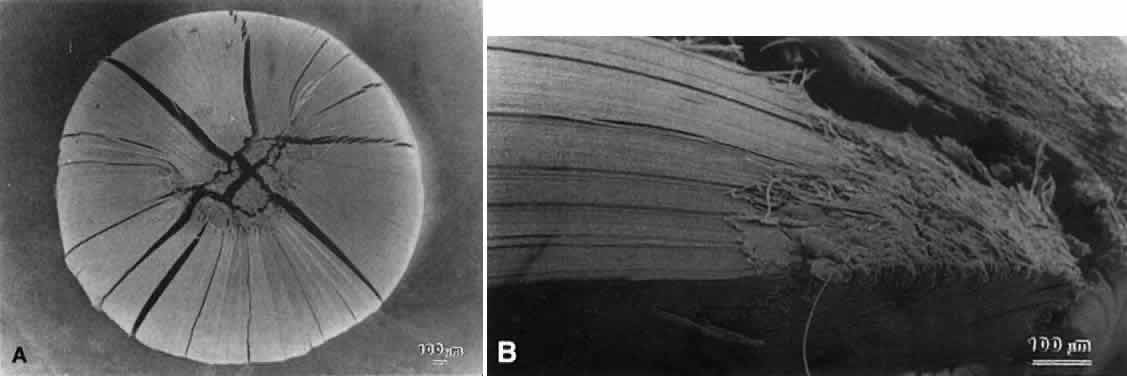
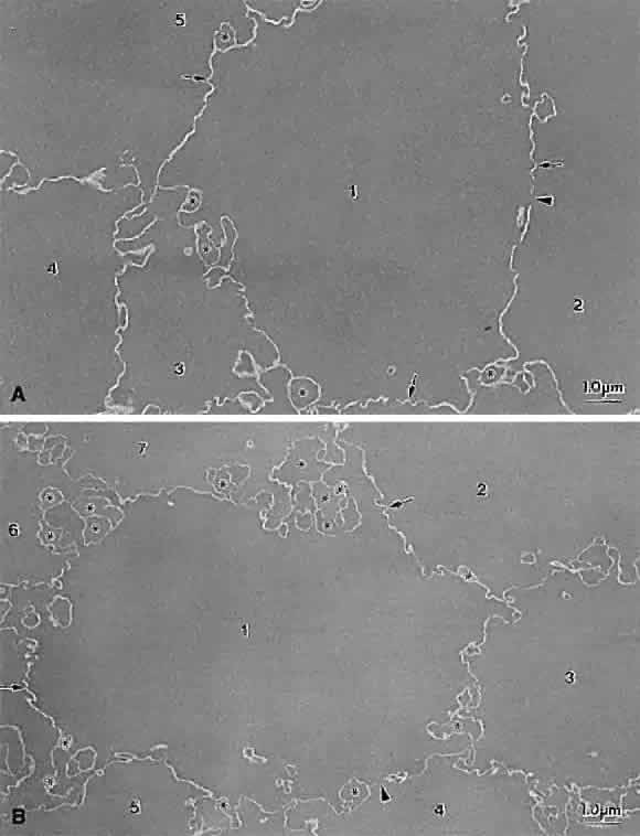
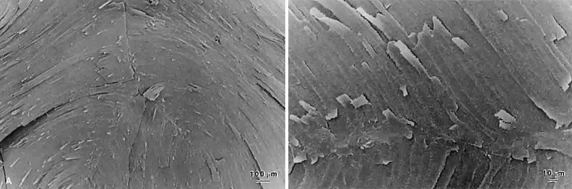
Lens sutures are formed by the overlap of fiber ends anteriorly and posteriorly in each shell.1 Although fibers generally are uniform in shape along their length, their ends are not uniform in shape or size. Thus, sutures are naturally occurring regions of disorder in the otherwise ordered lens. Indeed, although individual fibers cannot be resolved at low magnification, the sutures are recognized easily (Figs. 1, 2, 3). The higher visibility of sutures is related to the increased degree of scatter as light passes through these naturally occurring disordered regions. In contrast, the inability to resolve individual fibers at the same magnification is related to the minimal amount of scatter as light is transmitted through their uniform shape and ordered arrangement. Thus, an understanding of lens sutural anatomy is integral to comprehending lens function or dysfunction.

Fig. 1. Light micrographs of the anterior (A) and posterior surface (B) of a normal 40-year-old human lens. Faint, complementary, complex star suture patterns are visible. Although suture branches are resolvable, the individual fibers and their overlapped ends that make up the suture branches are not. The yellow coloration of this lens is an artefact of glutaraldehyde fixation rather than a function of age.

Fig. 2. Scanning electron microscopy (SEM) micrographic photomontage of an evolving juvenile nuclear suture pattern from a 6-year-old human lens. At this magnification, seven evolving suture branches of the star suture (denoted by arrowheads at the branches proximal ends) and three original Y suture branches extending to confluence at the pole are apparent.

Fig. 3. SEM micrographs showing the individual fibers and their overlapped ends forming suture branches from a 6-year-old human lens.

Although fibers are arranged end to end around a polar axis, they are not “meridians.” That is, fibers neither extend from pole to pole nor are they fusiform or tapered at their ends. Rather, there are two distinct types of mature secondary fibers: straight fibers and S-shaped fibers (Fig. 4). A straight fiber is crescent-shaped, with its entire length lying within a plane passed through the visual axis defined by its equatorial location. However, only one end of a straight fiber extends to a pole. An S fiber has a crescent shape, but in addition, its ends exhibit precise curvature away from the poles in diametrically opposite directions. This means that neither of the ends extend to a pole. Thus, the entire length of these fibers does not lie within a plane passed through the visual axis defined by their equatorial location.

Fig. 4. A series of scale (approximately 3:1) three-dimensional computer-assisted drawings (3D-CADs) showing normal human lens sutural anatomy as a function of fetal development (A), infantile and juvenile growth (B and C) and adulthood (D). Throughout fetal development, six straight fibers are positioned precisely to divide growth shells into six equal segments. Three of the straight fibers, oriented at 120° to one another, have one end that extends to confluence at the anterior pole, whereas the other three comparably oriented straight fibers have one end that extends to confluence at the posterior pole. All other fibers in any shell are S-shaped fibers, or fibers with opposite end curvature. Between any two straight fibers, are groups of S-shaped fibers with progressively variable degrees of opposite end curvature. Neither end of an S-shaped fiber reaches a pole, and because of the variable degree of opposite end curvature, the anterior and posterior ends of these fibers become aligned as offset (60°), anterior, and posterior latitudinal arc lengths. The ends of S-shaped fibers in neighboring groups overlap precisely to form suture branches. Although the anterior ends of S-shaped fibers in neighboring groups overlap to form an anterior suture branch because of opposite-end curvature, the posterior ends of these same fibers do not overlap with one another to form a posterior suture branch. In the fetal nucleus, the anterior and posterior ends of S-shaped fibers from all neighboring groups overlap to form three anterior and three offset (60°) posterior branches arranged respectively as Y and as inverted Y suture patterns. The key parameters of the progressively more complex generations of human lens suture patterns formed throughout life are shown in the second through fourth rows. From birth through infancy, 12 groups of S-shaped fibers become symmetrically positioned between 12 straight fibers to form a simple star. From juvenile through sexual maturation, 18 groups of S-shaped fibers become symmetrically positioned between 18 straight fibers to form a star suture. Finally, throughout adulthood, 24 groups of S-shaped fibers become symmetrically positioned between 24 straight fibers to form a complex star suture. As more suture branches are formed as a function of development, growth, and age, the degree of opposite end curvature and variation in intrashell fiber length decreases.

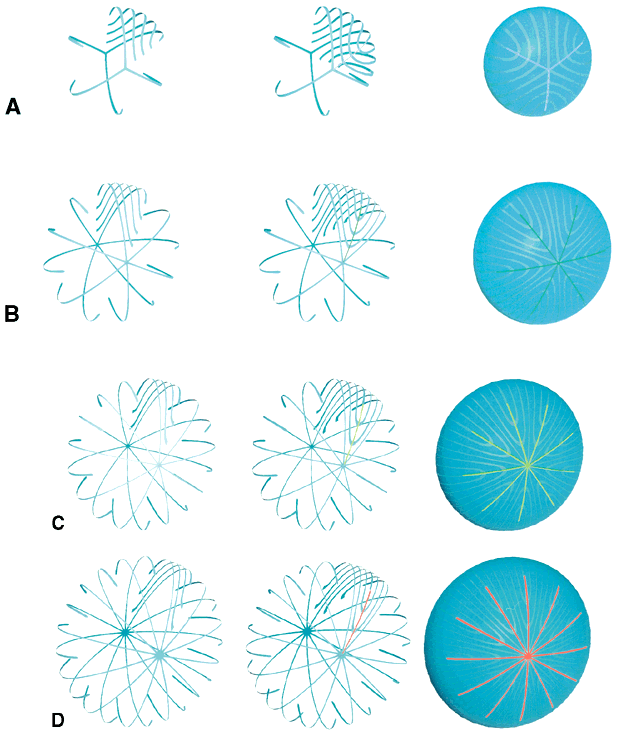
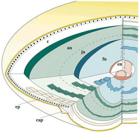
As a result of the variations in secondary fiber shape (failure of ends to extend to the poles and opposite end curvature), the ends of secondary fibers in all growth shells become aligned as specific longitudinal arc lengths. The overlapped ends of fibers in growth shells produce “suture branches” along these defined longitudinal arc lengths, and the origin of suture branches are defined by the ends of straight fibers. All the suture branches extend to confluence at the poles and combine to form discrete anterior and posterior suture “patterns.”

Throughout life, variably straight or S-shaped secondary fibers are arranged specifically to form suture branches and patterns in each growth shell. During the fetal period, as successive growth shells are formed, six straight fibers evolve in specific positions around the equator to subdivide shells into six equal segments. All other fibers evolve into S fibers arranged as six distinct groups, positioned between the straight fibers. Because of opposite-end curvature, the anterior and posterior ends of these fibers become aligned as offset anterior and posterior longitudinal arc lengths. The ends of two proximal groups of S fibers overlap to produce suture branches. The location and boundaries of suture branches are defined by the ends of straight fibers. Also, as a result of opposite-end curvature, while the anterior ends of fibers in proximal groups overlap to form anterior suture branches, their posterior ends overlap with different groups to form posterior branches.

In the anatomic position, the three anterior and three posterior suture branches are oriented at 120° angles to one another to produce respectively, upright Y and inverted Y suture patterns, that in turn are offset by 60°. In successive shells, suture branches formed during fetal development are positioned in identical locations so that continuous triangular suture planes are formed extending from the embryonic nucleus (primary fiber mass) to the lens periphery at birth. From an optical standpoint, the construction of suture planes has a significant negative effect on lens optics2–4 (i.e., increased spherical aberration or focal length variability [FLV] and sharpness of focus). The results of correlative laser scan, scanning electron microscopy (SEM), and three-dimensional computer-assisted drawing (3D-CAD) analysis of nonprimate lenses with Y sutures comparable to human lenses at birth, or of nonprimate lenses with “line” sutures, the simplest form of a branched suture pattern, have shown that FLV is minimal, and therefore, sharpness of focus is greatest, when a low-powered helium-neon laser passes through ordered radial cell columns. These studies also reveal that FLV is maximal, and therefore, sharpness of focus is least, when the beam passes through disordered suture planes.2–3

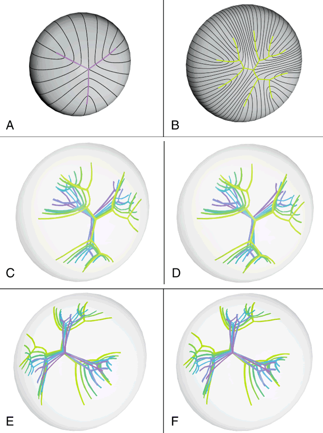
However, in comparable studies of primate lenses, it can be shown that an increased number of suture branches as a function of age results in superior optical quality. The reason for this paradox is that after birth, primates employ a similar but fundamentally different growth scheme than nonprimate lenses. After birth, humans produce progressively more complex sutures throughout distinct periods of growth. During the evolution of these sutures, the original six identical and symmetrically positioned Y suture branches formed throughout the fetal period serve as templates for the eventual formation of 12, 18, and 24 suture branches.

The second generation of sutures in humans, the “simple star” suture, is formed from birth through early childhood. Unlike the first generation Y suture, however, the fibers of successive shells are not identical in shape or position. As a result, these suture branches are out of register and thus, “discontinuous” suture planes are formed extending from the fetal nucleus to the lens periphery. Throughout adolescence and adulthood, “star” and “complex star” sutures—the third and fourth generation of sutures, respectively, are formed in a similar manner. In star and complex star sutures, respectively, 18 and 24 suture branches eventually are formed in successive shells. Thus, adult lenses have only small triangular suture planes within the fetal nucleus, overlain by progressively more complex suture patterns in successive infantile, juvenile, and adult nuclear and cortical shells. Because the branches of the progressively more complex suture patterns in successive shells are neither identical in size nor aligned in register, they form discontinuous suture planes extending from the fetal nucleus to the lens periphery. Thus, in human lenses, the negative influence exerted by suture planes on FLV is minimized effectively. In this manner, human lenses are optically superior to nonprimate lenses with line and Y suture lenses, at least in part, because of their more complex sutures.

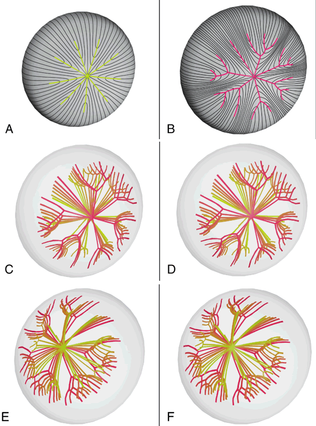
Also, the evolution of suture branches does not occur simultaneously within a growth shell. Suture formation normally commences in the inferonasal quadrant and then proceeds in turn in the superotemporal, inferotemporal, and finally, the superonasal quadrants of the lens. The specific starting point is not without developmental or pathologic precedent. Colobomas of the eye (e.g., retina, ciliary body, iris, choroid, lens, and zonules), the failure or arrest of normal embryonic fissure closure during embryogenesis, typically occur in the inferonasal quadrant.5 In fact, this embryologic defect is considered atypical if it occurs in another quadrant.

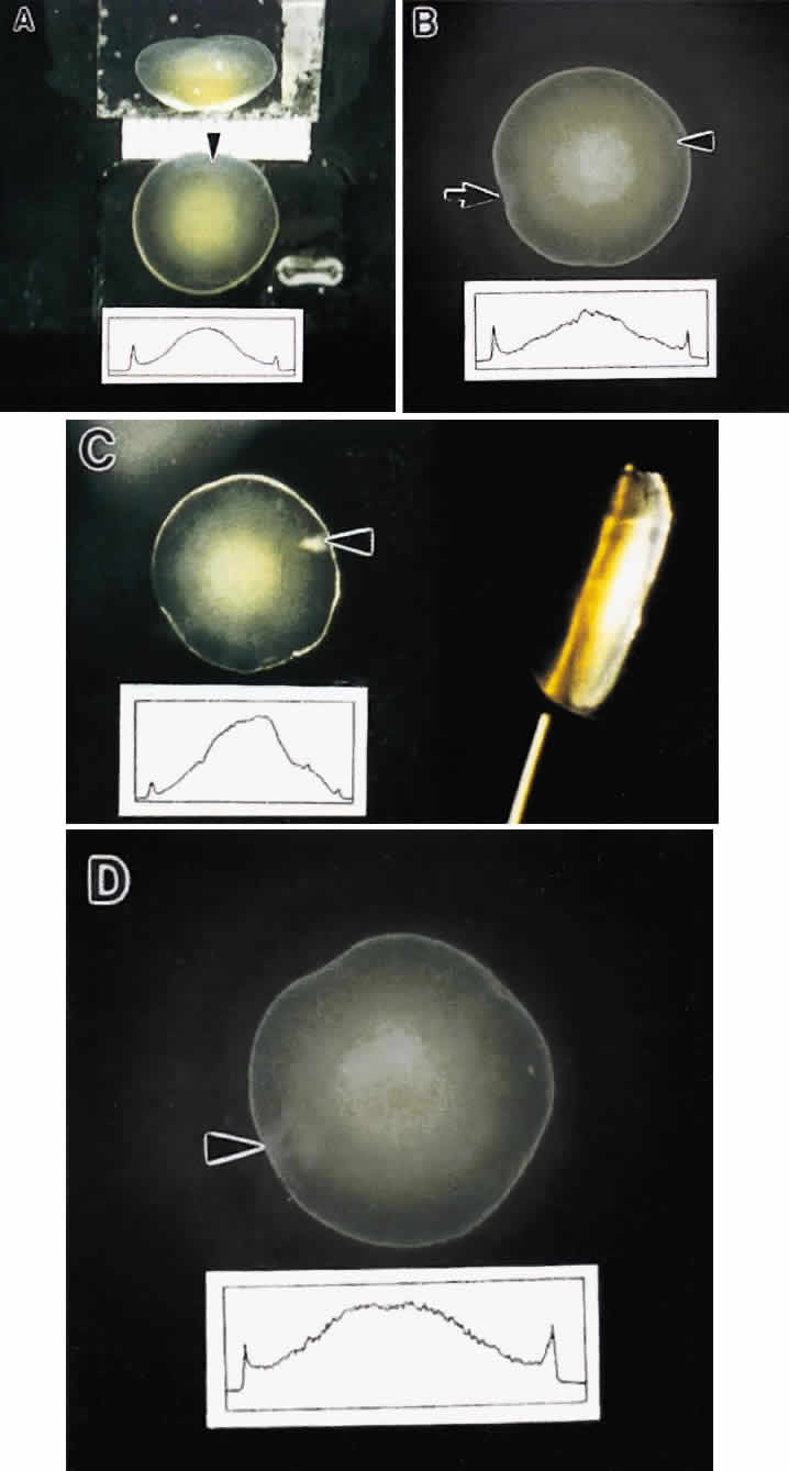
Slit-lamp biomicroscopy reveals four distinct and reproducible zones of discontinuity in aged emmetropic human lenses.6 These zones are formed by 4, 9, 19, and 46 years of age. The temporal development of the zones of discontinuity essentially is identical to the progressive iteration of the four generations of human lens sutures. Furthermore, the anatomic location and measure of the normal zones of discontinuity are coincident with the four distinct generations of sutures. This leads to the compelling argument that the zones of discontinuity and the distinct generations of sutures are one and the same. Indeed, the sharply demarcated zones of discontinuity are not coincident with alterations in human fiber membrane surface structure7–11 or variations in concentration and density of fiber crystalline and cytoskeletal proteins12,13 as a function of development, growth, and age. Furthermore, because the aforementioned changes in lens morphology as a function of age are common to both primate and nonprimate lenses, all lenses—not only primate lenses—should develop zones of discontinuity throughout life. Thus, the abnormal slit-lamp profiles of some cataractous lenses (particularly diabetic and cortical) are likely to be recordings of abnormal suture development. Correlative SEM and 3D-CAD analysis of human cortical cataract lenses surgically removed by the extracapsular technique confirm this premise.

ABNORMAL HUMAN SUTURE DEVELOPMENT AS A FUNCTION OF AGE

As a result of its inverted developmental and growth scheme, the lens retains every fiber formed throughout life within the concentric growth shells. By noting the axial dimensions of the lens at different ages, intact suture patterns formed at different ages can be retrieved for comparative studies between normal and human cortical cataracts.

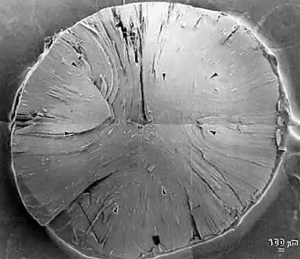
The first generation of sutures in cortical cataracts has normal Y patterns. However, examination of cortical cataracts removed from patients in the fourth through fifth decades reveal that the second through fourth generations of sutures failed to form normally (Fig. 5). In concentric growth shells, radially enlarging subbranches are formed in identical locations. As a result, continuous suture planes, in the shape of cones extending from the periphery to the pole, are formed over a period of 3 decades, beginning shortly after birth.

Fig. 5. Scale (3.5:1) schematic 3D-CADs depicting a normal human lens anterior suture pattern at birth (A) and an abnormal anterior suture pattern of a surgically removed (intracapsular technique) cortical cataract (37-year-old man; B). Because lens growth is inside-out throughout life, progressively younger to older growth shells can be removed to expose suture patterns formed during specific periods of development, growth, and age. The early cortical cataract had nine nonidentical and asymmetrically positioned suture subbranches extending from an irregular Y suture. By superimposing schematic 3D-CADs of suture patterns from representative growth shells extending from the adult nucleus to the superficial cortex in such a lens, the sutural anatomy of a cortical cataract lens can be reconstructed as it existed in situ from birth through the time of surgery. Stereo scale schematic 3D-CADs depicting the three-dimensional anterior (C and D) and posterior sutural anatomy (E and F) of the early cortical cataract shown in B. It can be seen clearly that abnormal suture formation, from birth through adulthood resulted in continuous abnormal suture planes of comparable shape (conical or cuneiform) and size (progressively larger as a function of depth or age), and in analogous locations as typical cortical opacities seen by slit-lamp biomicroscopy.

Examination of cortical cataracts removed from patients in the seventh through eighth decades reveal that although the first through third generations of sutures are normal, the fourth generation is abnormal (Fig. 6). In concentric growth shells formed after the third generation of sutures (the star suture), radially enlarging subbranches are formed in identical locations. As described previously, continuous suture planes, in the shape of cones extending from the periphery to the pole, are formed over three decades, but in this case the onset of abnormal suture formation does not occur until middle age. The production of continuous pyramid-like planes in concentric growth shells likely would cause a significant increase in both FLV and scatter.2–4

Fig. 6. Scale (3.5:1) schematic 3D-CADs depicting a normal young adult anterior suture pattern (A) and an abnormal aged anterior suture pattern from a surgically removed (intracapsular technique) cortical cataract (67-year-old man; B). The aged cortical cataract had 13 nonidentical and asymmetrically positioned suture subbranches extending from an irregular (7-branch) star suture. Stereo scale schematic 3D-CADs depicting the three-dimensional anterior (C and D) and posterior sutural anatomy (E and F) of the aged cortical cataract shown in B. In this case, it clearly can be seen that abnormal suture formation throughout adulthood resulted in continuous abnormal suture planes of comparable shape (conical or cuneiform) and size (progressively larger as a function of depth or age), and in analogous locations as typical aged cortical opacities seen by slit-lamp biomicroscopy.

In consideration of the aforementioned facts, the following should be noted:

  1. The evolution of identical but radially enlarged abnormal suture subbranches in concentric growth shells results in the formation of abnormal suture planes in cortical cataracts that essentially are identical in shape to the conical opacities observed clinically by slit-lamp biomicroscopy.
  2. The initial abnormal suture planes are seen most frequently in the inferonasal quadrant, as are the first conical opacities of cortical cataracts.
  3. The temporal formation of cortical opacities and abnormal suture planes are synchronous.
  4. Abnormal suture planes only involve the anterior or alternatively the posterior segments of fibers, and cortical opacities only occur in the anterior or posterior cortex.

Thus, a compelling argument can be made that abnormal suture planes and cortical cataract opacities are one and the same.

At surgery, the cortex of cortical cataract lenses is softer than the nucleus. Histologically, the cortex of these lenses feature swollen and liquefied fibers. It has been proposed that an increased uptake of water forces the cataractous cortical fibers apart between radial cell columns and concentric growth shells (lamellar separation). This proposition leads to speculation that the lens may be attempting to alter the structure of a localized site of increased scatter. Cortical opacities, seen to greatest advantage by retroillumination slit-lamp biomicroscopy, correspond to areas of lamellar separation that in turn are believed to correlate with “fiber folds” seen by SEM.14

Ultrastructurally, the opacities are characterized by variable amounts of degenerated cortical fiber fragments and ceroid bodies (lipofuscin), presumably the result of enzymatic digestion. Morgagnian cataracts are considered a special form of mixed cortical and nuclear cataract typified by complete liquefaction of the entire cortex. Again, it is tempting to speculate that the zones of liquefaction are an attempt by the lens to eliminate areas of scatter. This premise is strengthened by the fact that the nucleus in aged cortical cataract lenses has normal sutures and thus, would not need to undergo any internal alterations to improve light transmission. It also should be noted that to date, aged nuclear cataracts also seem to have normal sutural anatomy.

Back to Top
POSTERIOR SUBCAPSULAR CATARACT
In the older literature, age-related posterior subcapsular cataracts (PSCs) appropriately were referred to as cataract complicata, or cataracts arising as a complication of another pathology. Indeed, patients with retinitis pigmentosa or diabetes or patients undergoing chronic steroid therapy have a higher incidence of PSCs. In addition, long-term use of alcohol and cigarettes also is believed to predispose chronic users to PSC formation, and vitrectomy patients are at great risk of developing PSCs within a short period of time after surgery. Although the aforementioned diseases and conditions often are associated with age, young people with the same afflictions or in need of comparable treatment also are at increased risk for PSCs. Regardless of etiology, PSCs are described as displaying consistent ultrastructural changes in a common progression.

It generally is believed that all PSCs are characterized by a proliferation of dysplastic transitional zone lens epithelial cells.15–18 Transitional zone cells are normally high columnar cells that already have assumed the uniform hexagonal cross-sectional profiles that are maintained after fiber terminal differentiation. These cells normally are arrayed in ordered meridional rows just above the lens equator. As lens growth proceeds, these cells rotate 90° along their polar axis while simultaneously beginning the process of bidirectional elongation to become fiber cells. These dynamic events occur in the bow region of the lens, which can be shown to advantage in polar histologic sections.

In the earliest forms of PSCs, the meridional rows are uncharacteristically disorganized. As PSC formation proceeds, the transitional cells either rotate incompletely or not at all in the bow region. These cells then are believed to migrate progressively toward the posterior pole while simultaneously enlarging into ovate-shaped Wedl or bladder cells.18 PSCs often are seen overlain by additional strata of relatively normal elongating fiber cells. Ultrastructurally, the opaque regions of PSCs feature few organelles, degenerating nuclei, elaboration of crystallin proteins, a prominent cytoskeleton, and lateral membrane interdigitations between neighboring cells. Thus, the cataract cells of PSCs are considered to be characterized by dysplastic fibers that exhibit cellular alterations consistent with fiber terminal differentiation.

PSCs appear to have a preferential migration path that is associated with the lens sutures. Retroillumination slit-lamp images often reveal an asymmetrical stellate pattern of PSCs that is consistent with the continuously evolving star and asymmetrical star lens sutures that are formed as a function of age.

In a recent study19 of Royal College of Surgeons (RCS) rats, an animal model of autosomal recessive retinitis pigmentosa, correlative light, scanning, and transmission electron microscopic analysis clearly demonstrated that the posterior subcapsular opacity is the result of abnormal posterior suture formation rather than a proliferation of dysplastic transitional zone lens epithelial cells (Figs. 7 and 8). The RCS rat PSC is composed of markedly enlarged and irregular posterior fiber ends aberrantly curved away from the polar axis toward the vitreous rather than overlapping and abutting to form suture branches within and between concentric growth shells. Light microscopic analysis revealed evidence of progressively more numerous, enlarged, and irregular ovate cellular profiles at the posterior pole. However, there was no evidence of Wedl cells either within the meridional row region or along a migratory path from the equator to the posterior pole at any age. Transmission electron microscopy (TEM) analysis confirmed that the size and abnormal shapes of cellular profiles were consistent with SEM analysis and that nuclei never were observed within the plaque. Preliminary studies of transgenic mice developed as a model for autosomal dominant retinitis pigmentosa show PSCs resulting from sutural compromise and again give no indication of a proliferation of dysplastic transitional zone cells. The results of these studies raise important questions. Are there two types of PSCs, one that involves a proliferation of dysplastic cells and one that involves sutural malformations? Alternatively, are all PSCs a function of abnormal suture formation, a structural compromise that only recently has been elucidated by correlative ultrastructural analysis? Clinically, these issues are relevant because some PSCs have been noted to spontaneously regress and most RCS rat PSCs effect a form of recovery (internalization). These facts suggest that PSCs may be responsive to therapeutic intervention.

Fig. 7. Low-magnification SEM micrographs of a Royal College of Surgeons (RCS) rat posterior subcapsular cataract (PSC). A. There is the absence of a normal inverted posterior Y suture. B. At slightly higher magnification, it can be seen that there was no evidence of Wedl cells migrating from the periphery of the lens to the central opacity.

Fig. 8. Higher-magnification SEM micrographs of the RCS PSC shown in Figure 7. A. At this magnification, it can be seen that the PSC was the result of enlarged posterior ends of fibers turning up and away from the polar axis rather than overlapping within and between growth shells to form suture branches. It also can be seen that many of the posterior ends had blebbed off from the rest of the fibers. In any plane of section, these blebs would appear to be Wedl cells. B. A comparative view of posterior fiber ends beneath the posterior capsule of a normal adult rat lens.

At the time of surgical removal, the ultrastructure of mature PSCs are not dissimilar to those of mature cortical opacities. They are characterized by fragmentation, swelling, and degeneration of the ends of elongating and superficial cortical lens fibers. In addition, multilamellar membranous bodies in complex patterns also are typical of PSCs. Globules noted in some PSCs are likely to be necrotic fragments of fiber ends. Similar structural alterations frequently are seen in other types of cataracts. Dense bodies, consistent with lysosomal activity, have been found in varying amounts at sites of opacity in PSCs. In addition, acid hydrolases capable of eliminating membrane waste have been found to be localized at lens sutures. The presence of these lytic enzymes is believed to explain the “zone of liquefication” in PSCs. All the structural alterations noted to occur in PSCs are consistent with the theory that PSCs have a dynamic nature, with the lens attempting to repair itself more or less successively as a function of age and severity of PSC.

Back to Top
AGE-RELATED NUCLEAR CATARACTS

ARCHITECTURE OF THE AGED NUCLEUS

Most cataract extractions performed worldwide are due to nuclear opacification. Thus, if we hope to prevent the development or slow the progression of nuclear opacification, we must understand the cellular structure of the different regions of the normal nucleus and how structural changes within these regions cause nuclear cataracts.

The regions of the normal (noncataractous) aged human lens and their cellular structure, based on electron microscopic studies,20 are shown in Figure 9. The embryonic, fetal, juvenile, and adult nuclei, as well as “cortex” contain fiber cells, or simply fibers, produced at different rates during specific periods of development, growth and aging (Table 1). The embryonic nucleus, formed by the end of the first 7 weeks of embryogenesis, is the smallest region (up to 400 μm in equatorial diameter) and has the shortest fibers. However, although primary fibers are very irregular in shape, their average cross-sectional dimension, 80 μm2, is larger than in any other region. These cellular features, and the fact that the ends of primary fibers do not overlap to form sutures, distinguish the embryonic nuclear fibers from all subsequently formed secondary fibers. The fibers of the fetal nucleus, formed from 7 weeks until birth, grow at a very high rate and have an average cross-sectional area of 35 μm2. These fibers, only somewhat nonuniform in shape, are packed in short radial cell columns, and as described previously, their ends overlap as Y sutures. Secondary fibers formed after birth until puberty constitute the juvenile nucleus, and those formed after puberty until young adulthood constitute the adult nucleus. These fibers form at a slower rate, are more uniform in shape, and have smaller average cross-sectional areas, 14 and 7 μm2, respectively, partially because of pronounced fiber compaction during aging. As aforementioned, juvenile nuclear fiber ends overlap to form simple star sutures whereas adult nuclear fiber ends overlap to form star sutures. Fibers in the four different nuclear regions also have characteristic age-related structural alterations.20


TABLE 71B-1. Approximate Number of Cells in Each Region of the Lens and the Average Regional Growth Rate per Year*


RegionCell Size (μm)No. of CellsGrowth Rate(cells/yr)
Cortex2.24 × 14‡665,000133,000
Adult nucleus0.75 × 7.5‡4,460,000101,000
Juvenile nucleus1.7 × 9‡640,00053,000
Fetal nucleus5.0§700,0001,360,000
Embryonic nucleus10.0§8003500

*All calculations were made on cells in the equatorial plane.
†The number of years for each region was determined using the developmental time periods defined previously, applied to a 61-year-old lens. Five years was estimated for the cortex, and puberty was assumed to occur at age 12.
‡Thickness × width for an average cell.
§Diameter of equivalent circular cell with measured average area.
(Adapted from Taylor VL, Al-Ghoul KJ, Lane CW et al: Morphology of the normal human lens. Invest Ophthalmal Vis Sci 37:1396, 1996)


Fig. 9. Schematic scale (15:1) diagram of a 60-year-old aged noncataractous human lens. The embryonic nucleus (en) is enlarged 4× , and the epithelium (ep) and capsule (cap) are enlarged for clarity. Fibers are approximately to scale relative to each other (but not to the region thickness). The sutures are not shown. The fetal nucleus (fn), juvenile nucleus (jn), adult nucleus (an), and cortex (c) are composed of secondary fibers formed during specific periods of development and growth. (Taylor VL, Al-Ghoul KJ, Lane CW et al: Morphology of the normal human lens. Invest Ophthalmol Vis Sci 37:1396, 1996)

AGE-RELATED CHANGES IN THE NUCLEUS

The nucleus undergoes many age-related changes simultaneously. For example, the aging nucleus develops a yellow coloration, develops scattering centers that appear white, produces compacted fiber cells, and hardens throughout the whole nucleus. At this point, it is worthwhile to consider the distinction between aging, which is a normal process, and cataract formation, which implies a degenerative or pathologic process. Some of the changes in the nucleus observed during aging, such as yellow coloration and hardening of the nucleus, may be normal and protective,21 whereas other changes, such as cell disruption, membrane breakdown, and protein degradation, may lead to scattering centers that eventually result in impaired vision. The point at which scattering centers cause impaired vision is the point at which the pathology begins, even though the same underlying processes, such as oxidative damage, may be occurring in the lens before visual impairment. This is consistent with observations that most humans older than 75 years of age show an increased amount of nuclear light scattering even without obvious visual impairment.

The human lens nucleus becomes yellow in color with age, and the coloration becomes deeper, tending toward amber and brown, with increasing age. The yellow coloration often is present in aged lenses that show no observable nuclear scattering, and thus, the yellow color may not necessarily be related directly to nuclear cataract formation. However, the intensity of yellow coloration seems to parallel the increase in scattering during nuclear cataract formation. The most consistent observation is that pigments causing the yellow color are formed independently from and superimposed on the whitish scattering from scattering centers that cause nuclear cataracts. In mature cataracts, the color may approach dark brown or black, in which case the colored pigments may so extensively absorb light that light absorption becomes very significant in reducing visual function. The extent of yellow coloration corresponds exactly to the nucleus and is the same intensity throughout the nucleus. This property is another indication that the pigments are formed by a different mechanism than the scattering centers.

The classic view of light scattering in age-related nuclear cataracts, indicated by in vivo slit-lamp images22,23 or in vitro dark-field images,11,24 shows high light scattering from the embryonic and fetal nuclei (Fig. 10). Regardless of the age or stage of formation, the juvenile and adult nuclei usually show progressively less scattering up to the cortex/nucleus interface, where there is a pronounced decrease in scattering. The cortex often is clear in pure nuclear cataract or displays punctate localized white scattering or opacifications, as discussed previously. The amount of scattering often is judged using a variety of grading systems22,25; for example, some have scales from 1 for transparent to 6 for nearly complete opacification. The scales are slightly different for in vivo or in vitro classification, and the grading depends partially on subjective judgment and on the light source and the optics of the recording system. In most reported slit-lamp images of nuclear cataracts, the scattering uniformly and symmetrically increases as the grade progresses.22 Although this implies that the progression of the scattering in the patient follows the patterns reported for the grades, no systematic study reports the time course of the full range of scattering. Furthermore, there is no systematic documentation of the variations within each grade, such as the enhancement of scattering in one developmental region. For example, some variations in the white scattering from typical mixed cataracts are shown in Figure 10. Note that the scattering in some lenses is not symmetric about the optic axis (see Fig. 10A and B), and others have higher scattering in the embryonic and fetal nuclei with a decrease in scattering within the juvenile and adult nuclei and a further decrease at the cortex/nucleus interface (see Fig. 10C). In the early stages of cataract formation, scattering centers may be distributed uniformly throughout the nucleus. Because of the lens shape, this uniform distribution of scattering centers may give the impression of a greater amount of scattering from the lens center. In more advanced stages, views by slit lamp or dark-field photography confirm that the most common pattern is a gradual radial decrease in scattering from the embryonic nucleus through the adult nucleus. We note that the variety of the patterns of scattering may be more extensive than presently appreciated, and distinctive features of the patterns may lead to clues about the etiology of nuclear cataracts.

Fig. 10. Representative in vitro dark-field photomicrographs of surgically removed (intracapsular technique) age-related nuclear cataracts. Each lens has pronounced nuclear scattering and localized cortical opacities (arrowheads). The optical density scans (insets) demonstrate that the central regions of the lenses scatter the greatest amount of light. A. Anterior and side (prism) views from a 61-year-old cataractous lens. A calibration bar in millimeters is included. B. Anterior view of a 73-year-old cataractous lens with an asymmetric distribution of the whitish central scattering also indicated by the fluctuations at the apex of the density scan. The arrow indicates the probable attachment site of the cryoprobe used to extract the lens. C. Anterior (left) and in vitro slit-lamp (right) views of an 81-year-old cataractous lens. Note the markedly higher scattering from the central region corresponding to the embryonic and fetal nuclei. D. Anterior view of a 67-year-old cataractous lens with a very slight decrease in scattering at the lens center. (Al-Ghoul KJ, Lane CW, Taylor VL et al: Distribution and type of morphological damage in human nuclear age-related cataracts. Exp Eye Res 62:237, 1996)

POTENTIAL SCATTERING CENTERS IN NUCLEAR CATARACT

The structural characteristics of the scattering centers in age-related nuclear cataracts are unknown. Many theories have been proposed, and recent evidence can be used to evaluate some of the theories. For example, massive cellular damage throughout the nucleus was believed to be the cause of nuclear opacification. However, in a recent study11 of nuclear cataracts, minor cellular damage was shown convincingly to have occurred only in the adult nucleus. No evidence of extensive cellular damage ever was observed in the juvenile, fetal, or embryonic nuclei. In fact, fiber architecture of normal aged transparent (noncataractous) lenses is nearly indistinguishable from that of age-related nuclear cataracts (Figs. 11 and 12).

Fig. 11. Light micrographs of embryonic nuclear fiber cross-sectional profiles from a normal transparent 59-year-old lens (A) and from a nuclear cataractous 81-year-old lens (B). At this magnification, the morphology of normal and cataractous embryonic fibers essentially is indistinguishable.

Fig. 12. Transmission electron micrographs of embryonic nuclear fiber cross-sectional profiles from an aged transparent 62-year-old lens (A) and a nuclear cataractous 83-year-old lens (B). In several numbered fibers, gap junctions (space indicated by arrowheads), undulating membrane pairs (arrows), and interlocking edge processes (circular profiles marked with asterisks) are apparent. Cataractous embryonic nuclear fibers have quantifiably more numerous edge processes (asterisks) than aged embryonic nuclear fibers. (A, Al-Ghoul KJ, Costello MJ: Fiber cell morphology and cytoplasmic texture in cataractous and normal human lens nuclei. Curr Eye Res 15:533, 1996; B, Al-Ghoul KJ, Lane CW, Taylor VL et al: Distribution and type of morphological damage in human nuclear age-related cataracts. Exp Eye Res 62:237, 1996)

Other proposals for the source of scattering include protein-water phase separation, precipitation, and degradation of proteins. Although these changes probably do occur to some extent, TEM analysis reveals that these are likely to result in relatively minor changes of cellular architecture because the cytoplasm of fetal and embryonic nuclear fibers remain smooth and homogeneous with age.11,24 Oxidative damage to proteins and membranes is well documented especially for aged lenses, which have a decreased capacity for protection against reactive oxygen species. Extensive morphologic data demonstrate that damage and perhaps loss of membranes could be initiating events in age-related nuclear cataract formation.11,24 The implications are that the breakdown of membranes and subsequent loss or modification of cytoplasmic crystallins may result in the creation of localized scattering centers as a function of age. Furthermore, the structural evidence correlating fiber architecture with nuclear scattering in extracted nuclear cataract lenses indicates that the scattering centers result mainly from relatively minor changes in structure at the cellular interfaces and within the cytoplasm. We predict that the cumulative effect of these minor changes eventually leads to a gradual increase in scattering from numerous small scattering centers. This interpretation of the data is consistent with the observed late onset and slow progression of the nuclear cataracts.

Cell interfaces are very complex, particularly in the fetal and embryonic nuclei (see Figs. 11 and 12). Moreover, many images show a greater complexity of the interface in nuclear cataracts compared with controls (see Fig. 12). Notably, there are numerous circular profiles (edge processes) that are located between adjacent plasma membranes singly or in clusters20 (see asterisks in Fig. 12). These edge processes in SEM appear to be knobs that sometimes project extensively into adjacent cells (Fig. 13).

Fig. 13. SEM micrographs of embryonic nuclear fibers from aged transparent 57-year-old lens (A) and from a 77-year-old nuclear cataract (B). Interlocking edge processes (curved arrows), furrowed membrane domains (arrowheads), and anteroposterior fiber compaction (open arrows), resulting in accordion-like folds along the length of fibers, are apparent in both specimens. However, ongoing studies suggest that the anteroposterior compaction is quantifiably greater in nuclear cataract embryonic fibers than in aged embryonic nuclear fibers. (A, Taylor VL, Al-Ghoul KJ, Lane CW et al: Morphology of the normal human lens. Invest Ophthalmol Vis Sci 37:1396, 1996)

The gross interdigitation of adjacent cells is enhanced greatly by age-related compaction11,20 (see Fig. 9). Because of fiber compaction, which probably occurs through dehydration of the cytoplasm, the adult and juvenile nuclear fibers are flattened extremely whereas fetal and embryonic nuclear fibers have irregular borders and are compacted along their length (the optic axis) in accordion-like fashion (see Fig. 13). Compaction is a major process that affects the macroscopic features of the lens and accounts for how the human lens can maintain its appropriate size in view of continuous growth over more than 7 decades. Using light micrograph montages of the equatorial plane,26 for the first time we have a very clear view of the influence of compaction on fibers from different developmental regions. Ongoing studies suggest that the extent of compaction is greater in age-related nuclear cataracts compared with age-matched normal transparent lenses. These findings are consistent with the pronounced hardening of the nucleus with age-related cataract formation, as is obvious in dissections or surgical extractions. In addition, these results also are consistent with clinical observations of nuclear cataracts being thinner than age matched noncataractous lenses.

The morphologic data suggest that most nuclear cataracts simply may result from an acceleration or minor alteration of the normal aging process without a distinct pathologic event that transforms a normal lens into a cataract. It is also possible that, because the lens is a complex epithelial tissue with no turnover of its fibers, the development of age-related cataracts over decades probably is multifactorial. This implies that many nuclear cataracts may result from the gradual changes initiated during normal aging and others may have additional specific pathologic changes that accelerate the aging process or produce distinct scattering centers.

Back to Top
SUMMARY
In this chapter, we have illustrated previously unrecognized structural changes that occur in the human lens over the course of decades and result in age-related cataracts. We have identified a primary involvement between cortical cataractogenesis and abnormal formation of the progressively more complex lens sutures as a function of development, growth, and age. We also have described how altered posterior sutural growth during and as a consequence of retinal degeneration, rather than as a proliferation of dysplastic fibers, leads to PSCs. In addition, we have demonstrated that specific nuclear regions, again defined by development, growth, and age, normally show structural compromise that effect cytoplasmic and membrane changes, resulting in multiple small scattering centers or opacities. The challenge for future research is to identify the factors that cause normal age-related structural alterations to either become accelerated or altered to result in lens cataracts. That is, how do areas within the lens defined by segments of—rather than whole fibers—or regions of the lens—rather than the whole lens—become transformed into scattering centers that significantly impair vision?
Back to Top
REFERENCES

1. Kuszak JR: Development of sutures in the lens. Prog Retinal Eye Res 14:567, 1995

2. Kuszak JR, Sivak JG, Weerheim JA: Lens optical quality is a direct function of lens sutural architecture. Invest Ophthalmol Vis Sci 32:2119, 1991

3. Sivak JG, Herbert KL, Peterson KL et al: The inter-relationship of lens anatomy and optical quality: I. Non-primate lenses. Exp Eye Res 59:505, 1994

4. Kuszak JR, Peterson KL, Herbert KL et al: The Inter-relationship of lens anatomy and optical quality: II. Primate lenses. Exp Eye Res 59:521, 1994

5. Schwartz JS, Lee DA, Isenberg SJ. Ocular size and shape. In Isenberg SJ (ed): The Eye in Infancy, p 164. Chicago: Year Book Medical Publishers, 1989

6. Koretz JF, Cook CA, Kuszak JR: The zones of discontinuity in the human lens: Development and distribution with age. Vision Res 34:2955, 1994

7. Kuszak JR, Ennesser CA, Umlas J et al: The ultrastructure of fiber cells in primate lenses: A model for studying membrane senescence. J Ultrastruct Mol Struct Res100:60, 1988

8. Costello MJ, Oliver TN, Cobo LM: Cellular architecture in age-related human nuclear cataracts. Invest Ophthalmol Vis Sci 33:3209, 1992

9. Vrensen G, Van Marie J, Van Veen H et al: Membrane architecture as a function of lens fibre maturation: A freeze fracture and scanning electron microscopic study in the human lens. Exper Eye Res 54:433, 1992

10. Kuszak JR, Brown HG, Deutsch TA: Anatomy of aged and cataractous crystalline lenses. In Albert DA, Jacobiec FA (eds): The Principles and Practice of Ophthalmology, p 82. Philadelphia: WB Saunders, 1993

11. Al-Ghoul KJ, Lane CW, Taylor VL et al: Distribution and type of morphological damage in human nuclear age-related cataracts. Exp Eye Res 62:237, 1996

12. Benedetti EL, Dunia I, Ramaekers FCS et al: Lenticular plasma membranes and cytoskeleton. In Bloemendal H (ed): Molecular and Cellular Biology of the Eye Lens, p 137. New York: Wiley, 1981

13. Alcala J, Maisel H: Biochemistry of lens plasma membrane and cytoskeleton. In Maisel H (ed): The Ocular Lens: Structure, Function and Pathology, p 169. New York: Marcel Dekker, 1985

14. Vrensen G, Keppelhof J, Willekens B: Morphology of the aging human lens. Lens Eye Toxic Res 7:1, 1990

15. Eshaghian J, Streeten BW: Human posterior subcapsular cataract: an ultrastructural study of the posteriorly migrating cells. Arch Ophthalmol 98:134, 1980

16. Eshaghian J: Human posterior subcapsular cataracts. Trans Ophthal Soc UK 102:364, 1982

17. Greiner JV, Chylack LT: Posterior subcapsular cataracts: Histopathologic study of steroid-associated cataracts. Arch Ophthalmol 97:135, 1979

18. Wedl C: Atlas der pathologischen histologie des auges. Liepzig. Germany: Georg Vigan Verlag, 1860

19. Al-Ghoul KJ, Novak L, Kuszak JR: The structure of posterior subcapsular cataracts in the Royal College of Surgeons rats. Exper Eye Res 1998 (in press)

20. Taylor VL, Al-Ghoul KJ, Lane CW et al: Morphology of the normal human lens. Invest Ophthalmol Vis Sci 37:1396, 1996

21. Weale RA: Have human biological functions evolved in support of a life-span? Mech Ageing Dev 69:65, 1993

22. Chylack LT Jr, Wolfe JK, Singer DM et al: The lens opacities classification system: III. The Longitudinal Study of Cataract study Group. Arch Ophthalmol 111:831, 1993

23. Datiles MB: Clinical evaluation of cataracts. Duanes Clin Ophthalmol 1:Chapter 73B, 1992

24. Al-Ghoul KJ, Costello MJ: Fiber cell morphology and cytoplasmic texture in cataractous and normal human lens nuclei. Curr Eye Res 15:533, 1996

25. Sparrow JM, Bron AJ, Brown NAP et al: The Oxford clinical cataract classification and grading system. Int Ophthalmol 9:207, 1986

26. Al-Ghoul KJ, Costello MJ: Light microscopic variation of fiber cell size, shape and ordering in the equatorial plane of bovine and human lenses. Molecular Vision 1997;3:2 (web site publication ⟨http://www.emory.edu/molvis/v3/al-ghoul⟩)

Back to Top