Chapter 69C
Optics of Photodynamic Therapy
EDGAR L. THOMAS and DAVID H. CREAN
Main Menu   Table Of Contents

Search

PHOTOPHYSICS
PHOTOBIOLOGY
DEVICES AND DOSIMETRY
PHOTOSENSITIZERS
APPLICATIONS
CONCLUSIONS
REFERENCES

Photodynamic therapy (PDT) for the treatment of ocular tumors and choroidal neovascularization (CNV) associated with exudative age-related macular degeneration (AMD) has evolved from the earliest concepts of light activated dyes for treatment of cutaneous cancers in the early 1900s. Initial clinical trials of the therapy for CNV show early encouraging responses and merit further investigation.

PDT, also known as Photopoint, is based on the use of photosensitizers that absorb specific wavelengths of light, resulting in their excitation and leading to a therapeutic effect via a photochemical process. Treatment by PDT is a two-step procedure in which a photosensitizing drug is first administered to the patient, usually intravenously. After a delay to allow for accumulation and relatively selective binding within the diseased tissue, the targeted pathology is illuminated with light corresponding to a specific absorption peak of the drug. The photosensitizer absorbs the photons of energy, resulting in an excited state that transfers its energy to oxygen molecules within the tissue illuminated. The resultant energy transfer process creates excited oxygen species, such as singlet oxygen and hydroxyl radicals, which are therapeutic in treating the pathologic growths by means of oxidative damage to hyperproliferative targets.1 Minimal damage is seen in nonpathologic tissues due, in part, to differences in photosensitizer uptake and retention; thus, little to no damage occurs there. Successful application of PDT is dependent on three simple treatment factors: administered photosensitizer dose (surface area or per body weight calculation), time interval between drug and light application, and absorbed light dose at a specified irradiance level. Specific characteristics of the type and duration of light as well as beam spot sizes are reviewed in a later section.

The concept of PDT for the treatment of patients with disease dates from the early 1900s, when Jesionek and Tappeiner2 used eosin and sunlight to treat skin cancer. The first photodynamic studies using hematoporphyrin were carried out by Hausmann3 several years later. In the 1940s, Auler and Banzer4 and Figge and associates5 demonstrated the affinity of various porphyrins for hyperproliferating tissues. In attempting to improve the localization and photodynamic properties of hematoporphyrin, Lipson and Baldes6 developed a complex mixture of porphyrin species called hematoporphyrin derivative.

The first detailed clinical report of PDT with hematoporphyrin derivative was published in 1976 by Kelly and Snell,7 who treated a patient with recurrent superficial bladder carcinoma. The clinical development of PDT then was systematically pursued by Dougherty and others.8

By 1985, more than 2000 patients worldwide had received PDT for malignancies in a variety of sites, including the skin, the lung, the esophagus, the bladder, the brain, and the eye. Studies demonstrated that tumorous tissues could be eradicated or undergo significant regression without harming adjacent healthy tissues. The tumor responses seen in the studies were caused, in part, by a direct cellular effect as well as damage to proliferating endothelial cells. On noting the vascular damage mechanisms, there was a subsequent expansion of the modality to nontumor neovascular proliferations in the eye, such as CNV associated with AMD. Nonclinical and clinical studies have demonstrated the ability to cause vascular occlusion of proliferating neovessels under the fovea while preserving and/or improving visual acuity of patients afflicted with the disease.

Back to Top
PHOTOPHYSICS
If a photon of energy is absorbed by a photosensitizing molecule in its ground state (0S), a variety of reactions may take place (Table 1). One of the reactions involves the photodynamic process. Thus, the action of the photosensitizer is to absorb photons of the appropriate wavelength and intensity sufficient to elevate the photosensitizer to an excited state. For a molecule to absorb electromagnetic energy and advance from a stable ground state of energy to an electronically excited state, two criteria must be satisfied. First, the energy of the photon must match the energy difference of the allowable excited states of the electron bond in the molecule. For visible light, this corresponds to the electrons in the outer valence levels. Second, there must be a specific interaction of the electric vector of the light quantum, which induces the transition of the electron from its ground state orbital to its new orbital in the excited state. The absorption of the light quantum then raises the energy by promoting the electron to a higher energy state (singlet, 1S).


TABLE 69C-1. Photodynamic Process


0S + Light → 1SAbsorption of light
1S → 0S + LightFluorescence emission
1S → 0SInternal conversion
1S → 3SIntersystem crossing
3S → 0S + LightPhosphorescence
3S → 0SInternal conversion
3S + 3O20S + 1O2Energy transfer
1O2 + X → X'Oxidation of biological target


The excited molecule then may de-excite by a variety of competitive pathways, as shown in Table 1. The pathway chosen is independent of the photon used for excitation. The ability of a given wavelength of light to drive a photochemical reaction is determined by the absorption characteristics of the molecule.

In leading to the photodynamic process, one of the ways in which an excited molecule in the singlet state may de-excite is through a nonradiative process called intersystem crossing, in which the electron spin reverses, leading to an excited triplet state (3S). The triplet state, also called a metastable state, has a lower energy than the corresponding singlet state because of the repulsive nature of the spin-spin interaction between the electrons of the same spin. The photosensitizer, in its excited triplet state, then can undergo two kinds of reactions. It can react directly with substrates to form radicals or radical ions (type I reaction). Alternatively, it can react with endogenous ground state oxygen (3O2) by means of an energy transfer mechanism in producing excited oxygen species, such as singlet oxygen (1O2). This latter reaction is known as the photodynamic reaction and is a type II reaction. The excited oxygen species are highly reactive toward many biomolecules and, hence, toxic to cells and tissues.

Back to Top
PHOTOBIOLOGY
The photobiologic effects in the tissue are driven by the absorption of light by the photosensitizer. The probability of photon absorption (extinction coefficient, ep; cm-1 M-1) depends on the treatment wavelength. The absorption coefficient of the photosensitizer in tissue, uap, thus is given by uap = ep[C], where [C] is the molar concentration. For treatment with a monochromatic source at a specific wavelength, the rate of energy absorption by the photosensitizer is dependent on the product of the effective light fluency at a specified irradiance, the photosensitizer concentration, and the photodynamic efficiency in the targeted tissue. The newer, second-generation photosensitizers have singlet oxygen quantum yields up to 90%, leading to high efficiencies compared with first-generation compounds.9

For most photosensitizes, photodynamic effects are oxygen dependent (type II reactions). Cellular and tissue damage mediated in this fashion occurs close to the production of the excited oxygen species. Singlet oxygen (1O2) diffusion distance in a cellular environment has been estimated by Moan10 to be approximately 0.1 μm. Therefore, cell damage mediated by 1O2 occurs close to its site of generation and can affect most cellular components. Photosensitizers have been shown to be associated with or localized to membranous organelles, such as plasma, mitochondrial, lysosomal, and endoplasmic reticulum membranes.11

The most easily and rapidly discernible acute effects of PDT in vivo are of a vascular nature.12,13 Variable responses to PDT have been reported in poorly vascularized pathologies. The development of microscopic and macroscopic tissue damage has been well described for a number of photosensitizers, including Photofrin, purpurins, and phthalocyanines.12 The interval between initiation of damage and vascular occlusion may vary from tissue to tissue and with different photosensitizers, but eventual vessel occlusion appears to be the general phenomenon accompanying PDT.12

Major determinants for vascular therapeutic damage appear to be the level of circulating photosensitizer and binding to endothelial cells and erythrocyte membranes.12 Vascular occlusion can be induced on light exposure of tissues shortly after drug injection. With greater time allowed between photosensitizer administration and illumination, however, light doses necessary for vascular occlusion vary greatly.

Thus, the acute, lethal effects on cells and tissues depend on the localization of the photosensitizer on or within the cells, the photodynamic efficiency of the compound in that environment, the absorbed light dose reaching the targeted area, and the kinetics of vascular occlusion and oxygen supply.

Back to Top
DEVICES AND DOSIMETRY
Photodynamic therapy typically requires a light source, but not necessarily a laser system, coupled to light delivery systems pertinent to the application. Devices include broad-band light from incandescent or arc lamps or monochromatic light from lasers in the visible and near infrared region. The activating light can be delivered either directly from the source device or via fiber optics coupled to the device. The fiber optics can be composed of various specialty ends, which distribute light in a pattern advantageous to the tissue geometry. One distinct advantage of fiber optics is to allow clinicians to direct light in a minimally invasive manner during an intraoperative procedure. For applications of PDT in posterior and anterior segments of the eye, the activating light can be delivered via either fiber optics or optical devices coupled and adapted to diagnostic slit lamps (Fig. 1) to allow for transpupillary light delivery.

Fig. 1. A 664-nm semiconductor diode laser adapted for Haag Streit Slit Lamp Delivery (Miravant Systems, Inc., and Iris Medical Instruments, Inc.).

The optical accessibilities and properties of the eye are compatible with PDT. Visible light is transmitted to the retina with little absorption loss.14 All anterior segment components essentially are nonscattering, thus allowing their functioning as high-quality optical elements. In contrast, the sclera is highly scattering for the light reflecting from the retina and choroid. The effect of its back-scattering into the eye is diminished greatly by the pigmented epithelial layer.

PDT typically is performed with lower irradiances than those used in thermal laser photocoagulation, such as 150 to 200 mW/cm2, levels at which photochemistry predominates over thermal mechanisms. Mild hyperthermia may be produced at greater irradiances (e.g., 600 mW/cm2). If the optical power densities and energy densities are high enough, then a significant temperature increase can be produced in the exposed tissue, as in laser retinal photocoagulation. Synergism between PDT and hyperthermia has been reported by numerous investigations.15,16

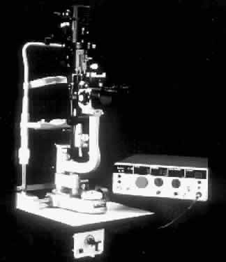
Practical considerations and differences in performing PDT treatment via the slit lamp versus thermal laser photocoagulation are the use of lower irradiance and power levels, longer durations of light application, and increased spot sizes. Because the duration of light ranges from seconds to minutes, proper alignment of the activating light beam must be maintained. Because the intensity of the light therapy is low, patients appear to tolerate the prolonged light treatment. In patients unable to fixate because of poor vision in the fellow eye, retrobulbar anesthesia for akinesia occasionally may be necessary. Typical powers required for PDT range from 5 to 50 mW, covering beam diameters of 0.5 to 3 mm spot sizes. Table 2 provides definitions and abbreviations for light terminology used in the treatment.


TABLE 69C-2. Light Terminology: Definitions and Abbreviations

  Joule (J): Unit of energy equivalent to 1 watt-second.
  Light dose: The total amount of energy given per unit area of surface treated. It is determined by multiplying power density (W/cm2) and treatment duraction (sec). It is expressed in J/cm2.
  Milliwatt (mW): Power equivalent to 1 watt/1000. A watt is a unit of power equivalent to the amount of energy (J) given off each second (sec).
  Power density: The power density, also known as irradiance, is defined as the power (mW) delivered to the tissue divided by the area (cm2) of the tissue being irradiated. It is determined by dividing the light dose (J/cm2) by the treatment duration (sec). It is expressed in mW/cm2.
  Power output: Power of light emitted through an optical fiber. It is a measurement of the rate at which energy is delivered. The number of watts (W) equals the amount of energy (J) given off each second (sec).
  Treatment time: The duration of time required for the light treatment; expressed in seconds (sec). It is equivalent to the light dose (J/cm2) divided by the power density (mW/cm2).
  Treatment area: Total surface area exposed to light during photodynamic therapy treatment. It is equivalent to π(radius)2 and is expressed as square centimeters (cm2).

  Radius (r): Radius of the light field. It is expressed in centimeters (cm).
  Diameter (d): Diameter of the light field. It is expressed in centimeters (cm).
  Area (A): Area of the light field. It is equivalent to π(radius)2 and is expressed as square centimeter (cm2).


  Wavelength: The wavelength is the distance between corresponding points on successive waves. It is expressed in nanometers (nm).


Back to Top
PHOTOSENSITIZERS
Administration of the photosensitizer, followed at the appropriate time by light application at a specific wavelength, can result in effective therapeutic treatment of the targeted tissue. The factors governing the effectiveness of a given photosensitizer are multifactorial and depend in part on its physiochemical and photophysical properties. As mentioned previously, the absorption spectrum of the drug dictates the wavelength of the illumination beam that may be used for PDT. In general, the wavelength selected coincides with an absorption maximum of the photosensitizer. Other important considerations include the metabolism, pH, pharmacokinetics, and route of administration of the photosensitizer. Photosensitizers, once administered to a patient, also exhibit a relative affinity for vascular and hyperproliferating tissues. This relatively selective accumulation, as well as the capacity for light activation, provides the basis for PDT over nonselective therapies such as thermal laser photocoagulation.

Several major classes of photosensitizers have been used in vitro and in animal models for mechanistic studies of photodynamic injury and to develop an optimal agent for human disorders. Among these agents, tetrapyrroles, phthalocyanines, bezophenoxazines, and xanthenes have been used for ophthalmic applications.17

Photofrin (porfimer sodium) is the photosensitizer that has been studied most extensively in medical applications of PDT, particularly for the treatment of a variety of cancers.18 Photofrin and its predecessor, hematoporphyrin derivative, are prepared from hematoporphyrin and essentially are complex mixtures of oligomeric esters and ethers of hematoporphyrin.

Despite the relatively good success of Photofrin PDT in treating various cancers of the bladder, lung, and esophagus, disadvantages with this first-generation photosensitizer have spawned an interest in producing more photodynamically efficient photosensitizers with less cutaneous phototoxicity. Photofrin has several drawbacks including: 1) there is a transient-induced photosensitivity of the skin for 6 to 8 weeks after injection; 2) there is a less than optimum activation wavelength (630 nm) for maximizing tissue penetration; 3) there is a relatively low extinction coefficient (2,800 M-1 cm1) at the activating wavelength; and 4) it is a multicomponent mixture of different photosensitizers.18

In recent years, a number of second-generation photosensitizers have been developed for use in PDT. Both hydrophobic and hydrophilic photosensitizers are being developed and proposed for treatment. Developments made in drug carrier systems, such as liposomes, lyophilization, and lipid emulsions, have made concerns of hydrophobic drug delivery less important while possibly offering some advantages over hydrophilic compounds.19 Two hydrophobic second-generation photosensitizers that have undergone extensive clinical testing are benzoporphyrin derivative (BPD) and Purlytin (tin ethyl etiopurpurin; SnET2).

BPD is a modified porphyrin formulated in liposomes that has an absorption maximum at about 690 nm and is phototoxic in vivo. It possesses a fast pharmacokinetic profile and produces minimal cutaneous photosensitivity at therapeutic doses.20 It currently is the subject of Phase III clinical trials for the treatment of exudative AMD, discussed in the following paragraph.20

Purlytin is a hydrophobic molecule that is formulated in an isotonic, iso-osmotic lipid emulsion suitable for intravenous use. It is a chlorin-type photosensitizer that has shown minimal cutaneous toxicity in nonclinical and clinical studies despite having a longer pharmacokinetic relative to BPD. Its formulation in a lipid emulsion facilitates association with lipophilic membranes, for example, endothelial membranes. Purlytin PDT is in multicentered clinical trials for the treatment of CNV secondary to AMD, as well in preclinical phases of development for corneal neovascularization and glaucoma.21–24

Back to Top
APPLICATIONS
Photodynamic therapy for ocular tumors has been used for more than 15 years for both malignant melanoma and retinoblastoma.25–27 The concentration of photodynamically active dye in proliferating endothelial cells producing a secondary necrosis after light therapy has produced significant tumor regressions. This response has led to the expansion of this therapy to nontumor neovascular proliferations in the eye.

Neovascularization, a complex response of vessels precipitated by an imbalance between stimulators and inhibitors of angiogenesis, is associated commonly with several ocular disorders, including AMD and diabetic retinopathy.28,29 Both conditions have a significant association with visual impairments and blindness. Presently, there are only preventative measures taken by clinicians to slow the progression of neovascularization. The ideal therapeutic modality would provide for highly selective and finely controlled destruction of the involved blood vessels and/or area while minimizing damage to adjacent normal tissues.

The potential for PDT to produce selective closure of ocular neovascularization with decreased or minimal collateral damage to other structures has been investigated in several experimental studies using both hydrophilic and hydrophobic photosensitizers. One of the first applications of using light-activated pharmaceuticals for neovascularization in the eye was reported in 1987.30 Closure of CNV in the subhuman primate model of Ryan31 was first demonstrated with subthreshold argon green PDT using dihematoporphyrin ether.30 In these preliminary experiments, closure of the subretinal and choroidal neovascular complex was seen to be produced by induced fibrin-platelet and erythrocyte plugs occluding the neovascular complex. Preservation of the choriocapillaris, retinal pigment epithelium, and overlying retina was found on electron microscopic evaluation post-PDT.

Miller and colleagues20,32,33 demonstrated the potential of BPD for occluding CNV in monkeys and humans. Choroidal neovascular membranes were treated with 690 nm light shortly after photosensitizer injection, resulting in subsequent occlusion of the neovascular membrane. Purlytin also has shown promise in the treatment of CNV and corneal neovascularization.21,22,24 Figures 2 to 3 demonstrate fluorescein angiographic documentation of initially successful PDT with Purlytin. The neovascular vessels in the choroid are seen to be in a subfoveal location (see Fig. 2). After systemic injection of Purlytin and photosensitization with 664 nm red light, complete occlusion of the neovascular vessels is seen in Figure 3.

Fig. 2. Fluorescein angiogram with the white arrows demarcating the borders of the hyperfluorescent choroidal neovascular complex in a eye with age-related macular degeneration before photodynamic therapy.

Fig. 3. Fluorescein angiogram of the same eye as Figure 2, 1 week after photodynamic therapy. The white arrows demarcate the treatment zone. There is an abscence of the hyperfluorescent neovascular complex previously seen in Figure 2.

In Phase I dose escalation clinical trials of Purlytin PDT, 89% of treated eyes showed evidence of treatment response within 7 days of therapy. Rapid resolution of subretinal fluid and improvement of visual acuity was observed in more than two thirds of the eyes responding with complete closure of the CNV in the first week after treatment. Despite angiographic staining of CNV 3 months after treatment, visual acuity remained at baseline or better for 64% of the eyes. This indicates recoverable photoreceptor and retinal pigment epithelial function.

Back to Top
CONCLUSIONS
Photodynamic therapy has been used for the treatment of ocular tumors and, more recently, neovascularization in both nonclinical and clinical settings. Initial clinical trials of the therapy for CNV show early encouraging responses. PDT produces selective closure of neovascular membranes without collateral damage to neurosensory retina and a potential concomitant preservation of visual acuity. Further randomized, controlled clinical trials may demonstrate the benefit of PDT in managing active, proliferating neovascularization.
Back to Top
REFERENCES

1. Wilson BC, Patterson MS: The physics of photodynamic therapy. Phys Med Biol 31:327, 1986

2. Jesionek A, Tappeiner VH: Zur Behandlung der Hautcarcinomi mit fluorescierenden Stoffen. Muench Med Wochenschr 47:2042, 1903

3. Hausmann W: Die sensibilisierende wirkung des hamatoporphyrins. Biochem Z 30:276, 1911

4. Auler H, Banzer G: Untersuchengen uber die rolle der porphyrine bei geschwulstkraken menschen und tieren. Z Krebsforsch 53:65, 1942

5. Figge FHJ, Weiland GS, Manganiello LOJ: Cancer detection and therapy: Affinity of neoplastic, embryonic, and traumatized tissues for porphyrins and metalloporphyrins. J Proc Soc Exp Biol Med 68:640, 1948

6. Lipson RL, Baldes EJ: The photodynamic properties of a particular hematoporphyrin derivative. Arch Dermatol 82:508, 1960

7. Kelly JF, Snell ME: Hematoporphyrin derivative: A possible aid in the diagnosis and therapy of cancer of the bladder. J Urol 115:150, 1976

8. Dougherty TJ, Grindley GB, Fiel R et al: Photoradiation therapy II: Cure of animal tumors with hematoporphyrin derivative and light. J Natl Cancer Inst 55;115, 1975

9. Fernandez JM, Bilgin MD, Grossweiner LI: Singlet oxygen generation by photodynamic agents. J Photochem Photobiol B Biol 37:131, 1997

10. Moan J: On the diffusion length of singlet oxygen in cells and tissues. J Photochem Photobiol B 6:343, 1990

11. Peng Q, Moan J, Neland JM: Correlation of subcellular and intratumoral photosensitizer localization with ultrastructural features after photodynamic therapy. Ultrastruct Pathol 20:109, 1996

12. Henderson BW, Dougherty TJ: How does photodynamic therapy work. Photochem Photobiol 55:145, 1992

13. Reed MWR, Miller FN, Wieman TJ et al: The effect of photodynamic therapy on the microcirculation. J Surg Res 45:452, 1988

14. Simpson ER, Wilson BC, Corriveau C et al: Thermal damage and hematoporphyrin derivative sensitized photochemical damage in laser irradiation of rabbit retina. Lasers Med Sci 2:33, 1987

15. Waldow SM, Dougherty TJ. Interaction of hyperthermia and photoradiation therapy. Rad Res 97:380, 1984

16. Waldow SM, Henderson BW, Dougherty TJ: Hyperthermic potentiation of photodynamic therapy employing Photofrin I and II: Comparison of results using three animal models. Lasers Surg Med 7:12, 1987

17. Haimovici R, Miller JW, Gragoudas ES: Photodynamic therapy for ocular tumors and neovascularization. In Benson WE, Ciscas G, Katz LJ (eds): Current Techniques in Ophthalmic Laser Surgery, p 135. Philadelphia: Current Medicine, 1994

18. Dougherty TJ. Photochemistry in the treatment of cancer. In Volman D, Hammond G, Neckers D (eds): Advances in Photochemistry, p 275. New York: John Wiley & Sons, 1992

19. Lin CW: Selective localization of photosensitizers in tumors: A review of the phenomenon and possible mechanisms. In: Kessel D (ed): Photodynamic Therapy of Neoplastic Disease, p 79. Boca Raton: CRC Press Inc, 1990

20. Miller JW, Walsh AW, Kramer M et al: Photodynamic therapy of experimental choroidal neovascularization using lipoprotein delivered benzoporphyrin. Arch Ophthalmol 113:810, 1995

21. Primbs GB, Casey R, Lin GC et al: Photodynamic therapy of corneal neovascularization using tin ethyl etiopurpurin (SnET2). Invest Ophthalmol Vis Sci 38:S702, 1997

22. Baumal CR, Puliafito CA, Pieroth L et al: Photodynamic therapy (PDT) of experimental choroidal neovascularization with tin ethyl etiopurpurin. Invest Ophthalmol Vis Sci 37:S122, 1996

23. Hill RA, Crean DH, Doiron DR et al: Photodynamic therapy for antifibrosis in a rabbit model of filtration surgery. Ophthalmic Surg Lasers 28:574, 1997

24. Peyman GA, Moshfeghi DM, Moshfeghi A et al: Photodynamic therapy of choriocapillaris using tin ethyl etiopurpurin (SnET2). Ophthalmic Surg Lasers 28:409, 1997

25. Murphree AL, Cote M, Gomer CJ: The evolution of photodynamic therapy in the treatment of intraocular tumors. Photochem Photobiol 46:919, 1987

26. Gomer CJ, Jester JV, Razum NJ et al: Photodynamic therapy of intraocular tumors: Examination of hematoporphyrin derivative distribution and long-term damage in rabbit ocular tissue. Cancer Res 45:3718, 1985

27. Bruce RA: Photoradiation therapy for choroidal malignant melanoma. In McCaughan JS (ed): A Clinical Manual: Photodynamic Therapy of Malignancies. Austin: RG Landes Co, 1993

28. Bressler SB, Bressler NM, Fine SL et al: Natural course of choroidal neovascular membranes within the foveal avascular zone in senile macular degeneration. Am J Ophthalmol 93:157, 1982

29. Patz A: Clinical and experiemntal studies on retinal neovascularization. Am J Ophthalmol 94:715, 1982

30. Thomas E, Langhofer M: Closure of experimental subretinal neovascular vessels with dihematoporphyrin ether augmented argon green laser photocoagulation. Photochem Photobiol 46:881, 1987

31. Ryan SJ: Subretinal neovascularization: Natural history of an experimental model. Arch Ophthalmol 100:1804, 1982

32. Kramer M, Miller JW, Michaud N et al: Liposomal BPD verteporfin photodynamic therapy: Selective treatment of choroidal neovascularization. Ophthalmology 103:427, 1996

33. Husain D, Miller JW: Photodynamic therapy of exudative age-related macular degeneration. Semin Ophthalmol 12:14, 1997

Back to Top