Chapter 69B
The Role of Lasers in Glaucoma Therapy
JACOB WILENSKY
Main Menu   Table Of Contents

Search

LASER IRIDECTOMY
LASER TRABECULOPLASTY
TREATMENT IN NEOVASCULAR GLAUCOMA
LASER SCLEROSTOMY
LASER IRIDOPLASTY
LASER CYCLOTHERAPY
LASER SUTURE LYSIS
OTHER LASER APPLICATIONS
REFERENCES

Since their initial utilization in the 1970s, lasers have been playing an ever-increasing and constantly changing role in the surgical treatment of glaucoma. Rapid developments in laser technology and instrumentation have led to new treatment techniques as well as refinements of old ones. Because of the rapid changes, it is difficult to stay abreast of this dynamic field. This indicates the current state of a number of the more widely used glaucoma laser therapies and to indicate some of the emerging techniques that may achieve greater utilization in the future.
Back to Top
LASER IRIDECTOMY
Laser iridectomy was one of the first laser techniques to achieve widespread application in glaucoma. It was initially performed using argon lasers. Creation of iridectomies with these continuous-wave, low-energy (500 to 1000 mW), long-duration (0.1 to 0.2 seconds) lasers was difficult and required precise technique. The introduction of high-energy, short-duration (nanosecond), pulsed Nd:YAG lasers converted this procedure into one that most trained ophthalmologists could easily perform. As a result, laser iridectomy has become the standard technique for treating papillary block angle-closure glaucoma except in those situations where lack of corneal clarity precludes delivery of the laser energy to the iris.1

Creation of a through-and-through hole in the iris is made easier by administration of a drop of 1% pilocarpine 15 to 20 minutes prior to the treatment. The resulting miosis will place the iris on stretch and enhance pupillary block causing more fluid to collect between the iris and the lens in the posterior chamber. Use of a high plus contact lens such as a Wise or Abraham Lens focuses and concentrates the laser energy at the point of treatment, and then more rapidly defocuses the energy so that if any of it gets through the iridectomy, it will not be sufficiently intense enough to cause any retinal burns. If bleeding occurs, placing pressure on the globe with the contact lens for 30 to 60 seconds usually will be sufficient to stop it.

Recently, a second indication for laser iridectomy has been suggested. It has been hypothesized that in pigmentary glaucoma reverse pupillary block leads to abrasion of pigmented epithelial cells on the back of the iris releasing pigment granules that then plug the trabecular meshwork and may cause elevated intraocular pressure (IOP).2 It has been demonstrated with ultrasonic high-resolution biomicroscopy (UBM) that laser iridectomy eliminates this reverse pupillary block and reduces concavity of the peripheral iris.3 It is believed that this will halt the release of additional pigment granules and as the existing pigment is cleared from the trabecular meshwork the IOP may decline. There have not yet been any long-term, controlled, prospective studies to validate the value of this treatment so its use is still the subject of much debate. If it is to be used, it probably should be reserved for eyes where there is noticeable posterior concavity of the peripheral iris.

Back to Top
LASER TRABECULOPLASTY
Laser trabeculoplasty is probably the most widely used laser technique for the treatment of glaucoma. In spite of its very extensive use since its introduction by Wise and Witter4 in 1979, there are still many unanswered questions regarding the mechanism(s) by which it lowers IOP. While a number of different types of lasers (e.g., pulsed, continuous wave) and different wavelength laser energies have been shown to be effective in lowering IOP, the large majority of experience with this technique has been with continuous-wave argon lasers.5

Laser trabeculoplasty is effective in approximately two-thirds to three-quarters of patients with primary open-angle glaucoma. In these eyes, it lowers the IOP approximately 25%. It also is beneficial in some secondary open-angle glaucomas such as exfoliation syndrome glaucoma and pigmentary glaucoma, in eyes that have had previous filtering surgery, and in eyes that have had surgical or laser iridectomies because of acute angle-closure glaucoma and have residual elevated IOP with open angles. There is still some question as to whether it is as effective in pseudophakic eyes as in phakic ones, but is generally effective in these eyes as well. It has been well documented that it is more effective in older patients than younger ones with the notable exception of pigmentary glaucoma, where it is effective in young individuals. There have been some reported cases of glaucoma made worse by laser trabeculoplasty in patients with juvenile glaucoma and there have been mixed results in angle-recession glaucoma.

There have been two major multicenter collaborative studies that have been conducted to help define the role of laser trabeculoplasty in glaucoma therapy. The first of these studies was the Glaucoma Laser Trabeculoplasty Trial (GLT) in which laser trabeculoplasty was compared to medical therapy as the initial treatment for primary open-angle glaucoma. The results of this study showed that laser trabeculoplasty as initial therapy was both safe and effective in the treatment of open-angle glaucoma, but in a large majority of patients it was not sufficient by itself to lower the intraocular pressure sufficiently to control the disease, and additional medical therapy was required.6 There was a trend to suggest that the laser-first eyes might have done slightly better than the medicine-first eyes but these differences were not statistically significant.

The second study was the Advanced Glaucoma Intervention Study (AGIS). In this study, patients with medically uncontrolled open-angle glaucoma were randomly assigned to either laser trabeculoplasty or surgical trabeculectomy for the treatment of their glaucoma. There was stratification within the study population based on race, and there was some evidence that there was a racial difference in the response to treatment with the black patients initially showing a slightly better visual field response to the trabeculoplasty first, while the white patients responded better to trabeculectomy first. In both racial groups, however, lower IOP levels were achieved by filtering surgery and fewer patients required additional surgical treatment in the trabeculectomy-first eyes.7

The use of laser trabeculoplasty has declined somewhat in recent years, probably as a result of several factors. One has been the introduction of more potent topical ocular hypotensive eyedrops, namely the prostaglandin-type agents. These have a greater pressure-lowering effect than laser trabeculoplasty and have a better side effect profile than the β-blockers that had been the mainstay of topical glaucoma therapy previously. A second factor was the recognition of the fact that the effect of laser trabeculoplasty seemed to decrease over time, such that approximately half of the patients treated have returned their IOP to pretreatment levels after an average of approximately 5 years.

In 2001, a new technique for laser trabeculoplasty was introduced and has been named selective laser trabeculoplasty (SLT). It utilizes a frequency doubled, pulsed Nd:YAG laser.8 In this technique, there is much less thermal damage to the trabecular meshwork than there is with argon laser trabeculoplasty. The initial IOP response to SLT appears to be approximately the same as that of argon laser trabeculoplasty (ALT). The proponents of this technique claim that because of the lack of thermal damage to the trabecular meshwork, it can be repeated over time with renewed effectiveness and without damage to the trabecular meshwork. There are several reports of case series where patients with previous ALT whose pressure had come back up were treated successfully with SLT. Long-term follow-up studies will be needed to define what the role of SLT will be in the future.

When performing laser trabeculoplasty, a mirrored Goldmann gonioscopy lens is used to focus the laser energy in the angle. The laser spot is aimed at the junction of the pigmented and nonpigmented trabecular meshwork. With ALT, a 50-μm spot is used and the duration of the burn is 0.1 second. Many adjust the laser energy to a level where they see a blanching of the trabecular meshwork but without bubble formation (usually 500 to 1000 mW). The author routinely uses 800 mW on all eyes whether there is blanching or not. With SLT and with diode laser trabeculoplasty, there is less visual response to the laser treatment so one cannot treat to a reaction point. Some initially treat 180 degrees of the meshwork with 50 laser applications and then treat the remaining 180 degrees with an additional 50 laser applications approximately a month later if there has not been a sufficient reduction in IOP. Others treat the entire 360 degrees of the angle with 100 applications in a single session.

Approximately 10% to 20% of eyes will have an acute rise in IOP 1 to 3 hours after the trabeculoplasty so the IOP needs to be monitored immediately posttreatment. The risk of such IOP spikes can be reduced by pretreating the eye with an α2-adrenergic agent such as apraclonidine or brimonidine prior to the laser treatment. Most ophthalmologists prescribe a topical anti-inflammatory agent such as a corticosteroid agent to their patients for several days after the treatment. The majority of the IOP reduction will be seen by 1 week but some additional effect may be noted over the following several weeks.

Back to Top
TREATMENT IN NEOVASCULAR GLAUCOMA
Lasers play a major role in the treatment of neovascular glaucoma; however, the treatment here is used more by retina specialists than by general ophthalmologists or glaucoma specialists. The main treatment technique in this area is panretinal photocoagulation (PRP) therapy. The high risk of developing rubeosis iridis and neovascular glaucoma after an ischemic central retinal vein occlusion has been well documented. Some individuals still recommend prophylactic PRP when extensive retinal ischemia has been documented by fluorescein angiography, however, most specialists delay the PRP until evidence of rubeosis becomes manifest. In the rubeosis that is seen with diabetes mellitus, there is fairly wide agreement that PRP should be reserved until there is evidence of neovascularization in the angle, because many people with diabetes with just rubeosis on the iris elsewhere, do not progress to angle closure. When PRP is performed, there is usually regression of rubeosis and more definitive forms of glaucoma therapy, such as trabeculectomy or shunt procedures can more safely and effectively be used if needed.

In the past, an additional form of laser therapy for rubeosis iridis was utilized. Abnormal vessels on the iris, particularly vessels bridging the angle were ablated with focal photocoagulation. This technique has been largely abandoned today because it does not work.

Back to Top
LASER SCLEROSTOMY
Numerous attempts have been made to use laser energy to create openings through the scleral wall from the anterior chamber to the subconjunctival space. Many of these have attempted to use noninvasive techniques so as to reduce the risk of endophthalmitis or other operative complications. Others have been designed with the idea that the opening created using a selected laser energy may have some advantages over those created with other surgical techniques.

One group of these procedures were ab interno laser sclerostomies that attempted to create an opening from the anterior chamber into the subconjunctival space by pulsing laser energy through the cornea using a special gonioscopy lens and focusing on the angle. While some limited success was reported, these procedures have not withstood the test of time and are not presently being utilized.9 Similarly, techniques were developed using endoprobes that were placed across the anterior chamber and used to create sclerostomies by opening channels from the anterior chamber to the subconjunctival space. Again, the advantages or benefits of these procedures have not been sufficient and these techniques have been largely abandoned.

A second group of procedures used external probes that were introduced into the subconjunctival space, advanced to the limbus and then used to create a channel through the sclera into the anterior chamber. The most extensive experience with this technology utilized a homium laser.10 For a while, the technique was used fairly extensively. Unfortunately, the high rate of iris plugging of the sclerostomy and other complications also has led to this technique largely being abandoned.

Back to Top
LASER IRIDOPLASTY
Continuous-wave argon laser energy when applied to the iris in relatively low-energy (200 to 500 mW), long-duration (0.2 to 0.5 seconds) burns causes contracture of the iris stroma that can change the configuration of the iris and/or pupil. This type of treatment can be used therapeutically in several different ways. The first of these is to flatten a prominent peripheral iris roll in patients with plateau iris configuration. The laser treatment widens the approach to the angle and draws the peripheral iris away from the cornea. The same treatment may be done to facilitate laser trabeculoplasty in eyes with a narrow approach to the angle. The second of these is a similar treatment in eyes with acute angle-closure glaucoma. Occasionally because of corneal edema or extreme shallowness of the anterior chamber, YAG iridectomies cannot be accomplished. In these eyes, iridoplasty pulls the iris away from the angle reestablishing outflow through the trabecular meshwork.11 Once the IOP has been reduced, the definitive laser iridectomy can be performed. A third use of iridoplasty is to enlarge a miotic pupil that has resulted from long-term miotic therapy. With the reduced use of miotics to treat glaucoma in recent years, this indication has diminished greatly. A fourth use of laser iridoplasty is laser synechiolysis.12 In some eyes with partial chronic angle closure, laser energy can be used to lyse peripheral anterior synechiae and open the angle. Although the early reports of laser synechiolysis employed argon lasers, I now recommend the Nd:YAG laser. The laser is focused on the peripheral iris just in front of the angle beginning at the site where the synechia begin. The shock wave from the laser pulse will pull the iris free from its attachment to the trabeculum adjacent to the treatment point. This process is repeated across the width of the synechia.
Back to Top
LASER CYCLOTHERAPY
A growing application of lasers to treat glaucoma is laser cyclotherapy.13 In this procedure, laser energy is used to damage the ciliary processes in an attempt to reduce the amount of aqueous being produced sufficiently to reduce the IOP to a desired level. Because a minimum amount of aqueous production is necessary to maintain adequate nutrition of the lens and cornea, if too much of the ciliary body is damaged, hypotony and/or phthisis may result. Moreover, there is a certain attrition of the ciliary body with aging with corresponding decrease in aqueous production. A ciliary body that provided adequate aqueous production at one time may not be sufficient years or decades later.

There are three approaches to laser cyclodestruction. The first of these is transpupillary focal photocoagulation of the ciliary processes.14 In many eyes with neovascular glaucoma, the iris is pulled forward sufficiently such that the ciliary processes are visible with a gonioscopy lens. The same may be true in some eyes with widely dilated pupils or in eyes with large iridectomies. In these eyes, the anterior face of the ciliary processes can be photocoagulated with an argon laser. The laser is set at 1000 mW for 0.2 seconds using a 100-μm spot, and as many processes as are visible are treated. Because the more posterior aspects of the processes are not visible and therefore not treated, the results of this treatment are very unpredictable and the eye may have to be treated several times. This technique is not very widely used.

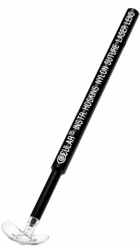
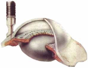
The second approach is transscleral laser cyclotherapy. In this technique laser energy is pulsed through the sclera to the ciliary body where the laser energy is absorbed by the melanin and converted into heat that damages the ciliary processes. At present, two different types of lasers are being used for this treatment: diode lasers and Nd:YAG lasers. Our experience and that of others has been that these two types of lasers are quite similar in both efficacy and side effects.15,16 In the beginning, the Nd:YAG lasers were used in a noncontact mode where the aiming beam was focused on the conjunctiva, but the laser energy was retrofocused deeper into the eye. Then a fiber-optic delivery system was developed with a synthetic sapphire tip that was placed on the eye that allowed contact administration of the laser energy. Similarly, a delivery system for the diode laser was developed with a special handle known as the G-probe that was especially designed for placement of the probe at the limbus (Fig. 1).

Fig. 1 The G-probe is used to perform transscleral laser cyclotherapy. The probe is placed at the limbus and the diode energy passes through the sclera and is absorbed by pigment in the ciliary body. (Photo compliments of Iridex Corp, Mountain View, CA)

The Nd:YAG laser uses slightly higher energy (6 to 8 W), shorter duration (0.7 second) pulses. Approximately 32 laser applications are evenly spaced around the 360 degrees of the limbus avoiding the 3 and 9 o'clock meridia. The tip of the probe is centered approximately 1 mm behind the limbus. After the treatment, dexamethasone solution is injected subconjunctivally and the eye is patched until bed time. Glaucoma medications are continued unchanged and topical corticosteroids are begun the next day. My patients rarely experience significant posttreatment pain, and I do not routinely prescribe an analgesic. Also, unlike others, I do not routinely prescribe cycloplegics. The patient is seen approximately a week later and the glaucoma medication is adjusted depending on the IOP response to the treatment. The topical corticosteroids are tapered based on the inflammatory response. In my experience, a majority of the eyes treated will experience a rebound in the IOP over weeks to months and the treatment will have to be repeated.

The treatment protocol with the diode laser is similar with some minor differences. The laser applications are longer in duration (1.5 to 2.5 seconds) but use lower energy (1.5 to 2.25 W) and an average of 25 applications are placed. The G-probe is placed with the foot plate at the limbus. The postoperative management is the same as with the Nd:YAG laser.

The third approach to laser cyclotherapy is focal photocoagulation of the ciliary processes using an endoprobe. The endoprobe contains one fiber-optic pipe that allows visualization of the tissues and a second one to deliver the diode laser energy. In general, there are two approaches that can be used. In the first approach, the endoprobe is entered into the anterior chamber via a limbal incision and then passed through the pupil behind the iris until the ciliary processes are visualized and then photocoagulated.17 This approach is particularly used in combination with phacoemulsification and posterior chamber intraocular lens (PC IOL) insertion. The second approach is posteriorly through the pars plana and is usually performed as part of a vitrectomy.18 The laser energy is adjusted to cause blanching and shrinkage of the processes but no vaporization of tissue. Usually approximately 180 degrees of the ciliary body is treated.

In the past, cyclodestructive procedures have been reserved for end-stage glaucoma eyes after everything else has failed. Recently, several reports have been published from Third World countries where is has been used earlier in the disease process.19 With short to medium follow-up, the results have suggested that it may be a reasonable alternative to trabeculectomy. Several of the early series of laser cyclotherapy cases with medium to long-term follow-up reported unexplained decreases in visual acuity and suggested that this was a side-effect of the laser treatment.20 Long-term follow-up of trabeculectomy eyes in the AGIS study and other case series have shown that some of these eyes have similar decreases in visual acuity.6,19 Thus, there is a growing question as to whether this side effect is specific to the laser therapy. I have performed laser cyclotherapy on patients with good visual acuity (20/80 or better) and the majority have maintained this vision after up to 6 years of follow-up.21 A second concern with laser cyclotherapy is the risk of sympathetic ophthalmia. Several cases of sympathetic ophthalmia in fellow eyes were reported following noncontact Nd:YAG laser cyclotherapy when this technique was first used,22 but no additional reports have appeared in spite of the great increase in laser cyclotherapy.

Back to Top
LASER SUTURE LYSIS
With the widespread use of antimetabolites such as 5-fluorouracil and mitomycin C as adjuncts to glaucoma filtering surgery, there has been a trend toward closing the scleral flap more tightly than was the case previously. Then during the first few days or weeks after the surgery, the scleral flap sutures are released one by one in an attempt to titrate the amount of filtration. Laser suture lysis is one of the popular methods of achieving this.23 Several special lenses have been designed to help facilitate this procedure (Fig. 2). The lenses help keep the upper lid out of the way, compress and blanch the overlying conjunctiva, and focus the laser energy on the suture. Most commonly, argon lasers are used for this procedure. The laser is set at approximately 500 mW for 0.05 seconds and a 100-μm spot is used. The laser must be focused carefully because if the focal plane is too anterior, it can burn a hole in the conjunctiva causing a bleb leak. If there is subconjunctival hemorrhage or excessive Tenon's capsule as is seen in some African American patients, it may not be possible to visualize the sutures sufficiently to perform this procedure.

Fig. 2 Illustrations of the Hoskins suture lysis lens. There is a high plus button in the center to magnify the suture and the flange helps retract the upper lid. (Courtesy of Ocular Instruments, Belleview, WA)

Back to Top
OTHER LASER APPLICATIONS
Hypotony caused by cyclodialysis clefts is not a form of glaucoma (although the intentional creation of such clefts has been used therapeutically to treat glaucoma), but the rare patients with this condition are often referred to glaucoma specialists for treatment because of their focus on IOP, gonioscopy, and laser therapy of the angle. Argon lasers have been used to create burns in the angle that cause scarring that results in closure of the cleft.24 Moderate power (approximately 500 mW), 100-μm sized burns of 0.1- to 0.2-second duration are delivered to the iris root beginning at the junction of the dialyzed and intact iris and then moving toward the center of the dialysis. The eye is cyclopleged and no steroids are used post treatment. Frequently, multiple treatment sessions are required to obtain closure of the dialysis. Recently, there has been a report of the use of transscleral diode laser for this purpose.

Various lasers have been used to treat some of the complications of filtration surgery.25–27 Among these have been the use of argon laser to treat leaking blebs, Nd:YAG lasers to treat dysasthetic or over filtering blebs, and various lasers to try to save failing blebs caused by fibrosis within the bleb or because of iris, ciliary processes, or vitreous incarcerated in the internal opening of the trabeculectomy wound. None of these applications has achieved extensive utilization to date.

There also have been a growing number of reports of the use of lasers in conjunction with nonpenetrating filtering surgery.28 The lasers have been used both in deep dissection to help unroof Schlemm's canal and to perforate Descemet's membrane ab interno when IOP control has been insufficient. With the recent increased interest in deep sclerectomy-type procedures, the use of such laser applications may become more frequent in the future.

Back to Top
REFERENCES

1. American Academy of Ophthalmology: Laser peripheral iridectomy for pupillary-block glaucoma. Ophthalmology 101:1749, 1994

2. Campbell DG: Pigmentary dispersion and glaucoma: a new theory. Arch Ophthalmol 97:1667, 1979

3. Potash SD, Tello C, Liebmann JM, et al: Ultrasound biometry in pigment dispersion syndrome. Ophthalmology 101:332, 1994

4. Wise JB, Witter SL: Argon laser therapy for open-angle glaucoma: a pilot study. Arch Ophthalmol 97:319, 1979

5. Reiss GR, Wilensky JT, Higginbotham EJ: Laser trabeculoplasty. Surv Ophthalmol 35:407, 1991

6. The Glaucoma Laser Trial Research Group: The glaucoma laser trial (GLT) and glaucoma laser trial follow-up study: results. The Glaucoma Laser Trial Report Number 7. Am J Ophthalmol 120:718, 1995

7. The AGIS Investigators: The Advanced Glaucoma Intervention Study (AGIS): 4. Comparison of treatment outcomes within races. Ophthalmology 105:1146, 1998

8. Latina MA, Sibayan SA, Skin DH, et al: Q-switch 532-nm Nd:YAG laser trabeculoplasty (selective laser trabeculoplasty): a multicenter, pilot, clinical study. Ophthalmology 105:282, 1998

9. Pappas RM, Higginbotham EJ, Choe HS: Advances in laser sclerostomy: how far have we come? Ophthalmic Surg Lasers 28:751, 1997

10. Iwach AG, Hoskins HDJr , Mora JS, et al: Update on the subconjunctival THG:YAG (Homium) laser sclerostomy ab externo clinical trial: a 4-year report. Ophthalmic Surg Lasers 27:823, 1996

11. Lam DSC, Lai JSM, Tham CCY, et al: Argon laser peripheral iridoplasty versus conventional systemic medical therapy in treatment of acute primary angle-closure glaucoma: a prospective randomized controlled trial. Ophthalmology 109:1591, 2002

12. Wand M: Argon laser gonioplasty for synechial angle closure. Arch Ophthalmol 110:363, 1992

13. Pastor SA, Singh K, Lee DA, et al: Cyclophotocoagulation: a report by the American Academy of Ophthalmology. Ophthalmology 108:2130, 2001

14. Shields S, Stewart WC, Shields MB: Transpupillary argon laser cyclophotocoagulation in the treatment of glaucoma. Ophthalmic Surg 19:171, 1988

15. Youn J, Cox TA, Herndon LW, et al: A clinical comparison of transscleral cyclophotocoagulation with neodymium YAG cyclophotocoagulation. Arch Ophthalmol 111:484, 1993

16. Zweifach E, Wilensky JT, Hillman D, et al: A prospective randomized trial of diode and Nd:YAG lasers in treating glaucoma. Invest Ophthalmol Vis Sci 38:S170, 1997

17. Mora JS, Iwach AG, Gaffney MM, et al: Endoscopic diode laser cyclophotocoagulation with a limbal approach. Ophthalmic Surg Lasers 28:118, 1997

18. Lim JL, Lynn M, Capone AJr : Ciliary body endophotocoagulation during pars plana vitrectomy in eyes with vitreoretinal disorders and concomitant uncontrolled glaucoma. Ophthalmology 103:1041, 1996

19. Egbert PR, Fiadoyor S, Budenz DL, et al: Diode laser transscleral cyclophotocoagulation as a primary surgical treatment for primary open-angle glaucoma. Arch Ophthalmol 119:345, 2001

20. Youn J, Cox TA, Allingham RR, et al: Factors associated with visual acuity loss after noncontact transscleral Nd:YAG cyclophotocoagulation. J Glaucoma 5:390, 1996

21. Wilensky JT, Kammer J: Long-term visual outcome of transscleral laser cyclotherapy in eyes with ambulatory vision. Ophthalmology (in press).

22. Lam S, Tessler HH, Lam BL, et al: High incidence of sympathetic ophthalmia after contact and noncontact neodymium:YAG cyclotherapy. Ophthalmology 99:1818, 1992

23. Macker P, Buys Y, Trope GE: Glaucoma laser suture lysis. Br J Ophthalmol 80:398, 1996

24. Ormerod LD, Baerveldt G, Sunalp MA, et al: Management of the hypotonous cyclodialysis cleft. Ophthalmology 98:1384, 1991

25. Geyer O: Management of large, leaking and inadvertent filtering blebs with the neodymium:YAG laser. Ophthalmology 105:983, 1998

26. Oh Y, Katz LJ: Indications and technique for reopening closed filtering blebs using the Nd:YAG laser—a review and case series. Ophthalmic Surg 24:617, 1993

27. Weber PA, Jones JH, Kapetansky F: Neodymium:YAG transconjunctival laser revision of late failing filtering blebs. Ophthalmology 106:2023, 1994

28. Verges C, Llevat E, Bardario J: Laser-assisted deep sclerectomy. J Cataract Refract Surg 28:758, 2002

Back to Top