Chapter 63
Principles of Ophthalmoscopy
AUGUST COLENBRANDER
Main Menu   Table Of Contents

Search

BASIC PRINCIPLES OF DIRECT OPHTHALMOSCOPY
FIELD OF VIEW IN DIRECT OPHTHALMOSCOPY
EXTENDING THE FIELD—INDIRECT OPHTHALMOSCOPY
FIELD OF VIEW IN INDIRECT OPHTHALMOSCOPY
IMAGING IN INDIRECT OPHTHALMOSCOPY
MAGNIFICATION IN DIRECT OPHTHALMOSCOPY
MAGNIFICATION IN INDIRECT OPHTHALMOSCOPY
COMPENSATION FOR REFRACTIVE ERROR
DESIGN OF LENSES FOR INDIRECT OPHTHALMOSCOPY
A CLOSER LOOK AT ILLUMINATION AND REFLECTIONS
ILLUMINATION IN INDIRECT OPHTHALMOSCOPY
LATITUDE OF BEAM PLACEMENT
MONOCULAR INDIRECT OPHTHALMOSCOPY
BINOCULAR INDIRECT OPHTHALMOSCOPY
PERIPHERAL VIEWING
LOCALIZATION IN THE FUNDUS
MEASUREMENT OF FUNDUS LESIONS
OTHER ACCESSORIES OF THE DIRECT OPHTHALMOSCOPE
ILLUMINATION LEVELS IN INDIRECT OPHTHALMOSCOPY
SLIT-LAMP EXAMINATION OF THE FUNDUS
REFERENCES

No other single invention has shaped the evolution of ophthalmology like the invention of the ophthalmoscope has. Ophthalmoscopy was introduced by Hermann von Helmholtz in December of 1850.1,2 However, Jan Purkinje (known for the Purkinje images) had described the complete technique and published it in Latin in 1823,3 but his audience apparently was not yet ready and his publication went unnoticed. A quarter of a century later, however, the situation changed.

Like many other important inventions, the ophthalmoscope was not based on any radically new concepts. Rather, it combined the appropriate application of various known principles with a recognition of its potential impact and presentation to an appropriate audience. Under the leadership of men like Bowman in London, Donders in Holland, and von Graefe and von Helmholtz in Germany, ophthalmology was emerging as the first organ-based specialty in medicine. Bowman (1816 to 1892) is known for Bowman's membrane and for his work in anatomy and histology. Donders (1818 to 1889) clarified the principles of refraction and accommodation (1864) and defined visual acuity as a measurable quantity. His coworker Snellen developed the Snellen chart. In Berlin, Albrecht von Graefe (1828 to 1870) was a leader in stimulating the clinical application of new techniques and the careful documentation of new findings. He is remembered for Graefe's knife and Graefe's Archives (1854) (one of the first ophthalmic journals), and he founded the German Ophthalmological Society (Heidelberg, 1857). Several workers had tried to visualize the inside of the eye but had fallen short of putting it all together. Kussmaul (known for “Kussmaul's airhunger”) described the imaging principles in a thesis in 18454 but failed to solve the illumination problem. Cumming5 (1846) in England and Brücke6 (1847) in Germany had shown that a reflection from the fundus could be obtained by bringing the light source in line with the observer, but they failed to solve the imaging problem. Babbage,7 the English mathematician, reportedly constructed an ophthalmoscope in 1847, but his ophthalmologist friend did not recognize the importance and did not publish it until 1854, when von Helmholtz' instrument was well known.

In the fall of 1850, von Helmholtz tried to demonstrate the inside of the eye to the students in his physiology class. On December 6, he presented his findings to the Berlin Physical Society1; on December 17, he wrote to his father8:

I have made a discovery during my lectures on the Physiology of the Sense-organs, which was so obvious, requiring, moreover, no knowledge beyond the optics I learned at the Gymnasium, that it seems almost ludicrous that I and others should have been so slow as not to see it…. Till now a whole series of most important eye-diseases, known collectively as black cataract, have been terra incognita…. My discovery makes the minute investigation of the internal structures of the eye a possibility. I have announced this very precious egg of Columbus to the Physical Society at Berlin, as my property, and am now having an improved and more convenient instrument constructed to replace my pasteboard affair…

Helmholtz' monograph on ophthalmoscopy was published in 1851 and soon was widely circulated. The next year there were several important improvements contributed by other workers. Rekoss,9 von Helmholtz' instrument maker, added two movable disks with lenses for easier focusing. Epkens, working with Donders in Holland,8 introduced a perforated mirror for increased illumination. Ruete10 in Germany did the same and also developed the indirect method of ophthalmoscopy. With these basic components in place, future generations provided technical improvements. In 1913, Landolt11 listed 200 different types of ophthalmoscopes. The most important changes are related to the change from candle light to gas light, to external electric light and, finally, to built-in electric light sources. This chapter concentrates on currently available forms of ophthalmoscopy. For additional information on the history of ophthalmoscopy the reader is referred to references 8 and 12.

Although the older generation found it difficult to adapt to the new instrument, the younger generation did so eagerly. One of them was Eduard von Jaeger (1828 to 1884) from Vienna, best known for his print samples that were based on the print catalogue of the Vienna State Printing House. He was the son of a well-known ophthalmologist and an artistically gifted mother. In 1855, at the age of 27, he published his first atlas; he continued to add to his collection of authoritative fundus paintings until his death in 1884.13

Back to Top
BASIC PRINCIPLES OF DIRECT OPHTHALMOSCOPY
The basic principle of direct ophthalmoscopy is simple (Fig. 1). If the patient's eye is emmetropic, light rays emanating from a point on the fundus emerge as a parallel beam. If this beam enters the pupil of an emmetropic observer, the rays are focused on the observer's retina and form an image of the patient's retina on the observer's retina. This is called direct ophthalmoscopy.

Fig. 1. Imaging in direct ophthalmoscopy. If patient and observer are both emmetropic, rays emanating from a point in the patient's fundus will emerge as a parallel beam and will be focused on the observer's retina.

However, there is a problem with this method: Sufficient light for visualization of the fundus emerges only if the patient's fundus is properly illuminated. Because of the optics of the eye (Fig. 2), incident light reaches only the part of the fundus onto which the image of the light source falls. Conversely, only light from the fundus area onto which the observer's pupil is imaged reaches that pupil. The fundus can be seen only where the observed and the illuminated areas overlap; in the emmetropic eye this can happen only if the light source and the observer's pupil are aligned optically. Under normal conditions this does not happen, and the pupil normally appears black. How a reflection can be seen under abnormal conditions is discussed later in this text.

Fig. 2. The illumination problem in direct ophthalmoscopy. If the light source and the observer are not aligned optically, the observer views a part of the fundus that is not illuminated.

There are several ways in which optical alignment of the illuminating and observing beams can be accomplished (Fig. 3). Von Helmholtz solved the problem with a semireflecting mirror made up of several thin parallel pieces of glass (see Fig. 3A). Epkens and Ruete used a perforated concave mirror, which places illuminating light rays all around the observation beam (see Fig. 3B). A modification of this arrangement is used in the fundus camera. Most hand-held instruments now have a small mirror or prism (see Fig. 3C), which uses the lower half of the patient's pupil for illumination and the upper half for observation.

Fig. 3. Illumination methods in direct ophthalmoscopy. A. Illumination with semireflecting mirror (Helmholtz). B. Illumination with perforated mirror (Epkens, Ruete). C. Illumination with mirror or prism (modern).

Back to Top
FIELD OF VIEW IN DIRECT OPHTHALMOSCOPY
If the patient's fundus is properly illuminated, the field of view is limited by the most oblique pencil of light that can still pass from the patient's pupil to the observer's pupil (Fig. 4). In direct ophthalmoscopy the retinal point that corresponds to this beam can be found by constructing an auxiliary ray through the nodal point of the eye.11 The point farthest from the centerline of view that can still be seen is determined by the angle α, that is, the angle between this oblique pencil and the common optical axis of the eyes.

Fig. 4. Field limits in direct ophthalmoscopy. The maximum field of view is determined by the most oblique pencil of rays (shaded) that can still pass from one pupil to the other.

Angle α, and therefore the field of view, is increased when the patient's or the observer's pupil is dilated or when the eyes are brought more closely together.

The more peripheral pencils of light use ever-smaller parts of each pupil. This means that, even if the patient's fundus is uniformly illuminated, the luminosity of the fundus image gradually decreases toward the periphery, so that there is no sharp limitation to the field of vision. In practice, therefore, the effective field of vision is determined by the illuminating system not by the viewing system. Most ophthalmoscopes project a beam of light of about one disc diameter.

Back to Top
EXTENDING THE FIELD—INDIRECT OPHTHALMOSCOPY
Even with appropriate illumination, direct ophthalmoscopy has a small field of view. Figure 5 shows that of four points in the fundus, points one and four cannot be seen because pencils of light emanating from these points diverge beyond the observer's pupil. To bring these pencils to the observer's pupil, their direction must be changed (Fig. 6). This requires a fairly large lens somewhere between the patient's and the observer's eye. This principle was introduced by Ruete10 in 1852 and is called indirect ophthalmoscopy to differentiate it from the first method, in which the light traveled in a straight, direct path from the patient's eye to the observer.

Fig. 5. Limited field of view in the direct method. Peripheral pencils of light do not reach the observer's pupil.

Fig. 6. Extended field of view in the indirect method. The ophthalmoscopy lens redirects peripheral pencils of light toward the observer.

The use of the intermediate lens has several important implications that make indirect ophthalmoscopy more complicated than direct ophthalmoscopy.

The primary purpose of the ophthalmoscopy lens is to bend pencils of light toward the observer's pupil. Figure 6 also demonstrates one of the most characteristic side effects of this arrangement: Compared with the image in direct ophthalmoscopy, the orientation of the image on the observer's retina is inverted. For the novice, this often causes confusion in localization and orientation. Figure 6 further shows that in this arrangement the patient's pupil is imaged in the pupillary plane of the observer. In optical terms the pupils are in conjugate planes. This fact is useful later in this discussion.

Back to Top
FIELD OF VIEW IN INDIRECT OPHTHALMOSCOPY
The field of view in indirect ophthalmoscopy is determined by the rays emerging from the patient's eye that can be caught in the ophthalmoscopy lens. With optimal placement of the lens and of the observer's eye, the distance from the patient's eye to the lens is only slightly more than the focal length of the lens. (The exact distance will be calculated later.) The field of view, therefore, is determined by the ratio of lens diameter and focal length. This ratio can also be written as a product:
Lens diameter/Focal length = Lens diameter × dioptric power

This provides an easy formula for comparing the field of view of various lenses.

Given lenses of equal power, a larger lens provides a wider field of view. If lenses have equal diameters, a stronger lens provides a wider field of view; however, because stronger lenses often have a smaller diameter, a stronger ophthalmoscopy lens does not always provide a larger field. A 20-diopter (D) lens of 30 mm provides about the same field of view as a 30-D lens of 20 mm or as a 13-D lens of 45 mm (because 20 × 30 = 30 × 20 = ±13 × 45).

Back to Top
IMAGING IN INDIRECT OPHTHALMOSCOPY
Figure 6 shows that light emerging from the patient's fundus is directed toward the observer's eyes. It does not specify whether the observer sees a focused image or just an unstructured red reflex. Figure 7 traces the rays within one of the pencils of light from the patient's fundus to the observer's retina.

Fig. 7. Imaging in the indirect method. A pencil of rays (shaded) is traced from the patient's fundus to the observer's retina. An intermediate, inverted image of the patient's fundus is formed in the focal plane of the ophthalmoscopy lens. The observer must accommodate on this image.

If the patient is emmetropic, the pencils emerging from the eye are composed of parallel rays, but this changes once the pencils pass through the ophthalmoscopy lens. In fact, because the rays within each pencil enter the ophthalmoscopy lens with zero vergence, they are brought to a focus in the focal plane of the ophthalmoscopy lens. Proceeding beyond that point, the rays within each pencil are divergent.

Considering all pencils emerging from the patient's eye together, an aerial image of the patient's fundus will be formed in the focal plane of the ophthalmoscopy lens. This image is inverted with respect to the patient's fundus, and it is this image that the observer is viewing. To focus the aerial image on his or her own retina, the observer must accommodate for the aerial image plane and hence cannot approach too closely.

It may be useful to recall the difference between tracing of pencils and tracing of rays. In any optical system, tracing of pencils is necessary to determine the limits of the field of view; tracing of rays is necessary to determine the position of the image plane.Optical diagrams may confuse the uninitiated, because they generally trace only one ray per pencil (see Figs. 5 and 6) and may use theoretic auxiliary rays beyond the physically existing pencils (see Fig. 4) to facilitate the construction of object and image planes.

In direct ophthalmoscopy, peripheral pencils of light are increasingly cut off by the observer's and patient's pupils (see Fig. 4). In indirect ophthalmoscopy (see Fig. 7) this does not happen; only the observer's pupil limits the diameter of the pencils that reach the observer's retina. The apparent luminosity of the fundus image, therefore, is constant throughout the field (provided, of course, that the fundus is illuminated evenly). This is one reason fundus cameras are built around the imaging principle of indirect ophthalmoscopy.

In summary, the purpose of the ophthalmoscopy lens in indirect ophthalmoscopy is to redirect diverging pencils of light emerging from the patient's pupil toward the observer's eye. In doing so, the lens also focuses parallel rays within each pencil into an inverted aerial image of the patient's fundus. The existence of an inverted aerial image is one of the most prominent and unavoidable characteristics of indirect ophthalmoscopy.

Another characteristic of indirect ophthalmoscopy is that it requires a considerable distance between the patient and the observer. This is in contrast to direct ophthalmoscopy, in which close approximation is advantageous. When tuberculosis and other respiratory infections were common, the arm's length distance between observer and patient was considered an additional advantage of the indirect method. In Europe, indirect ophthalmoscopy has been the preferred method; direct ophthalmoscopy is used mainly for additional detail. In the English-speaking countries the opposite has occurred; indirect ophthalmoscopy did not become popular until the introduction of the binocular indirect ophthalmoscope by Schepens.14

The indirect method offers a wider field of view than does direct ophthalmoscopy, but this advantage is at the expense of decreased magnification. How do the two methods compare?

Back to Top
MAGNIFICATION IN DIRECT OPHTHALMOSCOPY
If the patient and the observer are both emmetropic, the optical diagram for direct ophthalmoscopy (see Fig. 4) is completely symmetric. It is easy to see that the size of the retinal image in the observer's eye will equal the size of the fundus detail seen. In this sense the magnification is 1/1, that is, the image of the patient's disc will measure one disc diameter on the observer's retina.

Another more conventional way of defining magnification is to compare the observer's view of a given object with the view that would be obtained when looking at the same object from a standard distance. The usual standard for comparison is 25 cm. How much larger does the patient's disc appear than does the disc of a dissected eye viewed at 25 cm?

For this calculation the optics of the reduced eye (discussed elsewhere in these volumes) may be compared with a linen tester or other hand-held magnifier of 60 D (Fig. 8). Such a lens allows a viewing distance of 0.0167 m, 15 times shorter than the reference distance of 0.250 m. Thus, the viewing angle is 15 times larger, and the magnification is said to be 15 times.

Fig. 8. Magnification in the direct method. Viewing the fundus through the optics of the patient's eye (60 D in the reduced eye) can be compared with viewing a specimen under a 60-D magnifier.

If the patient and the observer are not both emmetropic, the calculations are more complex. Axial length of both eyes, refractive power of both eyes, and the position of the compensating lenses in the ophthalmoscope must all be considered; the eyes of myopic patients have extra plus power and the ophthalmoscope must carry a negative lens. This combination, in part, acts as a Galilean telescope for the observer, and fundus details are seen larger. In aphakia the reverse happens: fundus details are seen smaller, as through a reversed Galilean telescope.

In direct ophthalmoscopy the image on the observer's retina is about as large as the fundus detail viewed and is 15 times larger than it would be if the same fundus detail were viewed from 25 cm.

An additional 2× magnification can be achieved by placing a small Galilean telescope on the ophthalmoscope, but fundus microscopy with the slit lamp and a contact lens is a better way to achieve this level of magnification.

Back to Top
MAGNIFICATION IN INDIRECT OPHTHALMOSCOPY
Magnification in indirect ophthalmoscopy can best be understood if broken down into two components: magnification from fundus detail to aerial image and magnification from aerial image to the observer's retinal image. Magnification in the first step depends on the power of the ophthalmoscopy lens; magnification in the second step depends on the observation distance.

If the patient is emmetropic, the aerial image is formed in the focal plane of the lens (compare Fig. 7). Figure 9 shows that

Fig. 9. Magnification of the aerial image in the indirect method. Aerial image size is found through construction of an imaginary, auxiliary ray (dotted line). Fundus detail f × sin α. Aerial image is f × sin α.


Aerial image/Fundus detail = flens × sin α/feye × sin α = flens/feye

converting from focal length to diopters and assuming 60 D as the power of the eye


Aerial image/Fundus detail = flens × sin α/feye × sin α = flens/feye = Deye/Dlens = 60/lens power

Thus the aerial image formed by a 20-D lens will be 60/20, or three times larger than the corresponding fundus detail; with a 30-D lens it will be 60/30, or two times larger.

When the aerial image is viewed from 25 cm, no further magnification is involved, because 25 cm is the reference distance for magnification. A 25-cm viewing distance from the aerial image requires 4 D of accommodation on the part of the observer; a more common viewing distance is 40 cm, requiring 2.5 D of accommodation. Changing from 25 cm to 40 cm reduces the observer's retinal image size by 25/40 or 5/8.

Combining both steps we obtain the following: With a 20-D lens and a distance of 25 cm from aerial image to observer, the patient's disc is seen 3 timeslarger than the disc of a dissected eye at 25 cm. With direct ophthalmoscopy this would have been 15 times larger. Indirect ophthalmoscopy in this case provides five times less magnification than does direct ophthalmoscopy. For a 40-cm viewing distance the magnification becomes 5/8 × 3, which is approximately 2, or 8 times less than direct ophthalmoscopy.

Similar calculations can be made for other lenses. Figure 10 summarizes data for lenses of 30 D, 20 D, and 13 D. As the magnification becomes less, the area of the patient's fundus that can be imaged on a given area of the observer's retina increases quadratically; for instance, 8 times less linear magnification potentially results in a 64-times larger area seen. Whether this potential is realized depends on the factors mentioned in the discussion of the field of view in both methods: width of the illuminating beam in direct ophthalmoscopy and diameter of the ophthalmoscopy lens in indirect ophthalmoscopy.

Fig. 10. Total magnification in the indirect method. Total magnification depends on lens power and observation distance and is always less than in the direct method.

In summary, in indirect ophthalmoscopy the observer's retinal image is considerably less magnified than in direct ophthalmoscopy. The stronger the lens, the less magnified is the image. This is the price paid for the enlargement of the field of view. For any given ophthalmoscopy lens, some extra magnification can be gained by reducing the viewing distance, but this requires extra accommodation by the observer.

An alternative calculation compares the size of the observer's retinal image with the size of the patient's fundus detail. This ratio, as we have seen, is 1:1 in direct ophthalmoscopy. This calculation, which bypasses the size of the aerial image, is explained in Figure 11 and the accompanying table. The observer's retinal image size and the size of the corresponding fundus detail in the patient are determined by the angles α and β. These, in turn, are proportional to the distances a and b. The relationship of a and b follows from the requirement that the patient's and the observer's pupils must be in conjugate planes (in diopters: 1/a + 1/b = lens power).

Fig. 11. Calculation of distances and magnification for various lenses and viewing distances, in the indirect method.

The steps in the calculation are as follows. Given the patient's refractive error and the lens power, the distance c from the lens to the aerial image can be calculated. If the patient is emmetropic, c is the focal length. Given the observer-to-aerial-image distance (d), b can be calculated (b = c + d). Given b, a can be calculated, and subsequently a/b and a + b.

The last column (a + b) indicates the total distance from patient to observer. The a/b ratio indicates the ratio of the observer's retinal image to the patient's fundus detail. In direct ophthalmoscopy, as we have seen, this ratio is 1:1. The values in this column thus are the same as those in Figure 10. The a/b ratio will be used in later calculations on the latitude of beam placement.

Through these calculations the field of view formula can now be refined. The proper formula is lens diameter/a instead of lens diameter/focal length. The actual value of a, and hence the field of view, varies somewhat with the patient's refractive error and the observer's viewing distance. The effect of this refinement is small and the earlier formula remains a useful rule of thumb.

Back to Top
COMPENSATION FOR REFRACTIVE ERROR
The discussion so far has assumed that both the subject and the observer are emmetropic. This obviously is not always the case.

In direct ophthalmoscopy the problem can be overcome by having patient and physician wear their respective spectacle (or contact lens) correction. Each eye with its correction then acts as an emmetropic system. This method can be used to advantage in the case of high refractive errors and especially in the case of marked astigmatism. For small refractive errors, however, it is advantageous to remove the glasses because the eyes can then be approached more closely, resulting in an increased field of view. In this case a single lens in the ophthalmoscope must replace the mathematical sum of the patient's and the observer's correction. To do this conveniently, Rekoss9 (1852) devised a system of two disks, one carrying lenses with large steps and one with small steps, a miniature anticipation of the disks used in the phoropter. In indirect ophthalmoscopy, compensation for refractive error can be made without additional lenses. The data for Figure 12 are from recalculation of the table in Figure 11 for a 20-D lens, 45 cm between the lens and the observer, and various degrees of patient ametropia.

Fig. 12. Compensation for refractive error. With the indirect method, minor changes in the observer's accommodation can compensate for major changes in the patient's refractive error.

If the patient is emmetropic (E), the aerial image (E') will be 5 cm from the lens; if the observer is 45 cm from the ophthalmoscopy lens, he or she must accommodate for 40 cm (2.5 D). If the fundus detail observed lies in a plane (M) representing 5 D of myopia, the aerial image (M') will be at approximately 20 + 5 = 25 D = 4 cm. The accommodation required will be for 41 cm (2.45 D). A fundus detail representing 5 diopters of hyperopia (H) will form an aerial image (H') at 20 - 5 = 15 D = 6.6 cm, requiring an accommodative increase to 38.3 cm (2.6 D). Thus, minor changes in the examiner's accommodation can easily account for major refractive errors that the patient may have. The presbyopic observer, who cannot change accommodation, can compensate for the patient's refractive error by changing the observation distance or by using a near-vision add.

An interesting case exists for a patient with 20-D myopia. Here, the eye forms its own aerial image at 5 cm, that is, in the plane of the ophthalmoscopy lens. The ophthalmoscopy lens does not change the location of this image. This image could be viewed without the ophthalmoscopy lens, but the field of view in that case would be limited to the patient's pupil (Fig. 13). With the lens the field of view becomes far larger. This demonstrates that the field-enlarging function of the ophthalmoscopy lens can indeed be separated from its aerial image-forming function. Another example is found in the section on contact lens methods.

Fig. 13. Indirect ophthalmoscopy of a high myope. The myopic eye forms its own aerial image (dotted lines) without the help of the ophthalmoscopy lens. Without the lens, only the central part of this image would be visible (dashed lines, limited by the patient's pupil). With lens (solid lines) the image is limited by the lens rim.

Back to Top
DESIGN OF LENSES FOR INDIRECT OPHTHALMOSCOPY
In principle, ophthalmoscopy can be performed with any lens, but as the lens diameter is increased, the peripheral aberrations tend to increase and will degrade the clarity of the peripheral image. In binocular viewing it is important that there be no image distortion. A difference in distortion between the two eyes (which look through the lens in slightly different directions) would interfere with proper stereoscopic perception. Because of this, practically all ophthalmoscopy lenses, especially the larger and stronger ones used in binocular ophthalmoscopy, are now of aspheric design. They have two differently curved surfaces; the surface with the steeper curvature should face the examiner. A doublet lens may further reduce distortion but increases the number of reflecting surfaces. Rodenstock has made such a lens.

Because the illuminating light passes through the ophthalmoscopy lens, it will be reflected from the lens surfaces (reflections and scatter in the patient's eye will be discussed later). These reflections can be reduced, but not completely eliminated, by antireflective coating; most lenses today are coated to minimize reflections. Some lenses (Volk) are also colored to reduce unwanted radiation on the patient's retina.

When the lens is perpendicular to the line of viewing, the reflections from both front and back surfaces are in the center and are most bothersome. By tilting the lens a little, these reflections can be moved out of the line of view without degrading the image. Indeed, such tilting sometimes improves the image. When the peripheral retina is viewed, oblique astigmatism of the ocular media is present. Tilting the lens perpendicular to this astigmatism improves the image.

Back to Top
A CLOSER LOOK AT ILLUMINATION AND REFLECTIONS
Scattered light superimposed on the fundus image can seriously interfere with the visibility of fundus details in both direct and indirect ophthalmoscopy. Reflections not only arise at the ophthalmoscopy lens but also result because the illuminating beam and observation beam must pass through the same optical system of the patient's eye. These reflections are most bothersome when viewing along the optical axis, as is necessary in examination of the macular area.

For direct ophthalmoscopy, a solution was provided by Thorner15 (1899) and by Wolff16 (1900) who limited the illuminating beam to a small peripheral part of the patient's pupil. In 1910 Gullstrand17 gave the definitive account of the optical requirements involved: To avoid both reflection and scatter of illuminating rays into the viewing beam, both beams must be completely separated over the trajectory where such interference might occur. To avoid the corneal reflection, which is the most brilliant one, the two beams must be separated in the corneal plane. To avoid reflection from the lens surfaces and scatter by lens opacities, the beams must also be separated in their trajectory through the lens.

In a sense, this last requirement is the reverse of the illumination requirement discussed in the beginning of the chapter. The pupil seen without an ophthalmoscope appears black because the illuminating and viewing beams are totally separated at the retinal level and because there is nothing to reflect light in the areas where they do overlap (Fig. 14A). If a retinal detachment, a tumor, or a vitreous opacity reaches forward to the area of overlap, a reflection will be visible. When the pupil is dilated and/or when the light source and observation direction are brought closer together, the area of overlap will be extended farther toward the fundus and the spontaneous visibility of an elevated lesion will be increased (see Fig. 14B). Tumors, detachments, and other elements equivalent to high hyperopia may thus be seen without an ophthalmoscope.

Fig. 14. Spontaneous visibility of elevated lesions. A. When the pupil is small, the area of overlap (shaded) of illuminating and viewing beam is limited. B. When the pupil is large, the area of overlap extends farther backward, and elevated lesions may become visible without an ophthalmoscope.

For reflex-free ophthalmoscopy, we want to achieve the opposite: overlapping beams on the retina and separated beams through the cornea and lens where reflections and scatter may be bothersome.

Satisfying Gullstrand's requirement may call for a compromise in the construction of the ophthalmoscope. Bringing the illuminating and viewing beams close together (Fig. 15A) allows observation through small and undilated pupils but creates more visible reflections and scatter. Reflex-free viewing is achieved more easily if the beams are separated (see Fig. 15B), but separation of the beams sacrifices the ability to view through undilated pupils. A compromise can be reached by including a half-circle diaphragm in the illuminating beam. This diaphragm reduces the amount of reflection by intercepting the upper part of the illuminating beam (see Fig. 15C). As a result, only the lower half of the field of view is illuminated, but the entire field may be scanned by moving this illuminated area around while viewing. One ophthalmoscope on which this feature is available is the Propper instrument.

Fig. 15. Separation of viewing and illuminating beams for reflex-free direct ophthalmoscopy. A. Beams close: overlap in cornea and lens. B. Beams separated: Area of overlap moves backward, but a wider pupil is required. C. Beams close, but illuminating beam restricted: less overlap, yet smaller pupil possible.

This arrangement illustrates the advantage of equipping the illuminating system with its own optical system of a condensing lens (to intensify the beam), diaphragm, and projecting lens (to limit the total circumference of the beam). In modern hand-held ophthalmoscopes, this is always the case. In the older forms of ophthalmoscope with a mirror and external light source, this was not possible.

For maximum light effectiveness with small pupils and for the most even fundus illumination, the narrowest part of the illumination beam (the area where an image of the filament is formed) should be positioned within the patient's pupil, that is, 2 to 3 cm outside the ophthalmoscope head. Some ophthalmoscopes place it closer, for example, on the patient's cornea or even on the reflecting prism. The latter position is not optimal. In prefocused ophthalmoscopes the manufacturer has made the adjustments. In ophthalmoscopes that allow for some adjustment of the light bulb, the user can choose to adjust the illuminating beam either toward or away from the edge of the mirror or prism. Location toward the edge allows small pupil viewing at the expense of more reflections. Location away from the edge reduces reflections but requires more dilation. Some ophthalmoscopes have an illumination system that can slide up and down, thus allowing individual adjustment for each patient.

Back to Top
ILLUMINATION IN INDIRECT OPHTHALMOSCOPY
Separation of the observation and illumination beams is achieved more easily in indirect ophthalmoscopy than in direct ophthalmoscopy. In indirect ophthalmoscopy, the illuminating beam is usually mirrored into the optical path just in front of the observer's pupil and is projected through the ophthalmoscopy lens. For maximum light efficiency, the filament image should be located in the patient's pupil. Because the pupillary planes are conjugate planes, this requires that the filament (or an additional intermediate image) be located in or near the observer's pupillary plane.

The ophthalmoscopy lens projects the observer's pupil and the illuminating source as reduced images into the patient's pupil (Fig. 16A). These reduced images and the narrow pencils of light that generate them allow for more complete separation through the cornea and lens than is possible in the direct method. Because Gullstrand's requirement is more easily fulfilled in indirect ophthalmoscopy, media opacities are often more easily penetrated by this method.

Fig. 16. Illumination and observation beam placement in indirect ophthalmoscopy. The observer's pupil and the light source are placed within the image of the patient's pupil. Their image thus must fall within the actual patient pupil. A. Monocular method. B. Binocular method.

This greater latitude in beam placement allows the use of two observation beams, thus allowing binocular viewing and stereopsis. To achieve this, the images of the observer's pupils must fit within the actual patient pupil, or the observer's interpupillary distance (PD) must fit within the (enlarged) image of the patient's pupil. To make this possible the observer's PD is usually reduced through prisms or mirrors (see Fig. 16B).

The first binocular ophthalmoscope reportedly was made by Giraud-Teulon18 in France (1861). To reduce the observer's PD, he placed a set of prisms behind the perforated hand-held mirror commonly used in those days. Gullstrand's explanation of the principle of reflex-free ophthalmoscopy led to the construction of large table-mounted ophthalmoscopes (made by Zeiss in Europe and Bausch and Lomb in the United States), which were popular in clinical settings for many decades. These also allowed binocular vision. Yet binocular ophthalmoscopy did not gain wide acceptance until 1947, when Schepens14 introduced his binocular head-mounted ophthalmoscope with built-in light source. Today in the United States, binocular indirect ophthalmoscopy has largely replaced monocular indirect ophthalmoscopy.

Back to Top
LATITUDE OF BEAM PLACEMENT
Because the observer's pupil and the patient's pupil are in conjugate planes, it is optically equivocal whether we discuss the placement of the image of the light source and the image of the observer's pupil in the actual patient pupil (i.e., where reflections occur) or the position of the actual eyepiece and actual light source (where mechanical adjustments can be made) in the image of the patient's pupil.

The b/a ratio calculated earlier (see Fig. 11 and table) also indicates the magnification of the image of the patient's pupil. From this table it follows that the magnification of the pupil image resulting from a 30-D lens and a 40-cm viewing distance (47-cm total distance) is 12 times. Under these circumstances the image of a 7-mm pupil is 12 × 7 = 84 mm, and the fundus can be viewed binocularly even with a normal PD. At the same distance, a 4-mm pupil and 20-D lens provide an 8 × 4 = 32 mm pupil image, which is adequate for most binocular scopes in which the observer's PD is reduced to 15 to 20 mm. A 2-mm pupil provides an 8 × 2 = 16 mm pupil image, too small for the binocular scope but still adequate for the monocular method. With a 30-D lens the image of the 2-mm pupil would be 12 × 2 = 24 mm, so binocular visibility might be better than with the 20-D lens. In general, it should be remembered that a pupil that is too small for viewing with a low-power ophthalmoscopy lens may be penetrable if a higher power is used.

Back to Top
MONOCULAR INDIRECT OPHTHALMOSCOPY
Many ophthalmologists tend to forget (or never learned) that indirect ophthalmoscopy does not require binocular viewing and that monocular indirect ophthalmoscopy can penetrate pupils that are too small for direct ophthalmoscopy or binocular indirect viewing. The monocular indirect method also allows the use of low-power lenses through which small details are seen larger. It was noted earlier that low-power lenses do not need to have a smaller field of view if their diameter is large enough.

The original monocular, hand-held indirect ophthalmoscope used an external light source reflected by a mirror held in front of the observer's eye. Today the beam of a direct ophthalmoscope can be used, provided that the beam is strong enough and evenly concentrates all of its light output on the ophthalmoscopy lens. The Zeiss and Propper/Heine ophthalmoscopes provide good illumination, the latter in particular because of its halogen or fiberoptic light source. Ophthalmoscopes with a more divergent beam, such as the AO giantscope, are less desirable.

Alternatively, a special handle with built-in light source and prism can be used. The observer looks over the top or along the side of the prism (see Fig. 19). Oculus makes such a handle with rechargeable batteries. Propper/Heine makes one with a fiberoptic light source.

Fig. 19. Beam placement in monocular indirect ophthalmoscopy. A. For the 3 o'clock and 9 o'clock periphery, the observer looks over the top of the beam. B. For the 6 o'clock and 12 o'clock periphery, looking along the side of the beam is advantageous.

Using the direct scope as an indirect light source makes it easy to alternate the two methods for the exploration of peripheral details. The fundus is first scanned in the indirect mode, and the desired detail is centered. Then, the ophthalmoscopy lens is removed and the patient's eye is approached. If orientation and alignment are properly maintained, the ophthalmologist can automatically zero in for precise examination of the desired detail.

A major advantage of the monocular hand-held ophthalmoscope is that the positions of light source and viewing beam are variable. The light source can be brought very close to the observer's line of view to allow viewing through very small pupils; more separation can be used with wider pupils to reduce reflections and to avoid scatter by cataracts. In a patient with a peripheral iris coloboma, it is sometimes possible to view through the undilated central pupil, while maneuvering the illuminating beam through the coloboma (Fig. 17).

Fig. 17. Indirect ophthalmoscopy with a hand-held light. The hand-held light allows maximum flexibility of beam placement.

Back to Top
BINOCULAR INDIRECT OPHTHALMOSCOPY
In the more popular binocular, head-mounted indirect ophthalmoscope this maneuverability is sacrificed, but stereopsis and freeing of one hand are gained. The free hand can be used to steady or manipulate the eye. In this case, too, a compromise has to be reached. Bringing the beams close together allows viewing through a small pupil but increases reflections and reduces stereopsis. Separation of the viewing beams increases stereopsis, whereas separation of viewing and illuminating beams provides better compliance with Gullstrand's requirement; however, both types of separation require a wide pupil. In most ophthalmoscopes the distance between the viewing beams is fixed at 15 to 20 mm. The distance between illuminating and viewing beams can be varied by tilting the mirror. In all instances, binocular viewing requires a considerably larger pupil than does monocular viewing.
Back to Top
PERIPHERAL VIEWING
Examination of the fundus should not be limited to the posterior pole. The field of view discussed in the beginning of this chapter is the field without scanning. Scanning movements can considerably expand the area that can be seen. How far to the periphery can scanning movements in each method of ophthalmoscopy bring us? The most important constraint is that in peripheral viewing the patient's effective pupil assumes an elliptical shape and that both the viewing and illuminating beams must fit within this shape. The long axis of this oval remains the same as the diameter of the round pupil when seen frontally.

In direct ophthalmoscopy of the seated patient, the pupil becomes a vertically elongated oval when the patient looks to the left or to the right to allow us a view of the 3 o'clock and 9 o'clock periphery. With the ophthalmoscope held in its normal vertical position, the viewing and illuminating beams easily fit into this oval. In viewing of the 6 o'clock and 12 o'clock positions, however, the pupil becomes horizontally elongated and it is advantageous to shift the ophthalmoscope to a horizontal position. This is done almost instinctively because it also allows closer approximation of the pupils. For other meridians, the ophthalmoscope has to be tilted accordingly (Fig. 18).

Fig. 18. Beam placement in direct ophthalmoscopy. In peripheral viewing, the viewing and illuminating beams must fit within the narrowed appearance of the pupil. For the 12 o'clock and 6 o'clock positions, the ophthalmoscope is best tilted.

Peripheral viewing is possible until the projection of the pupil becomes too narrow to accommodate the beams. Peripheral viewing, therefore, is better the wider the patient's pupil is dilated and is usually achievable up to the equatorial area.

In indirect ophthalmoscopy the same restrictions apply, but, because of the narrower beam in the patient's pupillary plane, it is easier to reach more peripheral areas. In monocular indirect ophthalmoscopy of the seated patient, the same conditions apply as for direct ophthalmoscopy. To view centrally and in the 3 o'clock and 9 o'clock positions, the observer should look over the top of the illuminating beam. For the 6 o'clock and 12 o'clock positions, he or she will obtain better viewing if the illuminating beam and viewing beam are side by side (Fig. 19). If the observer uses the Oculus instrument, he or she will want to look along the side of the prism. If the observer uses the beam of a direct ophthalmoscope, he or she will want to change the ophthalmoscope to a horizontal position. Through a dilated pupil, a skilled ophthalmoscopist can readily visualize the ora serrata.

In binocular indirect ophthalmoscopy with a head-mounted scope, the same flexibility of beam placement is not available. Because the arrangement of the viewing and illuminating beams is generally horizontally elongated, viewing toward the 6 o'clock and 12 o'clock periphery of the eye of the seated patient will be relatively easier than toward the 3 o'clock and 9 o'clock periphery. Indirect binocular ophthalmoscopy of the peripheral retina is easier if the patient is reclining and looking upward and the observer can move around the patient's head. Under these conditions the patient's pupil always becomes a horizontally elongated oval and visualization up to the ora serrata is usually possible. Some ophthalmoscopes offer minor adjustments of the relative position of the illuminating and viewing beams through tilting of the mirror. The most flexible ophthalmoscope is the Schepens/Pomerantzeff model (Fig. 20). The adjustability is advantageous for the expert, but it also increases the possibility of inadvertent maladjustment. The novice should use an ophthalmoscope that is permanently adjusted for a reasonable compromise setting.

Fig. 20. Adjustments in the Schepens Pomerantzeff ophthalmoscope. A. Standard setting. B. Additional separation in an extra-wide pupil. C. Less separation in a narrow pupil. D. Illuminating beam shifted for peripheral viewing. (Redrawn from Schepens Pomerantzeff Ophthalmoscope: The Unique Optimized Ophthalmoscope. Waltham, MA: Medical Instruments Research Associates, Inc, 1977.)

To view the pars plana beyond the ora serrata, a technique first described by Trantas12 (1900) is useful. It brings the far peripheral areas into view by depressing the sclera, either with one's finger or, more commonly, with a thimble-mounted scleral depressor as described by Schepens14 (1950).

Back to Top
LOCALIZATION IN THE FUNDUS
To indicate the location of retinal lesions for descriptive reference, it has become customary to estimate the meridian in clock hours and the distance from the posterior pole in disc diameters or to refer to landmarks such as the equator and the ora serrata. Such localization obviously is only approximate.

Novice observers often find that attempting localization by indirect ophthalmoscopy is confusing. They should remember that only the central ray through the ophthalmoscopy lens passes undeflected. A lesion seen in the center of the lens is seen in the same direction in which it would be seen on direct ophthalmoscopy. It is around this center that the image is inverted (up is down, left is right) (Fig. 21). These relationships can easily be verified by viewing the inverted image of a room seen through an ophthalmoscopy lens.

Fig. 21. Relative localization in indirect ophthalmoscopy. Only the detail seen through the center of the ophthalmoscopy lens (ray 2) is seen in the proper direction. The rest of the image is inverted around this point.

In retinal surgery, precise localization is necessary for the treatment of retinal holes or for the removal of foreign bodies. This is not a simple matter, because the relationship between the ophthalmoscopic viewing angle (in degrees from the optical axis) and the external measurement (in millimeters behind the limbus) is not a linear one and varies according to the refractive error of the eye.

Considerable ingenuity has been applied to this problem. Measuring devices have been built to record the exact viewing angle under which a lesion is seen. Tables have been constructed to convert these data to external scleral measurements. Figure 22 summarizes the relationships for an emmetropic eye.

Fig. 22. Absolute localization through ophthalmoscopy. This figure summarizes the relationship of viewing angle and scleral localization. (Data from Arruga, quoted by Duke-Elder S: In System of Ophthalmology, The Foundations of Ophthalmology. Vol 7. Heredity, Pathology, Diagnosis and Therapeutics. 1962:290–325.)

Because these measurements have to be verified at the time of surgery, surgeons have generally preferred direct localization during surgery. During surgery the indentation made by a scleral depressor on the outside of the globe can be localized ophthalmoscopically and compared with the location of the tear or foreign body. The position of the depressor can then be adjusted until coincidence is reached. Fiberoptics have made it possible to use local transillumination for the same purpose. In dealing with metallic foreign bodies, the use of a metal detector during surgery is a further alternative or adjunct to ophthalmoscopic localization.

Back to Top
MEASUREMENT OF FUNDUS LESIONS
When the development of fundus lesions are followed over time, it often is desirable to obtain measurements for future comparison. This problem also stimulated the ingenuity of numerous observers, until the advent of fundus photography made comparison over time much easier. For most purposes a visual comparison of successive photographs is adequate. If absolute measurements are needed, photogrammetric techniques can be applied to obtain exact measurements, even in three dimensions if stereo photographs are available. Ultrasound has further extended the measurement capabilities.

In indirect ophthalmoscopy, nonphotographic fundus measurements can be made by engraving a scale on the surface of the ophthalmoscopy lens. In direct ophthalmoscopy, measurements of width can be made by projecting a scale or reticule in the illuminating beam. For absolute measurements, corrections have to be applied for axial length and for ametropia; for the follow-up of a specific lesion, relative measurements are sufficient. Projection of a reticule in the illuminating beam is simple if the eye to be observed is emmetropic. If this is not the case, the reticule will be out of focus and measurement will be difficult. Focusing of the reticule can be achieved in the following ways:

  1. An independently movable condensing lens can be incorporated in the illuminating optics. In this arrangement two focusing movements have to be made, one for the Rekoss disks to focus the viewing optics and one for the sliding lens to focus the projected image. Keeler has built an instrument of this type. Most ophthalmoscopes of this type lack a scale to read the sliding lens setting.
  2. The sliding lens and the Rekoss disk are connected through a contoured template. The two focusing movements are thus combined, and only one setting must be made. This mechanism is used in the Propper/Heine Autofoc ophthalmoscope (the coupling can be disconnected if desired). This simplification introduces a different restriction: No differential setting is possible, as may be required in the case of an ametropic observer. The observer who needs glasses is thus forced to keep them on.
  3. A third way to achieve combined focusing is to project the illumination beam through the Rekoss lenses. This is done in the Oculus Visuskop. This is more accurate than coupling by template, but a disadvantage is that more scatter may occur if dust accumulates on the Rekoss lenses.

To estimate depth, one may observe movement parallax when the direct ophthalmoscope is moved across the pupil or may judge stereopsis when a binocular indirect ophthalmoscope is used.

To measure depth in direct ophthalmoscopy, one may notice the difference in focusing required for details that lay in different planes, for example, the bottom of the disc, the normal retina, or an elevated lesion. For this measurement the observer must keep his or her accommodation constant, which is not easy for nonpresbyopic observers. Accurate measurement is facilitated if the focusing of a reticule or of fine lines can be observed. This technique (as is possible with the Oculus Visuskop and Propper Autofoc) eliminates the accommodative factor. The Visuskop is especially well suited for this measurement because it is can be focused in half-diopter steps throughout its -24 to +24 range.

All of the methods discussed previously involve relative measurement. If measurements are to be related to standard units, the following approximations can be used: for lateral measurement, in which 1 disc diameter is approximately 1.5 mm; for depth measurement: 3 D in an emmetropic eye is approximately 1 mm and 3 D in an aphakic eye is approximately 2 mm. For more exact conversions, elaborate corrections for ametropia have to be made. Ultrasound measurements offer an alternative that is independent of ophthalmoscopy.

Back to Top
OTHER ACCESSORIES OF THE DIRECT OPHTHALMOSCOPE
The construction of the hand-held electric ophthalmoscope has made it possible to incorporate various accessory functions in the illuminating beam. The use of a reticule for measurement and of a line figure such as an astigmatic dial for accurate focusing have been discussed. Other accessories include the following.

A fixation star, a dot or a star-shaped figure, may be used to determine the patient's fixation. This is useful in determining eccentric fixation not only in strabismic amblyopia but also in central retinal dystrophies or macular degeneration. In the latter, it may be found that the patient fixates with a point considerably outside the area of visible change, indicating that the area of functional deficit is larger than that of the ophthalmoscopically visible changes. Knowing which area is used for fixation and how stable this fixation can be maintained is also useful in the evaluation of macular scarring, and even minimal fixation nystagmus can easily be recognized. The patient will be more comfortable and cooperative during this test if the light level is reduced and/or a green filter is used.

A slit diaphragm is often provided to allow slit-lamp type observation of elevated retinal lesions. The value of this gadget is limited, because the angle between slit beam and observation beam is fixed at zero, precisely the angle at which no depth measurement on the slit lamp is possible. It may be used, however, as a hand-held slit lamp with observation from the side of the ophthalmoscope.

A pinhole or half-circle diaphragm may be used to reduce reflections by limiting the illumination beam as indicated earlier (see Fig. 15). It is also helpful in the observation of certain fine retinal details that are seen best in the transitional zone between illuminated and nonilluminated retina.

A “red-free” filter is often included. The spectral characteristics of various red-free filters vary, but all are low in transmission in the red part of the spectrum and high in the green and blue part. Lack of red light makes the red elements very dark so that vessels and pinpoint hemorrhages stand out more clearly. The relative abundance of shorter wavelengths, which are scattered more easily in the largely transparent superficial retinal layers, makes it easier to observe changes in these layers, such as incipient retinal edema and changes and defects in the retinal nerve fiber layer.

A blue filter may be provided to enhance the visibility of fluorescein, for use in fluorescein angioscopy and as a hand-held light source for fluorescein staining of the cornea.

A set of crossed polarizing filters in illuminating and viewing beam is sometimes used to reduce reflections if Gullstrand's requirement cannot be met. Light reflected off the cornea is not depolarized and can be filtered out by the viewing filter. Light diffusely reflected at the retina is depolarized and remains visible, but light that is specularly reflected, such as from the internal limiting membrane, is also filtered out. The use of these filters considerably reduces the effective light output of the ophthalmoscope.

Back to Top
ILLUMINATION LEVELS IN INDIRECT OPHTHALMOSCOPY
In indirect ophthalmoscopy the viewing beams occupy only a small part of the pupil. The advantages of this arrangement have been enumerated. The disadvantage is that only a small part of the light emerging from the fundus enters the viewing beams to reach the observer's eye. As stronger lenses are used, the percentage of light reaching the observer becomes less. To compensate for this, the intensity of the illuminating beam has to be increased.

Technologic advances in light source design have made it possible to deliver almost any amount of light to the fundus; this certainly does not improve patient comfort, and several studies have pointed at the side effects of prolonged intense ophthalmoscopy. Several attempts have been made to eliminate unnecessary radiation, particularly infrared. A heat filter can be used to absorb the infrared radiation; fiberoptic systems also provide “cooler” light. In the Exeter ophthalmoscope (Mentor), dichroic mirrors are used, which selectively reflect infrared out of the top of the lamp housing and visible light for viewing out of the bottom opening. Volk has introduced yellow-tinted ophthalmoscopy lenses, which are designed to absorb both infrared and blue light. The elimination of blue light also reduces scatter and enhances contrast.

Back to Top
SLIT-LAMP EXAMINATION OF THE FUNDUS
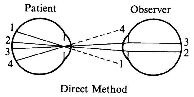
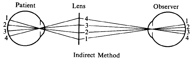
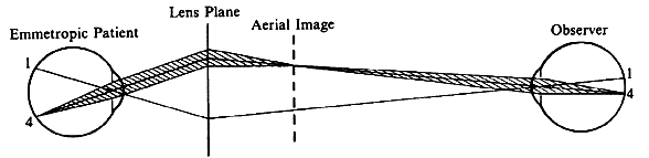
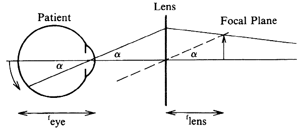
Although not generally considered as a method of ophthalmoscopy, fundus examination with the slit lamp offers an important addition to the traditional methods of direct and indirect ophthalmoscopy. It offers the advantage of high-power magnification through the microscope and flexible illumination with the slit-lamp beam. With appropriate contact lenses, it can offer higher magnification than direct ophthalmoscopy and a field several times wider than indirect ophthalmoscopy. These methods have become particularly important in combination with laser treatment.

Because the slit-lamp microscope has a fixed focus on a plane approximately 10 cm in front of the objective and because the image of the fundus 
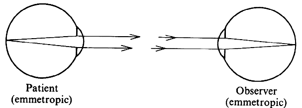
of an emmetropic eye appears at infinity, the fundus cannot be visualized without the help of additional lenses. There are several options.

NEGATIVE LENS

A negative lens placed in front of the objective of the microscope can move the microscope focus to infinity. The practical application of this principle was worked out by Hruby19,20 of Vienna (1942) with a lens known as the Hruby lens.

The optical principle is best understood if the lens is considered in conjunction with the eye, rather than as a part of the microscope. Parallel rays emerging from an emmetropic eye are made divergent by the Hruby 
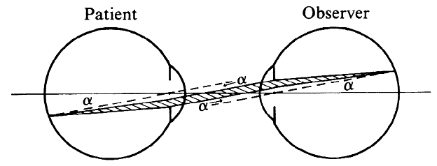
lens and seem to arise from the posterior focal plane of that lens (Fig. 23A). For a -50-D lens, this would be 20 mm behind the lens (the usual Hruby lens is -55 D). The slit-lamp microscope is thus looking at a virtual image of the fundus in a plane somewhere in the anterior segment and must be moved a little closer to the patient than it would be for the regular external examination.

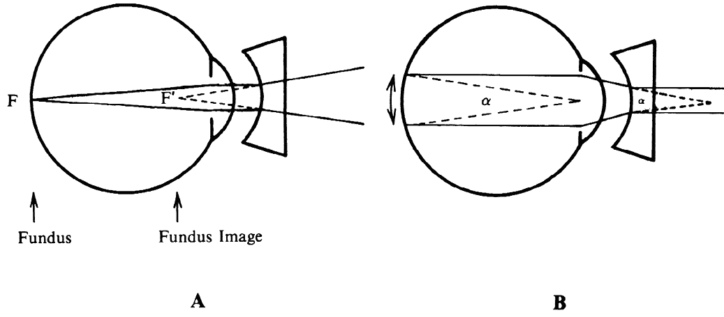
Fig. 23. Hruby lens. A. The fundus image (F') is formed in the posterior focal plane of the lens. B. The field of view is proportional to the size of the pupil as seen from the anterior focal point of the lens.

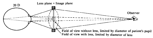
To estimate the field of view in this method, it may be assumed that only rays emerging parallel to the axis will reach the objective of the microscope and the observer's eye. When emerging from the eye, these rays must have been aimed at the anterior focal point of the Hruby lens. Fig. 23B, in which these rays are traced back to the retina, shows that the field of view (a) is proportional to the pupillary diameter as seen from the anterior focal point of the lens. This field is of the same order of magnitude as the field in direct ophthalmoscopy; it is largest when the lens is closest to the eye.

With the lens close to the cornea, the fundus image will be close to the fundus plane and approximately actual size. The magnification to the observer is thus largely determined by the magnification of the microscope. At 16×, the magnification is about equal to that of direct ophthalmoscopy; at higher settings, the magnification is greater. Binocular viewing and slit illumination are advantages over direct ophthalmoscopy, even at similar magnification. Limitation to the posterior pole is a disadvantage.

CONTACT LENS

When the Hruby lens is moved progressively closer to the eye, it will eventually touch the cornea and become a contact lens. If the curvature of the posterior lens surface equals the curvature of the anterior corneal surface, the image formation will not change, but two reflecting surfaces will be eliminated, and image clarity will increase.

The use of a contact lens for fundus examination was perfected by Goldmann21 of Berne, Switzerland (1938). His contact lens is known for the three mirrors incorporated in it. These mirrors positioned at different angles make it possible to examine the peripheral retina with little manipulation of the patient's eye or of the microscope axis (Fig. 24).

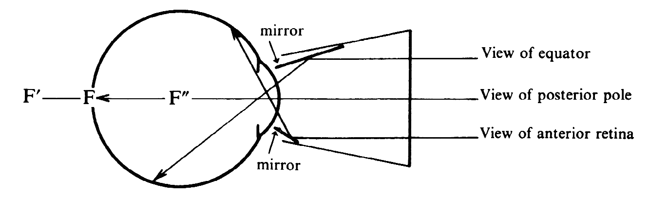
Fig. 24. Three mirror contact lens by Goldmann. Two of the three mirrors are shown. They allow visualization of different parts of the fundus.

The refractive power of the cornea is eliminated in the contact lens. The only effective refractive element left would seem to be the far less powerful crystalline lens. The retina is situated well within the focal length of this lens, and the crystalline lens will therefore form a virtual image of the fundus (F) in a plane (F') behind the globe. How can the microscope focus on an image that far back? We overlooked one other refracting surface: the plano front surface of the contact lens. F' is seen through plastic and vitreous. To the observer in air F' appears at F", through the same effect that makes a swimming pool appear shallower than it is. Because of this, the microscope again must focus on a plane inside the globe. As with the Hruby lens, magnification is largely determined by the microscope.

Thus, contact lens fundus microscopy extends our range of examination methods to details beyond the reach of ordinary direct ophthalmoscopy.

“INDIRECT” SLIT-LAMP MICROSCOPY

The use of the Hruby lens and Goldmann contact lens is comparable to direct ophthalmoscopy, because no real intermediate image is formed. The equivalent of indirect ophthalmoscopy can be achieved by focusing the microscope on the real image formed by a high-power plus lens.

El Bayadi22 introduced the use of a +60-D lens for this purpose. The inverted image formed by this lens is situated 16 mm (0.0167 m) in front of it. A practical problem with some older slit lamps is that they cannot be pulled back far enough to observe this image.

Compared with the Hruby (-55 D) lens, the El Bayadi (+60 D) lens offers the same major advantage as does indirect ophthalmoscopy: a larger field of view. With proper placement of the lens, the field is about six disc diameters (40 degrees), compared with the one- or two-disc diameter field of the Hruby lens.

With a 60-D lens the aerial image is as large as the fundus; thus the magnification is approximately equal to the microscope magnification (similar to that with the Hruby lens).

CONTACT LENS FOR THE INDIRECT METHOD

Can the field of view be widened even further? This is possible by using a contact lens of very high plus power with some additional optical tricks. Figure 25 illustrates the Rodenstock Panfunduscope, based on a design by Schiegel.23

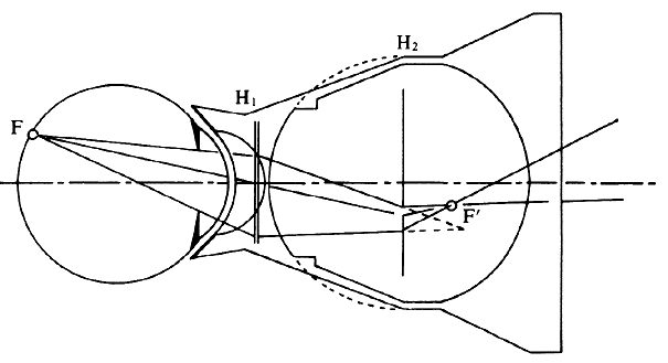
Fig. 25. Contact lens arrangement for wide-angle indirect biomicroscopy. A high-power contact lens forms an inverted image (F') inside a spherical element, which redirects the light toward the observer.

The unit contains a high plus contact lens, which forms an inverted fundus image (F') located inside a second, spherical glass element.

In this arrangement, as in the previous example of a high myope (see Fig. 13), the image-forming and field-widening functions of the ophthalmoscopy lens are separated again. The contact lens forms the image; the spherical element serves to flatten the image and to redirect the diverging pencils of rays toward the observer. Because these elements are so close to the eye, the field of view can be very wide. Indeed, without moving the lens, the view reaches 200 degrees, that is, from equator to equator, 4 to 5 times the diameter (16 times the area) of regular indirect ophthalmoscopy or of the El Bayadi lens.

The size of the image inside the front lens is 70% of the retinal size; for detailed examination, therefore, 50% more microscope magnification is required than with the other slit-lamp methods. However, the principal use of this lens is not for its magnification but for its overview, an overview previously achievable only in fundus drawings or photocompositions.

Similar contact lens arrangements are used in specially designed fundus cameras that allow fundus photography of areas 100 degrees or more in diameter. With lenses such as these, the spectrum of our examining methods can be extended not only toward higher magnification than with direct ophthalmoscopy but also, at the other end, toward an overview of the fundus considerably beyond that obtainable with regular indirect ophthalmoscopy.

As the technology to calculate, design, and manufacture lenses with aspheric surfaces has improved, it has been possible to make lenses with higher powers and better light gathering abilities. The number and variety of lenses for indirect ophthalmoscopy and of contact lenses for slit-lamp microscopy has grown accordingly.

RELATED IMAGING TECHNIQUES

Several related techniques to produce retinal images will be discussed briefly.

Fundus Photography

Fundus cameras have greatly improved the ability to document and follow fundus lesions. Eduard von Jaeger often spent countless hours drawing a single fundus, but today a photographic image is available in a fraction of a second. For reasons mentioned earlier, fundus cameras are built on the principle of indirect ophthalmoscopy. The observer's lens and retina are replaced by a camera lens and film. Because all components are enclosed in a rigid housing, more accessories can be built in. This includes a dual illumination system, which includes a constant light source for focusing and a flash for photography, and filters such as for fluorescein angiography. Rather than placing the viewing 
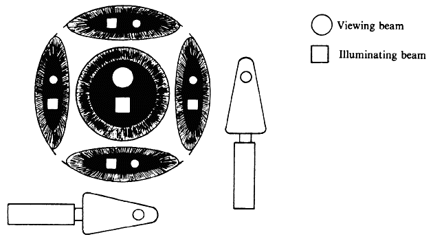
and illumination beams side by side, the illumination beam generally uses the periphery of the pupil and leaves the center for the observation 
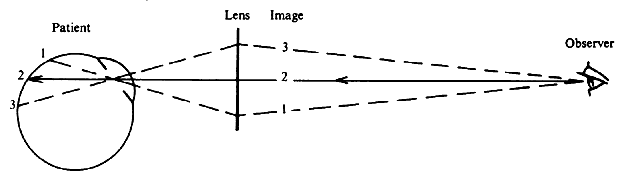
beam.24

An angled glass plate that can be flipped to the right or to the left can be used to slightly deviate the observation beam to the right part or the left part of the patient's pupil to produce photo pairs that can be viewed stereoscopically.

Because newer lens designs have allowed the construction of wide-angle cameras, a special challenge has been to construct the optical system in such a way that the curved retina is imaged in a plane that can be captured on a flat film.

Adaptive Optics

The optics of the eye are not perfect. Even if major errors are corrected with spherical and cylindrical lenses, small irregularities across the pupillary opening persist. The technique of adaptive optics was developed for astronomical telescopes to counteract image degradation by atmospheric irregularities. An adaptive optics system uses a grid to divide the pupillary opening into many small areas and determines a separate small correction for each area. The information is fed to a slightly deformable mirror with microactuators. Thus the image quality can be enhanced to the point at which the cone mosaic can be clearly visible. The setup is too laborious for use in routine photography. Because the corrective system has to be fixed in relation to the pupil, it cannot be implemented in glasses or contact lenses. However, the technique, also known as wavefront analysis, has found a place in the refractive sculpting of the cornea.25

Digital Imaging

The advent of digital cameras has replaced the use of film in many applications. The advantages include easier storage and manipulation, as well as greater sensitivity, so that less light can be used or fainter images can be captured.

Scanning Laser Ophthalmoscope (SLO)

This device takes the advantages of digital imaging one step further. In conventional photography all points of the object are illuminated and imaged onto corresponding points of the film simultaneously. In the Scanning Laser Ophthalmoscope (SLO) the points on the retina are illuminated sequentially by a scanning laser beam; the diffusely reflected light is not imaged but collected on a photocell that can collect light from the entire pupillary area. This allows another significant increase in light sensitivity. In this process the topographic information is transformed into a sequential modulation of signals over time. The image information is recovered by feeding the signals to a video monitor, in which the beam moves in the same way as did the scanning beam.26

An interesting application results from the fact that the illuminating beam can also be modulated to put an image on the retina. Thus it becomes possible to perform microperimetry, relating the areas where a stimulus is seen or not seen directly to the retinal image and to the presence of retinal scarring.27

Optical Coherence Tomography (OCT)

A different form of optical imaging is involved in Optical Coherence Tomography (OCT). This technique does not produce a fundus image but uses a low-coherence light beam to provide cross-sectional information in a way that is analogous to ultrasound B-scan. Because the wavelength of light is so much smaller than ultrasound wavelengths, it can produce far higher resolution and can be used to identify the relationship between layers of the retina and choroid.28

Back to Top
REFERENCES

1. Meeting of the Physikalische Gesellschaft, Berlin, December 6, 1850 (von Helmholtz' paper, read by his friend Dubois-Raymond, has been lost)

2. von Helmholtz H: Beschreibung eines Augenspiegels zur Untersuchung der Netzhaut im lebenden Auge, Berlin (1851). Translation in Arch Ophthalmol 46:565, 1951

3. Albert DM, Miller WH: Jan Purkinje and the ophthalmoscope. Am J Ophthalmol 76:494, 1973

4. Mark HH: The first ophthalmoscope? Arch Ophthalmol 84:520, 521, 1970

5. Cumming W: On a luminous appearance of the human eye, and its application to the detection of disease of the retina and posterior part of the eye. R Med Chir Soc Lond 29:283, 1846

6. Brücke E: Ueber das Leuchten der menschlichen Augen. Arch Anat Physiol Wissensch Med 25, 1847

7. Jones W: Report on the ophthalmoscope. Br Med Chir Rev 14:425, 1854

8. Rucker WF: A History of the Ophthalmoscope. Rochester, MN: Whiting, 1971

9. von Helmholtz H: Ueber eine neue einfachste Form des Augenspiegel. Arch Physiol Heilk 2:827, 1852

10. Ruete CGT: Der Augenspiegel und das Optometer für practische Aerzte. Gottingen, Prussia: Dieterich, 1852

11. Landolt E: Die Untersuchungsmethoden. Berlin, 1:234, 1913

12. Duke-Elder Sir Steward: System of Ophthalmology. Vol 7. Foundations of Ophthalmology: Heredity, Pathology, Diagnosis and Therapeutics. St. Louis: Mosby, 1962, 290–325

13. Albert DM: Jaeger's Atlas of Diseases of the Ocular Fundus. Philadelphia: Saunders, 1972

14. Schepens CL: A new ophthalmoscope: Demonstration. Trans Am Acad Ophthalmol Otolaryngol 51:298, 1947

15. Thorner W: A new stationary ophthalmoscope without reflexes. Am J Ophthalmol 16:330 and 16:376, 1899

16. Wolff: Report of the XII International Medical Congress, Ophthalmology Section. Z Augenheilkd 4:327, 1900; Ueber die zentrische reflexlose Mikro-ophthalmoskopie. Z Augenheilkd 28:307, 1912

17. Gullstrand A: Neue Methoden der reflexlosen Ophthalmoskopie. Ber Dtsch Ophthalmol Gesellsch 36:75, 1910; Die reflexlose Ophthalmoskopie. Arch Augenheilkd 68:101, 1911

18. Giraud-Teulon MALF: Ophthalmoscopie binoculaire on s'exercant par le concours des yeux associés. Ann Ocul 45:233, 1861

19. Hruby K: Spaltlampenmikroskopie des hinteren Augenabschnittes ohne Kontakglas. KIm Monatsbl Augenkeilkd 108:195, 1942

20. Hruby K: Spaltlampenmikroskopie des hinteren Augenabschnittes. Vienna: Urban and Schwarzenberg, 1950

21. Goldmann H: Zur Technik der Spaltlampenmikroskopie. Ophthalmologica 96:90, 1938

22. El Bayadi G: New method of slit lamp micro-ophthalmoscopy. Br J Ophthalmol 37:625, 1953

23. Schlegel HJ: Eine einfache Weitwinkeloptik zur spaltlampen-mikroskopischen Untersuchung des Augenhintergrundes. Doc Ophthalmol 26:300, 1969

24. Pomerantzeff O, Webb RH, Delori FC: Image formation in fundus cameras. IOVS 18:630, 1979

25. Roorda A: Adaptive optics ophthalmoscopy. J Refract Surg 16:S602, 2000

26. Woon WH, Fitzke FW, Bird AC et al: Confocal imaging of the fundus using a scanning laser ophthalmoscope. Br J Opthalm 76:470, 1992

27. Schuchard RA, Fletcher DC: Preferred retinal locus and scanning laser ophthalmoscope. In Albert DM, Jakobiec FA (eds): Principles and Practice of Ophthalmology. 2nd ed. Philadelphia: WB Saunders, 1998

28. Hee MR, Izatt JA, Swanson EA: Optical coherence tomography of the human retina. Arch Ophthalmol 113:325, 1995

Back to Top