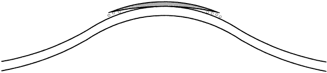
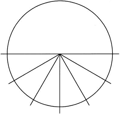
Chapter 54
Rigid Lens Fitting
PETER R. KASTL
Main Menu   Table Of Contents

Search

TERMINOLOGY
BASIC CONCEPTS
BASIC RIGID LENS FITTING
TORIC LENS FITTING
SPECIALTY LENS FITTING
BINOCULAR BALANCING
REFERENCES

In recent years, rigid contact lens fitting has been believed by some to be superfluous as cheap, disposable soft lenses became available and refractive surgery began making inroads into the patient base previously reserved for contact lens fitting. However, not only has there remained a place for rigid lenses, but also refractive surgery has created a need for specialty fitting of a new type of patient, the postrefractive surgery patient.

Corneal physiology or tear physiology is not explained in this chapter because these topics are covered elsewhere in this volume. In this chapter, an attempt is made to explain the nomenclature of rigid lenses and the fitting of rigid lenses of all types. When the reader has finished studying this chapter, he/she should be able to fit rigid contact lenses.

Back to Top
TERMINOLOGY
At the outset, it is important to differentiate between a “hard” lens and a “rigid” lens. Although the terms actually are synonymous, the patient hears the term “hard lens” and associates it with polymethylmethacrylate (PMMA) lenses (the original hard lenses). Nearly all new contact lens patients have heard of hard lenses and equate these lenses with pain.1–5 Therefore, instead of using the term “hard lenses,” the practitioner should refer to “rigid lenses,” meaning rigid gas-permeable (RGP) lenses, to diminish any patient misgivings. Because patients often are not familiar with RGP lenses, the practitioner has an opportunity to educate them.
Back to Top
BASIC CONCEPTS

CORNEA

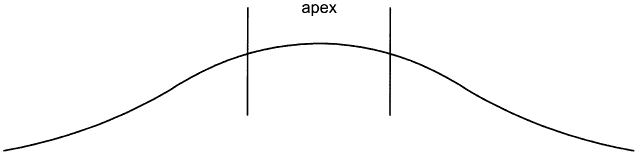
To fit contact lenses well, one must understand the contour of the cornea. The corneal surface is not a sphere; rather, it is aspheric, with an apical zone6–9 (Fig. 1). This zone is the area of the cornea over which the corneal curvature is regular or constant. Several topography devices have been invented to enable the practitioner to understand the contour of a particular patient's cornea.

Fig. 1. Sagittal drawing of a cornea, demonstrating that the cornea is an aspheric, rather than a spherical object.

KERATOMETRY

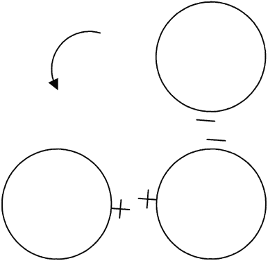
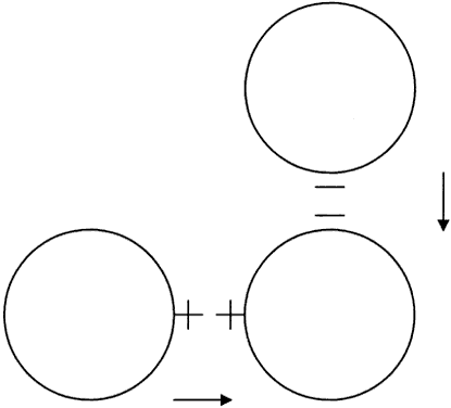
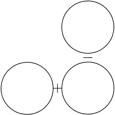
Although topography devices are available, rigid lenses still are fitted based on keratometry. We perform keratometry to measure the curvature of the apical zone. Never forget that the keratometer actually measures the distance between two points 3 mm apart on the cornea. The keratometer produces patterns that are reflected back from the cornea; these are viewed as mires. Mires first must be aligned by rotating the barrel of the keratometer (Fig. 2). Once the barrel of the keratometer is rotated, then the mires are partially aligned (Fig. 3). The next step is to rotate each drum (horizontal and vertical) until the “+ ” and “-” mires are superimposed (Fig. 4). After the mires are aligned, each of the two drums on the keratometer yields a meridional reading in both millimeters and diopters (D). In this chapter, diopters are used. An example of such a reading is 42.00 D @ 180° and 44.00 D @ 90°. The final keratometry readings (“K readings”) are written in a shorthand form as the flatter (smaller) reading in diopters, followed by the steeper (larger) reading in diopters, followed by the meridian of the steeper reading, for example, 42.00/44.00 @ 90°. The flatter meridian is called “K,” and rigid lens fitting always is based on this number. In this example, K is 42.00 D.

Fig. 2. Keratometric mires before any alignment has taken place. The arrow shows the direction in which the keratometer's barrel must be rotated to begin alignment.

Fig. 3. Partial alignment of keratometric mires. The arrows show that the horizontal and vertical drums of the keratometer must be adjusted to align the mires.

Fig. 4. Mires aligned.

ASTIGMATISM

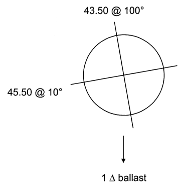
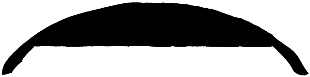
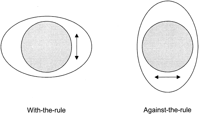
Keratometry can show whether corneal astigmatism is with the rule or against the rule. With-the-rule astigmatism means that the corneal “egg” is lying on its side; against-the-rule astigmatism means that the egg is standing on end (Fig. 5). Here are keratometric examples:

Fig. 5. Hypothetical “eggs” showing with-the-rule and against-the-rule astigmatism. Arrows indicate lens movement with blinking.


With-the-rule:42.00/44.00 @ 90°
Against-the-rule:42.00/44.00 @ 180°


Rigid contact lenses have a tendency to move along the steeper meridian. Thus, with-the-rule astigmatism is good for rigid contact lens fitting because the lens moves up and down with the blinking of the upper lid; against-the-rule astigmatism is not as good because the lens tends to move side to side with each blink. Usually, corneal astigmatism is with-the-rule, hence the name.

Thus far, we have considered only corneal astigmatism, which is measured with a keratometer. In clinical practice, we also measure refractive astigmatism. Whereas keratometry measures only corneal astigmatism, refraction measures a combination of both corneal and residual astigmatism. Residual astigmatism also is called lenticular astigmatism because it is believed to originate from a tilted crystalline lens. We decide which contact lens to fit based on how well the refractive astigmatism and the keratometric astigmatism match.

Following is an example of matching astigmatism, where the corneal shape is responsible for the entire refractive astigmatism; a simple rigid lens would work well because a rigid lens cancels out corneal astigmatism:


Keratometry:42.00/44.00 @ 90°
Refraction:-3.00 + 2.00 × 90°


Following is an example of residual astigmatism. Note that there is only 0.50 D of corneal astigmatism but 1.50 D of refractive astigmatism. The difference between the two, 1.00 D, constitutes the residual astigmatism. This difference requires toric lens fitting because a spherical rigid lens cancels out only corneal astigmatism:


Keratometry:42.00/42.50 @ 90°
Refraction:-3.00 + 1.50 × 90°


LENS TYPES AND PARAMETERS

Lenses either are soft or hard. Soft contact lenses (SCLs) are “all alike” because they are made from hydrogels, water-containing soft material. Rigid lenses currently are made from RGP materials; the “old” hard lenses were made of PMMA, a plastic that was not oxygen permeable.

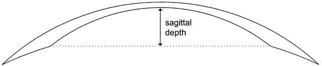
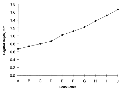
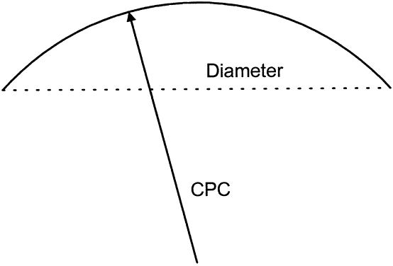
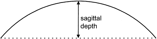
All contact lenses share the same important parameters: diameter, central posterior curve (CPC—also known as base curve, or BC), and power (Fig. 6). Diameter is measured in millimeters, CPC is measured in diopters (for rigid lenses) and millimeters (for SCLs), and power is measured in diopters. Diameter and CPC determine the sagittal depth of a lens (Fig. 7). No one has proven a numerical relationship between sagittal depth and lens fit, but most practitioners believe that an increase in sagittal depth for either a soft or rigid lens results in a more tightly fitting lens.10–13 Thus, the lens itself tightens as the sagittal depth increases. Increasing the diameter of a lens increases the sagittal depth to a great degree and also results in tight fitting.

Fig. 6. Line drawing of a contact lens, demonstrating the diameter and central posterior curve (CPC).

Fig. 7. Line drawing of a contact lens, demonstrating sagittal depth.

TEAR LENS

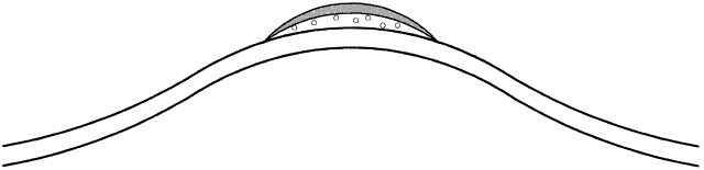
Lens power constitutes most, but not all, of the effective power of a rigid lens. Depending on its relationship to the cornea, the CPC also contributes to the effective power because it creates a “tear lens.” If a lens is “steeper than K,” it produces a tear lens that functions as a plus lens. This plus tear lens must be corrected in the final or prescribed lens power, by adding minus power (Fig. 8). Conversely, if the rigid lens is fitted “flatter than K,” then a minus tear lens is created, which needs to be corrected in the final or prescribed lens power by adding plus power (Fig. 9). The actual correction for the tear lens can be remembered as a mnemonic: SAM FAP—steeper, add minus; flatter, add plus.

Fig. 8. Contact lens fitted “steeper than K.” Note the “plus” tear lens beneath the contact lens.

Fig. 9. Contact lens fitted “flatter than K.” Note the “minus” tear lens beneath the contact lens.

MINUS CYLINDER THINKING

When we work with rigid lenses, we always convert to minus cylinder, then drop the cylinder. Why? We can answer this question in two steps:

  1. Because the cornea is a convex surface, any corneal astigmatism is a plus cylinder. Such a refractive error is neutralized by an equal and opposite power, that is, minus cylinder. Thus, we start by writing the refraction in minus cylinder form.
  2. When we place a rigid contact lens onto the eye, the resulting concave tear film is a minus cylinder, which automatically corrects the plus cylinder of the astigmatic cornea. Thus, we can “drop the minus cylinder” and consider only the resulting sphere power for the contact lens.

Thus the rule, “Convert to minus cylinder and drop the cylinder.” An example of this idea is to take a refraction of -3.00 + 1.00 × 90°. We convert to minus cylinder form: -2.00 - 1.00 × 180°. By dropping the cylinder, we end up with a rigid contact lens power of -2.00 D. Remember that this calculation assumes that all refractive astigmatism is corneal in origin.

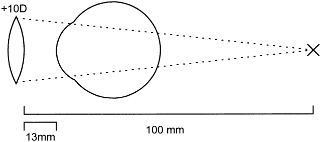
VERTEX DISTANCE CORRECTION

Any spectacle correction intended to be used for contact lens fitting needs to be corrected for vertex distance if the refraction is greater than + 4.00 or -4.00 D.

This concept is easier to understand with a couple of examples. We shall consider first a + 10 D and then a -10 D lens, both located 13 mm (vertex distance) from the eye.

In the first example, the focal length of a + 10 lens is (1000 mm/10) = 100 mm (Fig. 10). By placing a contact lens onto the eye, the focal length becomes shorter: 100 - 13 = 87 mm. Thus, the power of the contact lens is (1000/87) = + 11.50 D (rounded to nearest eighth of a diopter).

Fig. 10. Schematic representation of a + 10 D spectacle lens before contact lens fitting.

In the second example, the -10 lens has the same focal length (100 mm), but in the opposite direction (Fig. 11). When it is placed onto the eye as a contact lens, the focal length increases: 100 + 13 = 113 mm. Thus, the contact lens has a power of (1000/113) -8.87 D (rounded to nearest eighth of a diopter).

Fig. 11. Schematic representation of a -10 D spectacle lens before contact lens fitting.

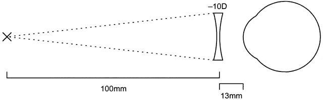
In both cases, for either plus or minus lenses, always add plus power when correcting for vertex distance.

LENS FITTING EXAMPLE

This is the type of problem seen on written examinations. We want to fit a rigid lens on a patient with the following readings:


Keratometry:42.00/43.50 @ 90°
Refraction:-9.00 + 1.50 × 90°


Suppose the refraction was done with a vertex distance of 13 mm and that we want to fit 0.50 D steeper than K. What is the power of the final lens?

This type of problem is solved in three steps:

  1. Convert to minus cylinder. First we convert the refraction to minus cylinder and drop the cylinder: -9.00 + 1.50 × 90° becomes -7.50 - 1.50 × 180°. Dropping the cylinder leaves -7.50.
  2. Correct for vertex distance. The focal distance of a -7.50 D lens is (1000/7.5) = 133 mm. In converting for 13 mm vertex distance, the focal length for the contact lens is (133 + 13) = 146 mm. The power of such a lens is (1000/146) = -6.85 D. Note that we do not round off the results at this point.
  3. Correct for lens fit. Because the lens is fitted 0.50 D steeper than K, we add minus power (remember: SAM of SAM FAP): -6.85 - 0.50 = -7.35. Rounding to the nearest eighth of a diopter yields the final answer: -7.37 D.

The K readings were not necessary to solve this problem. They often are included on examinations as red herrings. Also, I do not advise the actual fitting of lenses using this theoretical method of calculating the final lens directly from the refraction and keratometry. Nothing beats the actual placement of diagnostic contact lenses onto the patient's eyes. The following fitting methods use this philosophy.

Back to Top
BASIC RIGID LENS FITTING
When soft lenses first appeared, many observers predicted eventual obsolescence for hard lenses; however, the advent of RGP lenses has given rigid lenses a new lease on life.

A correct method for fitting rigid lenses is described in the following sections. The wrong way to fit RGP lenses is to perform keratometry and a refraction, telephone these readings to the contact lens laboratory, and expect a pair of well-fitting lenses in the return mail. This procedure leaves the actual fit of the lenses to the laboratory. The laboratory goes through a calculation identical to the aforementioned lens-fitting example.

Proper rigid lens design requires the practitioner to determine exactly the “big three” parameters: diameter, CPC, and power, in that sequence. Diagnostic lenses are placed onto the patient's eyes.

DIAMETER

Before the advent of RGP lenses, lens diameters had to be relatively small because corneal oxygenation depended solely on the tear pump mechanism to flush oxygen-laden tears beneath a PMMA lens. During that time, diameter could be determined by measuring the corneal apex with a device called the Topogometer, which attached to the keratometer. The contact lens diameter was made 2 mm larger than the longer diameter of the apical cap of the cornea. Such lenses were in the 7- to 8-mm range.

When RGP lenses were released, diameters could be made larger because oxygen diffuses through such a lens. Thus, one can obtain a diameter based on the horizontal visible iris diameter (HVID). Subtract 2.3 mm from HVID. A “standard” HVID of 11.5 mm gives a lens diameter of 9.2 mm. One should try to use the same diameter for both eyes; otherwise, the patient may feel a difference between the two lenses and may decide something is wrong with one of them.

CENTRAL POSTERIOR CURVE OR BASE CURVE

If the diameter is to be less than 9 mm, make the CPC either 0.50 D steeper than K or one half of the corneal astigmatism steeper than K, whichever is steeper. The rationale is that for a small lens, the final lens possesses a CPC of at least 0.50 D steeper than K.

For example, if a patient's keratometry is 42.00/42.50 @ 90°, K is 42.00 D, and one half of the corneal astigmatism is (42.50 - 42.00 D)/2 = 0.25 D; thus,


0.50 D steeper than K is42.50 D
½ of corneal astigmatism added to K is42.25 D
The steeper number is42.50 D


Therefore, the final CPC is 42.50 D.

As a further example, suppose instead that the keratometry was 42.00/44.00 @ 90°. K still is 42.00 D, and half of the corneal astigmatism is (44.00 - 42.00 D)/2 = 1.00 D. Thus,


0.50 D steeper than K is42.50 D
½ of corneal astigmatism added to K is43.00 D
The steeper number is43.00 D


Therefore, the final CPC is 43.00 D.

These examples illustrate that as corneal astigmatism grows larger, the CPC of a rigid lens grows steeper, providing stability on an increasingly astigmatic cornea.

Thus far, we have considered CPCs for lenses where the diameter is less than 9 mm. If the diameter is larger than 9 mm, make the CPC either 0.25 D steeper than K or one fourth of the corneal astigmatism steeper than K, whichever is steeper. Remember that a large lens is a tight lens, so the CPC is made relatively flatter than before to compensate for the large diameter. The final lens possesses a CPC of at least 0.25 D steeper than K.

Using the first example, where the keratometry is 42.00/42.50 @ 90°, K remains 42.00 D, but one fourth of the corneal astigmatism is (42.50 - 42.00 D)/4 = 0.12 D. To compute,


0.25 D steeper than K is42.25 D
¼ of corneal astigmatism added to K is42.12 D
The steeper number is42.25 D


Therefore, the CPC is 42.25 D.

The second example is computed in the same fashion. The keratometry is 42.00/44.00 @ 90°. K remains 42.00 D, and one fourth of the corneal astigmatism is (44.00 - 42.00 D)/4 = 0.50 D. Thus,


0.25 D steeper than K is42.25 D
¼ of corneal astigmatism added to K is42.50 D
The steeper number is42.50 D


Therefore, the final CPC is 42.50 D.

POWER

Thus far in the fitting process, we have performed calculations only. At this point, we diverge from theory into practice by placing actual contact lenses on the patient. Always use diagnostic lenses on both eyes simultaneously. First, place topical anesthetic into both eyes to eliminate excess tearing, which can change refractive power by increasing the tear lens beneath the contact lens. Next, place diagnostic lenses in both eyes, approximating the previously determined CPCs. Refract both eyes (called overrefraction) and binocularly balance (see Binocular Balancing section). For each eye, calculate its total optical power first by summing the diagnostic lens CPC and power and then adding the overrefraction. To obtain the desired power of the final contact lens, subtract the intended CPC of the lens, which was calculated in the previous step.

By summing the diagnostic lens CPC and power with the overrefraction, one arrives at a constant for that eye.

For example, a patient presents with the following keratometry and refraction:


Keratometry:41.00/42.00 @ 085°
Refraction:-3.00 + 1.00 × 085°


After measuring the HVID and determining the diameter and CPC, we decide that the final lens should have a CPC of 41.25 D; however, our fitting set has lenses of only whole diopters and one power, -1.00 D. Therefore, we put on a diagnostic lens of 41.00 D CPC and power -1.00 D. Assume we get an overrefraction of -0.75 D:


Diagnostic lens CPC41.00 D 
Diagnostic lens power-1.00 D 
Overrefraction-0.75 D 
Sum of the above three39.25 D(constant)
Subtract final CPC-41.25 D 
Power of final lens-2.00 D 


Thus, we would order a lens of CPC 41.25 D and power -2.00 D. By using this concept of a constant, one avoids the use of the SAM FAP rule. This calculation automatically takes tear lens correction into account. In addition, this example demonstrates that any diagnostic lens can be applied to establish the precise power of the final lens. However, if the overrefraction is greater than -4.00 D or + 4.00 D, it should be corrected for vertex distance.

While the diagnostic lenses are on the patient's eyes, you should examine them for fit, assuming that their CPCs are close to those of the final lenses. Well-fitting lenses are drawn upward by a blink, fall rapidly to the central cornea, and slowly drift downward.

Patients with large pupils may require larger lenses to prevent them from looking through the lens periphery in the dark. Patients with large amounts of corneal astigmatism (more than 3 D) or residual astigmatism may require a toric lens.

AVOIDING PROBLEMS

Many rigid lens problems are caused by poor blinking.14–16 The normal blink rate is once every 5 seconds. People tend to blink less frequently when reading, studying or otherwise concentrating on something, playing video games, working at computers, looking through microscopes, when fatigued, or when drinking alcohol. In addition, at certain times, tears may evaporate, causing problems, such as while sailing or skiing or while in an automobile with the air conditioning running.

Before dispensing rigid lenses, make sure they have been inspected for diameter, CPC, and power, and that all specifications are correct. The patient should be wearing the lenses 8 hours per day before you should consider making major changes in the lens specifications. In other words, give the eye time to adapt to the lens (decreased corneal sensation and decreased tearing).

Back to Top
TORIC LENS FITTING
In brief, bitoric or back toric lenses are used for large amounts of corneal astigmatism. Front toric lenses are used for eyes with spherical corneas but with residual astigmatism. Bitoric lenses with prism are used when an eye has only moderate corneal astigmatism and residual astigmatism.

BITORIC AND BACK TORIC

Indications

Some patients have large amounts of corneal astigmatism that require a contact lens with a back toric surface for the lens to be stable.17–21 If there is no residual astigmatism, then the lens has a back toric surface and a front spherical surface; such a lens is called a back toric lens. If there is residual astigmatism present, then the lens has both a back toric and front toric surface (the front toric surface corrects the residual astigmatism); this lens is called a bitoric lens. For either lens, corneal toricity should be greater than 3 D.

Rationale

These lenses are fitted “on K,” that is, parallel to the corneal meridians. Because keratometric astigmatism has two curvatures of different radii, a rigid lens that has these same radii on its posterior surface matches those of the cornea and fits as a glove does a hand. Therefore, its fit restrains the lens from rotating and keeps any anterior cylinder on the lens from rotating.

Method

Determine diameter first, then CPCs and powers; as stated previously, the two curves for each lens are on K in both meridians. Diameter should be determined by the fitter's usual method, such as the aforementioned HVID method. However, the diameter should be greater than 9 mm to minimize lens rotation by tightening the lens.

The CPC for each meridian is the eye's K readings (each is fitted parallel to each meridian). The power in each meridian is determined by refracting over a spherical PMMA diagnostic contact lens. A spherical PMMA lens is used as the diagnostic lens because such rigid material does not flex on an astigmatic cornea.

An optical power cross is constructed by a simple method (described in the following example). Vertex distance must be corrected, if either value in the cross is greater than + 4.00 D or -4.00 D. By adding the cross powers to the diagnostic lens CPC and power in each meridian, the optical constant of each meridian is obtained, similarly to a spherical lens, as described previously. Subtracting the K readings gives the power of the final lens for each meridian. This process is best explained with an example.

Example

A patient had the following measurements:

Keratometry


OD42.50/47.00 @ 100°
OS42.50/48.75 @ 84°


Refraction


OD-2.75 + 4.75 × 100°
OS-4.25 + 7.25 × 83°


Use of the HVID rule resulted in a lens diameter of 9.2 mm. Next, the following diagnostic lenses were placed onto each eye, with the resulting overrefractions (note that this patient had significant residual astigmatism in addition to his corneal astigmatism):


OD44.50/-1.00 Dplano + 2.00 × 110°
OS45.50/-1.00 D-0.25 + 2.25 × 77°


To calculate the power crosses for each eye, the overrefractions first were converted to minus cylinder notation:


OD+ 2.00 D - 2.00 × 20°
OS+ 2.00 D - 2.25 × 167°


The cross in each instance was calculated as the sphere on the left and the sum of sphere and cylinder on the right (the reason for left and right is given later):


The final results were obtained by summing the CPC and power of each diagnostic lens with the cross power at each meridian, to derive the optical power at that meridian. Subtracting the keratometry measurement at that meridian gave the final lens power at that meridian (Table 1).


TABLE 54-1. Example of Bitoric and Back Tone Lenses


OD OS 
42.5047.00Meridians42.5048.75 
44.5044.50Dx lens CPC45.5045.50 
-1.00-1.00Dx lens power-1.00-1.00Add these three
+2.00planoPower cross+ 2.00-0.25 
45.5043.50Optical total46.5044.25 
-42.50-47.00Subtract “K”-42.50-48.75Final lens parameters (D)
+3.00-3.50Final power+ 4.00-4.50 


The flat meridian of each eye is placed on the left, and the steep meridian is placed on the right. In this manner the proper arm of the power cross lines up with the proper meridian.

Thus, the following final bitoric lenses (fitted on K at each meridian) were ordered and the resulting acuities obtained:


OD42.50/+ 3.00 20/25 + 2
 47.00/-3.50
OS42.50/+ 4.00 20/20
 48.75/-4.50


Comments

The best type of diagnostic lens to use is a spherical PMMA lens; this type of lens does not flex on a highly astigmatic cornea. If a gas-permeable lens is used, flexure may occur, resulting in an incorrect overrefraction. Also, refract with the room lights on to constrict the pupils; otherwise, the patient actually may see around the lens during fitting because the diagnostic lens may ride eccentrically. During fitting, the cylindrical axis of the overrefraction should line up with the K readings within 10°; otherwise, the astigmatism is oblique, and the lens cannot be made by standard laboratory techniques. Note that the first number of the optical cross is added to the column of the flatter meridian; thus, the final lens always should have the most plus or least minus at the flatter meridian. These lenses do not work on corneal astigmatism less than 1.50 to 2.00 D because they rotate. They can be made in gas-permeable material with up to 10 D of toricity.

FRONT TORIC

Front toric rigid lenses are indicated when the patient has a spherical cornea and residual astigmatism.21–23 Front toric rigid lenses have a spherical back surface and contain a cylindrical correction; this correction is kept in proper alignment by the use of a prism ballast in the contact lens. Prism ballast acts as a weight to hold the lens in position.

Fitting Method

This fitting method begins with the same steeper than K method explained previously for spherical RGP lenses. The diameter is determined first. Next, the CPC is chosen based on the diameter. For a diameter smaller than 9.0 mm, CPC is 0.50 D steeper than K or one half of the astigmatism steeper than K, whichever is steeper; for a diameter larger than 9.0 mm, CPC is 0.25 D steeper than K or one fourth of the astigmatism steeper than K, whichever is steeper. Power then is determined by refracting over a diagnostic prism ballasted contact lens, with a dot indicating its base. The overrefraction is converted to minus cylinder form, and the spherical power for the final lens is calculated by adding the spherical part of the overrefraction (in minus cylinder form) to the sum of the diagnostic lens CPC and power and subtracting the CPC of the final lens. The orientation of the cylindrical portion of the refraction is corrected for lens rotation by using the acronym LARS: left, add, right, subtract. For each clock hour of rotation, 30° either is added or subtracted from the refractive axis (Fig. 12). For instance, in Figure 13, a lens is seen to rotate to the 7-o'clock position. This rotation is one clock hour to the practitioner's left. Thus, 30° is added (left, add) to the refractive axis.

Fig. 12. Lens rotation template for front toric lenses.

Fig. 13. Front toric diagnostic lens rotating one clock hour to the left, to the 7-o'clock position.

Example

In this example, diameter is assumed to be 8.8 mm, the same as the diagnostic lens that is used. A patient's eye had the following keratometry and refraction:


Keratometry:42.75/43.50 @ 100°
Refraction:-1.50 + 1.50 × 10⁵°


A diagnostic lens of CPC 43.00 D, diameter 8.8 mm, power + 0.50 D, and prism ballast 1.5Δ was placed onto the eye. Overrefraction was -0.50 + 1.00 × 100°, and the lens was rotated to the 4:30 position (Fig. 14).

Fig. 14. Front toric diagnostic lens rotating one and a half clock hours to the right, to the 4:30 position.

Because the diameter of the final lens was to be 8.8 mm, the CPC of the final lens was determined to be 43.25 D (0.50 D steeper than K). The power of the final lens was calculated by first changing the overrefraction to minus cylinder form:


Plus cylinder:-0.50 + 1.00 × 100°
Minus cylinder:+ 0.50 - 1.00 × 10°


Final lens sphere power was calculated, as follows:


Diagnostic lens CPC43.00 D
Diagnostic lens power+ 0.50 D
Overrefraction sphere (minus cylinder form)+ 0.50
Total sphere power44.00 D
Subtract final CPC-43.25 D
Final sphere power+ 0.75 D


Thus, the spherocylinder lens power (uncorrected for rotation) was + 0.75 - 1.00 × 10°. Because the diagnostic lens rotated one and a half clock hours to the right, 45° was subtracted from the axis of the astigmatism (10° + 180° - 45° = 145°). Therefore, the final lens power was + 0.75 - 1.00 D × 145°.

Comments

Make sure the diagnostic lens set is made from an RGP material and is manufactured by the same laboratory that will make the final lens. The newer high Dk gas-permeable materials are lighter than PMMA and may rotate differently than PMMA, so an RGP fitting set is required. Prism ballast typically comes in 0.75Δ, 1.5Δ, and 2.5 Δ; 1.5 Δ is the amount usually used in this type of lens.

BITORIC WITH PRISM

Indications

The rare patient presents with moderate corneal astigmatism and moderate lenticular or residual astigmatism. This patient manifests the following dilemmas:

  • There is residual astigmatism; therefore, a spherical rigid lens will not work.
  • There is too much corneal astigmatism for a front toric lens to work well (i.e., >1 D).
  • There is not enough corneal astigmatism for a bitoric lens to work well (i.e., 2 D).

This is a job for a bitoric lens with prism ballast.24,25 In other words, a bitoric lens is fitted on K, but this lens is ordered with prism ballast in it to help the lens maintain proper axis orientation. Thus, the combined effects of the toric back surface and the prism ballast serve to hold the lens in place on the cornea, allowing the astigmatic correction of the lens to line up properly.

Example

A patient presented with the following refraction and K readings in one eye:


Keratometry:43.50/45.50 @ 10°
Refraction:-2.50 + 2.75 × 4°


There is more refractive astigmatism than keratometric astigmatism, indicating residual astigmatism. There is not that much corneal astigmatism (2 D); thus, a simple bitoric lens will not hold in place very well.

A diagnostic lens of CPC 42.00 D (only a few PMMA diagnostic lenses were available at the time, so this one had to be used), diameter 9 mm, power + 0.50 D was placed onto the eye, and the following overrefraction was obtained: -0.25 + 1.00 × 5°. Slightly more residual astigmatism was present than was anticipated.

To begin calculations, the overrefraction first must be converted to minus cylinder, then to a power cross, in the same manner as for a standard bitoric lens:


The final lens powers were then calculated the same as for a standard bitoric lens:


Meridians43.50 D45.50 D
Fitting lens CPC42.00 D42.00 D
Fitting lens power+ 0.50 D+ 0.50 D
Power cross+ 0.75 D-0.25 D
Optical total43.25 D42.25 D
Subtract K-43.50 D-45.50 D
Final power-0.25 D-3.25 D


The final lens CPCs and powers were ordered, as follows:

  43.50/-0.25 D
  45.50/-3.25 D

However, in addition, the lens was ordered with 1Δ of prism ballast. To order this ballast, the K readings were specified, with the meridians. The laboratory fabricated a bitoric lens with 1Δ of prism ballast oriented at the 6-o'clock position, which held the lens in place better than if it had been a plain bitoric lens (Fig. 15). The diameter was enlarged to 9.2 mm to give further stability to the lens (remember that larger lenses are tighter). Final visual acuity was 20/20.

Fig. 15. Bitoric with prism lens demonstrating that the back toric surface aligns with the keratometric meridians, whereas the 6-o'clock prism ballast helps stabilize the lens.

Comment

The prism ballast should be ordered in the ¾-1Δ range; just as with front toric lenses, too much prism will be uncomfortable. The K readings must be specified with the meridians.

Back to Top
SPECIALTY LENS FITTING

KERATOCONUS

Keratoconus is a corneal disease in which a metabolic defect leads to a corneal cone.26,27 This disease produces irregular astigmatism, myopia (from anterior corneal displacement), and central scarring; the first two can be treated with a contact lens. Because of the irregular astigmatism, the keratometer is useless, except as a possible indicator of relative severity.

I believe that the best way to rehabilitate the keratoconus patient is to fit Soper-type keratoconus lenses.21,28 This type of lens is a bicurved lens; thus, a steep central posterior curve (CPC) vaults the cone, and a relatively large peripheral curve fits the peripheral cornea outside the cone (Fig. 16). This lens is fitted based on the concept of sagittal depth, and calibrated diagnostic lenses are placed onto the eye until a proper fluorescein pattern is observed.

Fig. 16. Soper keratoconus lens. Note the steep central posterior curve (CPC) and the flatter peripheral curve. Sagittal depth is calculated for the diameter of the CPC, not the diameter of the entire lens.

If a diagnostic lens produces an air bubble or extreme fluorescein pooling, then that lens has too large a sagittal depth (too steep), and a lens with a smaller sagittal depth should be tried. A flat lens produces apical touch, requiring that a steeper lens be tried next. The correct lens barely vaults the cone, demonstrating a uniform central fluorescein pattern, as demonstrated in Figures 17 through 19.

Fig. 17. Soper keratoconus lens, the sagittal depth of which is too great. Copious fluorescein is pooling centrally. If this lens were any steeper, it would trap an air bubble centrally

Fig. 18. Soper lens bearing on the cone. This lens's sagittal depth is too small. Occasional air bubbles may be trapped peripherally when a keratoconus lens is too flat.

Fig. 19. Well-fitting Soper keratoconus lens. Fluorescein pattern is uniform over the cone.

Because these lenses are fitted strictly from a diagnostic set and calibrated by sagittal depth (Fig. 20), the CPC and diameter of the dispensed lens are taken from the properly fitting diagnostic lens itself. Therefore, the only calculation necessary is the power of the lens. This calculation is performed similarly to the method for standard rigid lens fitting. Note, however, how the CPC is expressed for this type of lens: central curve/peripheral curve, for example, 54.00/45.00 D.

Fig. 20. Sagittal depth for the Soper keratoconus lens diagnostic set. Each lens has a letter engraved on it for identification purposes. The CPC and diameter of the set is varied lens by lens to achieve a near linear sagittal depth relationship.

Example

A keratoconus patient desires contact lens fitting. A diagnostic lens of CPC 52.00/45.00 D and power of -16.50 D was tried on the patient, and the overrefraction was found to be -3.00 D. A lens of CPC 56.00/45.00 D next was tried, and this lens fit better, as the first lens bore on the cone. Thus, the following calculation was performed:


Diagnostic lens CPC52.00/45.00 D
Diagnostic lens power-16.50 D
Overrefraction-3.00 D
Optical need (sum) 32.50 D
Subtract final CPC-56.00/45.00 D
Final power21.50 D


This calculation is identical to the previous one for RGP lenses. Only the central part of the CPC (52.00 D) notation is used for calculation; the peripheral part (45.00 D) applies to the fit only. Thus, the final lens was CPC 56.00/45.00 D, power -23.50 D, and diameter of the better fitting lens. Keratometry was not necessary to fit this keratoconic eye.

POSTGRAFT

Background

Although penetrating keratoplasty (PKP) helps certain patients with corneal disease, transplantation does not necessarily leave a cornea that is as visually ideal as an “original” clear cornea.29 Large amounts of regular and irregular astigmatism produce unacceptable optical results. It is the graft-host interface that is the crucial area of the cornea. If this interface is not uniform, fitting can be difficult.

Normally, the practitioner attempts to fit the patient with spectacles or a soft contact lens; however, if neither gives adequate acuity, then any irregular astigmatism requires a rigid lens.28,30,31 Of course, keratometry may be impossible to perform on such an irregular surface. These patients are fitted similarly to keratoconus patients: one places a diagnostic lens onto the eye, refracts over it, and evaluates the fit with fluorescein. Just as in keratoconus patients, several lenses may have to be tried on, so refracting over the first one offers the best refraction. Later lenses that are tried during the fitting process may be accompanied by much tearing after they have been taken on and off several times.

Fitting Set

Because the patient has a circular piece of donor tissue in the middle of his or her cornea, a large RGP lens is the best one to fit, one that will vault the graft. If the lens rides on the graft, it may irritate it to the point of graft rejection. Thus, a good-fitting set has lenses with large diameters, that is, 9.5 to 10.5 mm, and with plano and aphakic powers, the latter for the occasional patient with an aphakic graft.

Method

Obtaining good keratometry often is impossible in graft patients, due to the resultant irregular astigmatism. Nevertheless, one often can obtain a so-so K and use this value to start. Place onto the eye a rigid lens with a CPC somewhat steeper than K. Refract over this lens, and evaluate the fit with fluorescein.

Other lenses may have to be tried after this first lens. As in keratoconus patients, an air bubble indicates that the lens is too steep; too much bearing on the graft indicates the lens is too flat. Rarely does a fit look perfect. A good fit bears on the host and not on the graft.

After a lens is found to fit, the power is calculated, as shown in the following example. CPC and diameter are taken from the properly fitting diagnostic lens.

Example 1

A posttransplant patient had K readings of 45.00/49.00 @ 120° and could be refracted no better than 20/400 visual acuity, probably because of irregular astigmatism. The following diagnostic lens was placed onto the eye: CPC 45.00 D, diameter 10.0 mm, and power + 0.50 D. Refraction over this lens was + 1.00 D, giving a visual acuity of 20/30. This lens was noted to bear on the graft, however (no fluorescein was present centrally under the lens). Therefore, another lens of CPC 47.00 D and 10 mm was tried; this lens fit well.

The CPC and diameter of the lens to be ordered was to be that of the final, well-fitting lens (47.00 D, 10 mm). The final power was calculated as follows:


Diagnostic lens CPC45.00 D
Diagnostic lens power+ 0.50 D
Overrefraction+ 1.00 D
Optical constant of eye (sum)46.50 D
Subtract final CPC-47.00 D
Final power-0.50 D


Thus, the final lens was CPC 47.00 D, diameter 10 mm, and power -0.50 D.

The Flat Graft—Piggyback Fitting

The flat graft presents a fitting problem. The topography of a flat graft is similar to that of a radial keratotomy: the central cornea is flatter than that of the periphery, causing a rigid contact lens to decenter. If a large rigid lens fails to work, then a piggyback fitting should be considered.

Piggyback fitting refers to fitting a soft lens and then a rigid lens on top of the soft lens. In this manner, a really flat or irregular cornea first is steepened or smoothed to some degree by the soft lens, and then refracted optimally by the rigid lens. The trick is to use the proper soft and rigid lens.

Use an aphakic soft lens and a Soper keratoconus rigid lens. Because of its shape and thickness, the aphakic soft lens produces a better, smoother surface on which to fit the rigid lens, especially on flat grafts. The Soper cone lens fits this thick soft lens better than a simple spherical rigid lens.

Example 2

A patient has a flat graft that was not fitted adequately with a rigid lens alone. A + 13.00 D soft lens was allowed to equilibrate on the eye. Because the rigid lens fits the soft lens, keratometry is performed over the soft lens.

K readings of the soft lens were 55.00/55.50 D. A Soper keratoconus lens of CPC 56/45 D, diameter 9.5 mm, and power -12.50 D was placed onto the soft lens, and the resultant overrefraction was + 3.50 D. The power of the rigid lens was calculated as follows (we want to fit the rigid lens on K):


Diagnostic lens CPC56.00/45.00 D
Diagnostic lens power-12.50 D
Overrefraction+ 3.50 D
Optical constant of eye (sum)47.00 D
Subtract K of SCL-55.00 D
Final power of rigid lens-8.00 D


Thus, the final lenses for the patient were the same soft lens (+ 13.00 D) and a rigid lens of CPC 55.00/45.00 D, power -8.00 D, and diameter 9.5 mm.

Complications

PKP patients wearing contact lenses must be observed for possible rejection, vascularization, and/or ulceration. Fortunately, these complications are fairly rare. However, if a patient is wearing a piggyback system, then he or she must be observed more closely than usual.

AFTER REFRACTIVE SURGERY

When radial keratotomy (RK) fails to give the desired result, the reason is usually an over- or undercorrection or irregular astigmatism.32–35 Correcting these problems requires the use of an RGP contact lens—not a soft lens because a soft lens can cause wound vascularization. The RGP lens needs to be large and flat because the resultant post-RK topography is a flat cornea.

FITTING METHOD

Always use diagnostic lenses during fitting; never try to guess the final lens from refraction and keratometry (which will be difficult to obtain). The fitting set of lenses should have flat lenses with CPCs starting in the low 30-D range. A typical set is made of PMMA for durability. All possess plano power and 10-mm diameters. CPCs are 30, 32, 34, 36, 38, and 40 D.

Try to obtain keratometry anyway and place on the eye a lens with a CPC that approximates these readings. Refract over this first lens. Next instill fluorescein to ascertain adequacy of fit: if the lens bears on the cornea, it is too flat; if too much fluorescein pools or if an air bubble is trapped beneath the lens, then the lens is too steep.

Rarely does a lens center perfectly, because of the distorted corneal topography. Nevertheless, the patient should see quite well.

Example

A 44-year-old attorney had RKs performed simultaneously on both eyes while he was traveling. No keratometry was obtained at the time. His pre-RK refractions were as follows:


OD-5.50 + 2.50 × 78°20/25
OS-3.25 + 0.75 × 72°20/20


He required a + 1.25 add for reading at that time.

He presented for lens fitting 10 months post-RK with uncorrected acuities of 20/25-2 and 20/100. He reported postoperative anisometropia. His refractions were as follows:


OD-0.25 + 1.50 × 120°20/25
OS-+ 2.00 + 0.75 × 150°20/20


Keratometry was as follows:


OD36.12/39.25 D @ 136°
OS35.50/38.00 D @ 163°


A diagnostic lens of CPC 38 D, power plano, and diameter 10 mm was placed on the right eye. Overrefraction was + 0.50 D (20/20). Because this lens seemed to be somewhat flat, a second lens of CPC 40 D was placed; this lens fit well. Thus, the final lens power was calculated as follows:


Diagnostic lens CPC38.00 D
Diagnostic lens power0.00 D
Overrefraction+ 0.50 D
Optical constant of eye (sum)38.50 D
Subtract final CPC-40.00 D
Final power-1.50 D


Therefore, the final right lens was CPC 40.00 D, power -1.50, and diameter 10 mm.

A similar procedure was followed for the left eye, for which a diagnostic lens of CPC 36 D, power plano, and diameter 10 mm yielded an overrefraction of + 2.50 (20/20). A second lens of CPC 38 D fit better, however. The same calculation as for the right eye was done to determine power:


Diagnostic lens CPC36.00 D
Diagnostic lens power0.00 D
Overrefraction+ 2.50 D
Optical constant of eye (sum)38.50 D
Subtract final CPC-38.00 D
Final power+ 0.50 D


Thus, the final left lens was CPC 38.00 D, power + 0.50, and diameter 10 mm. Final visual acuities were 20/20 in both eyes; combined visual acuity was 20/15.

NEW LENS DESIGNS

Because standard spherical RGP lenses tend to decenter due to post-RK corneal topography, new RGP designs have been devised to surmount this problem. One is shown in Figure 21. The flat back surface supposedly more accurately matches the post-RK cornea.

Fig. 21. A specially designed lens for postradial keratotomy (post-RK) fitting. This figure was taken from company literature and demonstrates a flat back surface to match the altered cornea.

In summary, post-RK patients can do well with contact lenses, if their expectations can be made realistic—that is they will have to wear RGP lenses, and the fit will not be perfect.

Back to Top
BINOCULAR BALANCING
When refracting a patient's eyes, the practitioner tends to overminus the first eye refracted. This overcorrection forces one eye to try to accommodate more than the other. At the least, the patient experiences blurred vision in one eye; occasionally, he or she may have headaches. This mistake can be eliminated by the technique of binocular balancing.36,37
  1. After performing the best refraction on each eye, have the patient look through both eyes at the 20/40 line.
  2. Add + 0.75 D to each eye's refraction.
  3. Alternately occlude each eye repeatedly, asking which has the better acuity. Pause only 1 second at a time.
  4. Add + 0.25 D to the better eye. Continue steps 3 and 4 until the acuities are equal.
  5. Begin removing power from both eyes simultaneously, in 0.25-D steps, until the patient's acuity no longer improves or until he or she states that the letters are getting smaller. Do not stop at the 20/20 line because the patient may be able to see 20/10.
  6. If, in step 4, the acuities do not become equal, let the patient's dominant eye retain the better acuity. To find the dominant eye, cut a quarter-sized hole in a piece of paper and let the patient hold this piece of paper in his or her lap. Tell him to fixate on a small object across the room and quickly raise the paper at arm's length, sighting the object through the hole. Then ask him to bring the paper to his face. Whichever eye is looking through the hole is the dominant eye.
Back to Top
REFERENCES

1. Sarver MD, Polse KA, Harris MG: Patient responses to gas-permeable hard (Polycon) contact lenses. Am J Optom Physiol Optics 54:195, 1977

2. Simon DR, Bradley ME: Corneal edema in divers wearing hard contact lenses. Am J Ophthalmol 85:462, 1978

3. Simon DR, Bradley ME: Adverse effects of contact lens wear during decompression. JAMA 244:1213, 1980

4. Bennett ES: Immediate refitting with gas permeable lenses. J Am Optom Assoc 54:239, 1983

5. Yamane SJ: Are hard lenses superior to soft? The advantages of soft lenses. Cornea 9:S12, 1990

6. Mandell RB: Position and curvature of the corneal apex. Am J Optom Arch Am Acad Optom 46:25, 1969

7. Tomlinson A, Schwartz C: The position of the corneal apex in the normal eye. Am J Optom Physiol Optics 56:236, 1979

8. Edmund C: Location of the corneal apex and its influence on the stability of the central corneal curvature: A photokeratoscopy study. Am J Optom Physiol Optics 64:846, 1987

9. Patel S, Marshall J, Fitzke FW et al: The shape of thecorneal apical zone after excimer photorefractive keratectomy. Acta Ophthalmologica 72:588, 1994

10. Bibby MM, Tomlinson A: A model to explain the effect of soft lens design specifications on movement. Am J Optom Physiol Optics 60:287, 1983

11. Cedarstaff TH, Tomlinson A, Bibby MM: Validation of a model describing soft lens movement as a function of lens specification. Am J Optom Physiol Optics 60:292, 1983

12. Young G, Holden B, Cooke G: Influence of soft contact lens design on clinical performance. Optom Vis Sci 70:394, 1993

13. Hostetter TA: Learning the “numbers game” of today's contact lens fitting: clinical and business pearls. [Review] [7 refs]. J Ophthalmol Nurs Tech 15:148, 1996

14. Holly FJ: Tear film physiology. Am J Optom Physiol Optics 57:252, 1980

15. Holly FJ: Tear film physiology and contact lens wear: II. Contact lens-tear film interaction. Am J Optom Physiol Optics 58:331, 1981

16. Efron N, Carney LG: Effect of blinking on the level of oxygen beneath hard and soft gas-permeable contact lenses. J Am Optom Assoc 54:229, 1983

17. Sarver MD, Kame RT, Williams CE: A bitoric gas permeable hard contact lens with spherical power effect. J Am Optom Assoc 56:184, 1985

18. Kastl PR, Kirby RG: Bitoric rigid gas permeable lens fitting in highly astigmatic patients. CLAO J 13:215, 1987

19. Molinari JF, Boggess EA, Caplan L: Clinical evaluation of Boston IV custom toric contact lenses. J Am Optom Assoc 58:888, 1987

20. Weissman BA, Chun MW: The use of spherical power effect bitoric rigid contact lenses in hospital practice. J Am Optom Assoc 58:626, 1987

21. Kastl PR, Johnson WC: Fluoroperm extended wear RGP contact lenses for myopia, hyperopia, aphakia, astigmatism, and keratoconus. CLAO J 15:61, 1989

22. Gilbert ML, Kastl PR: Front toric gas permeable contact lens fitting for residual astigmatism. CLAO J 14:73, 1988

23. Kastl PR, Johnson WC: Fluoroperm extended wear RGP contact lenses: II. Front toric fitting. CLAO J 16:53, 1990

24. Bergenske PD: Correction of residual astigmatism on an against-the-rule cornea of moderate toricity. J Am Optom Assoc 57:212, 1986

25. Gonce MA, Kastl PR: Bitoric rigid contact lens with prism fitting in rare cases of moderate corneal and residual astigmatism. CLAO J 20:176, 1994

26. Cameron JA, Al-Rajhi AA, Badr IA: Corneal ectasia in vernal keratoconjunctivitis. Ophthalmology 96:1615, 1989

27. Lawless M, Coster DJ, Phillips AJ et al: Keratoconus: Diagnosis and management. Aust New Z J Ophthalmol 17:33, 1989

28. Soper JW, Jarrett A: Results of a systematic approach to fitting keratoconus and corneal transplants. Contact Lens Med Bull 5:50, 1972

29. Williams KA, Ash JK, Pararajasegaram P et al: Long-term outcome after corneal transplantation: Visual result and patient perception of success. Ophthalmology 98:651, 1991

30. Weinstock FJ: Contact lens fitting: A clinical text atlas. Philadelphia: JB Lippincott, 1989

31. Speaker MG, Cohen EJ, Edelhauser HF et al: Effect of gas-permeable contact lenses on the endothelium of corneal transplants. Arch Ophthalmol 109:1703, 1991

32. Shivitz IA, Arrowsmith PN, Russell BM: Contact lenses in the treatment of patients with overcorrected radial keratotomy. Ophthalmology 94:899, 1987

33. Haverbeke L: Is wearing contact lenses always possible following a radial keratotomy? [French]. Bull Soc Belge Ophtalmol 234:29, 1989

34. Nizam A, Waring GO3, Lynn MJ et al: Stability of refraction and visual acuity during 5 years in eyes with simple myopia: The PERK Study Group. Refract Corneal Surg 8:439, 1992

35. DePaolis MD: The role of contact lenses in the management of the radial keratotomy patient. Optom Clin 4:25, 1994

36. Amigo G: Binocular balancing techniques. Am J Optom Arch Am Acad Optom 45:511, 1968

37. Shapiro IJ: Parallel-testing infinity balance: Instrument and technique for the parallel testing of binocular vision. Optom Vis Sci 72:916, 1995

Back to Top