Chapter 52
Fresnel Optics
MERTON C. FLOM and ANTHONY J. ADAMS
Main Menu   Table Of Contents

Search

THE FRESNEL PRINCIPLE
TYPES OF PRISMS AND LENSES
OPTICAL CONSTRUCTION
PERFORMANCE
USES OF PRISMS
TREATMENT OF PHORIA
USES OF FRESNEL LENSES
REFERENCES

Any thick lens or prism can create practical problems because of its weight, ie, the higher the power and the larger the diameter, the greater the weight problem becomes. Interestingly, the thickness of a lens contributes relatively little to the refractive power which is determined primarily by the relative angle between the two surfaces of the lens. The critical refracting elements of a planoconvex lens are shaded in the upper half of Figure 1.* The slabs of material underneath each of these small shaded lenslets have little functional significance. In a Fresnel (silent “s”) lens only the small lens-lets are retained and the nonfunctional thickness of material is effectively removed. These small lenslets approximate prismlets which are shaded in the Fresnel lens in the lower half of Figure 1. Buffon in 1748 was probably the first to suggest that large diameter lenses could be made in concentric lenslets or prismlets, which reduced the thickness of the lens1. In 1822 Fresnel's hand-ground concentric groove lenses, which were used extensively in lighthouses along the French coast, popularized the concept. Subsequently, this type of lens came to be known as a Fresnel lens.
* All illustrations in this chapter were provided by the Multimedia Communications Center, School of Optometry, University of California, Berkley.

Fig 1. Principle of a Fresnel lens. Refracting or bending power of the lens depends primarily on the relative angle between its surfaces and not on physical thickness. The small shaded lenslets are the effective refracting elements of the planoconvex lens shown in the upper half of the figure. They are represented in their equivalent Fresnel lens form as small shaded prismlets in the lower half of the figure.

Molded glass Fresnel lenses are now widely used as signal and head lamps where large diameter and relatively light weight are required. Narrow grooves, which are necessary for good optical quality, cannot be ground and polished by conventional means; unfortunately, the high surface tension of glass does not permit fine details of a mold to be reproduced. Consequently, molded glass Fresnel lenses are of poor optical quality. Plastics have made possible the molding of narrow-grooved Fresnel lenses which have highly polished and accurate surfaces. These narrow-grooved plastic lenses have been used primarily in the viewing screens of microscopes and micro film readers. The emergence of high quality dies and new plastics which has permitted the production of Fresnel lenses and prisms.

Back to Top
THE FRESNEL PRINCIPLE
The basis of the Fresnel principle is to remove most of the nonrefracting portions of a conventional lens; this process results in a relatively lightweight, large diameter optical element. Since light is refracted (bent) only at the surface of an optical element and travels in a straight line elsewhere, the refracting power of a lens or prism depends primarily on the relative angle between the two refracting surfaces. The angle between the surfaces is unchanged across a prism, but changes across a lens. A lens, then, can be considered as a series of prisms of increasing apex angle as one moves away from the optical center of the surface. Indeed, Fresnel lenses are most commonly created by narrow, flat-surface, prism grooves of increasing apex angle.

The Fresnel principle for a prism with flat surfaces is illustrated in Figure 2. It is a common clinical observation that the smaller the spectacle frame, the thinner is the base of a conventional prism. Figure 2 shows that reducing the base-apex dimension of a prism from 40 mm to 20 mm reduces the base thickness from 10 mm to 5 mm. Reducing the prism to the very small size of only 4 mm would result in a base thickness of only 1 mm. In each case, the deviating power of the prism is the same since the apex angle is constant. If the base-apex size were reduced to only 2 mm, it is seen in the shaded area of Figure 2 that the base of the prism would be only 0.5 mm thick. A Fresnel prism can be imagined to be a series of small prisms lying adjacent to each other on a platform creating a thin membrane.

Fig 2. A Fresnel prism, much thinner than a conventional ophthalmic prism of the same power, can be imagined to be a series of small plastic prisms (see shaded prisms) lying adjacent to each other on a thin platform of plastic. (Modified from Jampolsky A, Flom MC, Thorson JC: Membrane Fresnel prisms: A new therapeutic device. In Fells P (ed): Proceedings of the First Congress of the International Strabismological Association. London: Kimpton, 1971.)

The Fresnel prism in the example has the same deviating power as a conventional prism of the same material; however, the Fresnel prism is only 1 mm thick (0.5 mm prism base and 0.5 mm platform), one tenth the thickness of the conventional prism of the same power.

A Fresnel lens consists of similar flat ridges in the form of concentric rings. The shaded area of Figure 3 represents the Fresnel equivalent of a conventional planoconvex lens. It is important to notice that the narrower the grooves are, the more closely each facet approaches a prism with a flat surface.

Fig 3. Fresnel equivalents of a conventional planoconvex lens.

Back to Top
TYPES OF PRISMS AND LENSES
The first ophthalmic applications of the Fresnel principle were prisms. Hard acrylic prisms of relatively high prismatic power (more than 10Δ) were developed primarily for the optical treatment of strabismus and were designed to clip onto existing glasses (manufactured by Essilor International, Paris and Universal Optical Co., Dallas, TX)2 (Fig 4). The principle advantage of the Fresnel wafer prism is the reduction in weight when compared to conventional prisms of the same power. The principle disadvantage is the conspicuous Fresnel grooves.

Fig 4. Clip-on wafer prisms of 15Δ base out each eye.

In 1970, thin flexible membrane Fresnel prisms were developed for ophthalmic use (Optical Sciences Group, Inc., San Rafael, CA)3. The membranes are made of clear polyvinyl chloride and are designed to be applied without adhesive to the back surface of a conventional ophthalmic lens. Because the membrane is extremely thin (0.8 mm) and the grooves very narrow (16 per inch), they are cosmetically superior to the hard acrylic wafer prisms. Their relatively large (64 mm) diameter allows them to be cut to conform to the shape of most spectacle carrier lenses (Fig 5). The refractive index (1.525) is very close to that of optical crown glass. A range of powers, from 0.5Δto 30Δ enables them to be used for the optical treatment of both heterophoria and strabismus. These prisms have been used clinically as a permanent correction and as a direct substitute for the conventional prism; however, they have found most common application in temporary use (eg, for diagnostic use or where the prism requires frequent changing).

Fig 5. Fresnel membrane prism of 12Δ being cut out to correspond to the shape of a patient"s glasses.

Flexible Fresnel membrane aspheric lenses have also been developed in powers up to 20 diopters (Optical Sciences Group, Inc., San Rafael, CA). They are similar in physical characteristics to the membrane prisms. For low spherical powers there are approximately 16 grooves per inch; they increase to approximately 80 per inch for the 14-D lens.

The Fresnel membrane lenses and prisms are designed for in-office application; they are cut with scissors to the appropriate shape for adhesiveless application to the back surface of a conventional glass or plastic spectacle lens. Both the membrane and the spectacle lens are immersed in a bowl of water, and the smooth side of the membrane is slid against the back surface of the spectacle lens. When air bubbles or dust particles have been removed from between the Fresnel and the lens and the membrane is properly positioned on the lens, the combination is removed from the water and excess moisture is gently squeezed out. Several hours later the residual layer of water completely evaporates through the polyvinyl chloride (PVC) leaving the membrane tightly adhering to the spectacle lens carrier. Membranes applied in this fashion remain attached to the carrier lens indefinitely, and in the event that they need to be removed, they can be peeled off, starting at the edge. The membrane adheres to the lens for the same reason that two fiat pieces of glass bond together when the air layer between them has been removed. This glueless application is not injurious to the spectacle lens, and successive Fresnel membrane applications to the same carrier lens are possible.

Fresnel membranes applied to the back surface of an ophthalmic lens have been shown to provide added eye safety from shattered glass following lens fracture4. The membrane seems to act as a barrier between the fragments and the eye (Fig 6).

Fig 6. Fresnel membrane prism pressed onto the back surface of a spectacle lens provides added eye safety from shattered glass following lens fracture by acting as a barrier between the lens fragments and the eye. (From Kors KK, Flom MC, Adams AJ: Fracturing and safety properties of glass spectacle lenses having back surface vinyl membranes. Am J Optom 49:471, 1972.)

Back to Top
OPTICAL CONSTRUCTION
Fresnel optics are made either by injection molding or by the more precise compression molding of plastics. In a lens mold the grooves are usually cut with a precision diamond tool that forms a concentric pattern in the surface. The tool has a straight cutting edge; over the very small width of one groove the flat surface is a close approximation to a curved surface, ie, the arc and chord are almost indistinguishable. Successive grooves away from the center of a concentric engraving have slightly increased apex angles for lenses. The angle of each successive groove is independently set so that virtually any spherical or aspheric Fresnel surface can be generated. Present technology employs computer control of the lathe cutting tool and, hence, also the groove angles. The production of aspheric

Fresnel surfaces is, unlike that for conventional aspheric lens surfaces, no more difficult or expensive than production of spherical surfaces. This fact is important to the Fresnel lens designer because he can utilize the flexibility of Fresnel lens surface design to eliminate or reduce many optical aberrations.

“Groove by groove” design has been used successfully in industrial applications where specific aberrations needed to be eliminated. Industrial Fresnel lenses are commonly produced by grooving one surface of a slab of flat material. Here the degree of freedom of lens “bending” is lost in the design of aberration-free optics. If the Fresnel principle is applied to the design of both surfaces of a lens in order to recapture the bending factor, objectionable moiré patterns result from the optical interference of the two groove patterns (ie, similar to the effect one gets when viewing one picket fence behind another).

When a Fresnel lens is made of thin flexible material with the grooves in one surface, then the resulting membrane “lens” can be applied to a curved optical surface of a conventional lens thus taking advantage of the additional degree of freedom found in “bending” a lens. This method has been used in the application of Fresnel optics to ophthalmic prescriptions.

Ophthalmic Fresnel membrane lenses and prisms are made from lens or prism molds which are cut in metal (commonly brass). Individual molds are then mounted together into a base or matrix of 14 different molds. Sheets of specially formulated plasticized PVC (plasticized in order to soften the PVC and cause it to adhere to a spectacle carrier lens) are pressed into the mold at high temperature and pressure. After cooling, the lenses are sealed by local heat to a rigid frame which provides support for the membrane. At the time that individual membrane lenses are punched out with their rigid frame, the border of the lens is perforated to allow subsequent removal when the membranes are to be applied to the surface of a conventional spectacle lens.

Back to Top
PERFORMANCE

OPHTHALMIC PRISMS

Wafer prisms of hard acrylic plastic have been used for approximately 9 years. These prisms have been used almost exclusively to clip onto existing glasses. Flexible membrane Fresnel prisms (and lenses) have also been manufactured to press onto an existing ophthalmic lens.

The distortions of conventional prisms and Fresnel membrane prisms have been compared by Adams et al5. Their study covered a range of base curves (0 to 9 D) and angular fields of view ( ±20° ). Five distortions described by Ogle6,7 were considered: horizontal magnification, vertical magnification, curvature of vertical lines, asymmetric horizontal magnification, and change in vertical magnification with horizontal angle (Fig 7) (Table 1).

Fig 7. The five distortions of a prism base left. (From Adams AJ, Kapash RI, Barkan E: Visual performance and optical properties of Fresnel membrane prisms. Am J Optom 48:289, 1971.)


TABLE 1. Comparison of Conventional and Fresnel Prisms


 Conventional Glass PrismsFresnel Membrane Prisms
Horizontal magnificationIncreases rapidly with increasing prism power and increasing base curve, eg, 15 Δ on 9 D base yields 6%Small for all prism powers and virtually unchanged for different base curves, eg, 15 Δ on 9 D base yields 1.5%
Vertical magnificationIncreases rapidly with increasing prism power and increasing base curve, eg, 15 Δ on 9 D base yields 4.5%Small for all prism powers and virtually unchanged for different base curves, eg, 15 Δ on 9 D base yields 0%
Curvature of vertical linesDecreases slowly with increasing base curve; no visually significant difference
Asymmetric horizontal magnificationCan be made to disappear completely with proper choice of base curve, eg, 15 Δ on 9 D base yields 0%Becomes smaller with increasing base curve; cannot practically be made zero but can be greatly reduced with proper choice of base curve, eg, 15Δ on 9 D base yields 0.0008%
Change in vertical magnification with horizontal angleDecreases slowly with increasing base curve; no visually significant difference


Except for horizontal and vertical magnification (ie, overall magnification), each distortion is reduced with increasing base curve up to 9 D for both conventional and Fresnel membrane prisms. There is little difference in these distortions for the two prism types; the Fresnel type is slightly better in two and slightly worse in one distortion.

For conventional prisms most of the distortions were reduced with high base curves (which are prescribed clinically for this reason), but horizontal and vertical magnification (ie, overall magnification) becomes quite large with increasing base curve. Figure 8 illustrates this point and also shows that the overall magnification created by Fresnel prisms is small and relatively unaffected by change in base curve.

Fig 8. Comparison of Fresnel and conventional prisms in inducing horizontal and vertical magnification. (From Adams AJ, Kapash RJ, Barkan E: Visual performance and optical properties of Fresnel membrane prisms. Am J Optom 48:289, 1971.)

If a practitioner desires to prescribe a conventional prismatic correction that is greater in power for one eye than the other, then the consequences of an induced difference in retinal image size must be considered. For example, if a patient were to wear a 15Δconventional prism on a 9 D base curve on only one eye, there would be an induced image size difference of at least 5%. The same prism power and base curve in a Fresnel prism produces only approximately 1.5% image size difference.

Prismatic distortion and its correction have received much attention6–9. However, the oblique astigmatism and power error found in ophthalmic prisms are also important clinically. These aberrations have received little attention until Barkan and Kapash10 compared them in conventional and Fresnel prisms. Oblique astigmatism and spherical power vary across the surface of both Fresnel membrane prisms and conventional prisms. The rate of increase with viewing angle is less for Fresnel membrane prisms. However, astigmatism is usually higher at any given viewing angle for the Fresnel prism. Interestingly, the astigmatism across a Fresnel prism is almost constant with the viewing angle; therefore, a cylindric correction incorporated into a carrier lens could offset most of the oblique astigmatism created by a Fresnel membrane prism. However, the spherical error for any viewing angle is always less in a Fresnel prism than in a conventional prism. For example, a conventional 15Δprism ground on a 9 D base curve varies in induced spherical power from + 1.25 D for a viewing angle of 20° toward the base to -0.75 D for a viewing angle of 20° toward the apex, a total of 2 D of power variation across a 40° span of the lens. The equivalent Fresnel membrane lens varies from +0.75 D to -0.50 D across the same angle giving 1.25 D of power variation.

Many patients note that Fresnel prisms “reduce their vision” although the comparison made by the patient is not usually between a Fresnel and conventional prism but rather between a Fresnel prism and no prism. Visual acuity decreases slightly as the power is increased in either a conventional or Fresnel prism11–13. Much of this acuity loss is due to the distortions and chromatic aberration associated with all prisms.

However, there appear to be additional acuity-reducing factors in Fresnel prisms. Reflections at the prism facets and increased chromatic dispersion (when the optical material is PVC) produce a loss in contrast of objects viewed through a Fresnel prism. For the physiologic range of pupil sizes, a slight decrease in acuity can occur when the groove width in prisms becomes less than approximately 2 mm, an effect that occurs because of diffraction of light. Interestingly, acuity through Fresnel prisms compared to conventional prisms is usually reduced by less than a line on a Snellen chart where the contrast is approximately 90% (for lower contrast objects the difference would be larger).

OPHTHALMIC LENSES

While scattering by the grooves can produce a slight reduction in vision with Fresnel lenses, the most important factor in determining resolution through these lenses is groove width. With a flat groove, no matter how small, collimated light from a distant point is merely bent by the small prism facet and fails to bring a narrow pencil of light to an exact focus, as would be the case if the facet had a curved surface. From this consideration, one would predict that the narrower the Fresnel grooves, the more closely the pencils of light through the facets would approximate exact focus; therefore, the resolution is better. This is true from the point of view of geometric optics. However, as the grooves are made finer, diffraction begins to limit resolution in the same way as it does with Fresnel prisms. Therefore, the groove width selected must represent a trade-off between exactness of geometric focus and absence of image deterioration from diffraction. Kapash and Barkan14 have shown that for best resolution with flat-faced facets the optimum groove width of ophthalmic Fresnel lenses increases directly with the square root of the focal length.

They have also shown that the resolution of an object with Fresnel lenses is dependent on the object's orientation. Components of an object which are parallel to the orientation of the grooves have poorest resolution (approximately of the magnitude described by Miller et al15) while components at right angles to the grooves have resolution comparable to that of a conventional lens. In the ophthalmic Fresnel lens where only a small section about the size of the pupil is used for a particular line of sight, the grooves approximate straight lines of one particular orientation. Therefore, some straight line segments of objects are well resolved while those at right angles are poorly resolved.

Back to Top
USES OF PRISMS
Although Fresnel prisms are used as permanent prescriptions, their widest application has been for temporary prescriptions when there is uncertainty as to the prism prescription, when the oculomotor condition is variable or is expected to recover, or when there is a long delay in obtaining the prism in the conventional form. The greatest advantages of Fresnel prisms for clinical use are their thinness (Figs 4 and 9), light weight, large aperture, localized use on spectacles, and in-office application and modification. Their disadvantages are a noticeable loss of contrast, a slight loss of acuity, reflections and scattered light from the prism facets, and visibility of the grooves. These last two features are especially disadvantageous in Fresnel wafer prisms where the prismlets are wide and the grooves are deep (Fig 4). The choice in clinical practice of one prism type over the other usually means deciding between these advantages and disadvantages. For some circumstances the conventional prism is unquestionably indicated; in other cases, the Fresnel prism form is the only form available.

Fig 9. Base-out prisms of 15Δ in conventional form on the right, in Fresnel membrane form on the left.

Back to Top
TREATMENT OF PHORIA
A common use of prisms is to provide easier maintenance of binocular fusion and to relieve phoria-associated symptoms. The range of prism powers prescribed for phoria is usually limited to approximately 0.5Δ to 10Δ. Both vertical and horizontal prisms can be provided with the application of a single Fresnel prism at an oblique axis either as a diagnostic aid or as a correction (Fig 10). When the phoria is significantly different in two or more directions of gaze (anisophoria), conventional prismatic treatment may be difficult or impossible. Fresnel membrane prisms can be cut out and placed on any region of a spectacle lens to provide a good solution to the problems of anisophoria. Two representative situations are when a different horizontal prism may be required in the straight ahead and reading gaze (Fig 11) as in so-called A, V, and X syndromes, and when anisophoria is optically induced at the reading level by differences in refractive correction in the two eyes (Fig 12).

Fig 10. When both horizontal and vertical prismatic power are required in a single lens, a Fresnel membrane prism can be applied at an oblique axis to provide the resultant prism. In the lens on the left, a 7Δ prism has been applied with its base at 45° down and out. This application provides a 5Δ base down and a 5Δ base out. The lens on the right has a 5Δ prism applied with its base out.

Fig 11. For patients requiring a different prismatic prescription in primary and down gaze, there are two different solutions with Fresnel membrane prisms. The first consists of grinding prism in the carrier lens, and adding the additional prismatic power regionally with a Fresnel prism. The lens on the left is an example of this method. The lens on the right consists of a carrier lens of no prismatic power that has two Fresnel prisms of different powers applied to it.

Fig 12. In this anisometropic prescription with executive-style bifocals, the vertical imbalance encountered at the reading level has been neutralized with the application of Fresnel prisms base up on the right and base down on the left. Therefore, although the power is clearly different for the two lenses, the ruler behind the spectacles is seen to have little relative displacement. Fresnel prisms can be applied as shown as a temporary diagnostic tool prior to prescribing the final slab-off prism.

Prismatic treatment of phorias occasionally requires subsequent change in the prescribed prism power. Variable measurements of phoria at subsequent office visits can be induced to stabilize with an initial prism prescription for several weeks followed by more refined prescriptions. The initial prism prescription and subsequent refinements can be made with Fresnel membrane prisms pressed temporarily onto the patient's glasses.

TREATMENT OF STRABISMUS

The angle of deviation in strabismus is often large, requiring large prism powers which, in the conventional form, have associated problems of weight and appearance (Fig 13). In these cases, Fresnel prisms can be of considerable advantage (Fig 14).

Fig 13. Conventional ophthalmic clip-on prisms of 15Δ base out each eye.

Fig 14. Fresnel membrane prisms of 15Δ base out each eye.

In addition to helping to obtain normal fusion, Fresnel membrane prisms can provide a purely cosmetic improvement of a blind strabismic eye by an optical displacement of the eye and orbit as seen by others. This “inverse prism” (a base-in prism would be prescribed before an esotropic eye) treatment16 provides an apparent displacement of the eye of approximately 1 mm for each 8Δof prism applied to the spectacle lens.

In noncomitant squint the angle of deviation can be sufficiently different in the straight-ahead and downward (reading) gaze to require different corrective prism power in these two important directions of gaze (Fig 15 ). Sometimes this problem can be avoided by prepresbyopic squinters if they learn to read with the head depressed and the visual material elevated. However, for the presbyope with this noncomitancy problem, consideration must be given to providing different prism power in the major and bifocal portion of the lenses. In this case, Fresnel membrane prisms can be cut and readily applied to the appropriate portion of the lens.

Fig 15. A patient who suffered from trauma to the right side of his head and orbit developed a constant right hypertropia which increased on down-gaze. The prismatic correction necessary to eliminate the diplopia in the primary direction of gaze was insufficient to correct down-gaze. In order to eliminate diplopia in the primary direction of gaze, it was necessary to prescribe 4Δ of base down prism before the right eye. An additional 8Δ of base down prism was required to eliminate the diplopia for down-gaze. This was applied in the form of a Fresnel membrane prism over the lower half of this lens.

Ocular torticollis (head tilt, turn, or elevation or depression) that sometimes accompanies noncomitant oculomotor deviations can be as important cosmetically as the “eye-turn” itself. Prisms to correct the oculomotor deviation can be tried in various combinations before the two eyes to see which formulation provides the most normal-appearing head carriage17. Fresnel membrane prisms are useful in establishing the optimal prism combination.

ORTHOPTICS OR VISION TRAINING

Amblyopia has been treated with Fresnel prisms. It has been proposed that low power prisms be used “in reverse” in combination with graded occlusion of the preferred eye for amblyopes with eccentric fixation to change the sensorimotor relationship and help produce central fixation. Treatment of amblyopia has also included the complete occlusion of the sound eye and the prescription of an “inverse” prism before the amblyopic eye that is slightly greater in power than the size of the eccentric fixation. For example, a base-in prism of 6Δ would be prescribed before an amblyopic eye with 4Δ of nasal eccentric fixation while the non-amblyopic eye would be totally occluded.

The purpose is to encourage the principal visual direction to return to the fovea and thereby regain central foveal fixation. As this change occurs, the Fresnel membrane prism power is reduced but the inverse prism orientation is maintained.

Anomalous retinal correspondence has been treated with Fresnel prisms. Using 8Δ BU before one eye and 8Δ BI before the other to “dissociate” the eyes has been proposed. As the eyes adapt to the prisms, the prism orientation and prism power are changed; this process is continued until the correspondence becomes normal. Frequent change in prism orientation and power is easily accomplished with round Fresnel membrane prisms. Prisms of power greater than the squint angle can be used to image the target of interest on the opposite hemiretina of the deviating eye for the purpose of eliminating suppression and normalizing retinal correspondence.

Motor fusional training can be effectively accomplished by having patients read or perform other vision tasks through Fresnel prisms which stimulate a specific fusional movement. Clip-on wafer Fresnel prisms or membrane Fresnel prisms are most appropriate here because the prism is intended as a training device and is unlikely to be incorporated in the final prescription. This procedure is most effective in increasing positive fusional vergence for exophoria by using base-out prisms. For some esophores, surprisingly good results can be obtained with training glasses containing fairly small amounts of base-in prisms; the improvement is more in the quality than in the quantity of fusion exhibited. When the training is complete, the Fresnel prisms are readily removed from the patients' glasses.

TREATMENT OF NYSTAGMUS

Nystagmus sometimes is reduced in one or more directions of gaze with the patient turning his head to improve resolution of objects which are straight ahead. Fresnel prisms can be beneficial in such cases. For example, prisms prescribed binocularly with bases left can be effective in reducing the nystagmus, improving acuity, and minimizing head turn in patients whose nystagmus was least in right gaze.

TREATMENT OF DIPLOPIA

Insuperable diplopia can be made less bothersome by Fresnel prismatic prescriptions that either direct the “extra” image into a suppression area or into the retinal periphery where it can be more easily ignored. If the diplopia is of recent origin, as occurs in muscle paresis associated with stroke, Fresnel correcting prisms can be prescribed to overcome the diplopia for straight-ahead gaze, and they can be prescribed in a regimen to stimulate the use of the affected muscle and to minimize secondary contracture18.

AID TO BEDRIDDEN PATIENTS

Bedridden patients who may be forced to read or watch television in extreme downgaze because of their recumbent position can often be helped with base-down prisms of 15Δ to 30Δ. Such prisms in the Fresnel form can provide a more normal direction of gaze for these patients (Fig 16). After the patient has recovered and is no longer bedridden, the Fresnel membrane prisms can readily be removed.

Fig 16. Fresnel prisms of large power (15Δ to 30Δ) applied base down to both lenses of a patient"s spectacles can provide the bedridden patient with the opportunity to read or view television from a reclining position. Similarly, special occupational needs can be met with a regional application of vertical prism, for example, in the upper sections of glasses.

TREATMENT OF VISUAL FIELD DEFECTS

Visual field defects, particularly of the homonymous variety or those present in one-eyed patients, frequently pose unique problems of eye movement. Such patients typically experience difficulty in moving the eye very far into a large “blind” area. A prism with the base oriented toward the blind area and covering only the portion of the spectacle lens corresponding to the blind area can produce a favorable result19. For example, a patient with sight in only the left eye and a left hemianopia would be helped in “seeing” objects in his left field with a base-out Fresnel prism applied to the temporal portion of the left lens. With a small eye movement from the primary position (no prism) to the left (into the prism), the visual field would be shifted by the amount of the eye movement plus the power of the prism. Thus, a small flick of the eye to the left would provide the subject with a large view of the visual scene normally lying in his “blind” area (Fig 17).

Fig 17. In a monocular patient with temporal hemianopsia, eye movement into the blind field was inexact and troublesome. Regional application of a Fresnel membrane prism of 12Δ base temporal substantially improve the patient"s fixational movements into that field.

Back to Top
USES OF FRESNEL LENSES
Like Fresnel prisms, Fresnel lenses provide a thin and lightweight alternative to their conventional glass or plastic counterpart (Fig 9). Again, the practitioner must trade off the Fresnel lens advantages of light weight, large aperture, regional application, and in-office application against the disadvantages of reduced contrast and slight loss of acuity.

There are at least three situations where Fresnel lenses offer distinct advantages to the practitioner and patient. First, when the spherical refractive error is changing or when a trial or diagnostic lens is needed, the temporary application of spherical lens power is easily accomplished with Fresnel lenses. Second, with very large refractive errors, the lightweight and cosmetic features of Fresnel lenses can be of benefit to the patient (Fig 18)20,21. Third, certain situations call for lens powers in forms that are difficult to obtain or are unavailable in other than the Fresnel form (Fig 19).

Fig 18. A patient"s prescription (OD: --25.00 +2.00 × 90; OS: --20.50 + 0.75 × 55) fabricated in minus lenticular glass lenses (bottom) and Fresnel membrane lenses (top).

Fig 19. These glasses were prescribed for a draftsman with low vision. They consist of a +4 D Fresnel membrane add in an executive-style bifocal having a segment 30 mm high which is considerably higher than available in conventional bifocals.

CORRECTION OF REFRACTIVE ERRORS

Fresnel lenses have been used permanently in correcting large refractive errors (especially where the frame eyesize is large) and temporarily for postoperative aphakia (Fig 20), transient ametropia, latent hyperopia, and progressive myopia. In all of these instances the astigmatic portion of the correction is contained in the “carrier” lens and the sphere component is contained in the Fresnel membrane. When there is little or no astigmatism, the spherical equivalent of the refractive error has been applied in Fresnel membrane form to underwater diving masks, gas masks, helmet visors, and ski goggles.

Fig 20. After cataract surgery and before ordering the final prescription, a plus power aspheric Fresnel membrane lens can be applied to the patient"s own preoperative glasses. Shown on the right is a temporary postoperative prescription using a 12 D Fresnel lens. The probable final lenticular form of the prescription is shown on the left.

ADDED LENSES

Presbyopes

The most widely used added lenses are plus adds in bifocal form in the power range of + 1.00 to +2.50 D for presbyopes. For the beginning presbyope who has yet to wear bifocals, a pair of + 1.00 D Fresnel bifocal membrane segments can be applied to the patient's existing single-vision glasses to demonstrate the advantages of multifocal lenses. Fresnel membrane segments are available in precut form (D-shape, 25 mm wide) or they can be cut by the optician in a variety of shapes and sizes including the entire lower portion of the spectacle lens. The segment height can be changed until it best suits the patient. If regular bifocals are obtained, the patient can use his old glasses with the Fresnel bifocals as a spare pair of glasses to use in case of breakage or for shop work, gardening, or similar activities. Or, Fresnel plus lenses can be applied entirely over the lenses of the old glasses to provide the patient with reading glasses. Previous bifocal wearers who obtain new glasses with an upgraded reading add can retain their old glasses as usable spares by having, for example, a pair of +0.50 D Fresnel segments applied over the existing bifocal areas (Fig 21 ). Existing bifocal glasses can be converted to trifocals with Fresnel lenses (Fig 22). Presbyopes with special occupations or hobbies that require unusual position, shape, or size for the bifocal segment can be benefited by a plus Fresnel membrane that is cut to suit the patient's particular needs (ie, barbers, ceiling workers, and golfers).

Fig 21. Bifocal wearers can have the power of their reading segment modified in the office by applying a low power plus Fresnel lens as shown on the left. This additional +0.50 D serves the patient while he awaits his new bifocal glasses and afterward as a spare pair of updated bifocals. A variation of this procedure is to apply the Fresnel lens so that it covers the existing bifocal and extends above it to create an intermediate focal area shown on the right.

Fig 22. Patients wearing bifocals of +1.75 D or more may experience difficulty seeing at intermediate distances. The uncertainties of patient acceptance of trifocals may be reduced by using Fresnel lenses. A patient"s existing bifocals can be convened into trilocals by applying a lens strip above the bifocal as illustrated on the left. On the right a large-field trifocal has been created by applying to a single vision carrier lens a Fresnel lens for the reading segment and a second Fresnel lens to create the intermediate area.

Prepresbyopes

Fresnel lenses have been used for prepresbyopic patients. Esodeviations at near vision (tropia or phoria) can be helped with a Fresnel membrane bifocal which can later be reduced in power as the negative fusional vergence is increased or as the patient learns to underaccommodate. Exodeviations, particularly intermittent exotropias, can be aided by stimulating accommodation (and, thus, accommodative vergence) with added minus lenses applied overall to the spectacle lenses or in a segment form for either distance or near viewing17. Latent hyperopia can be made to become manifest by causing relaxation of accommodation at near vision with the temporary use of regular bifocal segments. As more hyperopia becomes manifest, plus Fresnel membranes are added overall to the spectacle lenses; the power of the bifocal add remains the same and the bifocals are discontinued when all of the latency is uncovered. Positive fusional vergence can be “trained” (increased) in patients who have exophoria at near vision by having them read through added plus Fresnel lenses of +2,5 to 5 D (applied regionally or overall) and slowly pushing the printed material further away than the clearest position (pulled nearer if the accommodative convergence/accommodation (AC/A) ratio is less than 2Δ/1 D) . This modified Updegrave technique relaxes accommodative vergence, produces an increased exophoria at near vision, and causes increased use of positive fusional vergence.

Near Magnification

Nearly all low-vision (partially sighted) patients and some normally sighted patients require additional magnification of the retinal image to perform a specified near vision task. Moving the material closer to the eye increases the size of the retinal image, but accommodation may be inadequate (especially for adults) to focus on the very proximal target. High power plus Fresnel adds have been applied in membrane bifocal form to permit comfortable focusing on objects placed close to the eye so that image magnification can be obtained. Precut Fresnel bifocal segments for near magnification are available in powers from +3 to +8 D and on factory order to +20 D. These Fresnel membrane segments contain sufficient base-in prism so that binocular patients can obtain comfortable, bifoveal fixation. The + 8 D (2 × magnification) precut segment contains a 8.5Δ base-in; thus, a patient wearing a pair of these bifocals and binocularly viewing a target at 12.5 cm (that normally requires about 42Δ of convergence) will have the demand on convergence reduced by 17Δ, the sum of the base-in prisms. Monocular, high-power membrane adds can be positioned anywhere on the spectacle lens; therefore, it is relatively simple, for example, to apply a + 10 D add as a disk in the upper right portion of the right lens of a machinist's glasses to allow him to inspect on occasion a small part or a fine vernier ruling (Fig 23).

Fig. 23. It is possible to create high-power, out-of-the-way segments to propvide occasional magnification for persons such as machinists and parts inspectors by using Fresnel membrane lenses.

As added lens power is increased, the target must be held closer to the eye and the magnification becomes greater. However, a special problem arises in terms of the patient's head shadowing the very close target and the discomfort in having to hold material so near the eye for sustained periods. If the distance between the “added” lens (eg, 10 D) and the target is kept fixed (at 10 cm) and both are moved away from the eye by the same amount (eg, 30 cm), the magnification and accommodation are unchanged. The patient can now hold, or have positioned in a stand, a rigid Fresnel lens of + 10 D (2.5x) that is 10 cm in front of printed material lying on a desk or upright in a holder. In order to increase the field of view through the remotely positioned Fresnel lens, the diameter of the lens must be made larger. Because of their thinness and light weight, large diameter high-power Fresnel acrylic lenses are very useful (especially for elderly patients) as aspheric magnifiers that provide a large and relatively undistorted field of vision.

Back to Top
REFERENCES

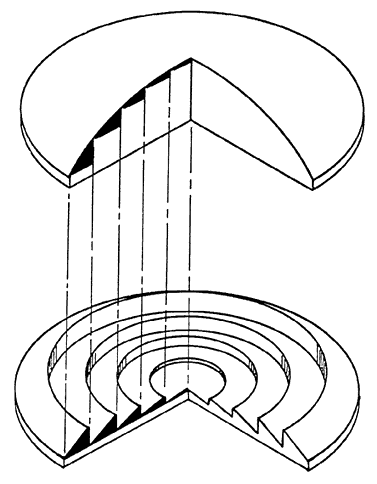
1. Fresnel, cited by Miller OE, McLeod JH, Sherwood WT: Thin sheet plastic Fresnel lenses of high aperture. J OptSocAm 41:807, 1951

2. Woodward F: Unlikely looking prisms. Read before the American Association of Certified Orthoptists at the 70th Annual Session of the American 
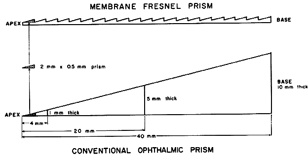
Academy of Ophthalmology and Otolaryngology, Chicago, 1965

3. Jampolsky A, Flora MC, Thorson JC: Membrane Fresnel prisms: A new therapeutic device. In Fells P (ed): Proceedings of the First Congress of the International Strabismological Association. London: Kimpton, 1971, p 183

4. Kors KK, Flora MC, Adams AJ: Fracturing and safety properties of glass spectacle lenses having back 
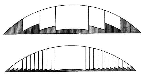
surface vinyl membranes. Am J Optom 49:471, 1972

5. Adams AJ, Kapash RJ, Barkan E: Visual performance and optical properties of Fresnel membrane prisms. Am J Optom 48:289, 1971

6. Ogle KN: Distortion of the image by prisms. J Opt Soc Am 41: 1023, 1951

7. Ogle KN: Distortion of the image by ophthalmic prisms. Arch Ophthalmol 47:121, 1952

8. Miles PW: Eliminating distortion due to prisms in glasses. Am J Ophthalmol 34:87, 1951

9. Morgan MW: Distortions of ophthalmic prisms. Am J Optom 40:344, 1963

10. Barkan E, Kapash RJ: Visual performance and optical properties of Fresnel membrane prisms: I. Residual astigmatism and spherical power. Read before the Annual Meeting of American Academy of Optometry, New York, 1972

11. Wild BW: Single vision lenses. In Borish IM: Clinical Refraction, ed 3. Chicago: Professional Press, 1970, p 1086

12. Clippers C: Comments on paper presented by Jampolsky et al. In Fells P (ed): Proceedings of the First Congress of the International Strabismological Association. London: Kimpton, 1971, p 190

13. Arruga A: Aplicaciones de los prismas membranosos en terapeutica estrabologica. Arch Soc Esp Oftal 31:381, 1971

14. Kapash RJ, Barkan E: Fresnel optics and human visual performance. Paper presented at the Annual Meeting of the American Academy of Optometry, Ontario, 1971

15. Miller OE, McLeod JH, Sherwood WT: Thin sheet plastic Fresnel lenses of high aperture. J Opt Soc Am 41:807, 1951

16. Hirsch M J: Prism in spectacle lenses for cosmesis. Am J Optom 45:409, 1968

17. Flom MC: Treatment of binocular anomalies of vision. In Hirsch MJ, Wick R (eds): Vision of Children. Philadelphia: Chilton, 1963, p 197

18. Guibor G: Squint and Allied Conditions. New York: Grune & Stratton, 1959

19. Weiss NJ: An application of cemented prisms with severe field loss. Am J Optom 49:261, 1972

20. Wechsler S, Weisman B: Fresnel lenses to correct twenty-four diopters of myopia. Am J Optom 49: 1030, 1972

21. Wick B: Uses of minus Fresnel press-on(tm) lenses to improve eyewear appearance in moderate to high myopia. Am J Optom 51:343, 1974

Back to Top