Chapter 36
Correction of Ametropia with Spectacle Lenses
SUSANNAH B. MARCUS and MELVIN L. RUBIN
Main Menu   Table Of Contents

Search

OPTICAL PRINCIPLES
CORRECTION OF MYOPIA
CORRECTION OF HYPEROPIA
CORRECTION OF ASTIGMATISM
EFFECTS OF SPECTACLE PRESCRIBING ON ACCOMMODATION
EFFECTS OF SPECTACLE PRESCRIBING ON BINOCULARITY
CORRECTION OF ANISOMETROPIA AND ANISEIKONIA
SPECTACLE DESIGN CONSIDERATIONS
REFERENCES

The provision of spectacles to correct vision is based on both science and art. Prescribing appropriate corrective lenses depends on the ability of the patient to adapt to change and the patient-specific relationship between accommodation and vergence. But first and foremost, a thorough understanding of the optical principles of lenses and of the eye is necessary. Armed with that knowledge and supplemented by clinical experience, you can attain true competency in providing optimal visual correction for each of your patients.
Back to Top
OPTICAL PRINCIPLES

OBJECT/IMAGE CONJUGACY IN AMETROPIC EYES

An object and the image of that object created by any optical system are said to be conjugate to one another. That is, the positions of the object and its image could trade places if the light rays were reversed. (This principal of object–image interchangeability evokes the ancient concept of the “visual spirit,” which was regarded as a vital force emanating from the eye of an observer to create visual perception.)

In a nonaccommodating emmetropic eye, a distant object is conjugate to the retina (Fig. 1); that is, a distant object is crisply imaged on the retina. Conversely, if the retina were the object, its image would be located at an infinite distance in front of it. If the eyes' optical elements do not create conjugacy between the retina and a distant object, ametropia exists. In the myopic eye, the sharply focused image of a distant object is not on the retina but located in front of it (Fig. 2). This occurs when the optical power is too strong relative to the existing axial length. This can happen either when the optical power of the eye is too great, the axial length is too long, or some combination of both factors. On the other hand, when an eye is hyperopic, light rays from a distant object strike the retina before they have converged to form a sharp image, and this can occur because the eye has insufficient optical power, a shorter than optimal axial length, or both (Fig. 3).

Fig. 1 In emmetropia, light rays coming from infinity are brought to focus on the retina.

Fig. 2 In myopia, the optical power of the eye is too strong relative to its axial length.

Fig. 3 In hyperopia, the optical power of the eye is too weak relative to its axial length.

Far Point and Optical Corrections

If the retina of an eye is thought of as an object, the image of the retina created by the eye's optical system will be located at the far point plane.1 The far point denotes the location at which the far point plane intersects the optical axis. That point is conjugate to the fovea of any nonaccommodating eye. In the emmetropic eye, the far point plane is located at optical infinity (beyond 6 meters). In the myopic eye, the far point plane is located not at infinity but somewhere in front of the eye. The higher the degree of myopia, the closer the far point is to the eye. The position of the far point plane relative to the eye, actually the reciprocal of its distance in meters from the cornea, indicates the exact amount of refractive error in diopters. In the hyperopic eye, the far point plane is virtual; that is, it is located behind the eye.

The far point forms the basis for optical correction of any ametropia. Why that is so requires another important optical principle: Any lens brings light from an object located at a distance and images it at that lens' secondary focal plane. Therefore, if that lens is positioned in front of the eye so that its secondary focal plane is superimposed on the eye's far point plane, that lens becomes a “corrective lens.” The lens creates a focused image of a distant object and puts it at the far point plane. That image then becomes the object for the eye's optical elements, which take over and bring a sharply focused image onto the retina (Fig. 4).

Fig. 4 Far point plane in myopia is in front of the eye. A lens is corrective if its anterior focal point coincides with the far point plane.

Lens Effectivity

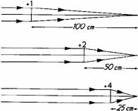
Almost any lens can become a corrective lens if its secondary focal plane is placed to coincide with the ametropic eye's far point plane. In other words, lenses of different powers may accomplish the same goal of providing sharply focused retinal images as long as this optical condition exists. All such lenses are said to have equal optical effectivity for distant objects. In fact, lenses that focus light from an object to the same point or plane are said to have equal effectivity (Fig. 5). This is an important principle because corrective spectacle lenses may be fit at different distances from the eye (or in the case of contact lenses, directly on the cornea) if their powers are appropriately adjusted. The distance between the posterior surface of a corrective lens and the anterior surface of the cornea is known as the vertex distance (Fig. 6). The effective power of a low-powered corrective lens will not vary significantly with small changes in vertex distance; however, the proper corrective power of high-powered lenses, such as those needed for high myopes, is very dependent on the lens' distance from the eye. In this latter situation, you need to specify both the corrective power and the vertex distance if proper retinal focus is to be attained.

Fig. 5 Lenses of +1.00, +2.00, and +4.00D positioned to bring parallel rays of light to focal points in the same plane. These lenses are said to have the same effectivity.

Fig. 6 Vertex distance (V.D.) is the distance between the anterior surface of the cornea and the back surface of the eyeglass or trial lens.

Example of prescribing a high-powered lens:

An aphakic patient's right eye is refracted at a vertex distance of 14 mm and found to be +12.00D. What power contact lens would provide the same optical correction (i.e., the same effectivity) (Fig. 7)?

Fig. 7 Contact lens versus spectacle corrected hyperopia.

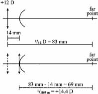
Both the +12.00D spectacle lens placed 14 mm in front of they eye and the +14.50D contact lens placed on the cornea will “correct” this eye because both lenses have their focal points coincident with the eye's far point. Again, both lenses will have the same optical effectivity (though the +12 lens will produce a larger retinal image).

Effect of Changing Lens Position

If a given corrective lens is moved along the visual axis, its effectivity is changed; the image of a distant object created by the lens will no longer be coincident with the far point, which is fixed in position relative to the eye and so will result in blurring the retinal image. An optician who fits spectacles at a vertex distance different from that prescribed by the refracting clinician must appropriately alter the power of the lenses to maintain equal effectivity. Alternatively, patients whose refraction shifts over time may find that pulling their old glasses further down their noses or pushing them closer to their eyes may improve visual clarity. What they are actually doing is positioning the lenses' secondary focal point closer to the new position of the eyes' shifted far point. This indicates the need for a new prescription. As an example, if a patient's myopia has gradually increased, as might occur with developing nuclear sclerosis, the eye's previous far point will have moved closer. The now undercorrected myope will find that pushing her glasses closer to her eyes will bring the lens' secondary focal point closer and coincident with the new far point position, and this will improve her distance visual acuity. An undercorrected hyperope will find that moving the glasses farther from his eyes will provide similar improvement.

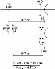
Example of how a change in spectacle vertex distance can compensate for a refractive change:

A patient who has been wearing -6.00D eyeglasses finds that, in order to see clearly, he must push his eyeglasses 1 cm closer to his eyes. What is his new refractive correction if he wants to use his current frame?

As illustrated in the diagram (Fig. 8), moving the lens closer to the eye has also moved its secondary focal point closer to the eye, making it coincident with the new far point of the eye. A -6.37D lens located at the initial vertex distance would allow correction of this new ametropia without having to adjust the position of the original frame and lens position. The patient has become 0.37D more myopic.

Fig. 8 Vertex distance alteration may compensate for a change in refractive error.

Back to Top
CORRECTION OF MYOPIA

Basic Optics

As previously mentioned, the far point plane of the myopic eye is located at a certain distance in front of the eye and determines the degree of myopia. The greater the myopia, the closer the far point will be. A real object placed at that distance (far point plane) will be in sharp focus on the retina. Thus, an uncorrected myope can hold an object at the eye's far point plane and see it clearly. The term nearsighted is an accurate description of the myope's visual condition. By measuring the distance at which a nonaccommodating myope holds reading material, one can make a good estimation of the degree of myopia present. For example, if a presbyopic patient holds reading material at 20 cm, he is likely to be a 5D myope.

For the myope, all objects located further away than the far point plane will result in blurred retinal imagery. The far point plane for even low degrees of myopia will be relatively near to the patient; 1 meter away for 1D of myopia, 4 meters away for only 0.25D of myopia. This accounts for the difficulty even “low” myopes have in recognizing a friend's face from across the street. To be seen really clearly, the friend must be no further away than the far point plane.

By definition, the lens that corrects a given degree of myopia images an object at the far point plane of the eye. A “minus” lens, a lens that diverges light, is necessary to accomplish this task. From objects at a distance, parallel light rays traveling from left to right will encounter the minus lens, the light rays will be diverged, and a virtual image will be formed to the left of the lens, at the lens' secondary focal point. When that point is located at the far point plane of the eye, that minus lens will “correct” the myopia (refer back to Fig. 4). Again, minus lenses of different powers can correct this eye as long as each is placed at the necessary vertex distance to keep its secondary focal point precisely positioned at the eye's far point plane. Each will have the same optical effectivity, and therefore, any of them may be used to correct this myopic eye. The closer to the eye the corrective lens is placed, the less divergence power it needs to allow its secondary focal plane to be coincident with the eye's far point plane, therefore necessitating a weaker minus corrective lens. Obviously, a contact lens will not need to be as strong as the corresponding spectacle lens to correct the same degree of myopia. Because of the small difference in power induced by changes in vertex distance in low refractive errors, it is not typically necessary to make corresponding prescriptive changes unless the myopia is 4D or greater.

Full Versus Partial Correction of Myopia

To create a truly clear retinal image of a distant object, the full extent of myopia must be corrected. However, several issues need to be considered. First, the length of the examination room is significantly shorter than infinity. So, when refractions are performed without cycloplegia, a slight undercorrection of the full myopia may result, but this is typically no more than 0.25D. This 0.25D discrepancy may be irrelevant, but it depends on the individual's tolerance for blur and his or her visual requirements. Usually for myopes, prescribing the actual refractive findings will be sufficient to improve vision satisfactorily.

Another factor is night myopia, which results from a reduction in contrast induced by low illumination. This causes the patient's focus to drift toward a “resting” level of accommodation that is not zero, thus inducing some degree of residual myopia. In such patients, this increase in myopia may be symptomatic, and patients may report blurred vision or halos around lights at night. Symptomatic night myopia requires correction, such as wearing nighttime driving glasses to correct 0.50D to 1.50D more than the degree of myopia you identify during your office refraction.2

Yet another prescribing consideration involves the patient's visual needs. A myope who is bedridden, for example, and infrequently leaves her home may not have a need for clear vision beyond a 10 ft distance. In addition, if the same patient is significantly presbyopic, undercorrecting the myopia may allow her to function comfortably with a bifocal instead of a trifocal or progressive power lens.

If a significantly myopic patient has never worn a correction before, prescribing the full correction may result in significant asthenopia. The patient may feel dizzy and uncomfortable and end up rejecting the glasses. In such cases, especially in older patients, partial correction of the myopia will likely dramatically improve vision, yet permit easier adaptation. It is a matter of clinical judgment whether or not to prescribe the full prescription to start with or, alternatively, to prescribe only a partial correction of the myopia. Either choice has the potential to provide a satisfactory visual result for the patient.

Myopic Progression and Spectacles

The topics of myopia progression and the effects of corrective lenses on this progression have been, and continue to be, hot topics for the ophthalmic community. Some think that the development of myopia is environmentally induced, and that excessive near work perhaps combined with accommodative or binocular dysfunctions are to blame. Such associations are supported in the literature.3,4 In such patients, many believe that what starts as accommodative spasm due to nearpoint stress may become imbedded into a permanent myopic condition. If this early stage of myopia is treated with “vision therapy” or orthoptic training instead of full myopic spectacle correction, this “pseudomyopia” might be reversible. On the other hand, others believe that the development of refractive error is based on a hard-wired inheritance pattern, and that attempted manipulations to alter progression are not indicated. Several recent studies have found heredity to be the most important factor in the development of both juvenile and adult onset myopia.5,6 Numerous studies utilizing methods for influencing accommodation (bifocals and/or pharmaceutical agents) have been undertaken in an attempt to address the topic.

For many years, some clinicians have prescribed bifocals for their emerging myopes in the hopes of staving off further myopic development. The COMET study (Correction of Myopia Evaluation Trial) was designed to evaluate this clinical practice and its outcome. Children were fit with either progressive lenses or single vision lenses and were followed for 3 years using cycloplegic autorefraction. Study results revealed that the group wearing progressives had developed only about 0.2D less myopia, occurring primarily during the first year of lens wear. There was a corresponding greater increase in axial length in the single vision lens group compared to the progressive lens group.7 The Myopia Progression Study examined a much smaller group, looking at the effect of bifocal prescribing on the myopic progression of esophoric children specifically. A similar outcome occurred in this study: The group that had been prescribed bifocals showed a slowing of myopic development over the first two years (0.1D less development per year for the first 2 years) that then stopped with a maintenance of the intergroup difference through the end of the study period.8 One study examining the effect on myopic progression of combination bifocal and atropine therapy found a lessened increase in myopia of, respectively, 0.15D/year (median treatment interval of 3.62 years) in the atropine/bifocal prescribed group as compared to the nontreatment group.9 Note, however, that not all prior studies found any benefit from the prescription of bifocals to control myopia development,10 and as previously indicated, those that did found only very little useful effect. The full answer is not yet in hand.

Back to Top
CORRECTION OF HYPEROPIA

Basic Optics

The far point plane (the image of the retinal plane) of the hyperopic eye is located behind the eye and, so, is virtual. That means that there is no real position in front of the eye of a nonaccommodating hyperope where an object can be placed so that a clear image can be created on the retina. A corrective lens for hyperopia is a “plus” lens. A plus lens converges rays of light from a distant object to its secondary focal plane, which, when it is made to coincide with the far point plane of the eye, will allow the eye's optical elements to form a clearly focused image on the retina (Fig. 9). As described previously with minus lens corrections, many different plus lenses offer equal effectivity, that is, will be corrective when placed at the appropriate vertex distance from the eye. If the hyperope is contact lens-corrected, the contact lens is positioned closer to the far point plane than any corrective spectacle lens would be, so the lens must be of higher plus (convergent) power to allow rays to be focused at the far point. As with myopic correction, this difference in necessary corrective power with varying vertex distances is irrelevant with low-power lenses, but can become quite significant in high hyperopic refractive errors, such as in aphakia. In such cases, the vertex-adjusted power can be easily determined mathematically or with the use of a table.

Fig. 9 Far point plane in hyperopia lies behind the eye. A lens is corrective if its anterior focal point coincides with the far point plane.

Full versus Partial Correction of Hyperopia

When and how to prescribe corrective lenses for the hyperope depends on the patient's age, degree of hyperopia, and accommodative and binocular status.

Young hyperopes innately discover that accommodation can be used to compensate or overcome mild to moderate degrees of refractive error. By using this accommodative ability to keep their retinal images clear, most hyperopic patients develop and maintain a degree of habitual accommodative tone at all times. For this reason, many hyperopes will be initially uncomfortable if you prescribe their full refractive correction, even in adulthood.

The manifest refraction of the hyperopic patient may be deceptive because the hyperope's habitual accom-modation has not been fully suppressed; although cycloplegic refraction often reveals a greater degree of hyperopia than is evident without cycloplegia, the hyperope may still not be able to sufficiently relax accommodation to accept the latent (hidden) component of the refractive error when the cycloplegia has worn off. The amount that will be accepted must be determined on a postcycloplegic refraction performed at least a week after the cycloplegic refraction. The “new” manifest refraction (which is aided by the information gained during the prior cycloplegia) provides a measure of the best visual acuity in the noncycloplegic state.

In the presence of high hyperopia, and especially when accompanied by a high AC/A ratio, attempts to accommodate may create significant esophoria or accommodative esotropia. This may result in diplopia and asthenopia that is worse at near. In such patients, prescribing maximum tolerated plus lenses, often in the form of bifocals, is indicated to normalize binocularity. If the patient is frankly esotropic, careful cycloplegic refraction should be done to suppress as much latent hyperopia as possible to determine the accommodative component of the esodeviation.

As an undercorrected hyperope ages and accommodative amplitude decreases, he is less able to overcome all the hyperopic blurring. This often first becomes evident with attempts to focus at near. Such patients may present at a young age with presbyopic symptoms, which the examiner will discover are merely due to uncorrected hyperopic refractive error. Simply correcting the distance ametropia will often completely alleviate the patient's near point symptoms. As he ages and his accommodative ability decreases further, the need for bifocals will be inevitable.

Example of an emerging presbyopic hyperope's first symptoms:

A 38-year-old patient presents with a complaint of blurred vision and eyestrain after reading for a short period of time. She explains that she has never worn glasses.

Entering VAs (uncorrected):

  OD 20/25-
  OS 20/30

Manifest refraction:

  OD +0.75 sphere 20/20
  OS +1.00+0.50 × 180 20/20

Cycloplegic refraction:

  OD +2.00 sphere 20/20
  OS +2.50 sphere 20/20

There are multiple prescribing options for this patient, but here are two.

  Option A: If the patient rejects the notion of wearing glasses full time, consider prescribing single vision reading glasses, such as OD +1.50, OS +2.00.
  Option B: If the patient prefers the idea of wearing a single vision pair of glasses full time, consider prescribing OD +1.00 sphere, OS +1.50 sphere.

Such a patient is frequently unable to wear the full cycloplegic refraction and requires more correction at near than at distance. However, the patient will likely reject any recommendation of bifocals.

Back to Top
CORRECTION OF ASTIGMATISM

Basic Optics

The astigmatic eye is unique in that it has two far point planes, one for each of the two principal meridians of the refractive error. In simple myopic astigmatism, one plane is located at infinity and the other is at a finite distance in front of the eye. In compound myopic astigmatism, the two far point planes are located at different distances in front of the eye (Fig. 10). Similarly, in simple hyperopic astigmatism, one plane is located at infinity, and the other is located behind the eye, whereas in compound hyperopic astigmatism, both planes are located at different distances behind the eye. In mixed astigmatism, one far point plane is located in front of the eye, and the other is located behind the eye.

Fig. 10 In compound myopic astigmatism there are two far point planes, corresponding with the two principal meridians of astigmatism.

For any object point, an astigmatic eye creates a complex bowtie-shaped image that is called the conoid of Sturm. The waist (narrowest point) of the conoid is called the circle of least confusion. At that location, the size of the blur circle formed from a point object is the smallest.

To correct astigmatic refractive error, cylindrical lenses are used. Such lenses create two line images, each with a specific orientation, from an infinitely located point object. In compound or mixed astigmatism, two simple cylinders oriented 90 degrees apart are used to create image lines at each of the far point planes (Fig. 11). The combination of two cylinders results in a spherocylindrical lens. The spherical equivalent of that lens is the spherical power that is dioptrically halfway between the highest and lowest of the lens' principle meridional powers.

Fig. 11 Two corrective cylindric lenses (or a spherocylindric combination) are needed to correct an eye with myopic astigmatism.

In with-the-rule astigmatism, the focusing elements of the eye require more convergent power in the 180° meridian, which is corrected by plus cylinder axis 90° (or minus cylinder axis 180°). In against-the-rule astigmatism, the focusing elements of the eye require more convergent power in the 90° meridian, which is corrected by plus cylinder axis 180° (or minus cylinder axis 90°).

Patients with astigmatism cannot achieve perfect retinal clarity by holding an object at any single position, as the myope is able to do. Neither will accommodative effort help the astigmat achieve proper focus as it will for the prepresbyopic hyperope. For patients with astigmatism, optical correction is the only option for creating sharp retinal imagery. The amount of astigmatism required to produce blur symptoms varies greatly among patients, but low degrees of astigmatism are rarely symptomatic.

Full versus Partial Correction of Astigmatism

As with hyperopes and myopes, visual correction of the astigmat is not often as clear-cut as the determination of a refractive endpoint. As with plus and minus lenses, spherocylindrical lenses also create image magnification or minification, but to different relative extents according to the specific attributes of each of the principle meridians. This nonuniform magnification or minification results in meridional aniseikonia, which sometimes produces disturbances in spatial orientation and asthenopia, particularly in the first-time spectacle wearer. A common complaint is that flat surfaces appear tilted. However, almost always, the patient will rapidly adapt to these initial symptoms, which cease to be annoying. If not, simply reducing the amount of astigmatic correction, while maintaining the spherical equivalent, is often all that is necessary to alleviate such symptoms. Maintaining the spherical equivalent ensures that the circle of least confusion formed by any residual uncorrected astigmatism will be located at the retinal plane, minimizing resultant blur. Previous spectacle wearers requiring significant changes in cylinder power or axis may experience similar perceptual disturbances, but again, adaptation is almost always rapid. Still, occasionally the correcting cylinder power will need to be reduced to facilitate lens acceptance.

Example of cutting the cylinder while maintaining the spherical equivalent:

A 68-year-old woman complains of a gradual worsening of her distance vision with her current eyeglasses. She also reports a history of maladaptation to changes in her eyeglass prescription in the past.

Entering distance VAs:

  OD -0.50+1.25 × 180 20/50
  OS -1.00+1.00 × 170 20/50

Manifest refraction:

  OD -1.00+2.75 × 180 20/20
  OS -1.50+2.50 × 170 20/20

To facilitate adaptation, the clinician may choose to cut the cylinder while maintaining the spherical equivalent. Maintaining the spherical equivalent ensures that the circle of least confusion remains on the retina providing the clearest possible vision, despite the existence of residual astigmatism. One-half the amount of the cylinder reduction should be added to the sphere power to maintain the spherical equivalent.

Tentative prescription:

  OD -0.50+1.75 × 180 20/30
  OS -1.00+1.50 × 170 20/30

The cylinder is reduced by +1.00D in each eye, and +0.50D is added to the sphere power in each eye to ensure the circle of least confusion remains on the retina.

Back to Top
EFFECTS OF SPECTACLE PRESCRIBING ON ACCOMMODATION
When prescribing spectacles, you should always consider how the refractive correction may affect the accommodative function of each patient, especially those with accommodative dysfunction or those with early or established presbyopia. A careless refractive exam may result in prescribing more minus than is actually needed, an especially common problem in the young, actively accommodating patient. Such overminused prescriptions will necessitate excess accommodative effort, especially during near tasks. Such accommodative stress may lead to asthenopia or even avoidance of near tasks such as reading. Particularly in the pediatric population (but actually important for all patients), a cycloplegic refraction is necessary to suppress accommodation during testing. As patient age increases and accommodative amplitude decreases, the negative impact of myopic overcorrection becomes even more marked. Such faulty prescribing may result in creating early presbyopia that erroneously requires premature prescription of multifocal lenses, much to the dismay of the patient. In fact, when early presbyopia occurs in a myope, we suggest you slightly undercorrect the myopia to delay the need for bifocals or progressives.

Similarly, the hyperope often benefits from the “pushing of plus,” which minimizes the adverse effects on accommodation from undercorrecting the refractive error. As in young myopes, hyperopic children always need a cycloplegic exam to uncover the total refractive error and expose any latency. Interestingly, even middle-aged hyperopes can have significant amounts of latent hyperopia. As with myopes, maximizing the hyperopic correction helps delay the need for multifocals in emerging presbyopia. Given the habitual accommodative tone that many hyperopes maintain and find comfortable, the clinician attempting to pick the ideal lens endpoint may find him- or herself on a slippery slope. This is where the art of prescribing is called upon.

Patients who require resorting to a partial correction of high astigmatism because of maladaptation to their full correction are prone to develop asthenopia because their intervals of Sturm are broad and result in continuous accommodative flux.

Back to Top
EFFECTS OF SPECTACLE PRESCRIBING ON BINOCULARITY
Because accommodation and binocularity are neurologically linked, one cannot regard one without the other. Because spectacles affect accommodation, the prescribing clinician needs to understand the resultant effects on binocularity, especially in patients with esophoria or esotropia.

All esophoric and esotropic patients should first have a cycloplegic refraction to uncover any accommodative spasm or latent hyperopia, and then appropriate treatment with lenses should be instituted. Once corrective lenses restore binocularity at distance, additional plus power may need to be prescribed in the form of bifocal lenses to lessen accommodative demand and any induced esotropia at near. Whether uncorrected hyperopia or a high AC/A ratio is at fault in inducing the eso tendency, spectacle lenses are frequently helpful in restoring binocular function.

In contrast, inadvertent overcorrection of myopia or undercorrection of hyperopia may result in a significant iatrogenic esophoria. Obviously, this should be avoided.

In exophoria and exotropia, the overcorrection with plus lenses or the undercorrection with minus lenses will likely exacerbate the condition because accommodation is relaxed. Here, prescription of the least plus, or sometimes even an overcorrection with minus lenses, may help to restore proper binocular posture. By inducing a state in which the patient must actively use some amount of accommodation, the linked convergence is activated and may be sufficient to correct the dysphoria or strabismus.

Back to Top
CORRECTION OF ANISOMETROPIA AND ANISEIKONIA
Anisometropia represents the condition in which a patient's refractive error significantly differs between the two eyes. To what degree the term significant implies is unclear, as the threshold of inequality resulting in adverse symptoms is unique to any given individual. Most commonly, a refractive difference of 2 diopters is considered significant in defining the presence of anisometropia, but the clinician's judgment is likely superior to any random designation.

Unfortunately, the asymmetric spectacle lenses that are prescribed in an attempt to equalize visual acuity create their own asymmetrical effects on magnification, and a discrepancy between the retinal image sizes between the two eyes is the final consequence. Aniseikonia (the perception of an image size difference between the two eyes) can create significant adaptation problems and prove to be a management challenge. These patients may complain of headache, eye ache, tearing, and decreased reading stamina. Patients may also experience vertigo and spatial distortions, sometimes so severe that ambulation may be hazardous, as steps and curbs may appear tilted (Fig. 12). Asymmetric astigmatism may result in meridional aniseikonia creating similar symptoms. Surprisingly, some patients are able to accept very anisometropic spectacle prescriptions without any symptoms at all, a testament to the remarkable adaptability of the human visual–perceptual system.

Fig. 12 Distortion of spatial perception results when retinal image in one eye is enlarged, as would occur if a plus cylinder at axis 90° were held before the right eye. Diagram shows binocular projection in this situation. Frontal-parallel plane appears to have rotated around the point of fixation.

In addition to the problem of lens-induced aniseikonia, anisophoria (differential prism effect between the two eyes and quantifiable by Prentice's Rule) occurs with off-axis viewing through the asymmetric spectacle lenses, which is required when looking downward through bifocal segments. Asymmetric prismatic effects between the eyes may result in both asthenopia and diplopia. Anisophoria is of little concern for anisometropic patients who wear single vision lenses because they can always avoid the lens periphery and look through the lens optical centers, where there is no differential prism effect.

When symptoms of lens-induced aniseikonia do occur, they can be reduced somewhat by minimizing the vertex distance or by fitting and prescribing contact lenses. Unfortunately, not all patients are good contact lens candidates, so spectacles may invariably have to be prescribed. Axial anisometropia, which is due to differing axial lengths between the two eyes, can be minimized by placing the corrective lenses close to the anterior focal planes of the eyes (15.7 mm in front of the corneas), as indicated by Knapp's Rule. However, most anisometropia represents a combination of both axial and refractive causes.11

The difficulty in solving the aniseikonia problem starts with the inability to exactly quantify its degree. The Space Eikonometer (American Optical Co.), an instrument that was designed to measure small amounts of aniseikonia, is no longer manufactured, though there is some newly available computer software, Aniseikonia Inspector (www.opticaldiagnostics.com), which permits a clinical measurement of aniseikonia to be made. However, even if it is not actually measured, a reasonable estimation may be made by assuming a 1% image size difference per diopter of anisometropia. Threshold tolerance of aniseikonia varies among individuals, so the determination of how much magnification difference a given patient will tolerate is elusive. It is said that image size differences between 1% and 5% are more likely to generate symptoms and interfere with fusion.12 Once image size difference is determined, spectacle lenses can be designed to minimize this discrepancy. By manipulating base curve, thickness, and vertex distance, image magnification can be beneficially affected and aniseikonia lessened.13

Back to Top
SPECTACLE DESIGN CONSIDERATIONS
The spectacle lens market, like most others, is driven by competition. As such, companies are continuously developing new and more desirable lens and frame materials to satisfy an ever-demanding public. Lens designs are being improved to provide better visual comfort and cosmesis. Newly available lenses and frames are more lightweight and comfortable. Instead of merely prescribing refractive findings, the clinician is in a prime position to recommend the best lens designs and materials.

Although lens weight and thickness have never posed a significant problem for patients with low refractive errors, historically those with significant degrees of refractive error have been quite encumbered by the very glasses they depended on. Over the last several decades, new lens materials, including high-index plastics, have emerged that have greatly improved lens appearance. Materials have been designed with protective qualities such as impact resistance and UV protection, allowing the clinician the opportunity to become active in promoting ophthalmic safety.

Polycarbonate and Trivex are plastic polymers that are excellent choices for correcting high refractive errors because they are lightweight and can be fabricated to very thin profiles. In addition, these materials have excellent impact resistance and inherent UV absorption, making them fine choices for children, for whom eye safety is a significant concern and who spend significant amounts of time out of doors. Choosing an impact-resistant material is equally important for patients who are monocular and those with hazardous vocations or avocations.

Proper lens design can also improve cosmesis and visual comfort. Aspheric lenses made with flatter base curves, thinner edge or center thicknesses, and decreased lens volumes facilitate the fabrication of thinner, lighter finished lenses. Frames are now made increasingly flexible, lightweight, durable, and with hypoallergenic materials, such as titanium and genium.

The prescribing clinician can now utilize both the art and science of refraction and choose from an arsenal of ophthalmic materials and designs, to provide a wide variety of products that correct vision both effectively and comfortably.

Back to Top
REFERENCES

1. Rubin ML: Optics for clinicians. 2nd ed. Gainesville: Triad Publishing Company, 1974:132–158

2. Milder B, Rubin ML: Night myopia. In The fine art of prescribing glasses. 3rd ed. Gainesville: Triad Publishing Company, 2004:87–89

3. Goss DA: Variables related to the rate of childhood myopia progression. Optom Vis Sci 67(8):631–636, 1990

4. Parssinen O, Hemminki E, Klemetti A: Effect of spectacle use and accommodation on myopic progression: final results of a three-year randomized clinical trial among schoolchildren. Br J Ophthalmol 73(7):547–551, 1989

5. Mutti DO, Mitchell GL, Moeschberger ML, et al: Parental myopia, near work, school achievement, and children's refractive error. Invest Ophthalmol Vis Sci 43(12):3633–3640, 2002

6. Iribarren R, Iribarren G, Castagnola MM, et al: Family history and reading habits in adult-onset myopia. Curr Eye Res 25(5):309–315, 2002

7. Gwiazda J, Hyman L, Hussein M, et al: A randomized clinical trial of progressive addition lenses versus single vision lenses on the progression of myopia in children. Invest Ophthalmol Vis Sci 44(4):1492–1500, 2003

8. Fulk GW, Cyert LA, Parker DA: A randomized clinical trial of bifocal glasses for myopic children with esophoria: results after 54 months. Optometry 73(8):470–476, 2002

9. Chiang MF, Kouzis A, Pointer RW, et al: Treatment of childhood myopia with atropine eyedrops and bifocal spectacles. Binoc Vis Strabismus Q 16(3):209–215, 2001

10. Grosvenor T, Perrigin DM, Perrigin J, et al: Houston Myopia Control Study: A randomized clinical trial. Part II. Final report by the patient care team. Am J Optom Physiol Opt 64(7):482–498, 1987

11. Milder B, Rubin ML: Anisometropia. In The fine art of prescribing glasses. 3rd ed. Gainesville: Triad Publishing Company, 2004:225–26

12. Linksz A, Bannon R: Aniseikonia and refractive problems. International Ophthalmology Clinics 5: 515–534, 1965

13. Milder B, Rubin ML: Anisometropia. In The fine art of prescribing glasses. 3rd ed. Gainesville: Triad Publishing Company, 2004:235–36

Back to Top