Chapter 20
Ophthalmoplegic Syndromes and Trauma
PAUL R. MITCHELL and MARSHALL M. PARKS
Main Menu   Table Of Contents

Search

DUANE RETRACTION SYNDROME
BROWN SYNDROME
MOEBIUS SYNDROME
FIBROSIS SYNDROMES
VERTICAL RETRACTION SYNDROME
STRABISMUS FIXUS
THYROTOXICOSIS OPHTHALMOPLEGIA
OPHTHALMOPLEGIAS
MYASTHENIA GRAVIS
TRAUMA
ORBITAL FRACTURE
SURGICAL TRAUMA
LOST MUSCLE
SLIPPED MUSCLE
REFERENCES

DUANE RETRACTION SYNDROME
Prior to publication of Alexander Duane's description in 19051 this syndrome had been described by Stilling2 in 1887 and Turk3 in 1896 and, consequently, should more correctly be called the Stilling-Turk-Duane syndrome. Duane emphasized thatretraction of the adducted involved globe is an essential clinical feature of this syndrome, and because this feature is so diagnostic, common usage of the designation Duane retraction syndrome is deeply entrenched and should be continued.

Clinical presentation of Duane retraction syndrome is varied. Invariably, in addition to retraction of the adducted globe, the patient exhibits a defect in horizontal motility. In addition, a vertical motility defect frequently occurs in adduction. Various contributors to the literature have categorized Duane syndrome4–10 into subtypes, but this may have its limitations, given that one type may merge with another. Furthermore, the various subclassifications are confusing because some are based on clinical findings4–6 whereas others are based on electromyographic findings.7–12 Huber,5 using electromyography, classified Duane retraction syndrome into three types:

  [*] Type I: Absence of abduction, normal or restricted adduction with associated retraction of the globe, and widening of the palpebral fissure on attempted abduction. Electromyography shows absence of electrical activity in the lateral rectus muscle on abduction but paradoxical electrical activity on adduction.
  [*] Type II: Exotropia with restricted adduction and abduction and retraction of the globe on adduction. Electromyography reveals electrical activity with contraction of the lateral rectus muscle on both abduction and adduction.
  [*] Type III: Severe restriction of both abduction and adduction, with either minimal esotropia or exotropia or near orthophoria and retraction on adduction and widening of the palpebral fissure on abduction. The electromyogram demonstrates cocontraction of the horizontal rectus muscles on both adduction and abduction.

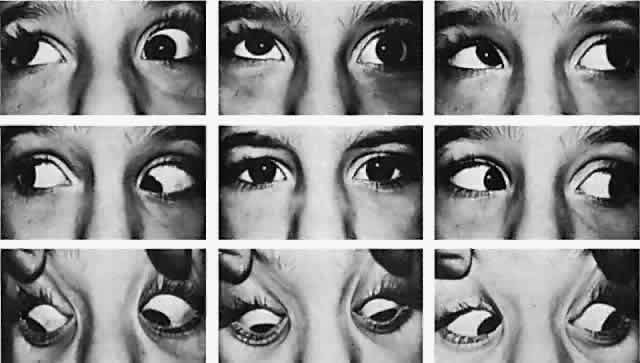
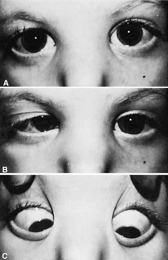
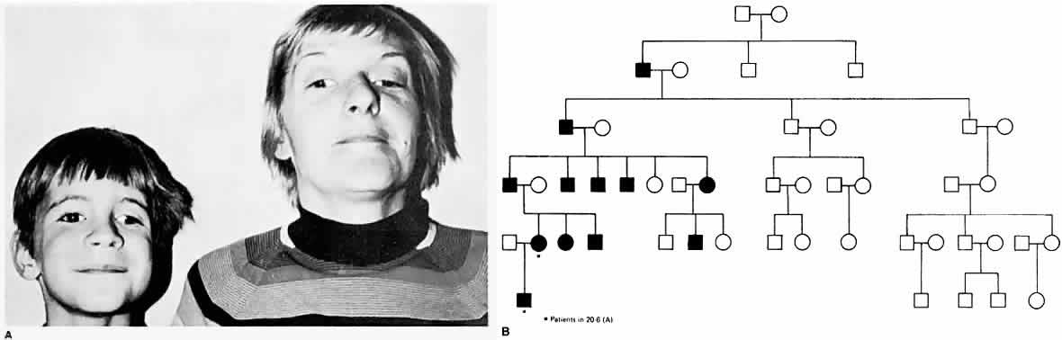
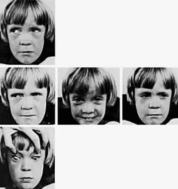
The most characteristic clinical presentation of Duane syndrome is an absence of abduction of an eye with some degree of restricted adduction and retraction when an attempt is made to adduct (Fig. 1). The retraction is variable: it is conspicuous in some but minimal in others. Additionally, either an upshooting or downshooting, or both, of the adducted eye frequently occurs, particularly as the adducting eye begins to move in the oblique position of up and in or down and in. This overshoot simulates overaction of the inferior and superioroblique muscles. Occasionally, the upshoot or down-shoot is so marked that the cornea is driven completely out of the palpebral fissure, hiding behind the upper or lower lid. Some patients manifest only the upshoot and a few only the downshoot, but most patients have various degrees of both vertical abnormalities. Magnetic resonance imaging (MRI) has been used in the cine mode13 to assess muscle contractility. In a patient with Duane type II the restriction of movement caused by the cocontraction of the horizontal rectus muscles was evident with the cine motion picture created by the MRI scanning unit.

Fig. 1. Duane retraction syndrome, left eye.

Duane retraction syndrome may be bilateral,14–20 and although it most commonly involves the left eye, it may involve only the right eye. Duane retraction syndrome occurs more frequently in female than in male patients. Several excellent indepen-dent statistical studies have confirmed these facts.Kirkham15 determined that 1% of the people with strabismus have Duane retraction syndrome. In his study of 100 patients, 65 were female. The left eye was involved in 60% of the patients, the right eye was involved in 22%, and there was bilateral involvement in 18%. In reporting on 186 patientswith Duane syndrome, Pfaffenbach and partners16 found that 57% of the patients were female; the left eye was involved in 60% of the patients, the right eye in 21%, and both eyes in 19%. Isenberg and Urist18 reported on 101 patients and found 57% female; 84% were unilateral and the left eye wasinvolved in 66% of the unilateral cases, and 16% ofthe bilateral cases. In a review of 97 patients,O'Malley and associates19 found 62% female; 82% were unilateral, of which 67% involved the left eye, and 18% were bilateral. Raab20 reviewed 70 patients, of whom 64% were female; 90% were unilateral, with the left eye involved in 67%, and 10% were bilateral.

In Duane retraction syndrome, there is frequently a concurrent Klippel-Feil anomaly, which occurred in 4% of the patients from Kirkham's series15 and in 3% of the patients from Pfaffenbach and coworkers' series.16 Congenital labyrinthine deafness is alsooften associated with this syndrome; 11% ofKirkham's patients and 7.5% of the patients inPfaffenbach and coworkers' series manifested this association. Ro and associates21 found evidence of hearing impairment in 15.9% when testing a series of 44 patients with Duane syndrome with audiograms and auditory brainstem responses. Duane retraction syndrome, the Klippel-Feil anomaly, and congenital labyrinthine deafness constitute the syndrome of Wildervanck22 Kirkham15 has suggested that Duane syndrome, perceptual deafness, and the Klippel-Feil anomaly are inherited as a single gene trait, with incomplete penetrance and incomplete expressivity. The clinical presentation in the heterozygote is a gene with pleiotropic effects inherited in an irregularly dominant manner. The abnormal gene may be partly gender limited in such a way that male patients are more resistant, and female patients more susceptible to the action of the gene.

Several articles23–27 have reported localizations of genes for patients with Duane syndrome, including 8q insertion and deletion,23 localization to chromosome 2q31,24,25 chromosome loci at 4q, 8q, and 22q,26 and 22q11 deletion syndrome.27

The epibulbar dermoids and preauricular skin tags of Goldenhar syndrome occur more frequently in patients with Duane syndrome than in people without Duane syndrome. Most cases of Duane syndrome seem to be sporadic; however, the syndrome has been reported by some and observed by one of the authors of this chapter (Parks) in three instances as a dominant inherited defect and has been reported by others, either as a unilateral28 or bilateral29 inherited syndrome. Chung and coworkers30 found 25 affected family members, distributed in an autosomal dominant pattern, in studying 110 members of 114 living relatives in an extended family with Duane syndrome. Twenty-four patients had bilateral Duane retraction syndrome, with a broad spectrum of severity. Amblyopia was present in 48% and strabismus in 76%. Other associated findings included cranial nerve (CN) IV palsy, partial CN III palsy, nystagmus, seizures, and deafness. Strabismus and amblyopia are far more common in bilateral than in unilateral Duane syndrome. Within families, a great deal of phenotypic variability is possible, and the genes responsible may affect the development of several CNs. Duane syndrome, associated with numerous congenital anomalies, probably results from dysgenetic events in the middle of the first trimester of pregnancy.31 The frequent association of nonocular abnormalities, such as the Wildervanck syndrome, with Duane retraction syndrome, suggests a teratogenic event that occurs between the fourth and eighth weeks of gestation.32 Acquired Duane retraction syndrome has been reported after palsy of CN VI33 as well as following Kronlein's lateral orbitotomy.34 Entrapment of the medial rectus muscle by a blowout fracture of themedial wall of the orbit, or by metastatic cancer, has been described by Duane and colleagues.35 This “pseudo-Duane retraction syndrome” differs in that there may be a history of trauma, diplopia is common, there is lid retraction on attempted abduction, and the prognosis is good when the entrapment can be reduced surgically. Various other findings and anomalies seen in association with Duane retraction syndrome include Holt-Oram syndrome,36 atrial septal defect and hand anomalies with autosomal dominant transmission; crocodile tears, anomalous lacrimation or paradoxical gustatory-lacrimal reflex, in unilateral37 and bilateral38 case reports; bilateral Duane syndrome, bilateral paroxysmal lacrimation and the Klippel-Feil anomaly39; familial perceptual deafness in five generations and Duane syndrome in two generations40; with myasthenia gravis41; with anisometropia and amblyopia42; with cleft palate43; with nevus of Ota and axial anisometropia44; with optic nerve hypoplasia45; with morning glory syndrome46 an unusual congenital optic nerve dysplasia, characterized by a funnel-shaped optic disc, annulus of surrounding pigment disturbance, and anomalous retinal vessels; with marfanoid hypermobility syndrome47; and with Marcus Gunn jaw-winking,48 fetal alcohol syndrome,31 Rubinstein-Taybi syndrome,49 and with de Morsier syndrome.50 Combined horizontal and vertical retraction syndrome has been reported.51 Duane syndrome has also been described in association with cat-eye syndrome52,53 with a supernumerary chromosome, probably derived from chromosome 22. Postmortem studies54–56 have shown absence of abducens nuclei and nerves from the brain stem, and partial innervation of the lateral rectus muscles by the oculomotor nerves54 and another study55 showed absence of the left abducens nerve and innervation of the left lateral rectus in part by branches from the inferior oculomotor nerve. Parsa and coworkers57 verified the absence of the abducens nerve in vivo in a 36-year-old woman with Duane syndrome type I in her left eye by MRI.

Most patients with Duane syndrome have straight eyes in the primary position, at least during infancy and childhood. The minority gradually develops an increasing esodeviation in the primary position that is offset by the restricted adduction of the involved eye. These patients find it possible to continue their normal binocular status; they adopt a compensatory turn of the head toward the side of the involved eye, thus offsetting the lateral gaze position that the eyes must assume to continue normal binocular vision. Usually, the quantity of abduction of the involved eye is nil; in some patients the eye cannotbe abducted even to the zero straight ahead position. Yet, a few patients with Duane syndrome can abduct the involved eye many degrees; they may have severe restriction of adduction in this eye with retraction on abduction, rather than adduction. This latter variety is designated as “inverse Duane retraction syndrome” by many, or Huber type II.5 The possibility of Duane retraction syndrome having progressive features was documented by Noonan and O'Connor58 in their adult reexaminations of 21 patients with known childhood Duane syndrome. The incidence of severe retraction on adduction, the presence of enophthalmos in the primary position, and the presence of upshoots and downshoots were significantly higher in adults with Duane type I than in children. The awareness that clinical features may become more severe with increasing age is important for future counseling and possible treatment.

Until the advent of electromyography,7–10,19,20 Duane retraction syndrome was attributed to replacement of the normal contractile substance within the lateral rectus muscle with fibrous tissue. This thesis supposedly explained the abduction deficiency, the restricted adduction, the retraction, and the frequently encountered upshoot and downshoot of the adducting eye. Biopsy specimens of the lateral rectus muscle often revealed an increase in fibrous tissue, possibly resulting from a change that occurs secondary to abnormal innervation. Huber5 revealed a paradoxical innervation of the lateral rectus as the pathologic mechanism in all forms of the Duane retraction syndrome through simultaneous electromyographic recordings of the medial and lateral rectus muscles of the affected eye. In 19 of 20 patients studied by Strachan and Brown10 gross abnormalities of the firing pattern of the lateral rectus were noted, varying from paradoxical innervation to innervation with incomplete inhibition in adduction and recruitment in abduction. Thecocontraction of the horizontal rectus muscles onadduction causes the retraction of the globe.9 Thevarious degrees of the paradoxic innervation abnormality determine whether any abduction of the involved eye occurs and also determine the degree of retraction of the eye on adduction. Furthermore, secondary anatomic changes may occur in the abnormally innervated lateral and medial rectus muscles, producing the positive traction test finding and the gradual esodeviation change that occurs in the primary position in some patients.

Auditory brain-stem response testing in one series59 suggested a pontine anomaly in 64%, always ipsilateral to the affected eye, or bilateral when bilateral Duane syndrome was present. Another series,60 however, did not reveal any consistent relationship between the affected eye and the auditory brain-stem response.

Unless the patient has adopted an unsightly compensatory head posture, surgery for Duane syndrome is contraindicated, because it cannot correct the anomalous innervation. Most patients eventually learn to mask the unsightly appearance caused by their motility defect by turning the head rather than the eyes to view in lateral gaze, thus revealing their concomitant esotropia on side gaze only when making a sudden lateral gaze movement when startled. However, surgery is indicated if a compensatory head posture develops to offset a primary position horizontal tropia. Diplopia is occasionally described as a symptom of Duane syndrome.53–56,59–63 Although surgery has been recommended to avoid diplopia in lateral gaze or to reduce asthenopia,63 most authors do not report diplopia and asthenopia as a common complaint, nor as an indication for surgical intervention. MacDonald and associates62 were unable to plot a suppression scotoma with binocular perimetry and concluded that patients with Duane syndrome possess a sensory adaptation to their incomitant strabismus. Some form of suppression must be used to ignore the extra image without producing a scotoma that could be plotted on a screen. The purpose of the surgery is to restore the eyes to a parallel alignment in the primary position, making the unsightly compensatory face turn unnecessary. The prognosis is excellent, provided that the esotropic eye has been surgically aligned to orthophoria in primary position. The literature on surgical correction of Duane syndrome begins in 1955, when Nutt64 recommended a recession of the medial rectus of the involved eye in cases of uncomplicated Duane syndrome with limitation of abduction.

At surgery, the medial rectus muscle of the involved eye is usually hypertrophied and taut. After the medial rectus muscle is disinserted, the positive traction test that manifested resistance to passive abduction becomes negative. Patients receiving surgery for the compensatory head posture resulting from esodeviated retracted eye require maximal recession of the medial rectus muscle. In addition to the recession, if the medial rectus is extremely tight, a Z-tenotomy can be performed on this muscle at the same time that it is maximally recessed. The lateral rectus muscle should not be resected, because an increase in the retraction of the globe follows this procedure. If retraction of the globe is marked and there is extreme narrowing of the palpebral fissure on attempted adduction, the lateral rectus muscle can be recessed at the same time that the medial rectus muscle is weakened. Recessing the lateral rectus muscle does not augment the deficiency of abduction, but it decreases retraction of the globe that occurs on adduction. Whatever surgery is performed to eliminate the primary position esodeviation in Duane retraction syndrome also further aggravates the adduction weakness. Transposition of the muscles,65–67 which entails moving all or portions of the vertical rectus muscles temporally to assist the deficient pull power of the rectus muscle, with the intention of giving the patient abduction beyond the zero straight ahead position, should not be considered, because this causes further embarrassment of adduction. The minimal gain in abduction from such procedures as the Hummelsheim or Jensen or total vertical rectus muscle temporal transpositions is unjustified when it is weighed against the significant adduction loss. However, in one series, Foster68 performed full vertical rectus muscle transpositions for lateral rectus palsies and for 5 patients with Duane syndrome type I. A full tendon transfer of the superior and inferior rectus muscles to the lateral rectus muscle was performed, along with a 5-0 Dacron scleral fixation suture,16 mm from the limbus, placed at the borders of the lateral rectus, and incorporating 2 to 3 mm of the transposed vertical rectus muscles. The medial rectus muscle was not recessed. There was no postoperative limitation of adduction, and 80% (4 of 5) had elimination of the preoperative face turn, whereas 20% had 5 degrees of residual face turn.All patients had a -3 to -3.5 limitation of abduc-tion postoperatively, and four patients had smallhorizontal or vertical deviations. Although aKestenbaum transposition has been advocated for the treatment of Duane syndrome,69 this procedure should be reserved for torticollis and compensatory head posture secondary to nystagmus. Secondary exotropia may be more likely to occur after medial rectus recession in bilateral Duane syndrome,70 and therefore simultaneous medial and lateral rectus recessions may be the treatment of choice to avoid this complication. Treatment of consecutive exotropia after medial rectus recession for Duane syndrome type I has been reported.71 The use of a posterior fixation suture72 has been reported for use in the recession of the medial rectus of the fellow eye in Duane syndrome type I to improve concomitance into the field of action of the involved eye. Despite a large recession of the medial rectus muscle in Duane syndrome with limited abduction, there usually is no significant increase in abduction postoperatively, nor is there a problem with overcorrections, despite large medial rectus recessions, as reported by Pressman and Scott.73

Although ocular alignment in the primary position can generally be achieved by recession of the ipsilateral medial rectus in esotropic patients with Duane type I, and by recession of the ipsilateral lateral rectus muscle in Duane type II,8,17,32,73–75 this further limits horizontal ductions in the operated eye. Therefore, the field of single binocular vision may potentially not improve, and may actually worsen.73,74,76 For esotropia greater than 25 prism diopters in the primary position, Kraft75 has recommended recession of the medial rectus, both eyes, especially when recessing the ipsilateral lateral rectus for globe retraction. Kraft suggests that the recession of the normal medial rectus muscle can stabilize the postoperative results because of Hering's law of equal innervation. For exotropia greater than 25 prism diopters in the primary position, recession of the lateral rectus of both eyes is recommended, for the same reasons.

Saunders and coworkers76 also recommend surgery on the normal eye in patients who lack severe duction deficiencies preoperatively, which not only helps restore primary position alignment but also expands the field of single binocular vision by creating compensatory duction limitations. Sprunger77 described another benefit of recession of both horizontal rectus muscles, by the treatment of significant globe retraction in the primary position in Duane syndrome, and in the improvement of enophthalmos.

Surgery for the leash effect of overelevation and overdepression of the involved eye in adduction is best directed at preventing the tight lateral rectus muscle from snapping around the dorsal surface of the adducted globe (simulating overaction of the inferior oblique muscle) or from leashing around the ventral surface of the adducted eye (simulating an overacting superior oblique muscle). Tenotomizing the superior oblique muscle or recessing the inferior oblique muscle does not reduce the excursion of vertical displacement of the globe caused by the leashing of the tight lateral rectus muscle. The leashing of the lateral rectus is eliminated by placement of a permanent posterior fixation suture on the superior and inferior one third of the muscle width 14 mm posterior to the insertion.78 In addition to the placement of the posterior fixation suture, the lateral rectus also may be recessed to reduce retraction of the globe alone or combined with the restricted adduction that results from the tight muscle. Neither the placement of the posterior fixation suture nor the recession further reduces the already deficient abduction in the involved eye in Duane syndrome. Jampolsky proposed splitting the end of the lateral rectus into a Y configuration, as a means of reducing the upshoot or downshoot in adduction.79 Rogers and Bremer80 demonstrated a marked decrease in upshoot or downshoot in their series of patients who had a Y splitting of the lateral rectus, with or without recession of the ipsilateral medial rectus. In the presence of marked enophthalmos, the authors suggest a recession of the lateral rectus in addition to the Y splitting. Von Noorden81 recommended recession of both horizontal rectus muscles in the presence of both upshoot and downshoot in type III Duane syndrome. The bridle effect was reduced by placing the insertions posteriorly, in relation to the center of the globe.

When surgical intervention is required in Duane retraction syndrome, the approach should not be based purely on classification type, but on the alignment in primary position, the presence and degree of abnormal head position, the severity of retraction, and the pattern of upshoot, downshoot, or accompanying A, V, or X pattern.75

Back to Top
BROWN SYNDROME
The motility defect manifest by an inability to raise the adducted eye above the midhorizontal plane was first described by Brown.4,82,83 Less elevation restriction is usually apparent in the midline, and even a smaller elevation deficiency is detectable in abduction. Slight downshoot of the adducting involved eye is often present, although no overdepression, simulating overaction of the superior oblique muscle, occurs. A widening of the palpebral fissure on adduction is associated with this elevation restriction (Fig. 2). Exodeviation usually increases as the eyes are moved upward in the midline.

Fig. 2. Brown's syndrome.

Brown84 has redefined this syndrome, recognizing that it is more complex than originally thought because it constitutes a spectrum of varying degrees and different causes. Brown initially reasoned that the simulated inferior oblique muscle palsy comprising this syndrome resulted from an innervational deficit to this muscle, with secondary contracture of the anterior sheath of the superior oblique tendon. Electromyography proved this concept erroneous by invariably demonstrating normal innervation to the inferior oblique on attempted upgaze.84–86 Electromyography of the oblique muscles in the superior oblique tendon sheath syndrome87 did not reveal any evidence of cocontraction but did demonstrate smooth recruitment of the electrical activity from the relaxed to the maximally contracted state. However, Brown describes the true sheath syndrome as congenital, permanent, and invariably associated with a positive traction test result manifested by inability to passively elevate the adducted involved eye. Brown syndrome has been reported in monozygotic twin girls,88 with reversed asymmetry (mirror imaging), with one child having Brown syndrome, left eye and amblyopia, right eye, and the second child with Brown syndrome, right eye, and amblyopia and esotropia in her left eye. Bilateral Brown syndrome has been reported in three siblings in one family pedigree, with other family members unaffected.89 Bilateral Brown syndrome with delayed onset in the second eye has been reported by Kraft and coworkers.90 All six patients were initially diagnosed with unilateral congenital Brown syndrome, without evidence of Brown syndrome in the contralateral eye. Brown syndrome is bilateral in about 10% of cases.91–93 The right eye seems to be involved more than the left eye according to some reports,92,93 but evidence is not conclusive to support a predilection for either sex or laterality.94 Most patients lack vertical misalignment in the primary position, but in some the involved eye is hypotropic and fusion occurs only in downgaze, causing a compensatory chin-up face posture. Some patients with Brown syndrome lack full fusion and stereopsis, even with an accompanying head posture, and the torticollis may persist even if one or the other eye is patched. Amblyopia may be present.91 Incising or removing the “tendon sheath” has not produced good results in improving the compensatory head posture or in overcoming the primary position vertical tropia or the elevation deficiency of the adducted involved eye. These poor results have provoked some doubt that the taut anterior sheath is the cause of this syndrome.95–98 In fact, the use of the term sheath is a misnomer. The superior oblique tendon does not possess a sheath98; instead, it penetrates the Tenon capsule, to which it is attached by an elastic connective tissue sleeve. The terminal half of the tendon is below the Tenon capsule, and this portion is completely surrounded by intermuscular septum. Superior oblique tendon weakening surgery should be confined to the sub-Tenon portion, which should be approached visually, rather than blindly. The surgical technique should leave the Tenon's capsule unopened beyond 10 mm from the limbus to avoid the orbital fat: the intermuscular septum should be left intact except at the point of tenotomy, and the superior oblique insertion should remain undisturbed. When surgery on the superior oblique tendon is not carefully performed under direct vision, Tenon's capsule and the intermuscular septum invaginate around the tip of the muscle hook and encircle the superior oblique tendon, producing a thickened layer of tissue erroneously interpreted as a “sheath.” Unfortunately, the word sheath remains ingrained in the ophthalmic literature. Improvement has resulted from tenotomizing the superior oblique tendon, but this procedure has in turn produced a new problem (i.e., palsy of the tenotomized superior oblique muscle associated with the opposite vertical tropia in downgaze that is offset with torticollis).96–101 Undoubtedly, a taut superior oblique tendon is the cause of this syndrome.95–101 Scott and Knapp102 removed the sheath tissue from around the superior oblique tendon and sutured the eye with coarse nonabsorbable material that was passed through the horizontal rectus muscles near their insertion to the nasal portion of the upper lid, fixing the eye in an upturned adducted position for several days. The results produced by this procedure have been equivocal and controversial.96–99,103

A group of patients who manifest resistance to elevation in adduction and who on occasion can elevate the adducted involved eye are included in the spectrum of this motility defect. The elevation is abrupt and associated with an audible snap that both the examiner and the patient can palpate in the superior nasal orbit. Pressing the nasal orbital tissue with the index finger often assists the abrupt elevation of the involved eye as though this pressure releases whatever is restraining the movement. This intermittent inability to elevate the adducted eye may be acquired at any age and also may eventually disappear.82–84,99–113 The traction test in this latter type of patient may be normal or at least less unequivocally abnormal than it is in patients with the more severe variety of this syndrome. Brown considers these patients to have something other than the true sheath syndrome; hence, he describes them as having a simulated sheath syndrome.84 According to Brown, their motility disorder may be congenital; he attributes the disorder to a thickened area in the superior oblique tendon posterior to the trochlea or some firm attachment between the posterior tendon and the posterior sheath, either of which interferes with the forward movement of the tendon through the trochlea. Brown considers the acquired simulated sheath syndrome to be secondary to an inflammatory process extending from the contiguous ethmoidal cells to the posterior sheath and tendon. One of the authors of this chapter has operated on a patient who developed Brown syndrome secondary to surgery for frontal sinusitis; at surgery the superior oblique tendon was found to be extremely taut and the tissue of the Tenon capsule covering the superior oblique tendon and superior rectus muscle was thick, opaque, and yellowish. The superior oblique tendon was as taut as the tendon encountered in nine other patients who had a severe degree of congenital Brown syndrome with manifest chinelevation and vertical strabismus in the primary position.98 Rheumatoid arthritis has been associated with this entity, and it is presumed that formation of some nodules on the posterior tendon of the superior oblique muscle accounts for the transient or permanent motility defect.108–120 Therapy has included topical and oral steroids113,114 and local injection of methylprednisolone into the trochlear region.115,118 A clinicopathologic study121 of an acquired superior oblique tendon sheath syndrome did not reveal any inflammation or scarring of the trochlea and superior oblique muscle and tendon. Perisheath adhesions anterior to the trochlea were presumed to be the cause, rather than an intrasheath pathologic state. Tucking of the superior oblique tendon also produces Brown syndrome.122,123 An adherence phenomenon in either the superior or inferior nasal quadrant of the anterior orbit produces the same motility defect as found in Brown syndrome. Raab124 found an anomalous fibrous band between the superior oblique tendon sheath and upper nasal intermuscular quadrant. Improvement was observed after surgical division of the fibrous band. An insertion anomaly of the superior oblique muscle in the superior nasal quadrant has been described as well.125,126 The simulated sheath syndrome may also be seen following orbital floor fracture,127 with focal metastasis to a superior oblique muscle,128 with orbit metastasis,129 after blepharoplasty,130 after frontal sinus surgery,131 after blunt orbital trauma,132 in association with frontal sinus osteoma,133 and in association with systemic lupus erythematosus.134 A patient with Brown syndrome with cyclic characteristics has been reported.135 A case of acquired Brown syndrome has been reported,136 in association with Hurler-Scheie syndrome. This possibly was caused by shortening of the superior oblique tendon, associated with shortening of the long tendons of the arms and feet, commonly occurring in Hurler-Scheie syndrome.

The motility defect as described by Brown is a definite entity with many different causes. After studying 54 embryos and fetuses, Sevel137 suggested that the fine trabeculae between the superior oblique tendon and trochlea, which persist into adulthood, most likely act like tethering strands to control and limit the excursions of the tendon within the trochlea. Helveston and associates138 have suggested that fluid accumulation, concretion in the bursa-like space, or vascular distension in the sheath might cause limitation of superior oblique tendon motion through the trochlea and bring about an acquired Brown syndrome. Computed tomography (CT) scanning may appear normal114 or may reveal thickening in the reflected portion of the superioroblique tendon118 in acquired Brown syndrome. Magnetic resonance imaging (MRI) in congenital Brown syndrome139 reveals enlargement of the tendon-trochlea complex, irregularity in shape, and intermediate signal intensity. However, MRI does not provide predictive information on the outcome of the disease process. The cause of this defect in most patients remains undetermined, but the “sheath” of the superior oblique tendon is probably blameless, although the tendon itself is undoubtedly short and taut in many of these patients. Therefore, Brown syndrome is an appropriate designation for the disorder in these patients, but the term sheath syndrome is probably inaccurate and misleading.

Spontaneous cure or improvement is rarely discussed in the literature. Gregersen and Rindziunski140 studied 10 patients with Brown syndrome an average of 13 years. Spontaneous cure occurred in three patients, who developed normal motility. In six patients who improved in varying degrees, there was reduction in the initial hypotropia and depression of the adducted eye. Kaban and coworkers141 studied 71 patients retrospectively, finding only 2 (5%) who demonstrated worsening of primary po-sition hypotropia, and 6 (10%) who experiencedcomplete spontaneous resolution of the elevationdeficiency. Waddell109 reported improvement or resolution of Brown syndrome in 24 of 36 patients (67%) who were examined from 1 to 14 years after the initial diagnosis of Brown syndrome. Most congenital Brown syndrome cases are constant, are less likely to improve spontaneously, and if severe, may require surgery. Most cases of acquired Brown syndrome are intermittent and are more likely to respond to medical management or to improve spontaneously, although there are exceptions to these generalizations.94 Brown syndrome associated with overaction of the contralateral inferior oblique muscle142 probably begins as bilateral Brown syndrome, followed by spontaneous improvement of the Brown syndrome on one eye, and subsequent secondary inferior oblique overaction.

Surgery can eliminate Brown syndrome if thesuperior oblique tendon is tenotomized. SinceBerke's143 description of superior oblique tenotomy, several other weakening procedures have been described along with variations of the Berke tenotomy.95–100 Recession of the superior oblique tendon from 12 to 14 mm has been described by Parks98 and others.144 Surgical complications seen with superior oblique tenotomy145 include severing a vortex vein with subsequent orbital hematoma, transient or permanent ptosis, failure to find the tendon, weakening only a portion of the tendon, or inadvertent severing of the superior rectus muscle instead of the superioroblique tendon.146 Grave and irreversible complications147 occurring after superior oblique tenotomy or tenectomy include not only superior oblique palsy, but also may include diplopia worse in downgaze, incomitant vertical deviation with significant torsion, and anomalous head postures. By carefully avoiding the orbital fat cushion148 10 mm from the limbus, fibrofatty proliferation and the adhesive syndrome can be avoided. The standard weakening procedure for Brown syndrome has been the superior oblique tenotomy or tenectomy.96–99 However, the high incidence of superior oblique palsy (66% to 85%) has led to the need for secondary surgery in most patients.98–100 When secondary surgery has been required, the ipsilateral inferior oblique is usually recessed because of overaction, and occasionally, recession of the yoke inferior rectus has been required. Parks and Eustis101 had recommended simultaneous superior oblique tenotomy and inferior oblique recession in the treatment of Brown syndrome to prevent reoperation for iatrogenic superior oblique palsy. A good to excellent result had been achieved in 94% of the operated eyes, and this combined procedure was previously their treatment of choice in Brown syndrome. Sprunger and coworkers149 advised superior oblique tenotomy without simultaneous weakening of the ipsilateral inferior oblique muscle as an initial procedure, because 42% of their series observed for at least 1 year postoperatively did not develop superior oblique palsy. Superior oblique tendon expander, with a segment of 240 silicone retinal band, sutured to the cut ends of the superior oblique tendon, first reported by Wright150 has been used by others151–156 to modify and improve on the superior oblique tenotomy in Brown syndrome and in superior oblique overaction. Stereoscopic vision has either been maintained or has improved in those who have had the silicone spacer, compared with those who have had tenotomy alone,151,152,154 in whom stereoscopic vision has been lost in some. In one report,152 adhesions of the silicone spacer and the nasal border of the superior rectus muscle alone or combined with the sclera has led to downgaze restriction and diplopia in the reading position, requiring reoperation. In a report of 66 cases using a silicone expander for overacting superior obliques,153 1 patient acquired a Brown-like restriction, with inability to elevate in adduction past the midline. In a study of long-term results of the silicone expander (6 to 10 mm) for moderate and severe Brown syndrome (Brown syndrome “plus”), Stager and coworkers154 reported on 20 eyes of 19 patients. All patients had resolution of the down-shoot in adduction, with partial or full ability to elevate the eye in adduction. Reoperation for over-correction was required in 20% of patients, generally by using silicone expanders of shorter length (5 to 8 mm). Downgaze restriction was not noted postoperatively. There may be an initial undercorrection, with improvement continuing for up to three years. Wright156 reported long-term results in 15 consecutive patients with Brown syndrome who underwent the silicone expander tendon elongation procedure, from 1987 to 1997. On a scale of 1 to 10, with 10 being the best outcome, 87% of the patients were graded 7 to 10 with a single surgery. Of the 15 patients, two did not have true Brown syndrome, but they had limited elevation in adduction not caused by a tight superior oblique tendon. Therefore, of the 13 with true Brown syndrome, 12 of 13 (92%) had a good outcome (graded 7 to 10), and 10 of 13 (77%) had normal postoperative versions. Use of the silicone expander reduces the need for simultaneous or subsequent inferior oblique recession, and represents a true advance in the treatment of Brown syndrome.154,156

Back to Top
MOEBIUS SYNDROME
The abnormal ocular motility involved in Moebius syndrome is only a portion of this relatively extensive malady. Von Graefe and Saemisch157 first described a case of congenital paralysis of CNs VI and VII in 1880, but the reports and classification of congenital ophthalmoplegias by Moebius in 1888 and 1895 have given this syndrome its name.158,159 Horizontal versions are congenitally absent in this syndrome. Congenital esotropia is often present; yet some patients are orthophoric in the primary position. A or V patterns are common in Moebius syndrome, and if straight eyes can be achieved in either upgaze or downgaze, a compensatory head posture is adopted to continue binocular vision. Most reported cases have been sporadic, with both genders affected with equal frequency. Autosomal dominant, autosomal recessive, and X-linked pedigrees have been reported.160,161 Deletion of chromosome 13 in two reports162,163 suggests that the Moebius gene is located in the region of chromosome 13.

Esotropia and inability to abduct the eyes are the usual reasons for the patient to be referred to an ophthalmologist. Horizontal versions are lacking, but vertical version and convergence are intact (Fig. 3). Because of the inability to move the eyes horizontally in response to horizontal version stimulation, the patient may maintain fixation of the laterally moving target by voluntarily converging. Pupillary constriction and increasing accommodation occur as the voluntary convergence increaseswhile the patient visually tracks the target farther and farther into either right or left gaze. Normal reading despite limited eye movements has been reported in two patients with Moebius syndrome, suggesting that normal eye movements are not required for the development of reading skills, and that efforts to normalize eye movements in poor readers by vision therapy may be misdirected.164

Fig. 3. Möbius' syndrome. A. Primary position. B. Convergence. C. Downgaze.

Bilateral palsy of CN VII is manifest in the orbicularis muscles by laxity of the lower lids, which allows the tears to pool between the lower lid and the bulbar conjunctiva. Forced closure of the lids may be inadequate to even approximate the upper and lower lid, but if they can be closed, the examiner can easily pry them apart (Fig. 4). Palsy of CN VII is further manifest by the expressionless smooth face, absent nasolabial folds, round mouth, and the inability to grin and wrinkle the forehead normally.

Fig. 4. Möbius' syndrome. A. Forced closure of lids. B. Examiner prying lids apart.

The most consistent associated defects encountered in Moebius syndrome are palsy and hypoplasia of the tongue, due to hypoglossal nerve paralysis, which is manifest by an abnormal sucking reflex during infancy. The history provided by the parents indicates that the neonatal difficulty in maintaining the infant's weight occurred because of the feeding problem. In patients with multiple central nervous system defects, a tracheotomy and a feeding gastrostomy and possibly a Lindeman laryngeal diversion may be necessary to counter life-threatening aspiration.165 Tongue palsy is also manifest in the abnormal phonation and the retarded development of speech. The terminal third of the tongue is atrophied, characterized by being furrowed, fissured, narrow, and pointed (Fig. 5). Some degree of mental retardation may be present,165–167 and frequently there is an associated deafness. Osteologic defects159,165–169 are common in the form of supernumerary digits, syndactyly, brachydactyly, clubbed feet, rocker-bottom feet, atrophy of the peronealmuscles, and a peculiar gait. Associated defects in the musculature of the neck and chest may be found159,165–167,169,170 Congenital absence of the pectoralis muscles alone, or with absence of breast or ipsilateral hand deformity (the Poland anomaly) is noted occasionally in association with the Moebius syndrome.169–171 One series of 17 children with Moebius syndrome found 40% with many or all of the symptoms typical of autistic disorder.172 The high frequency of autistic symptoms may be a marked overrepresentation and may suggest a common underlying neurobiologic deficit at the level of the brain stem. Acute onset Bell's palsy with resolution in a patient with Moebius syndrome has been reported.173

Fig. 5. Tongue in Moebius syndrome.

The motility, facial, and tongue findings in Moebius syndrome are due to aplasia of the nuclei of the abducens, facial, and glossopharyngeal nerves.161 The medial longitudinal fasciculi are probably also defective.159,166,174,175 Abnormal brain-stem auditory evoked potentials have been reported,176 indicating a dysfunction of the auditory tract at a supranuclear level. This suggests a more general involvement of the central nervous system than only the CN nuclei.

Because patients with Moebius syndrome often have esotropia at birth, they must be differentiated from those with the usual type of congenital esotropia. Although surgery for strabismus should be performed at a young age, as advocated for primary type congenital esotropia, invariably the traction tests and the character of the muscles encountered at surgery in patients with Moebius syndrome are abnormal. The positive traction test shows pas-sive resistance to both adduction and abduction, whereas vertical ductions are normal. The horizontal muscles are taut, thickened, and fibrotic177 and resemble the medial rectus muscle usually found in the involved eye in Duane syndrome. Aplasia of the extraocular muscles with complete absence of horizontal rectus muscles and fibrous tissue replacement has been reported.178 Miller and Stromland179 suggest the term Moebius sequence rather than syndrome, because sequence defines a cascade of secondary events, occurring after an embryonic insult from many heterogeneous causes. Associated findings in Moebius sequence may help reveal the location and timing of the developmental disturbances. The concept of a lesion found only in a nucleus of CN VI is not an adequate explanation for the wide range of ocular motility patterns.

Back to Top
FIBROSIS SYNDROMES
Tissue of the extraocular muscle is abnormal because of the replacement of the normal contractile substance with fibrous tissue, interfering with movement of the eye.180–182 The movement interference may result from either lack of pull power within the fibrotic muscle or resistance offered by the fibrotic muscle to its normally contracting antagonist. The latter has been speculated to produce retraction of the globe; this is the old thesis used to explain Duane retraction syndrome. Fibrosis syndromes are extremely varied: they range from congenital to acquired, may be unilateral or bilateral, and may affect all of the muscles of both eyes or only one muscle,181 or two muscles,182 and the degree of fibrosis of a muscle is total or partial.183,184 CT has been used in the diagnosis of congenital fibrosis syndrome185 showing marked loss of muscle volume in affected muscles. Genetic studies of multiple families with classic congenital fibrosis of the extraocular muscles (CFEOM) have demonstrated linkage to the centromere region of chromosome 12, now known as CFEOM-1.186–188 CFEOM-2, inherited exotropic strabismus fixus, is linked to markers on chromosome 11q13, and is known as CFEOM 2.189 A nonprogressive eye movement disorder, CFEOM-3, with variable expression of ptosis and restrictive ophthalmoplegia has been localized near the telomere of the long arm of chromosome 16.190

GENERALIZED FIBROSIS SYNDROME

Brown191 first used the description of general fibrosis syndrome while reporting 3 sporadic cases with complete bilateral ptosis and fibrosis of all extraocular muscles. There were adhesions between the globe and Tenon's capsule, so that depression and elevation of the eyes was not possible and lateral motion was extremely limited. All muscles, including the levator muscle, tend to be involved in this bilateral syndrome, although the muscles of one eye may be more involved than those of the other. Some muscles may be minimally fibrotic whereas others are maximally involved. The inferior rectus muscles are usually the most prone to maximal involvement, causing the eyes to be drawn downward and offering resistance to elevation in abduction and midline. Ptosis is associated with the motility defect in the eye, although a case of unilateral congenital fibrosis syndrome has been reported, presenting with hypertropia and minimal ptosis.192 Horizontal movements of the eye may be limited to a few degrees in one or both directions or there may be no horizontal movement. Because of the downturned fixed po-sition of the globes, the patients usually adopt achin-up position to fixate straight ahead centrally. This is a dominantly inherited condition179,182,193–195 affecting multiple family members (Fig. 6), it but may also be sporadic.194,195 Amblyopia may occur and is treated with occlusion when appropriate.179

Fig. 6. A. Generalized fibrosis syndrome. B. Autosomal dominant inheritance.

The surgical approach to center these eyes and to improve the compensatory head posture must be unorthodox. Recessions of the inferior rectus muscles of 8 mm or more are required to allow the eyes to come to a relatively level position. Bilateral frontalis suspension surgery for the ptosis is the safest approach because of the absence of a Bell phenomenon. The surgeon should purposely undercorrect the ptosis to prevent cornea damage from exposure in the absence of a Bell phenomenon. Surgery for the ptosis can be combined with the recession of the inferior rectus muscles. Scott196 described transposition of the superior oblique tendon to a position between the superior and medial rectus muscle insertions, in association with other extraocular muscle surgery for correction of hypotropia and exotropia in congenital fibrosis

CONGENITAL FIBROSIS OF THE INFERIOR RECTUS MUSCLE

Congenital fibrosis of the inferior rectus muscle183 is probably a variant of the generalized fibrosis syndrome, but only the inferior rectus muscle becomes involved, because of its peculiar predilection for becoming fibrotic. The condition may be unilateral or bilateral, but if it is bilateral, it is usually asymmetric, with greater involvement of one eye. Vertical strabismus is inevitable with this disorder, and if the patient maintains binocular vision, a compensatory chin-up posture is adopted.

Congenital fibrosis of the inferior rectus muscle must be differentiated from double elevator palsy in which the traction test result is normal; however, with fibrosis of the inferior rectus muscle, it is impossible to elevate the eye passively. Surgical correction of this disorder requires maximal recession of the inferior rectus muscle. As soon as the inferior rectus muscle is disinserted, the traction test is converted to normal.

Back to Top
VERTICAL RETRACTION SYNDROME
Khodadoust and von Noorden197 described a vertical retraction syndrome comprising inability to depress either eye while abducted and also restricted elevation of either eye in abduction. In addition to the vertical duction limitation in each eye of two siblings, they described the retraction of each eye during attempted depression with the eye in the abducted position. The traction test result was positive in both patients for both upgaze and downgaze limitation. One patient had exotropia on upgaze and the other patient had exotropia on downgaze; otherwise, horizontal alignment in the primary position was satisfactory. These authors speculated that fibrous replacement of the contractile substance within the superior rectus muscles accounted for the absence of elevation and retraction of the globes on downgaze. Three patients with a rare variantof Duane retraction syndrome have been reportedby Weinacht and coworkers.198 Electrooculographyand electromyography demonstrated V-incomitancetwitch abduction with vertical saccades. Lateral rectus muscle firing occurred during downgaze and upgaze. Synergistic innervation between lateral rectus muscle and ipsilateral vertical acting muscles were documented. Few cases of vertical retraction syndrome have been reported.199
Back to Top
STRABISMUS FIXUS
Strabismus fixus is a marked fixed position of either an in-turned or an out-turned eye or eyes. Tightness of the medial rectus muscle or muscles, which are fibrous bands, causes the eye or eyes to turn inward, and tightness of the lateral rectus muscle or muscles causes the widely fixed eye or eyes to turn outward. The eyes may be so firmly fixed in their far deviated horizontal position that it is impossible to displace them horizontally, either actively by the patient or passively by the traction test. These patients benefit from tenotomy of the fibrous bands, but it is difficult to gain access to them surgically. Because this is a congenital disorder, these patients are devoid of any form of binocular vision and free of any diplopia. If the tenotomy permits the eyes to move around toward a straight position, allowing surrender of the severe adopted horizontal compensatory head posture required to see straight ahead, the patient is appreciative of both the visual and the comfort gain.195,200

Acquired forms of strabismus fixus convergens have been reported after bilateral paralysis of the lateral rectus muscles, amyloidosis, high myopia, and of undetermined etiology.201

An acquired strabismus fixus convergens in an adult, high myope, -17 OU, as a rare case of my-opic myositis has been reported.202 Subsequently, Bagolini and coworkers203 reported on six adult patients who developed convergent strabismus with high myopia. An extreme degree of esotropia can develop because of paralysis of one or both lateral rectus muscles. The condition may be caused by reduction in muscle function related to progressive myopathy, caused by the lateral rectus muscle's being pressed between the enlarged myopic eye and the anterior lateral orbit wall. This has been documented by echographic and CT scans. When hypotropia is also present with unilateral high myopia, the term heavy eye is also used.203,204 A rare case of divergent strabismus fixus has been reported by Gillies and coworkers.205 CT scanning and histopathologic specimens found the lateral rectus to be fibrotic and hypoplastic, with normal muscle only at the orbit apex on CT scanning. Recession of the lateral rectus and resection of the medial rectus improved the exotropia, but there was limited adduction and abduction.

Back to Top
THYROTOXICOSIS OPHTHALMOPLEGIA
The gross exophthalmos of thyrotoxicosis caused by the increase in the intraorbital tissue, mainly the muscles that are enlarged, reduces the motility of the eyes to practically zero. The elevation of the eye is particularly limited, and this probably leads to fibrosis of the inferior rectus muscles. The presence of mucopolysaccharides within the extraocular muscles and within orbital fat is a distinctive feature of Graves' ophthalmopathy.206 The predominant mucopolysaccharide is hyaluronic acid, which in combination with the interstitial edema and inflammatory cell reaction causes the orbital contents to increase in mass and subsequent proptosis occurs. CT scanning provides excellent visualization of the contents of the orbit.206,207 Documentation of enlarged extraocular muscles and increased amounts of retrobulbar connective tissue is possible. The presence of unilateral or bilateral orbit inflammation can be readily revealed, which is especially important if a unilateral mass lesion is suspected in the differential diagnosis.

As the thyrotoxicosis is brought under control and exophthalmos subsides, the muscles return to their normal size. However, fibrosis of the inferior rectus muscle may have developed, causing hypotropia as cicatrization occurs during the ensuing months. The traction test is positive, revealing inability to passively elevate the involved eye.208 Any one or several of the rectus muscles may be involved, but because the inferior rectus muscle has a predilection for becoming fibrotic, it is most frequently involved. These patients to see on the straight level or to fuse if the condition is unilateral ordinarily use a chin-up compensatory head pos-ture.

After the condition stabilizes, sufficient weakening of the involved muscles is required to allow the eye to come straight in the primary position.206–211 Generally, the inferior rectus muscles must be recessed and rather heroic quantities may be required to convert a positive traction test to normal. Unequal involvement of the two inferior rectus muscles is the principal reason that the surgical balance of uplifting forces in the two eyes is rather difficult. Some surgeons have resorted to the adjustable suture technique,212 hoping to reduce the need for reoperation by adjusting the quantity of recession according to the cover test findings disclosed several hours after the surgery. Regardless of the technique, the objective of the surgery is to allow the eye or eyes to come to the straight level position. Although a limitation of upgaze persists, it is not disabling. A prospective study by Prendiville and coworkers213 was designed to identify factors predictive of operative success or failure for vertical muscle surgery in Graves ophthalmopathy. The difference between restriction of the contralateral opposing vertical rectus muscles, specifically the right superior rectus and the left inferior rectus, as well as the right inferior rectus and the left superior rectus, was not only the best predictor of preoperative hypertropia but also the most significant predictor of surgical success. The second most frequently involved are the medial rectus muscles. Inadequate surgery to eliminate the esotropia is generally the result unless large recessions of the medial rectus muscles are performed. Excyclotropia from involvement of inferior rectus and inferior oblique muscles, with diplopia from unequal restriction of upward gaze, has been reported in one large series.214 Thyroid myopathy is almost universally restrictive, when significant in degree. Paretic thyroid myopathy is a very rare finding.215 The occurrence of Graves disease is reported in 3% to 10% of patients with myasthenia gravis,216,217 but myasthenia occurs in far less than 1% of patients with thyroid disease.217,218 New onset of diplopia caused by varying degrees of exotropia, along with varying degrees of ptosis were presenting signs suggestive of myasthenia in a series of patients with dysthyroid ophthalmopathy.217 All patients responded to corticosteroids and to edrophonium, indicating that the ocular motility disturbance was not solely due to restrictive thyroid myopathy.

Back to Top
OPHTHALMOPLEGIAS
The ophthalmoplegias comprise a group of motility disorders associated with transient or permanent changes occurring either at the myoneural junction or within the muscle fiber.

PROGRESSIVE EXTERNAL OPHTHALMOPLEGIA

Progressive myopathy of the extraocular muscles is invariably bilateral, but it may affect one eye before the other. In the end stage, the condition is remarkably symmetric. It may be either sporadic or hereditary; if it is hereditary, it is usually autosomal dominant.219

Acquired ptosis is usually the first sign of the disorder, and the other extraocular muscles may not reveal their condition until some years later. It often starts toward the latter part of the first decade of life during adolescence, but it may be delayed until after the first 20 or 30 years of life. The important feature of this disease is progression. The medial rectus muscle is the most frequently involved of the extraocular muscles, and if strabismus appears, exotropia is common. Elevation weakness usually appears before depression weakness, and thiscauses the patient to adopt a compensatory chin-upposture; however, as the condition progresses, the motility of the eyes eventually decreases to zero. The intraocular muscles of the eye are not involved.

Progressive external ophthalmoplegia is often associated with involvement of other muscle groups, particularly the sternomastoid muscles, the deltoid, and the trapezius. The related condition involving the various muscle groups of the body is hereditary progressive muscular dystrophy, and it may be difficult to separate these two disorders. Except for one report220 of external ophthalmoplegia in association with genetic, clinical, histologic, and electromyographic evidence of myotonic dystrophy, it is not related to the hereditary chronic progressive myotonic dystrophy that most frequently affects the muscles of the upper portion of the body before progressing to the other skeletal muscles. The latter condition is associated with cataracts that are usually acquired in the latter part of the second decade of life, and it is also frequently associated with retinal dystrophy. Carta and coworkers221 performed ultrastructural investigation and molecular analysis of mitochondrial DNA on extraocular muscle samples from a sporadic case of chronic progressive external ophthalmoplegia. Focal areas of mitochondrial disruption and abnormality were found by electron microscopy in only some of the muscle fibers, producing selective vacuolization. This case resembled the findings in Leber hereditary optic neuropathy, another mitochondrial disease, with selective damage only in the papillomacular bundle of the retina, but sparing peripheral axons, which suggests some common anatomic and physiologic factors in both diseases.

The electromyogram is diagnostic for this condition and shows normal recruitment but low amplitude. Treatment entails supporting the lids either by crutch spectacles or surgery for ptosis. Because the cause is unknown, there is no specific medication for this disorder. External ophthalmoplegia in association with pigmentary retinopathy and cardiomyopathy has been described extensively by Kearns and Sayre222,223

Back to Top
MYASTHENIA GRAVIS
Myasthenia gravis is a chronic disease of neuromuscular transmission, causing fluctuating fatigue of muscle groups. Acetylcholine receptors are reduced through IgG-class antibodies by modulation and pharmacologic blockade, as well as by complement-mediated destruction of junctional folds.224 Myasthenia gravis has a predilection to first affect the ocular muscles before spreading to the other muscle masses. Palsy of various extraocular muscles is the usual presenting symptom, and the levator muscle is usually the first muscle involved.225 Of patients, 20% demonstrate signs and symptoms confined to the eye muscles and are classified as ocular myasthenia gravis. Any involvement beyond the ocular muscles is classified as generalized myasthenia gravis.226 The easy fatigability of the muscle is manifest by an enhancement of the muscle palsy that is evident on forced use and also associated with general body fatigue. This causes a diurnal variation, with the ptosis and muscle palsies minimal or nonexistent in the morning but increasing as the day proceeds. Any type of strabismus that may appear is ever changing and almost impossible to assess accurately. However, weaknesses of convergence and upgaze are more common than any other gaze palsies. The inferior rectus muscle is commonly palsied intermittently, and this is unusual as an isolated entity. The lateral rectus muscle also is frequently palsied, and this may first be considered to be palsy of CN VI rather than the true diagnosis of myasthenia gravis.

There are three distinct myasthenic syndromes of childhood: neonatal, congenital, and juvenile, based on presentation rather than age.224 Transient neonatal myasthenia presents within a few hours of birth in 66% of cases, and by 3 days in all cases. Ocular involvement, in the form of ptosis, is noted in under 15% of children. Congenital myasthenia227 includes type I, autosomal recessive, including familial infantile myasthenia, acetylcholinesterase deficiency, and acetylcholine (ACh) receptor deficiency type II, autosomal dominant, the classic slow-channel syndrome, and type III, sporadic, with no family history of myasthenia. Congenital myasthenia syndromes encompass 1% of all myasthenia, and 8% to 13% of childhood myasthenia. Juvenile myasthenia is autoimmune myasthenia with onset before 18 years of age, and similar to adult myasthenia. This accounts for 10% to 15% of all patients with myasthenia in North America.224

Patients with congenital myasthenia syndromes (CMS) vary in the degree of severity.228 In a retrospective study at the Hospital for Sick Children in Toronto, 34 children with myasthenia were studied. In all patients with CMS, strabismus, ophthalmoplegia, and ptosis were the main ophthalmologic findings and remained constant. Compared with autoimmune myasthenia gravis (AMG) the ophthalmologic signs and symptoms were usually permanent. Visual signs and symptoms were prominent with AMG, but those in remission were asymptomatic. Over 50% with juvenile AMG had ocular symptoms. After 2 years of treatment, patients usually entered remission and were visually asymptomatic. Long-term permanent damage to the extraocular muscles from juvenile AMG is rare.

Onset of the ptosis or extraocular muscle involvement may occur at any age, even during infancy. There is an association of greater risk of respiratory crisis or death with myasthenia with increasing age of onset, whereas a greater chance of a benign outcome occurs with onset of myasthenia at a younger age.229 The ophthalmologist is usually the first medical examiner with the opportunity to make this diagnosis. The ophthalmologist can easily fatigue the patient by demanding forceful, voluntary, rapid, repetitive version movement that cannot be sustained.

The electromyogram is diagnostic; as the muscle fatigues, action potentials begin to drop out to the point that the myogram becomes silent. The diagnosis is proved by injection of edrophonium (Tensilon) or administration of neostigmine either systemically or topically. Combining administration of these drugs with the electromyography discloses an absolute improvement in the myogram; this increases the diagnostic capability because these drugs do not invariably totally eliminate the ptosis or strabismus, often giving an equivocal test response without the electromyogram.

Different pathogenic mechanisms may cause myasthenia gravis.230 Neonatal myasthenia is presumably due to passive transfer across the placenta of maternal antibody to the ACh receptor (AChR)231 despite absence of elevation of the acetylcholine receptor antibody level (AchR Ab).232 Most likely it results from a genetically transmitted autosomal recessive defect in neuromuscular transmission.233 Most juvenile and adult myasthenia is a result of active induction of an autoimmune response to AChR at the neuromuscular junction.234 AChR Ab elevation is present in 87% of patients with these forms of myasthenia.235 Thymus gland abnormalities are common, with thymoma evident in 10% of patients more than 30 years of age. Thymic hyperplasia with germinal centers in the medulla and cortex is frequently seen in patients without thymoma and younger patients, but it is rarely observed in the elderly.226 Because thymic hyperplasia is a systemic disease, it falls outside the realm of the ophthalmologist, instead it is treated by endocrinologists, neurologists, and surgeons.

Back to Top
TRAUMA
Trauma is a common source of motility abnormality. Trauma may be produced by an external force disrupting the orbit, and there may be associated damage to the extraocular muscles, causing strabismus. Ophthalmic involvement in craniofacial trauma in 104 patients was reviewed.236 There was a male preponderance of 82%, with road or traffic accidents in 37%, and industrial accidents 21%. Fractures seen were predominantly blow-out orbital fractures, complex fractures, Le Fort II and III, and panfacial and frontobasilar skull fractures. The most common presenting feature was diplopia in 40%, visual acuity disturbance of less than 6/60 in 23%, traumatic optic neuropathy in 20% and serious eye injury in 9%. Despite treatment, 15% had significant diplopia in one or more gazes, and 12.5% had vision less than 6/60. Trauma may also be produced by the surgeon operating either directly on the extraocular muscles or performing surgery in their vicinity for some other reason (e.g., retinal detachment surgery). Probably one of the most traumatizing motility events that can occur is the surgeon either cutting the wrong muscle or losing a muscle.
Back to Top
ORBITAL FRACTURE
Orbital fractures237,238 may be caused either by direct trauma to the bones of the orbit or by blunt objects larger than the diameter of the orbit (e.g., a tennis ball), striking the eye with sufficient force to drive it back into the orbit, causing an intraorbital pressure wave that blows out the thin friable orbital bones. The blow-out fracture occurs in the floor, the medial wall, and the orbital roof where the orbital bones are paper thin. Blow-out fractures of the orbit are discussed with particular reference to their effect on ocular motility and the treatment for this disabling defect. The surgeon must always remember that more is involved in the fracture than simply a motility defect, and until proved otherwise, intraocular injury and scleral rupture must always be suspected.

Blowout fracture of the orbital floor is the most common of the various orbital blowout fractures.239 Immediately after the injury, ecchymosis of the lids appears and there is congestion of the orbital contents that causes proptosis. Pain is localized in the orbital floor, especially on vertical ocular movement attempts, particularly upgaze. Normal elevation and depression of the globe are restricted, and diplopia is noted. The vertical movements are most embarrassed with the eye in the abducted position if the inferior rectus muscle is the primary muscle involved in the fracture; however, with involvement of the inferior oblique muscle, vertical movement may be interfered with both in adduction and in abduction. If the fracture site is near or includes the infraorbital groove, the function of the infraorbital nerve is compromised; this causes either hypoesthesia or anesthesia over the lower eyelid, the cheek, and the lateral side of the nostril, extending down to the upper lip.

The motility findings are varied according to the nature of the fracture, its size and location, and the specific tissue displaced and caught within the fracture site. The inferior oblique muscle and the inferior rectus muscle are either individually or both entrapped within the fracture.240 Depending on which muscle is entrapped, the extent of the entrapment determines whether in the primary position the eye is hypotropic, hypertropic, or neither; whether the restriction of ocular movement is greater in upgaze or in downgaze; and whether the restriction is equal in both adduction and abduction or maximal in one vertical plane. The fracture may be a simple linear break without impingement on the orbital tissue, or it may be hinged open long enough to catch tissue driven into it at the time of the initial impact, although no bony defect is apparent in the floor at the time of surgery. The fracture site may be a relatively large bony defect filled with orbital contents and spicules from the comminuted bones of the orbital floor. After the edema subsides, the initial proptosis may be replaced with enophthalmosis if orbital tissue is incarcerated within the fracture site.

CT scanning of the orbit and antrum should be obtained as soon as the patient's condition permits. Frequently, routine radiographs are negative despite an obvious floor fracture. A cloudy antrum is suggestive of trauma to the orbital floor, and this opacity is caused by accumulation of blood alone or blood plus orbital tissue within this space. Expertly performed CT scanning significantly aids in the diagnostic localization of a floor fracture. A positive forced traction test208,241 performed with the patient under either topical or general anesthesia is usually helpful in determining whether orbital contents are caught within a floor fracture, but it is not necessarily absolute. A patient known to have a floor fracture may have an equivocal traction test. Hemorrhage or edema within the muscle may cause transient paresis of the inferior rectus and inferior oblique muscles. Damage to the nerve entering the muscles or their myoneural junctions may cause the motility defect without entrapment of the muscle or orbital content. This is usually transient, with gradual improvement of the motility function during the first week to 10 days following the injury, but occasionally the paresis produced by this method is permanent. Therefore, the motility defect may arise from the orbital trauma and have no relationship to the contiguous floor fracture.

Orbital medial wall fracture may be isolated or associated with orbital floor or roof fractures. The nasal orbital contents, including the medial rectus muscle, are usually incarcerated within the fracture of the laminae papyraceae. This causes considerable restriction of the horizontal ocular movements and also of the vertical movements to a limited degree. CT scans and passive duction tests are helpful in the diagnosis.

In orbital roof fractures, there is associated involvement of the levator and superior rectus muscles. Hence, ptosis and hypotropia are the usual findings, along with an elevation restriction of the eye and maximal diplopia in upgaze.241

Proper management of an orbital fracture demands first knowing whether the orbital contents are incarcerated within the fracture. Incarceration is an absolute indication for surgical exploration to correct this derangement of tissue. In addition to the motility considerations, enophthalmos can be prevented only by coverage of an obvious orbital defect with an alloplastic implant.

Occasionally, the agony of making the decision to operate on a patient with persistent diplopia who is not improving within 10 days following the injury can be frustrating. Conservative treatment directed at rapidly reducing the ecchymosis and edema in the hope that restoration of normal motility function can be detected should be continued for the first 10 days. If the motility defect does not improve by 2 weeks following trauma, surgery is usually indicated despite a negative tomographic result, particularly if the traction test result is positive. The best results occur from early liberation of the incarcerated tissue from the fracture. Only a small amount of orbital tissue caught in the fracture interferes with motility. A small hinge fracture with only orbital fat caught is enough to interfere with normal motility. Fibrofatty proliferation results, and cicatrization gradually produces an increase in the eye alignment imbalance in the primary position. It is better to err on the side of early careful surgical investigation, finding nothing to explain the persistent motility defect, than to hope that the motility defect will disappear, only to learn that it does not as a result of entrapment of orbital tissue within the hinged fracture. The large blow-out fractures with obvious defects in the floor, medial wall, or roof are more definitely diagnosed on the basis of CT scans and clinical findings; hence, there is less torment confronting the ophthalmologist regarding the decision to operate.

Late repair of the orbital fracture is usually disappointing in regard to improvement of the motility defect. The physician should still attempt investigation and make a full assessment before designing secondary surgery either on other muscles of the involved eye or on the contralateral eye. If ptosis and superior rectus muscle palsy are permanent residuals of an orbital roof fracture, surgery on the levator muscle and recession of the contralateral inferior oblique muscle is the best combination to improve this defect. These patients are usually near orthophoria in the primary position and have no defect in downgaze, so the surgeon does not wish to perform any surgery that puts these positions at risk. Therefore, recession of the direct antagonist of the paretic superior rectus muscle and resection of the paretic muscle should not be performed. Recession of the contralateral inferior oblique muscle improves the vertical movement in the position where it is needed, and it also improves the excyclodeviation associated with this condition.

Back to Top
SURGICAL TRAUMA
Surgical technique is important if surgery is to be performed without harm to the muscles, allowing for reoperation several times if necessary. Vastly improved atraumatic surgical technique is possible with newer sutures, fine needles, improved instrumentation, and refined techniques. The particular technique used should not place the conjunctival and Tenon's capsule incision directly over the surface of the muscle; it should displace the incision to the limbus or near the fornix, which avoids considerable scarring.

Direct vision and proper identification of all muscles before cutting them is essential to prevent cutting the wrong muscle. For instance, blind sweeping for the inferior oblique muscle between the lateral border of the inferior rectus muscle and the insertion of the inferior oblique muscle can result in the inferior rectus muscle or the lateral rectus muscle being cut either along with or instead of the inferior oblique muscle. The same danger prevails with the blind sweep for the superior oblique tendon when the superior rectus muscle can inadvertently be cut either instead of or along with the superior oblique tendon. Hence, fishing blindly with a muscle hook into the incisional area, hoping to pull up into the operative field the inferior oblique muscle or the superior oblique tendon, should be condemned. These muscles should always be approached by direct vision, and techniques have been described to accomplish this.242–244

Surgery on the retina is not without trauma to the extraocular muscles, and placement of encircling bands and exoplants have distorted and traumatized muscles and produced motility defects. The superior oblique tendon is prone to be displaced forward to the nasal border of the insertion of the superior rectus muscle; this causes a kinked, taut tendon, producing incyclodeviation, hypotropia, limitation of elevation in abduction, and limitation of abduction.241

Severing the superior oblique tendon has occurred when the nasal horn of the levator muscle is blindly cut during the levator resection for ptosis. Creation of massive adhesions between the superior rectus and the levator muscles during surgery on the levator has also produced vertical deviations and elevation deficiency. The inverse of this problem is when ptosis results from massive orbital hemorrhage caused by blind sweeping for the superior oblique tendon or traumatic surgery on the superior rectus muscle.145

Traumatization of the neatly compartmented orbital fat pad that is arranged peripheral to the muscle cone, allowing the fat to mix with blood in the operative field, results in fibrofatty proliferation that attaches to the external muscles and sclera; this causes unanticipated embarrassment of motility. The fibrofatty proliferation undergoes cicatrization, forming taut bands causing a deviation of the eye that gradually increases with time, restricting the movement of the eye as it attempts to move in the direction opposite to the bands. The surgeon should make every effort to prevent traumatizing the orbital fat pads during extraocular surgery.98,240,245

Back to Top
LOST MUSCLE
The irretrievable lost muscle, usually the medial rectus, most frequently occurs during a recession procedure.246,247 It is more difficult to lose the other three rectus muscles, even if cut iatrogenically during oblique muscle surgery because of their connection to the oblique muscles. The superior rectus muscle is attached to the superior oblique tendon sheath; hence, it does not recoil back into the orbit when it is inadvertently cut. The lateral rectus and inferior rectus muscles are also attached to the inferior oblique muscle, and this usually locks them in a forward position where they can readily be found if cut. These rectus muscles coil into a small mass at the site of attachment to the respective obliques, but they can be uncoiled and brought forward and attached to the original insertion sites. However, because the medial rectus muscle has no attachment to any other muscle, it retracts through the Tenon capsule at its entry point as it proceeds from the origin to the insertion, making it almost impossible for the surgeon to find. A diligent prolonged search using wide exposure and a Desmarres lid retractor to spread the tissue apart represents the only chance of ever finding the muscle. The surgeon should use a fiberoptic operating headlamp before proceeding with the tedious search. If the rectus muscle cannot be found, the surgeon should not bring Tenon tissue up and attach it to the insertion site of the lost rectus muscle because this would create severe limitations of horizontal and vertical auctions.

Diagnosis of the lost muscle after surgery is based on a primary position deviation opposite to the preoperative alignment and absence of ocular duction into the field of action of the lost muscle. Additionally, the following sign is helpful: as the eye attempts to move into the field of action of the lost muscle, it protrudes and the palpebral fissure widens because of the relaxation of the ipsilateral antagonist and the absence of an intact opponent rectus muscle, thus reducing the retraction effect which the rectus muscles exert on the eye. As soon as the diagnosis is made, surgical exploration designed to correct the motility defect should be performed. If the surgery is performed before the suture absorption, the suture remnant may help find the disinserted recoiled muscle. If the muscle has disappeared through Tenon's capsule, the capsule should not be sacrificed, because this would allow the extraconal fat to infiltrate into sub-Tenon's space and thus create an unsightly scar and an adhesive syndrome. The integrity of the Tenon capsule should be maintained at all costs. If the muscle is declared lost, the nearest halves of the two adjacent rectus muscles should be transposed to the scleral insertion site of the lost muscle. In addition, a maximal recession of the direct antagonist of the lost muscle should be performed. Usually, this aligns the eyes satisfactorily in the primary position and does not diminish the ductions produced by the three intact rectus muscles even though half of the two adjacent rectus muscles have been transposed. Furthermore, no unsightly scar is produced by prolapsed fat over the lost muscle.

In a series of 25 lost muscles, Plager and Parks247 were able to retrieve 11. The lost muscles occurred after various procedures including: recession, resection, Z-myotomy, unknown procedure, inadvertent rectus muscle myotomy (rather than the intended oblique muscle), retinal detachment repair, removal of conjunctival cyst, and trauma. Ten of 15 (67%) lateral, superior, and inferior rectus muscles were retrievable, but only 1 of 10 (10%) medial rectus muscles was recovered. The lateral, superior, and inferior rectus muscles were retrievable primarily because their intermuscular septal attachments prevented recoil of the muscles through their normal penetration sites in the Tenon capsule. To avoid the adherence syndrome, the authors performed the Hummelscheim procedure, rather than excessively disrupting the Tenon capsule in search for the involved rectus muscle.

Back to Top
SLIPPED MUSCLE
A slipped rectus muscle is a less severe variation of the lost muscle problem.248–250 It may result from the surgeon's passing the suture through only the tendon capsule rather than the tendon substance, thus attaching the capsule to the sclera and allowing the tendon to recoil posteriorly within the capsule. A slipped rectus muscle may not be recognized at surgery because the muscle is limp during anesthesia, but when the muscle briskly contracts later, the tendon coils back within its capsule. At reoperation the thin-walled, empty capsule can be observed attached to the sclera and the coiled thick tendon can be located back or near the site of its penetration through the Tenon capsule. It can be recognized, pulled forward, and attached at the appropriate site on the sclera. A slipped muscle usually involves the medial rectus muscle and occurs as a complication of the recession procedure.

Plager and Parks248 reported on 62 slipped muscles in 52 patients. There were 48 medial rectus muscles, or 78%, compared with a total of 14 lateral, inferior, and superior rectus muscles, or 22%. The etiologies of the slipped muscles were primarily recessions53 followed by five resections, three free tenotomies, and one Z-myotomy. Fifty-seven slipped muscles were advanced and attached to the sclera, and five required a transposition procedure. Thirty patients required one procedure, 11 patients required two procedures, and one required a third operation. Of particular interest is that before referral when the slipped muscle was diagnosed, 36 unsuccessful reoperations had been performed on 24 patients.250 In these 24 patients, surgery had been performed on another rectus muscle in either the involved or in the opposite eye, rather than on the slipped rectus muscle. This underscores the importance of recognizing not only the clinical presentation of slipped muscles, but also the appearance of a slipped muscle at the time of surgical intervention. The slipped muscle presents as an overcorrection following rectus muscle surgery, usually associated with restricted duction in the involved muscle's field of action and widening of the palpebral fissure. Preoperatively, it may be indistinguishable from a lost rectus muscle. Only at surgery can it be determined that the muscle in question has slipped within its capsule and can be retrieved, or has disappeared through Tenon's capsule and should be declared lost.

Repair of the slipped or lost muscle can be corrected only by carefully and meticulously finding it and reattaching it to the sclera, rather than opening Tenon's capsule, fishing blindly for it, and creating a worse complication known as the adherence syndrome, with reduced rotation of the globe in all directions.251

Back to Top
REFERENCES

1. Duane A: Congenital deficiency of abduction, associated with impairment of adduction, retraction movements: Contractions of the palpebral fissure and oblique movements of the eye. Arch Ophthalmol 34:133, 1905

2. Stilling J: Untersuchung ëber die Entstehung der Kurzsichtigkeit, p 13. Wiesbaden, Germany: Bergman, 1887

3. Turk S: E¯ber Retraktionsbewegungen der Augen. Dtsch Med Wochenschr 22:199, 1896

4. Brown HW: Congenital structural muscle anomalies. In Allen JH (ed): Strabismus Ophthalmic Symposium 1. St Louis: CV Mosby, 1950:205

5. Huber A: Electrophysiology of the retraction syndromes. Br J Ophthalmol 58:293, 1974

6. Lyle TK, Wybar KC: Lyle and Jackson's Practical Orthoptics in the Treatment of Squint. 5th ed. London: Louis, 1967:573

7. Breinin GM: Electromyography: A tool in ocular and neurologic diagnosis. 11. muscle palsies. Arch Ophthalmol 57:165, 1957

8. Scott AB, Wong GY: Duane's syndrome: An electromyographic study. Arch Ophthalmol 87:140, 1972

9. Blodi FC, van Allen MW, Yarbrough JC: Duane's syndrome: A brain stem lesion. Arch Ophthalmol 72:171, 1964

10. Strachan IM, Brown BH: Electromyography of extraocular muscles in Duane's syndrome. Br J Ophthalmol 56:594, 1972

11. Papst W, Esslen E: Symptomatology and therapy in ocular motility disturbances. Am J Ophthalmol 58:275, 1964

12. Nemet P, Ron S: Ocular saccades in Duane's syndrome. Br J Ophthalmol 62:528, 1978

13. Cadera W, Viirre E, Karlick S: Cine magnetic resonance imaging of ocular motility. J Pediatr Ophthalmol Strabismus 29:120, 1992

14. Alexander CM: Bilateral Duane's retraction syndrome. Am J Ophthalmol 60:907, 1965

15. Kirkham TH: Inheritance of Duane's syndrome. Br J Ophthalmol 54:323, 1970

16. Pfaffenbach DD, Cross HE, Kearns TP: Congenital anomalies in Duane's retraction syndrome. Arch Ophthalmol 88:635, 1972

17. Metz HS, Scott AB, Scott WE: Horizontal saccadic velocities in Duane's syndrome. Am J Ophthalmol 80:901, 1975

18. Isenberg S, Urist MJ: Clinical observations in 101 consecutive patients with Duane's retraction syndrome. Am J Ophthalmol 84:419, 1977

19. O'Malley ER, Helveston EM, Ellis FD: Duane's retraction syndrome-plus. J Pediatr Ophthalmol Strabismus 19:161, 1982

20. Raab EL: Clinical features of Duane's syndrome. J Pediatr Ophthalmol Strabismus 23:64, 1986

21. Ro A, Chernoff G, MacRae D et al: Auditory function in Duane's retraction syndrome. Am J Ophthalmol 109:75, 1990

22. Wildervanck LS: Een servico-oculo-acusticus-syndroom. Ned Tijdschir Geneeskd 104:2600, 1960

23. Calabrese G, Stuppia L, Morizio E et al: Detection of an insertion deletion of region 8q13-q21. 2 in a patient with Duane syndrome. Eur J Hum Genet 6:187, 1998

24. Appukuttan B, Gillanders E, Juo SH et al: Localization of a gene for Duane retraction syndrome to chromosome 2q31. Am J Hum Genet 6:1639, 1999

25. Evans JC, Frayling TM, Ellard S et al: Confirmation of linkage of Duane's syndrome and refinement of the disease locus to an 8. 8-cM interval on chromosome 2q31. Hum Genet 106:636, 2000

26. Gutowski NJ: Duane's syndrome. Eur J Neurol 7:145, 2000

27. Versteegh FG, von Liudern JS, Kemper J et al: Duane retraction syndrome, a new feature in 22q11 deletion syndrome? Clin Dysmorphol 9:135, 2000

28. Singh P, Patnaik B: Heredity in Duane's syndrome. Acta Ophthalmologica 49:103, 1971

29. Sevel D, Kassar BS: Bilateral Duane syndrome, occurrence in three successive generations. Arch Ophthalmol 91:492, 1974

30. Chung M, Stout JT, Borchert MS: Clinical diversity of hereditary Duane's retraction syndrome. Ophthalmology 107:500, 2000

31. Holzman AE, Chrousos GA, Kozma C et al: Duane's retraction syndrome in the fetal alcohol syndrome. Am J Ophthalmol 110:565, 1990

32. DeRespinis PA, Caputo AR, Wagner RS et al: Duane's retraction syndrome. Surv Ophthalmol 42:295, 1997

33. Smith JL, Damast M: Acquired retraction syndrome after sixth nerve palsy. Br J Ophthalmol 57:110, 1973

34. Sood GC, Srinath BS, Krishnamurthy G: Acquired Duane's retraction syndrome following Kronlein's operation. Eye Ear Nose Throat Monthly 54:308, 1975

35. Duane TD, Schatz NJ, Caputo AR: Pseudo-Duane's retraction syndrome. Trans Am Ophthalmol Soc 74:122, 1976

36. Ferrell RL, Jones B, Lucas RV Jr: Simultaneous occurrence of the Holt-Oram and the Duane syndromes. J Pediatr 69:630, 1966

37. Regenbogen L, Stein R: Crocodile tears associated with homolateral Duane's syndrome. Ophthalmologica 156:353, 1968

38. Nair KR: Bilateral Duane's retraction syndrome associated with crocodile tears syndrome. Neurol India 26:144, 1978

39. Brik M, Athayde A: Bilateral Duane's syndrome, paroxysmal lacrimation and Klippel-Feil anomaly. Ophthalmologica 167:1, 1973

40. Kirkham TH: Duane's syndrome and familial perceptive deafness. Br J Ophthalmol 53:335, 1969

41. Sharma OP, Tomar VPS: Retraction syndrome of Duane associated with myasthenia gravis. Acta Ophthalmol 47:1021, 1969

42. Kirkham TH: Anisometropia and amblyopia in Duane's syndrome. Am J Ophthalmol 65:774, 1970

43. Kirkham TH: Duane's retraction syndrome and cleft palate. Am J Ophthalmol 70:209, 1970

44. Holtz SJ: Congenital ocular anomalies associated with Duane's retraction syndrome, the nevus of Ota, and axial anisometropia. Am J Ophthalmol 77:729, 1974

45. Denslow GT, Sims M: Duane's retraction syndrome associated with optic nerve hypoplasia. J Pediatr Ophthalmol Strabismus 17:26, 1980

46. Kawano K, Fujita S: Duane's retraction syndrome associated with morning glory syndrome. J Pediatr Ophthalmol Strabismus 18:51, 1981

47. Rozen S, Rozenman Y, Arnon N et al: Marfanoid hypermobility syndrome associated with Duane's retraction syndrome. Ann Ophthalmol 15:862, 1983

48. Isenberg S, Blechman B: Marcus Gunn jaw winking and Duane's retraction syndrome. J Pediatr Ophthalmol Strabismus 20:235, 1983

49. Cruz OA, Mason DM, Eswara MS et al: Duane retraction syndrome associated with Rubinstein-Taybi syndrome. Ophthalmic Genet 16:171, 1995

50. Aguirre-Aquino BI, Rogers DG, Traboulsi EI: A patient with deMorsier and Duane syndromes. J AAPOS 4:243, 2000

51. Prakash P, Menon V: Combined horizontal and vertical retraction syndrome. Indian J Ophthalmol 29:113, 1981

52. Weleber RG, Walknowska J. Peakman D: Cytogenetic investigation of cat-eye syndrome. Am J Ophthalmol 84:477, 1977

53. Kalpakian B, Choy AK, Sparkes RS et al: Duane syndrome associated with features of the cat-eye syndrome and mosaicism for a supernumerary chromosome probably derived from number 22. J Pediatr Ophthalmol Strabismus 25:293, 1988

54. Hoyt WF, Nachtigaller H: Anomalies of ocular motor nerves: Neuroanatomic correlates of paradoxical innervation in Duane's syndrome and related congenital ocular motor disorders. Am J Ophthalmol 60:443, 1965

55. Hotchkiss MG, Miller NR, Clark AW et al: Bilateral Duane's retraction syndrome: A clinical-pathologic case report. Arch Ophthalmol 98:870, 1980

56. Miller NR, Kiel SM, Green WR et al: Clark AW: Unilateral Duane's retraction syndrome (type 1). Arch Ophthalmol 100:1468, 1982

57. Parsa CF, Grant PE, Dillon WP et al: Absence of the abducens nerve in Duane syndrome verified by magnetic resonance imaging. Am J Ophthalmol 125:399, 1998

58. Noonan CP, O'Connor M: Greater severity of clinical features in older patients with Duane's retraction syndrome. Eye 9:427, 1995

59. Jay WM, Hoyt CS: Abnormal brain stem auditory-evoked potentials in Stilling-Turk-Duane retraction syndrome. Am J Ophthalmol 89:814, 1980

60. Taylor MJ, Polomeno RC: Further observations on the auditory brainstem response in Duane's syndrome. Can J Ophthalmol 18:238, 1983

61. Keith CG: Duane's retraction syndrome. Am J Ophthalmol 51:1257, 1961

62. MacDonald AL, Crawford JS, Smith DR: Duane's retraction syndrome: An evaluation of the sensory status. Can J Ophthalmol 9:458, 1974

63. Waddell EM: Duane's retraction syndrome reconsidered. Br Orthopt J 37:56, 1980

64. Nutt AB: The significance and surgical treatment of congenital ocular palsies. Ann Royal Col Surg 16:30, 1955

65. Gobin MH: Aspects of horizontal muscle surgery, particularly in Duane's syndrome. Trans Ophthalmol Soc UK 92:685, 1972

66. Gobin MH: Surgical management of Duane's syndrome. Br J Ophthalmol 58:301, 1974

67. Feretis D, Papastratigakis J, Tsamparlakis J: Planning surgery in Duane's syndrome. Ophthalmologica 183:148, 1981

68. Foster RS: Vertical muscle transposition augmented with lateral fixation. J AAPOS 1:20, 1997

69. de Decker W: Kestenbaum transposition operation for treatment of the Duane I retraction syndrome. Trans Ophthalmol Soc UK 100:479, 1980

70. Goldstein JH, Sacks DB: Bilateral Duane's syndrome.J Pediatr Ophthalmol Strabismus 14:12, 1977

71. Kushner BJ: A case of consecutive exotropia after medial rectus recession for Duane syndrome. Binocular Vis Strabismus Q 13:188, 1998

72. Lingua RW, Walonker F: Use of the posterior fixation suture in type I Duane's syndrome. J Ocul Ther Surg 4:107, 1985

73. Pressman SH, Scott WE: Surgical treatment of Duane's syndrome. Ophthalmology 93:29, 1986

74. Nelson LB: Severe adduction deficiency following a large medial rectus recession in Duane's retraction syndrome. Arch Ophthalmol 104:859, 1986

75. Kraft SP: A surgical approach for Duane syndrome. J Pediatr Ophthalmol Strabismus 25:119, 1988

76. Saunders RA, Wilson ME, Bluestein EC et al: Surgery on the normal eye in Duane retraction syndrome. J Pediatr Ophthalmol Strabismus 31:162, 1994

77. Sprunger DT: Recession of both horizontal rectus muscles in Duane syndrome with globe retraction in primary position. J AAPOS 1:31, 1997

78. Eisenbaum AM, Parks MM: A study of various surgical approaches for the leash effect in Duane's syndrome. Presented at the Joint Session of the American Association for Pediatric Ophthalmology and Strabismus and the American Academy of Ophthalmology, Chicago, November 5, 1980

79. Jampolsky A: Discussion of AM Eisenbaum and MM Parks: A study of various surgical approaches for the leash effect in Duane's syndrome. Presented at the Joint Session of the American Association for Pediatric Ophthalmology and Strabismus and the American Academy of Ophthalmology, Chicago, November 5, 1980

80. Rogers GL, Bremer DL: Surgical treatment of the upshoot and downshoot in Duane's retraction syndrome. Ophthalmology 91:1380, 1984

81. von Noorden GK: Recession of both horizontal recti muscles in Duane's retraction syndrome with elevation and depression of the adducted eye. Am J Ophthalmol 115:685, 1993

82. Brown HW: Isolated inferior oblique paralysis: An analysis of 97 cases. Trans Am Ophthalmol Soc 55:415, 1957

83. Brown HW: In Haik GM (ed): Strabismus Symposium of the New Orleans Academy of Ophthalmology. St Louis: CV Mosby, 1962

84. Brown HW: True and simulated superior oblique tendon sheath syndromes. Doc Ophthalmol 34:123, 1973

85. Brenin GM: New aspects of ophthalmoneurologic diagnosis. Arch Ophthalmol 58:375, 1959

86. Feric-Seinwerth F: Report on the 2nd International Orthoptic Congress in Amsterdam, May 11, 1971. Int Strabismus Assoc Newslett 10(Nov 19):7 1971

87. Catford GV, Dean Hart JC: Superior oblique tendon sheath syndrome, an electromyographical study. Br J Ophthalmol 55:155, 1971

88. Katz NNK, Whitmore PV, Beauchamp GR: Brown's syndrome in twins. J Pediatr Ophthalmol Strabismus 18:32, 1981

89. Hamed LM: Bilateral Brown syndrome in three siblings.J Pediatr Ophthalmol Strabismus 28:306, 1991

90. Kraft SP, Nabi NU, Wilson ME et al: Bilateral idiopathic Brown's syndrome with delayed onset in the second eye. J AAPOS 3:158, 2000

91. Clarke WN, Noel LP: Brown's syndrome: Fusion status and amblyopia. Can J Ophthalmol 18:118, 1983

92. Eustis HS, O'Reilly C, Crawford JS: Management of superior oblique palsy after surgery for true Brown's syndrome. J Pediatr Ophthalmol Strabismus 24:10, 1987

93. Berek AT, Erkan D, Sener C et al: Congenital Brown's syndrome: Clinical and surgical approach. Eur J Ophthalmol 4:138, 1994

94. Wilson ME, Eustis HS, Parks MM: Brown's syndrome. Surv Ophthalmol 34:153, 1989

95. Parks MM, Brown M: Superior oblique tendon sheath syndrome of Brown. Am J Ophthalmol 79:82, 1975

96. Crawford JS: Surgical treatment of true Brown's syndrome. Am J Ophthalmol 81:287, 1976

97. Crawford JS: Incomitant strabismus, true Brown's syndrome: Surgical treatment. Am Orthop J 27:15, 1977

98. Parks MM: Doyne Memorial Lecture, 1977: The superior oblique tendon. Trans Ophthalmol Soc UK 97:288, 1977

99. von Noorden GK, Olivier P: Superior oblique tenectomy in Brown's syndrome. Ophthalmology 89:303, 1982

100. Crawford JS, Orton RB, Labow-Daily OC: Late results of superior oblique muscle tenotomy in true Brown's syndrome. Am J Ophthalmol 89:824, 1980

101. Parks MM, Eustis HS: Simultaneous superior oblique tenotomy and inferior oblique recession in Brown's syndrome. Ophthalmology 94:1043, 1987

102. Scott AB, Knapp P: Surgical treatment of the superior oblique tendon sheath syndrome. Arch Ophthalmol 88:282, 1972

103. Scott AB: The superior oblique tendon sheath syndrome, differential diagnosis of deficient elevation in adduction. In Moore S, Mein J, Stockbridge L (eds): Orthoptics, Past, Present and Future. New York: Stratton, 1976:463

104. Costenbader ED, Albert DO: Spontaneous regression of pseudoparalysis of the inferior oblique muscle. Arch Ophthalmol 59:607, 1958

105. Adler FH: Spontaneous recovery in a case of superior oblique tendon sheath syndrome of Brown. Arch Ophthalmol 61:1006, 1959

106. Lowe RF: Bilateral superior oblique tendon sheath syndrome. Br J Ophthalmol 53:466, 1969

107. Goldstein JH: Intermittent superior oblique tendon sheath syndrome. Am J Ophthalmol 67:960, 1969

108. Mein J: Superior oblique tendon sheath syndrome. Br Orthop J 28:70, 1971

109. Waddell E: Brown's syndrome revisited. Br Orthop J 39:17, 1982

110. Sandford-Smith JH: Intermittent superior oblique tendon sheath syndrome. Br J Ophthalmol 53:412, 1969

111. Sandford-Smith JH: Superior oblique tendon sheath syndrome and its relationship to stenosing tenosynovitis. Br J Ophthalmol 57:859, 1973

112. Sandford-Smith JH: Superior oblique tendon sheath syndrome. Br J Ophthalmol 59:385, 1975

113. Killian PI, McClain B. Lawless OJ: Brown's syndrome: An unusual manifestation of rheumatoid arthritis. Arthritis Rheum 20:1080, 1977

114. Roifman CM, Lavi S, Moore AT et al: Tenosynovitis of the superior oblique muscle (Brown syndrome) associated with juvenile rheumatoid arthritis. J Pediatr 106:617, 1985

115. Herrnann IS: Acquired Brown's syndrome of inflammatory origin, response to locally injected steroids. Arch Ophthalmol 96:1228, 1978

116. Beck M, Hickling P: Treatment of bilateral superior oblique tendon sheath syndrome complicating rheumatoid arthritis. Br J Ophthalmol 64:358, 1980

117. Kemp AS, Searle C, Horne S: Transient Brown's syndrome in juvenile chronic arthritis. Ann Rheumatic Dis 43:764, 1984

118. Mafee ME, Folk ER, Langer BG et al: Computed tomography in the evaluation of Brown syndrome of the superior oblique tendon sheath. Radiology 154:691, 1985

119. Moore AT, Morin ID: Bilateral acquired inflammatory Brown's syndrome. J Pediatr Ophthalmol Strabismus 22:26, 1985

120. Wang FM, Wertenbaker C, Behrens MM et al: Acquired Brown's syndrome in children with juvenile rheumatoid arthritis. Ophthalmology 91:23, 1984

121. Wright KW, Silverstein D, Marrone AC et al: Acquired inflammatory superior oblique tendon sheath syndrome, a clinicopathologic study. Arch Ophthalmol 100:1752, 1982

122. Harcourt B: Superior oblique muscle surgery. Aust J Ophthalmol 12:167, 1984

123. Reinecke RD: Symposium: The superior oblique, superior oblique surgery. Am Orthop J 24:23, 1974

124. Raab EL: Superior oblique tendon sheath syndrome: An unusual case. Ann Ophthalmol 8:345, 1976

125. Stanworth A: Superior oblique sheath syndrome with abnormal insertion. Trans Ophthalmol Soc UK 92:715, 1972

126. Babel J, Korol S, Forrer H: Une variation anatomique du syndrome de Brown. J Fr Ophthalmol 3:315, 1980

127. Zipf RF, Trokel SL: Simulated superior oblique tendon sheath syndrome following orbital floor fracture. Am J Ophthalmol 75:700, 1973

128. Salvin ML, Goodstein S: Acquired Brown's syndrome caused by focal metastasis to the superior oblique muscle. Am J Ophthalmol 103:598, 1978

129. Booth-Mason S, Kyle GM, Rossor M et al: Acquired Brown's syndrome: An unusual cause. Br J Ophthalmol 69:791, 1985

130. Wesley RE, Pollard ZF, McCord CD Jr: Superior oblique paresis after blepharoplasty. Plast Reconstr Surg 66:283, 1980

131. Blanchard CL, Young LA: Acquired inflammatory superior oblique tendon sheath (Brown's) syndrome. Arch Otolaryngol 110:120, 1984

132. Baker RS, Conklin ID: Acquired Brown's syndrome from blunt orbital trauma. J Pediatr Ophthalmol Strabismus 24:17, 1987

133. Biedner B, Monos T, Frilling F et al: Acquired Brown's syndrome caused by frontal sinus osteoma. J Pediatr Ophthalmol Strabismus 25:226, 1988

134. Whitefield L, Isenberg DA, Brazier DJ et al: Acquired Brown's syndrome in systemic lupus erythematosus. Br J Rheumatol 34:1092, 1995

135. Can I, Yarangumeli A, Kural G: Brown's syndrome with cyclic characteristic. J Pediatr Ophthalmol Strabismus 32:243, 1995

136. Bradbury JA, Martin L, Strachan IM: Acquired Brown's syndrome associated with Hurler-Scheie's syndrome. Br J Ophthalmol 73:305, 1989

137. Sevel D: Brown's syndrome: A possible etiology explained embryologically. J Pediatr Ophthalmol Strabismus 18:26, 1981

138. Helveston EM, Merriam WW, Ellis FD et al: The trochlea: A study of the anatomy and physiology. Ophthalmology 89:124, 1982

139. Sener EC, Ozkan SB, Aribal ME: Evaluation of congenital Brown's syndrome with magnetic resonance imaging. Eye 10:492, 1996

140. Gregersen E, Rindziunski: Brown's syndrome, a longitudinal long-term study of spontaneous course. Acta Ophthalmol 71:371, 1993

141. Kaban TJ, Smith K, Orton RB et al: Natural history of presumed congenital Brown syndrome. Arch Ophthalmol 111:943, 1993

142. Clarke WN, Noel LP: Brown's syndrome with contralateral inferior oblique overaction: A possible mechanism. Can J Ophthalmol 28:213, 1993

143. Berke RN: Tonotomy of the superior oblique muscle for hypertropia. Arch Ophthalmol 38:605, 1947

144. Astle WF, Cornock E, Drummond GT: Recession of the superior oblique tendon for inferior oblique palsy and Brown's syndrome. Can J Ophthalmol 28:207, 1993

145. McNeer KW: Untoward effects of superior oblique tenotomy. Ann Ophthalmol 4:747, 1972

146. Raymond WR, Parks MM: Transection of the superior rectus muscle during intended superior oblique tenotomy. Ophthalmic Surg 26:244, 1995

147. Santiago AP, Rosenbaum AL: Grave complications after superior oblique or tenectomy for Brown syndrome.J AAPOS 1:8, 1997

148. Parks MM: Atlas of Strabismus Surgery. Philadelphia: Harper & Row, 1983:6

149. Sprunger DT, von Noorden GK, Helveston EM: Surgical results in Brown syndrome. J Pediatr Ophthalmol Strabismus 28:164, 1991

150. Wright KW: Surgical procedure for lengthening the superior oblique tendon. Invest Ophthalmol Vis Sci 30:377, 1989

151. Wright KW, Min BM, Park C: Comparison of superior oblique tendon expander to superior oblique tenotomy for the management of superior oblique overaction and Brown syndrome. J Pediatr Ophthalmol Strabismus 29:92, 1992

152. Wilson ME, Sinatra RB, Saunders RA: Downgaze restriction after the placement of superior oblique tendon spacer for Brown syndrome. J Pediatr Ophthalmol Strabismus 32:29, 1995

153. Pollard ZF, Greenberg MF: Results and complications in 66 cases using a silicone tendon expander on overacting superior obliques with A-pattern anisotropias. Binocul Vis Strabismus Q 15:113, 2000

154. Stager DR Jr, Parks MM, Stager et al: Long-term results of silicone expander for moderate and severe Brown syndrome (Brown syndrome “plus”). J AAPOS 3:328, 1999

155. Clarke MP, Bray LC, Manners T: Superior oblique tendon expansion in the management of superior oblique dysfunction. Br J Ophthalmol 79:661, 1995

156. Wright KW: Results of the superior oblique tendon elongation procedure for severe Brown syndrome. Trans Am Ophthalmol Soc 98:41, 2000

157. von Graefe A, Saemisch T (eds): Handbuch der gesammten Augenheilkunde. Vol 6. Leipzig: Engelmann, 1880:60

158. Moeblus PI: Uber angeborenen doppelseitige Abducens-facialis-lahmung. Munchen Med Wochenschr 35:91–94, 108–111, 1888

159. Sogg RL: Congenital facial diplegia syndrome of Möbius. Arch Ophthalmol 4:4, 1961

160. McKusick VA: Mendelian inheritance in man. Catalog of autosomal dominant, autosomal recessive and X-linked disorders. Baltimore: Johns Hopkins University Press, 1988

161. Kumar D: Moebius syndrome. J Med Genet 27:122, 1990

162. Ziter FA, Wiser WC, Robinson A: Three generation pedigree of a Möbius syndrome variant with chromosomal translocation. Arch Neurol 34:437, 1977

163. Slee JJ, Smart RD, Viljoen DL: Deletion of chromosome 13 in Moebius syndrome. J Med Genet 28:413, 1991

164. Hodgetts DJ, Simon JW, Sibilia TA et al: Normal reading despite limited eye movements. J AAPOS 2:182, 1998

165. Cohen SR, Thompson JW: Variants of Möbius' syndrome and central necrologic impairment, Lindeman procedure in children. Ann Otol Rhinol Laryngol 96:93, 1987

166. Henderson JL: Congenital facial diplegia syndrome: Clinical features, pathology and etiology, review of 61 cases. Brain 62:381, 1939

167. Haslam RHA: Congenital facial diplegia. In: Birth Defects Atlas and Compendium. New York: Alan R Liss, 1979:426

168. Rogers GL, Hatch OF, Gray I: Moebius syndrome and limb abnormalities. J Pediatr Ophthalmol Strabismus 14:134, 1977

169. Parker DL, Mitchell PR, Holmes GL: Poland-Möbius syndrome. J Med Genet 18:317, 1981

170. Gadoth N. Biedner B, Torok G: Moebius syndrome and Poland anomaly: Case report in review of the literature.J Pediatr Ophthalmol Strabismus 16:374, 1979

171. Farina D, Gatto G, Leonessa L et al: Poland syndrome: A case with a combination of syndromes. Panminerva Med 41:259, 1999

172. Gillberg C, Steffenburg S: Autistic behaviour in Moebius syndrome. Acta Pediatr Scand 78:314, 1989

173. Budenz DL, Wulc AE, Galetta S et al: Bell's palsy in Moebius syndrome. Ophthalmol Plast Reconstr Surg 7:187, 1991

174. Bedrossian EH, Lachman BE: Congenital paralysis of sixth and seventh nerves. Am J Ophthalmol 41:304, 1956

175. Yasuna JM, Schlezinger NS: Congenital bilateral abducens: Facial paralysis (Moebius syndrome). Arch Ophthalmol 54:137, 1955

176. Stabile M, Cavaliere ML, Scarano G et al: Abnormal brainstem auditory evoked potentials in a family with Moebius syndrome: Evidence for supranuclear lesion. Clin Genet 25:459, 1984

177. Meyer E, Ludatscher RM, Zonis S: Möbius syndrome: Electron microscopic study of the extraocular muscles. Ophthal Paediatr Genet 4:91, 1984

178. Traboulsi El, Maumenee IH: Extraocular muscle aplasia in Moebius syndrome. J Pediatr Ophthalmol Strabismus 23:120, 1986

179. Miller MT, Stromland K: The Möbius sequence: A relook. J AAPOS 3:199, 1999

180. Apt L, Axelrod RN: Generalized fibrosis of the extraocular muscles. Am J Ophthalmol 85:822, 1978

181. Laughlin RC: Congenital fibrosis of the extraocular muscles. Am J Ophthalmol 41:432, 1956

182. Crawford JS: Congenital fibrosis syndrome. Can J Ophthalmol 5:331, 1970

183. van Noorden GK: Congenital hereditary ptosis with inferior rectus fibrosis. Arch Ophthalmol 83:378, 1970

184. Prakash P, Menon V, Ghosh G: Congenital fibrosis of superior rectus and superior oblique: A case report. Br J Ophthalmol 69:57, 1985

185. Hupp SL, Williams JP, Curran JE: Computerized tomography in the diagnosis of the congenital fibrosis syndrome.J Clin Neuroophthalmol 10:135, 1990

186. Engle EC, Kunkel LM, Specht LA et al: Mapping a gene for congenital fibrosis of the extraocular muscles to the centromeric region of chromosome 12. Nat Genet 7:69, 1994

187. Engle EC, Marondel I, Houtman WA et al: Congenital fibrosis of the extraocular muscles (autosomal dominant congenital external ophthalmoplegia): Genetic homogeneity, linkage refinement, and physical mapping of chromosome 12. Am J Hum Genet 57:1086, 1995

188. Sener EC, Lee BA, Turgut B et al: A clinically variant fibrosis syndrome in a Turkish family maps to the CFEOMI locus on chromosome 12. Arch Ophthalmol 118:1090, 2000

189. Wang SM, Zwaan J, Mullaney PB et al: Congenital fibrosis of the extraocular muscles type 2, as an inherited exotropic strabismus fixus, maps to distal 11q13. Am J Hum Genet 63:517, 1998

190. Doherty EJ, Macy ME, Wand SM et al: CFEOM3: A new extraocular congenital fibrosis syndrome that maps to 16q24. 2-q24.3. Invest Ophthalmol Vis Sci 40:1687, 1999

191. Brown HW: Congenital structural muscle anomalies. In Allen JH (ed): Strabismus Ophthalmic Symposium 1. St Louis: CV Mosby, 1950:229

192. Tandon RK, Burke JP, Strachan IM: Unilateral congenital fibrosis syndrome presenting with hypertropia. Acta Ophthalmol 71:860, 1993

193. Hansen E: Congenital fibrosis of the extraocular muscles. Acta Ophthalmol 46:469, 1968

194. Letson RD: Surgical management of the ocular congenital fibrosis syndrome. Am Orthop J 30:97, 1980

195. Hiatt RL, Halle AA: General fibrosis syndrome. Ann Ophthalmol 15:1103, 1983

196. Scott AB: Symposium: Incomitant strabismus, transposition of the superior oblique. Am Orthop J 27:11, 1977

197. Khodadoust AA, von Noorden GK: Bilateral vertical retraction syndrome. Arch Ophthalmol 78:606, 1967

198. Weinacht S, Huber A, Gottlob I: Vertical Duane's retraction syndrome. Am J Ophthalmol 122:447, 1996

199. Pesando P, Nuzzi G, Maraini G: Vertical retraction syndrome. Ophthalmologica 177:254, 1978

200. Duke-Elder S, Wybar K: Ocular Motility and Strabismus. Vol 6. St Louis: CV Mosby, 1973

201. Mohan K, Sharma A, Gupta R: Treatment of strabismus fixus convergens. J Pediatr Ophthalmol Strabismus 36:94, 1999

202. Prakash P, Gupta NK, Menon V et al: Myopic strabismus fixus convergens. Ind J Ophthalmol 37:24, 1989

203. Bagolini B, Tamburrelli C, Dickman A et al: Convergent strabismus fixus in high myopic patients. Doc Ophthalmol 74:309, 1990

204. Bagshaw J: Heavy eye phenomenon, a preliminary report. Br J Ophthalmol 23:73, 1966

205. Gillies WE, Brooks AMV, Scott M et al: A case of divergent strabismus fixus. Binoc Vis Eye Musc Surg Q 14:40, 1999

206. Sergott RC, Glaser JS: Graves' ophthalmopathy: A clinical and immunologic review. Surv Ophthalmol 26:1, 1981

207. Trokel SL, Jakobiec FA: Correlation of CT scanning and pathologic features of ophthalmic Graves' disease. Ophthalmology 88:553, 1981

208. Goldstein JH: The intraoperative forced duction test. Arch Ophthalmol 72:647, 1964

209. Langham EJ: The surgical approach to endocrine ophthalmopathy. Int Ophthalmol Clin 10:85, 1970

210. Schimek RA: Surgical management of ocular complications of Graves' disease. Arch Ophthalmol 87:655, 1972

211. Scott WE, Thalacker JA: Diagnosis and treatment of thyroid myopathy. Ophthalmology 88:493, 1981

212. Scott WE, Martin-Casals A, Jackson OB: Adjustable sutures in strabismus surgery. J Pediatr Ophthalmol Strabismus 14:71, 1977

213. Prendiville P, Chopra M, Gauderman WJ et al: The role of restricted motility in determining outcomes for vertical strabismus surgery in Graves' ophthalmopathy. Ophthalmol 107:545, 2000

214. Caygill WM: Excyclotropia in dysthyroid ophthalmopathy. Am J Ophthalmol 73:437, 1972

215. Hermann JS: Paretic thyroid myopathy. Ophthalmology 89:473, 1982

216. Kiessling WR, Fink ER, Kotulla P et al: Circulating TSH-binding inhibiting immunoglobulins in myasthenia gravis. Acta Endocrinol 101:41, 1982

217. Vargas ME, Warren FA, Kupersmith MJ: Exotropia as a sign of myasthenia gravis in dysthyroid ophthalmopathy. Br J Ophthalmol 77:822, 1993

218. Drachman B: Myasthenia gravis and the thyroid gland.N Engl J Med 266:330, 1962

219. Daroff RB: Chronic progressive external ophthalmoplegia. Arch Ophthalmol 82:845, 1969

220. Lessell S, Coppeto J, Samet S: Ophthalmoplegia in myotonic dystrophy. Am J Ophthalmol 71:1231, 1971

221. Carta A, D'Adda T, Carrara F et al: Ultrastructural analysis of extraocular muscle in chronic progressive external ophthalmoplegia. Arch Ophthalmol 118:1441, 2000

222. Kearns TP, Sayre GP: Retinitis pigmentosa, external ophthalmoplegia and complete heart block: Unusual syndrome with histologic study in one of two cases. Arch Ophthalmol 60:280, 1958

223. Kearns TP: External ophthalmoplegia, pigmentary degeneration of the retina in cardiomyopathy: A newly recognized syndrome. Trans Am Ophthalmol Soc 63:559, 1965

224. Davitt BV, Fenton GA, Cruz OA: Childhood myasthenia. J Pediatr Ophthalmol Strabismus 37:5, 2000

225. Cogan DG: Myasthenia gravis. Arch Ophthalmol 74:217, 1965

226. Seybold ME: Myasthenia gravis: A clinical and basic science review. JAMA 250:2516, 1983

227. Middleton LT: Congenital myasthenic syndromes. Neuromuscul Disord 6:133, 1996

228. Mullaney P, Vajsar J, Smith R et al: The natural history and ophthalmic involvement in childhood myasthenia gravis at the Hospital for Sick Children. Ophthalmology 107:504, 2000

229. Bever CT Jr, Aquino AV, Penn AS et al: Prognosis of ocular myasthenia. Ann Neurol 14:516, 1983

230. Seybold ME, Lindstrom JM: Myasthenia gravis in infancy. Neurology 31:476, 1981

231. Keesey J, Lindstrom JM, Cokely H et al: Anti-acetylcholine receptor antibody in neonatal myasthenia gravis. N Engl J Med 296:55, 1977

232. Vincent A, Newsom-Davis J: Absence of anti-acetylcholine receptor antibodies in congenital myasthenia gravis. Lancet 1:441, 1979

233. Bundy S: A genetic study of infantile and juvenile myasthenia gravis. J Neurol Neurosurg Psychiatry 35:41, 1972

234. Lindstrom JM: Autoimmune response to acetylcholine receptor in myasthenia gravis and its animal model. Adv Immunol 27:1, 1979

235. Linstrom JM, Seybold ME, Lennon VA et al: Antibody to acetylcholine receptor and myasthenia gravis: Prevalence, clinical correlates in diagnostic value. Neurology 26:1054, 1976

236. Amrith S, Saw SM, Lim TC et al: Ophthalmic involvement in cranio-facial trauma. J Craniomaxillofac Surg 28:140, 2000

237. Culbertson WW: Symposium on trauma to the head and the neck, diagnosis and management of ocular injuries. Otolaryngol Clin North Am 16:563, 1983

238. Ervin-Mulvey LD, Nelson LB, Freeley DA: Symposium on pediatric ophthalmology, pediatric eye trauma. Pediatr Clin North Am 30:1167, 1983

239. Lawless M, Pountney R, Porter W et al: Surfboard-related ocular injuries. Aust NZ J Ophthalmol 14:55, 1986

240. Parks MM: Transactions of the New Orleans Academy of Ophthalmology. In: Symposium on Strabismus. St Louis: CV Mosby, 1978:275

241. Metz HS: Restrictive factors in strabismus. Surv Ophthalmol 28:71, 1983

242. Parks MM, Helveston EM: Direct visualization of the superior oblique tendon. Arch Ophthalmol 84:491, 1970

243. Parks MM: A study of the weakening surgical procedures for eliminating overaction of the inferior oblique muscle. Trans Am Ophthalmol Soc 69:163, 1971; Am J Ophthalmol 73:107, 1972

244. Parks MM: Atlas of Strabismus Surgery. Philadelphia: Harper & Row, 1983:67

245. Wright KW: The fat adherence syndrome and strabismus after retina surgery. Ophthalmology 93:411, 1986

246. Knapp P: Transactions of the New Orleans Academy of Ophthalmology. In: Symposium on Strabismus. St Louis: CV Mosby, 1978, 301

247. Plager DA, Parks MM: Recognition and repair of the “lost” rectus muscle: A report of 25 cases. Ophthalmology 97:131, 1990

248. Plager DA, Parks MM: Recognition and repair of slipped rectus muscle. J Pediatr Ophthalmol Strabismus 25:270, 1988

249. Parks MM, Bloom IN: The “slipped” muscle. Ophthalmology 86:1389 1979

250. Bloom IN, Parks MM: The etiology, treatment, and prevention of the “slipped muscle.”J Pediatr Ophthalmol Strabismus 18:6, 1981

251. Parks MM: Slipped, disinserted or severed, and lost muscles. In Rosenbaum AL, Santiago AP (eds): Clinical Strabismus Management, Principles, and Surgical Techniques. Philadelphia: WB Saunders, 1999:529–538

Back to Top