Chapter 19
Cranial Nerve Palsies
PAUL R. MITCHELL and MARSHALL M. PARKS
Main Menu   Table Of Contents

Search

THIRD CRANIAL NERVE PALSY
FOURTH CRANIAL NERVE PALSY
SIXTH CRANIAL NERVE PALSY
REFERENCES

A palsy of any one of three motor cranial nerves (CNs) that supply the extraocular muscles presents with characteristic findings affecting ocular motility. A single nerve may be involved; there may be a degree of bilateral involvement in either the third, fourth, or sixth CNs; or various combinations of them may be involved in the lesion. The palsy may be congenital, due to some defect in the development of the nucleus or motor nerve fibers, or acquired. The lesion is located in or beyond the nucleus. If the motor fibers are affected, they may be interrupted either within or without the medulla; if outside the medulla, involvement may be intracranial (within the foramen through which the cranial nerve exits from the cranium) or extracranial (intraorbital).

The distribution and etiology of third, fourth, and sixth CN palsy at the Mayo Clinic have been reported by Rucker in 19581 and 19662 and Rush and Younge in 19813 (Table 1). Sixth CN palsy was noted in 45% of the cases, third CN palsy in 30%, fourth CN palsy in 11%, and involvement of multiple CNs in 14%. Each series of 1000 patients, of all ages, was classified into six broad categories of CN-palsy etiology: undetermined, head trauma, neoplasm, vascular, aneurysm, and other. In the most recent series, the etiology remained undetermined in 26% of the cases, or was due to head trauma in 20%, vascular in 17%, neoplastic in 14%, other sources in 15%, and aneurysm in 7%. These series are not typical of a general ophthalmology experience, because they review cases of patients treated at a large referral center by ophthalmologists, neurologists, and neurosurgeons. Nevertheless, the scope of these series provides important data in the etiology and classification of CN palsies.


TABLE 1. Patients With Cranial Nerve Paralysis


NerveRucker1Rucker2Rush and Younge3All 3 Series
III33527429030%
IV678417211%
VI40951541945%
Multiple18912711914%


A study at the same institution by Holmes and coworkers4 evaluated the population-based incidence of pediatric third, fourth, and sixth CN palsies. In a 15-year period, 36 cases of CN palsies were identified in 35 children younger than 18 years of age. In contrast with the three previous series of 1000 patients each, of all ages, this pediatric series noted the most commonly affected CN was the fourth, in 36%, followed by the sixth in 33%, third in 22%, and multiple nerve involvement at 9%. The most common cause for third and fourth CN palsy was congenital, for sixth CN palsy was undetermined, and for multiple CN palsies was trauma (see Table 1).

Back to Top
THIRD CRANIAL NERVE PALSY
Third CN palsy in children is more frequently a congenital disorder than an acquired disorder, whereas acquired third CN palsies appear more frequently in adults than in children.

However, in a retrospective study, Ing and co-workers5 evaluated 54 children with oculomotor nerve (CNIII) palsy, presenting over a period of 21 years, and found 38 isolated third CN lesions and 16 with additional CNs involved. Only 11 cases were congenital, and of the 43 that were acquired, 31 were traumatic, 7 related to infection, 2 ophthalmoplegic migraine, 2 neoplastic, and 1 vascular-hypertensive. In contrast with adults, this series did not include any third CN palsy secondary to aneurysm, diabetes, metastatic tumors, or pituitary lesions.

CONGENITAL

Congenital third CN palsy presents with varied degrees of extraocular involvement. Intraocular musculature is not usually affected in congenital third CN palsy, although pupil constriction may occur on attempted adduction in some cases of aberrant regeneration.6,7

The involved pupil may even be smaller than the uninvolved eye in congenital third CN misdirection syndrome.8 Although congenital third CN palsies are considered to be benign and isolated, Balkan and Hoyt9 reported other signs of focal neurologic damage, including pupillary involvement, oculomotor synkinesis, hemiplegia, seizures, and developmental delay.9 These congenital third CN palsies probably occur as a result of damage to both the peripheral nerve and the brain stem.9

Balkan and Hoyt found neurologic involvement or developmental delay in 7 of 10 patients studied. Hamed10 described neurologic involvement in 10 of 14 cases with congenital oculomotor palsy, and Tsaloumas and Willshaw11 reported 5 of 14 patients with congenital oculomotor palsy with significant neurologic abnormalities, including 2 patients using digital lid elevation to allow fixation with their affected eye.

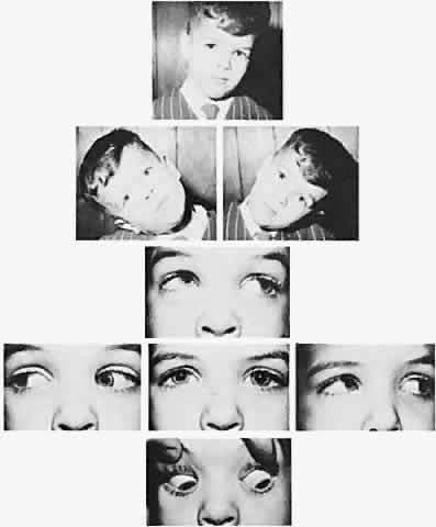
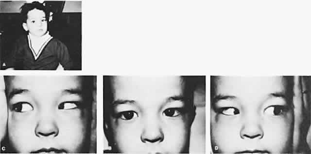
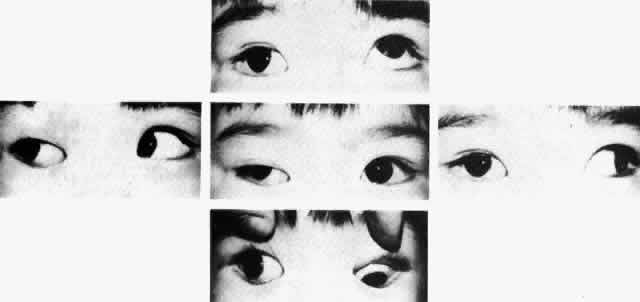
The degree of involvement of the levator muscle varies, but some function is usually retained. Therefore, ptosis is variable in this form of third CN palsy. The four extraocular muscles innervated by the third CN are also affected in various degrees. However, there is usually some trace at least of weakness of the medial, inferior, and superior rectus muscles, and of the inferior oblique muscle. The fourth CN is uninvolved and, consequently, the involved eye is exotropic and hypotropic (Fig. 1). Therefore, the clinician should always suspect congenital third CN palsy in an exotropic patient who has one low eye, intact pupillary and accommodation responses, and minimal ptosis of the involved eye with varied degrees of limitation of both elevation and depression in addition to diminished adduction. Many of these patients are able to develop single binocular vision and to maintain a compensatory malposition of the head that allows the alignment of the eyes to serve this purpose. When the eyes are moved into a position in which fusion is not possible, these patients experience diplopia if they have binocular vision with torticollis. Amblyopia of either eye may occur if the patient does not have binocular vision and does not maintain torticollis.

Fig. 1. Congenital right third cranial nerve palsy with exotropia, intact pupillary and accommodation responses, minimal ptosis, and diminished elevation, depression, and adduction of the involved eye.

Schumacher-Feero and coworkers12 reported a series of 49 children with third CN palsy, involving 53 eyes, and observed during a follow-up for a mean of 5.5 years. Amblyopia developed in 27 eyes, and at the last follow-up visit, in 56% of affected eyes, the visual acuity ranged from 20/15 to 20/40. Binocular function was difficult to restore or to preserve but was significantly improved after surgery. Horizontal alignment was initially good in 6 of 49 patients, improving to 30 with good alignment at their last evaluation. The vertical alignment improved from 24 initially to 35 with good alignment at their last evaluation. Only 1 child achieved fusion at distance and near after a single recession/resection procedure at 8 months of age. A complete palsy required a mean of 2.3 operations to align the eyes, and a partial third CN palsy required a mean of 1.5 operations over the 5.5-year period. In general, the surgery was a horizontal recession/resection procedure for exotropia, with graded supraplacement of the horizontal rectus insertions for the hypotropia.

The cause of congenital third CN palsy is unknown, but it is presumed to be due to a developmental defect in either the nuclear or the motor fiber portion of the third CN complex that innervates the levator muscle and the extraocular muscles. It is not an extremely rare motility disorder. We have seen many patients having only unilateral involvement.

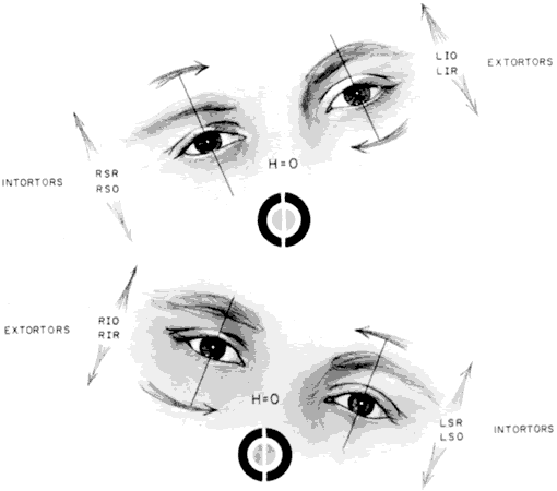
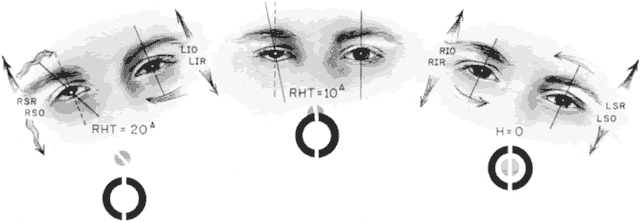
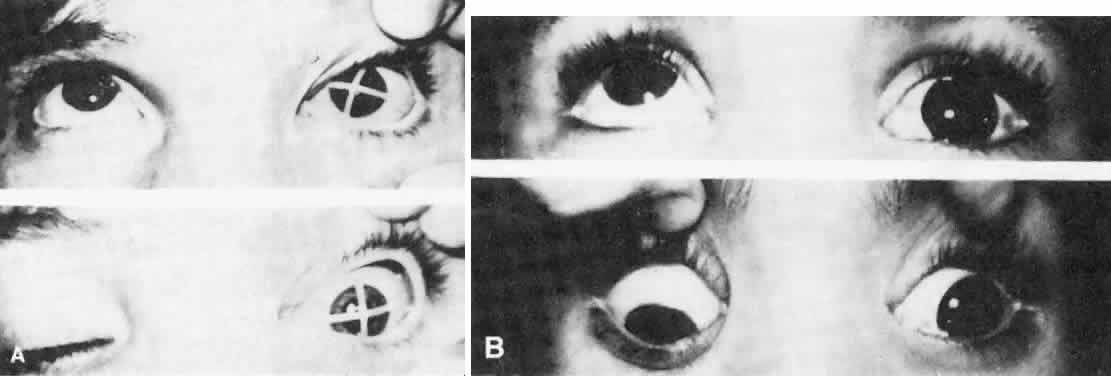
As in any third CN palsy, the absence of adduction of the involved eye makes it difficult to determine the intactness of the ipsilateral CN nerve. Clinically, the method used is to observe the crypts of the iris while the involved eye remains abducted and to ask the patient to look upward and downward. If the fourth CN is intact, the iris markings reveal a conspicuous intorsion as infraduction is attempted and extorsion as supraduction of the involved eye is attempted (Fig. 2). Dieterich and Brandt13 measured ocular torsion and subjective visual vertical tilts in acute and chronic oculomotor, trochlear, and abducens nerve palsies for each eye separately in the primary position with the head held upright. Unexpectedly, ocular torsion was abnormal in only 32% of third and fourth CN palsies involving oblique eye muscles, and normal in all abducens palsies. When measurable, pathologic ocular torsion was low, from 2 to 8 degrees, monocular, involving either the paretic or nonparetic eye. Subjective visual vertical tilts were abnormal in 67% of third and fourth CN palsies, mostly low in amplitude, between 1 and 6 degrees, and involving either the paretic or nonparetic eye, depending on the duration of the palsy. In contrast with acute unilateral brain-stem lesions with frequent binocular and conjugate tilts, third and fourth CN palsies cause only minor and unpredictable ocular torsion and subjective visual vertical tilts.

Fig. 2. Intact fourth cranial nerve in third cranial nerve palsy. A. Litmus paper marker on cornea. B. Pigmented scleral spots demonstrate intorsion as depression is attempted.

The forced duction test produces a negative result in third CN palsy; this rules out any adhesive phenomenon that limits motility of the eye. The degree of involvement of the third CN determines whether therapy is indicated. Involvement may be so minor and partial that no therapy is necessary, or it may affect only the elevators of an eye and is therefore known as double elevator palsy. Cadera and associates14 studied pathophysiology of double elevator palsy in two patients with magnetic resonance imaging (MRI) with volume scanning technique. They found the volume of the superior rectus muscle on the affected side to be less than half that of the normal eye, with other rectus muscles normal, suggesting either congenital hypoplasia or paresis of the affected superior rectus muscle. The inferior oblique muscles could not be evaluated by MRI. After a Knapp procedure in both patients, only minimal superior displacement of the medial and lateral rectus muscles was detectable posterior to the equator of the globe in both patients with MRI.

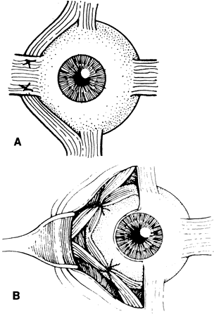
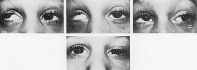
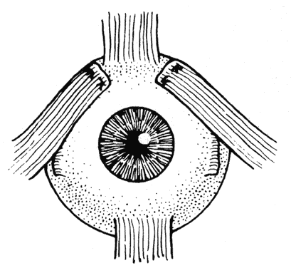
Double elevator palsy is usually rather complete, and it may also be associated with various degrees of ptosis (Fig. 3). The ptosis may be only pseudoptosis because of the hypotropia and because the lid position follows that of the eye. Fixating with the hypotropic eye causes the complete disappearance of the pseudoptosis; however, there may be a small degree of bona fide ptosis in addition to the pseudoptosis. The traction test result is normal in double elevator palsy. Treatment of double elevator palsy involves transposition of the insertions of the horizontal rectus muscles, placing the new insertions immediately adjacent to the insertion of the superior rectus muscle (Fig. 4). This does not produce normal elevation beyond the midline level, but it renders considerable improvement in, if not total elimination of, the hypotropia caused by this disorder.

Fig. 3. Double elevator palsy.

Fig. 4. Transposition of the insertions of the horizontal rectus muscles superiorly for treatment of double elevator palsy. (Helveston EM: Atlas of Strabismus Surgery, p 149. St Louis: CV Mosby, 1973.)

After a full tendon transfer of the lateral and medial rectus muscle for double depressor or double elevator palsy, Knapp obtained an average correction of 38 diopters in the primary position and movement of 25° in the field of action of the paretic muscle group from the primary position.15 In the presence of a poor or absent Bell's phenomenon, an accentuated lower lid fold of the hypotropic eye in attempted elevation, and a positive traction test result, Scott and Jackson recommend inferior rectus recession only as the initial procedure.16 With a negative forced traction test result, a full Knapp procedure is advised.

Complete congenital third CN involvement requires surgery for exotropia, hypotropia, and ptosis. Hypotropia is resolved by disinserting the tendon of the superior oblique muscle from the globe, which is tight and contracted. Maximal recession of the lateral rectus and resection of the medial rectus may be sufficient to reposition the involved eye in the horizontal plane satisfactorily. However, if this is inadequate, removing the superior oblique tendon from the trochlea, severing the reflected tendon of the superior oblique muscle from the muscular portion, and attaching the superior oblique muscle to the sclera at the insertion of the medial rectus muscle offer excellent correction of the horizontal defect created by the third CN palsy in the primary position.

This does not create normal adduction of the involved eye but is an effective technique for centering the eye.17 However, following transposition of the superior oblique muscle, when the patient depresses the involved eye, it adducts. Saunders and Rogers, who attempted correction of third CN palsy by superior oblique anterior transposition and advancement without trochleotomy, reported unsatisfactory results because of inadequate horizontal alignment, postoperative hyperdeviations, or paradoxical ocular movements.18 Superior oblique tendon transposition with trochleotomy causes the adherence syndrome, owing to violation of Tenon's capsule, which is unavoidable in removing the superior oblique tendon from the trochlea. Therefore, this procedure is no longer recommended. Scott and colleagues19 and Gottlob and associates20 described anterior transposition of the nasal portion of the superior oblique tendon, 2 to 3 mm anterior to the nasal border of the superior rectus muscle, without trochleotomy. In addition, Gottlob and associates20 performed large recessions of the ipsilateral lateral rectus, and in some patients, a recess-resect procedure on the horizontal rectus muscles of the contralateral eye. Orthophoria was achieved in 4 patients, a 10 prism diopter (PD) residual exotropia in 1 patient, and 2 patients required reoperations because of aberrant regeneration of the oculomotor nerve. In a much larger series, Maruo and coworkers21 compiled 280 cases of exotropia secondary to oculomotor palsy, between 1971 and 1993. There were 130 congenital and 150 acquired cases of oculomotor palsy. Surgery was performed 234 times in 138 patients with paralytic exotropia, with transposition of the superior oblique tendon and resection of the medial rectus muscle, with or without resection of the ipsilateral lateral rectus muscle. With a 4-year follow-up in 35 cases, the authors found similar results when transposing the superior oblique tendon in patients with complete palsy, and in resection of the medial rectus muscle in patients with incomplete palsy. There was no benefit adding a resection of the medial rectus when the superior oblique transposition was performed. However, recession of the lateral rectus muscle greatly improved the effectiveness of either the superior oblique transposition or the medial rectus resection. Mudgil and Repka22 reviewed retrospectively the ophthalmologic outcome of third CN palsy or paresis in 41 children younger than 8 years of age. Etiologies included congenital in 39%, traumatic in 37%, and neoplastic in 17%. Initial visual acuities were reduced in 71%, and the long-term outcome in the 20 who could be observed during follow-up, for a mean of 3.6 years, vision was reduced in 35% because of amblyopia, and in 25% because of nonamblyopic factors. In the congenital third CN palsy group, all patients improved to normal visual acuity. Despite improved alignment after surgery in 40% (8 of 20) of children with long-term follow-up, none obtained measurable stereopsis. Aberrant reinnervation occurred in 45% (9 of 20). Only 3 patients fully recovered and regained measurable stereopsis, with the etiologies of congenital, neoplastic, and traumatic third CN palsies.

Because of disruption in sensory fusion mechanisms, Elston has recommended a one-stage procedure of maximal lateral rectus recession, medial rectus resection, and simultaneous transposition of insertions to that of the superior rectus.23 The simplest procedure providing predictable cosmetic improvement has been recommended. Ptosis, however, was not addressed. A frontalis suspension of the ptotic lid is the indicated procedure for the associated ptosis. A synthetic material (e.g., 4-0 Supramid) is ideal because if the cornea cannot tolerate the relatively dry state after the lid is elevated, these synthetic sutures can easily be removed and the cornea not harmed permanently. The surgeon should be aware of the absence of Bell's phenomenon in these patients and be alert to postoperative corneal problems associated with this deficiency.

ACQUIRED

Acquired third CN palsy may be partial or complete and may involve only the extraocular muscles or both intraocular and extraocular muscles. The pupil is usually spared in third CN palsy associated with diabetes.

Acquired third CN palsy usually occurs rather precipitously with maximal involvement. Within days to weeks, there may be an indication of restoration of third CN function manifest by only partial involvement. Recovery is usually complete by 6 months following onset, and, consequently, no judgment should be rendered regarding the necessity of treatment until after the 6-month interval. Partial or complete recovery can be expected, depending on the etiology of the third CN palsy, in 48% of the patients.3 Many times such palsy results from relatively serious intracranial involvement, and this may determine whether therapy is indicated. Mark24 recommends MRI evaluation of patients with third CN palsy to include proton density and T2-weighted images through the brain in axial section, to study the brain stem for nuclear lesions, along with thin section T1-weighted images in the coronal and axial planes to evaluate cisternal, cavernous, and orbital segments of the third nerve. Gadolinium diethylenepentaminetetraacetic acid has also been found helpful in evaluation of third CN palsy. In posttraumatic third CN palsies, use of gradient echo images to detect hemorrhage is helpful. Ischemia of the oculomotor nerve causes most cases of nontraumatic oculomotor nerve palsy.25 MRI and lumbar puncture are helpful in diagnosing cases caused by inflammatory or neoplastic meningitis. A cerebral aneurysm, which can be fatal, can be diagnosed by cerebral angiography, but this test has a 1% to 2% morbidity and mortality rate. Magnetic resonance angiography is a variant of MRI that highlights blood vessels; however it is only 95% accurate in detection of aneurysms. Trobe25 therefore concludes that because pupil involvement occurs in 96% with aneurysms, and if anisocoria exceeds 2.0 mm, that catheter angiography is justified.

The causes can be classified as follows:

VII. M.

  1. Brain-stem lesion
    1. Benedikt's syndrome manifest by homolateral third CN paralysis and contralateral intention tremor
    2. Weber's syndrome manifest by homolateral third CN paralysis and contralateral hemiplegia
  2. Inflammatory conditions
    1. Meningitis
    2. Encephalitis
    3. Polyneuritis from toxins such as alcohol, lead, arsenic, and carbon monoxide, and from diabetes
    4. Herpes zoster infection
    5. Echovirus infection26
  3. Vascular lesions
    1. Aneurysms27,28
    2. Internal carotid artery (ICA) dissection,29 ICA stenosis30
    3. Carotid cavernous fistulas31
  4. Tumors1–3,32
    1. Glioblastoma multiforme33
  5. Demyelinating diseases
    1. Multiple sclerosis34
    2. Chronic inflammatory demyelinating polyneuropathy34,35
  6. Trauma
  7. Miscellaneous
    1. Anterior communicating artery aneurysm34,36
    2. Bilateral chronic subdural hematomas34,37
    3. Congenital toxoplasmosis38
    4. “Crack” cocaine39
    5. Diagnostic angiography34,40
    6. Eosinophilic granuloma of the optic nerve34,41
    7. Frontal sinus mucocele42
    8. Infectious mononucleosis43
    9. Leukemia34,44
    10. Measles immunization45
    11. Myasthenia gravis46,47
    12. Ophthalmoplegic migraine5,48–50
    13. Polyarteritis nodosa
    14. Porphyria
    15. Sarcoidosis
    16. Schwannoma34,51
    17. Temporal arteritis52
    18. Viagra therapy (sildenafil citrate)53

Partial third CN palsy in the form of isolated inferior rectus paresis may be the presenting sign of myasthenia gravis, with sudden onset of diplopia.47 The oculomotor nerve aberrant regeneration or misdirection syndrome is believed to result from extensive and haphazard growth that characterizes the regeneration of injured nerve fibers.8,23 The third CN misdirection syndrome includes the following:8

  • Retraction of the globe on attempted vertical gaze
  • Adduction of the globe on attempted vertical gaze
  • Upper lid retraction on attempted downgaze (pseudo-Graefe's sign)
  • Narrowing of the fissure on abduction, and widening of the fissure on attempted adduction (horizontal gaze lid dyskinesis)
  • Pseudo-Argyll Robertson pupil
  • Relative monocular vertical optokinetic responses

Treatment involves relief of the patient's diplopia, which usually is not a problem in complete third CN paralysis because of the associated ptosis covering the pupil. However, in partial involvement, the lid may sufficiently clear the pupillary space so that diplopia is a problem. Occlusion therapy is the best solution for the patient's diplopia. The patient usually wishes to have the involved eye occluded rather than the uninvolved eye. Surgery is indicated for associated strabismus and ptosis if the patient's general condition permits it and if a significant residual paralysis is present 6 months after onset of third CN palsy. The surgery described for congenital third CN palsy is also applicable for acquired third CN palsy. Kushner54 described surgical treatment in five patients with the rare finding of paralysis of the inferior division of the third CN. Clinical findings included a large exotropia and hypertropia of the affected eye, intorsion, and internal ophthalmoplegia. As described by Knapp,55 Kushner performed a superior oblique tenotomy along with transposing the superior rectus toward the insertion of the superior border of the medial rectus, following the spiral of Tillaux and transposing the lateral rectus toward the lateral border of the inferior rectus, following the spiral of Tillaux. In follow-up lasting between 3 and 10 years, all patients maintained satisfactory eye alignment and were free from diplopia in the primary position.

Back to Top
FOURTH CRANIAL NERVE PALSY
The most common isolated cyclovertical muscle palsy encountered by the ophthalmologist is involvement of the trochlear nerve. The cause of congenital trochlear nerve palsy is a defect in the nucleus or motor portion of the nerve; a defect in the motor nerve occurs either inside or outside the medulla. The most common cause of acquired fourth CN palsy is closed head trauma.1–3,56–61

Of 3000 cases of strabismus reviewed by Khawam and coworkers, closed head trauma ranging from minor to severe caused 68% of the 40 cases of acquired superior oblique palsy.56 Uncertain etiology was noted in 20%, and the remainder of the cases included cerebrovascular accident, diabetes, brain tumor, ethmoiditis, and mastoiditis. The cause of acquired bilateral superior oblique palsy following severe head injury is hemorrhage in the roof of the midbrain at the caudal end of the quadrigeminal plate, the area of decussation of the fourth CN.57 Even mild head trauma causing a mass lesion, a hematoma, may cause traumatic fourth CN palsy.58

In many cases of fourth CN palsy, no definite cause can be established despite extensive clinical and laboratory testing.1–3,62,63 Harley reviewed the etiology of paralytic strabismus in 121 children from birth to age 16 and found 67% of fourth CN paralysis to be of undetermined origin.63

Intracranial tumors and aneurysms also account for the diagnosis in some patients,1–3 up to 24% in the Mayo Clinic series.3 Displacement of the trochlea as a result of surgery or trauma to the orbit is another cause. Damage to the trochlear nerve may occur during intracranial or orbital surgery.34 Transient trochlear nerve paresis was postulated to have occurred from indirect traction on the trochlear nerve during surgery.34,64 Bilateral trochlear nerve palsy has been reported65 in association with cryptococcal meningitis in human immunodeficiency virus infection. Acute fourth CN palsy, which has been reported in association with pseudotumor cerebri in children ages 8, 11, and 15, may be a nonspecific sign of increased intracranial pressure.66

Plagiocephaly, or unilateral coronal suture stenosis, causes a shortening of the roof of the orbit, with posterior displacement of the trochlea. The unreflected portion of the superior oblique tendon is shorter than in the normal orbit, possibly with less efficient contracting power, with resultant overaction of the ipsilateral inferior oblique and ocular torticollis.67 This may not be a true palsy, but the clinical presentation is similar. However, palsy of the superior oblique muscle must be differentiated from palsy of the other cyclovertical muscles, which if paretic, manifest a combined cyclovertical phoria or tropia. Each of the muscles that move the eye in a vertical plane about the X axis of Fick, including the superior oblique muscle, also renders a torsional movement about the Y axis of Fick.

The nature of the eye movement produced by contraction of a cyclovertical muscle depends on the horizontal position of the eye. In abduction, the vertical rectus muscles move the eye in the vertical plane and the oblique muscles move the eye in the torsional plane. In adduction, the vertical rectus muscles act torsionally and the oblique muscles act vertically. Both the vertical rectus muscles and the oblique muscles deliver a combined vertical and torsional action in the primary position. The vertical actions and the cycloactions of the eight cyclovertical muscles in the primary position, dextroversion, and levoversion are shown in Figure 5.68 However, the action of the vertical rectus muscles is somewhat more vertical than torsional, whereas the action of the oblique muscles is more torsional than vertical. Therefore, weakness of a single cyclovertical muscle is characterized by vertical and torsional deviation in the primary position. The torsional deviation increases on lateral gaze to one side, while the vertical deviation increases on opposite lateral gaze. For example, given that the left superior oblique muscle is a depressor and intorter, weakness of this muscle results in left hypertropia that increases on right gaze and excyclotropia that increases on left gaze.

Fig. 5. Vertical and torsional actions of the cyclovertically acting muscles. (Parks MM: Isolated cyclovertical muscle palsy. Arch Ophthalmol 60:1027, 1958. Copyright ©1958, American Medical Association.)

The diplopia seen by patients with fourth CN palsy is a combined vertical and torsional set of images projected from the object of regard. The superior pole of the low image seen by the palsied eye is tilted inward compared with the superior pole of the high image seen by the normal eye. By sending inhibitory innervation to the palsied muscle, the diplopia disappears. Inasmuch as the superior oblique muscle depresses and intorts, its tone is diminished by upgaze and by tilting the head to the shoulder opposite the palsied muscle that extorts this eye. The typical ocular torticollis noted in patients with fourth CN palsy is chin depression and head tilt, as depicted in Figure 6.

Fig. 6. Right fourth cranial nerve palsy with compensatory head posture, secondary overacting right inferior oblique muscle, and positive Bielschowsky head-tilt test finding.

Spontaneous head tilt in superior oblique muscle palsy may be absent, or the tilt may be directed toward the same side as the eye with the superior oblique muscle palsy. The binocular vision may be inadequate to overcome a residual vertical deviation, or the presence of poor vision or amblyopia may preclude diplopia in the primary position or the achievement of binocular vision in some other gaze direction.69 Lewis and coworkers70 studied ocular alignment and saccades in seven patients with trochlear nerve pareses, before and after strabismus surgery. A position-dependent vertical ocular misalignment was present before surgery, and the downward saccades were hypometric in the paretic eye. The magnitude and the position-dependence of the static misalignment were reduced by strabismus surgery. In those patients with congenital pareses and in the patient with an acquired paresis, saccade conjugacy improved, but in those patients with traumatic pareses, less improvement occurred. The authors note that postoperative change in saccade conjugacy in relation to the change in static alignment correlated with preoperative vertical vergence, suggesting that the changes in saccade yoking depended on interaction between vertical vergence and saccades.

Soon after onset of fourth CN palsy, the cardinal field test reveals the disorder, because hypertropia is greatest in the vertical field of action of the superior oblique muscle (Fig. 7). However, as the palsy continues for many months, contracture of the ipsilateral inferior oblique muscle begins to manifest itself by eliminating the early cardinal field finding. On the side in which the vertical deviation of the eyes is greatest, hypertropia measures the same in upgaze and downgaze (i.e., the hypertropia becomes concomitant) (Fig. 8). Hence, vertical concomitance requires that some test other than cardinal field measurements be done to diagnose the palsied cyclovertical muscle correctly.

Fig. 7. Cardinal field measurements in right fourth cranial nerve palsy soon after onset.

Fig. 8. Cardinal field measurements in right fourth cranial nerve palsy late after onset.

With vertical concomitance established, another sign of contracture of the direct antagonist inferior oblique muscle is the overelevation of the adducted palsied eye (secondary overaction of inferior oblique muscle).

By having the patient move the eyes into the cardinal fields, the clinician can easily misdiagnose left superior rectus muscle palsy instead of correctly diagnosing right superior oblique muscle palsy and vice versa. The misdiagnosis occurs so frequently that it has received the auspicious name of inhibitional palsy of the contralateral antagonist.71 During cardinal field testing, as the patient fixates with the eye that is innervated by the palsied fourth CN, underelevation of the abducted opposite eye is conspicuous to the examiner, who may be inclined to attribute this condition to palsy of the superior rectus muscle of the sound eye. This error in diagnosis is less apt to happen while the patient fixates with the sound eye because it makes the full movement in elevation and abduction, but fixating with the palsied eye makes the less than normal elevation in abduction of the sound eye obvious. This apparent deficiency in the function of the sound eye in this particular cardinal field is due to the less than usual innervation being dispatched to the inferior oblique muscle of the fixating palsied eye to drive against the atonic palsied superior oblique muscle to continue pursuit of an elevating adducting target. By the law of Hering, equal innervation is dispatched to yoke muscles, resulting in deficient innervation to drive the sound eye upward against the normal tone in its inferior rectus muscle. The terms yoke and contralateral agonist are synonymous names for the same muscle. Both an agonist (palsied muscle) and a contralateral agonist (yoke to the palsied muscle) have antagonists (direct and contralateral). The superior rectus muscle of the sound eye is the contralateral antagonist to the palsied muscle. Because the mistaken diagnosis of palsy of the superior rectus muscle of the opposite eye in the patient with fourth CN palsy is due to inhibition of the normal innervation sent to carry out its normal function, the misdiagnosis is attributable to an inhibitional palsy of the contralateral antagonist, not to a bona fide palsy of an agonist.

To prevent erroneous diagnosis of fourth CN palsy, the Bielschowsky head-tilt test should always be done and the measurements recorded, because contracture of the direct antagonist of the ipsilateral palsied muscle does not influence findings.

To evaluate any isolated cyclovertical muscle palsy logically, including fourth CN palsy, the accumulation of three separate pieces of information followed by three separate reasoning steps invariably provides an accurate diagnosis. The clinician should develop a policy of applying three separate steps known as the three-step test, which follows:68

III. B. 1.

  1. Step 1. Determining whether there is right hypertropia or left hypertropia in the primary position eliminates four of the eight cyclovertical muscles as being causative. For example, right hypertropia signifies there is a
    1. Weak right depressor
      1. Right inferior rectus
      2. Right superior oblique
    2. Weak left elevator
      1. Left superior rectus
      2. Left inferior oblique
  2. Step 2. Determining whether the vertical deviation increases on right or left gaze eliminates one of two possibly faulty muscles in each eye. For example:
    1. Right hypertropia increases in left gaze, indicating that either the
      1. Right superior oblique is weak
      2. Left superior rectus is weak
    2. At this point, the two possibly faulty muscles are always either intorters or extorters. Never is one muscle an intorter and the other an extorter.
  3. Step 3. Bielschowsky head-tilt test accurately differentiates which of the two muscles from the preceding step is responsible (e.g., the right superior oblique or the left superior rectus).

The utricular reflex is stimulated by tilting the head. Tilting to the right causes the intorters of the right eye and the extorters of the left eye to contract, whereas the opposite combination contracts on tilting to the left. However, because the eyes remain in the primary position with reference to the skull, despite the change of the head position in the field of gravity, the contracting cyclovertical muscles render a combined torsional and vertical action. The two intorters or the two extorters of each eye that contract in response to head tilt have opposite vertical actions: one is an elevator and the other is a depressor. Normally, the opposite vertical actions cancel each other, whereas their identical torsional actions are additive; therefore, the eye remains on the same level in the vertical plane but cycloverts accordingly (Fig. 9). This physiologic feature is the source of step 3, as illustrated in Figure 10, depicting a palsied right intorter that is stimulated on right head tilt with its weak depressor action unable to offset the elevator action of its normal fellow intorter. Consequently, increased right hypertropia on right head tilt and decreased left tilt diagnoses the right superior oblique as the palsied muscle. Increased right hypertropia on left head tilt and decreased right head tilt diagnoses the left superior rectus as the palsied muscle.

Fig. 9. The utricular reflex normally produces no vertical movement of the eyes in the primary position as torsional movements are produced. (Parks MM: Isolated cyclcovertical muscle palsy. Arch Ophthalmol 60:1027, 1958. Copyright ©1958. American Medical Association.)

Fig. 10. The utricular reflex produces an increase in right hyperdeviation on right head tilt and a reduction on left head tilt in right superior oblique muscle palsy. (Parks MM: Isolated cyclovertical muscle palsy. Arch Ophthalmol 60:1027, 1958. Copyright ©1958, American Medical Association.)

Trochlear nerve palsies may be unilateral or bilateral; if they are bilateral, the involvement may be minimal on one side and maximal on the other. The minimally involved side may be masked entirely by the maximally involved eye until surgical therapy is performed to eliminate the motility defect, and only then is it truly apparent that there must be different degrees of bilateral involvement. If bilateral involvement can be detected, there is right hypertropia in left gaze and left hypertropia in right gaze; right hypertropia increases as the patient's head is tilted on the right shoulder, and the left hypertropia increases with the head tilted to the left shoulder. There is also greater excyclotropia (10 degrees or more) in bilateral involvement than in unilateral involvement.72

Fourth CN palsy is diagnosed clinically. An MRI may show lesions of the brain stem that may affect the fourth CN nucleus, such as demyelinization, hemorrhages, or infarcts.24 The long cisternal course of the fourth CN allows the nerve to become susceptible to trauma, but imaging is of minimal assistance because the nerve is barely visible normally. However, a high-resolution orbital MRI can demonstrate superior oblique muscle atrophy in chronic fourth CN palsy.24

Management of congenital fourth CN palsy attracts clinical attention by 6 months of age because of either an obvious vertical strabismus or torticollis, alone or combined. Surgery on the cyclovertical muscle is indicated as soon as possible, even prior to 1 year of age if the surgeon is confident of the findings. Early surgery to decrease the obstacles to fusion offers the best chance of developing or maintaining binocular vision, and it is the only rational means of preventing the permanent musculoskeletal changes of torticollis, facial asymmetry, and scoliosis. The facial asymmetry in long-standing torticollis present during childhood is manifest by a shallow, atrophied face on the low side; it is possibly due to reduced carotid artery blood flow as a result of pinching of the vessel, deviation of the nose toward the low side (possibly due to the effect of gravity), and a slanted mouth that seems to try to assume a horizontal orientation.

Wilson and Hoxie73 report that facial asymmetry is an underrecognized association or sequela of torticollis in congenital or in very early onset superior oblique muscle palsy. When present in an adult, this asymmetry may assist in confirming chronicity and may prevent an unnecessary neurologic evaluation. In a prospective study of superior oblique muscle palsy, 19 patients were evaluated. Of 12 unilateral superior oblique palsies presenting in adulthood, 9 were considered congenital, and 7 of 9 (77%) had facial asymmetry. Two children who presented at 18 months and at 3 years of age with a constant head tilt also had facial asymmetry. The authors suggest that correction of the torticollis before maturation of the facial structure may help the asymmetry to resolve, as previously noted by Parks.68 Both facial asymmetry and superior oblique tendon laxity are associated with congenital superior oblique palsy. Paysee and associates74 evaluated 29 patients with superior oblique palsy, and although 76% (16 of 21) with unilateral superior oblique palsy had facial asymmetry, and 81% (17 of 21) had tendon laxity, only 57% had tendon laxity on the side opposite the facial hypoplasia. This suggested that tendon laxity and facial asymmetry do not occur within the same developmental sequence, but that the facial asymmetry develops as a consequence of chronic head tilting from a young age.

Treatment of acquired fourth CN palsy during the first 6 months usually involves simply waiting to determine the degree of spontaneous recovery. After 6 months from onset, specific surgical correction of the acquired strabismus should be undertaken. Unless the hypertropia exceeds 20PD in the primary position, only one muscle should be operated on at a time, and the degree of improvement should be studied over a 3-month period before deciding whether further surgery is indicated. The objective is to improve the cyclodeviations and vertical deviations simultaneously. This can be achieved by operating on the following four of the eight cyclovertical muscles:

  1. Agonist (paretic muscle)
  2. Direct antagonist
  3. Contralateral agonist (yoke)
  4. Contralateral antagonist (direct antagonist of yoke)

If contracture of the direct antagonist is present, as manifested by its obvious overaction, and the vertical tropia is concomitant for upgaze and downgaze with the side of the vertical field of action of the palsied muscle, weakening the direct antagonist should be the first procedure. Otherwise, in the absence of obvious contracture, one may weaken the yoke of the palsied muscle or tuck the palsied muscle as the first procedure. In general, the longer the duration of the strabismus, the greater is the likelihood of contracture of the direct antagonist.

Knapp has provided a classification and treatment scheme for superior oblique palsy.75 In class 1, the superior oblique palsy is found where the greatest deviation is in the field of action of the direct antagonist inferior oblique muscle. Treatment consists of weakening the antagonist inferior oblique muscle.

In class 2, the greatest hypertropia is in the opposite and down oblique field, the field of action of the paretic superior oblique. Knapp recommends a tuck of the superior oblique as the method of first choice or, if there is difficulty in finding a superior oblique tendon or if there is little or no head tilt, recession of the yoke inferior rectus.

In class 3, hypertropia has spread upward from the field of action of the paretic superior oblique because of overaction of the direct antagonist inferior oblique muscle. If the deviation is 25PD or less, inferior oblique weakening or a superior oblique tuck is recommended. If the deviation is greater than 30PD, a weakening of the inferior oblique muscle plus a graded tuck of the paretic superior oblique or a graded recession of the yoke inferior rectus is recommended.

In class 4, the deviation is the same as in class 3 except for an additional hypertropia across the lower field of action because of underaction of the synergist inferior rectus muscle. Knapp recommends performing the same surgery as in class 3 but postponing the correction of the hypertropia in the field of a synergist inferior rectus because of possible spontaneous improvement. If the hypertropia does not improve, a resection of the inferior rectus is recommended.

In class 5, the greatest hypertropia is across the lower field, also described as double depressor paresis. If the deviation is 30PD or more in the lower field, Knapp recommends a tuck of the paretic superior oblique and a tenotomy of the fellow superior oblique muscle.

In class 6, bilateral superior oblique palsy, Knapp initially recommended a tuck of each superior oblique muscle along with infraplacement of the medial recti because of the V pattern. At present, the Harada and Ito modification produces a more satisfactory correction of the excyclotropia.72

In class 7, traumatic paresis plus restriction of relaxation of the superior oblique, also known as the canine tooth syndrome because it is most frequently caused by a dog-bite injury to the trochlear area, requires surgery to correct the Brown syndrome first, with a subsequent attempt at correcting paresis of the superior oblique if possible.

Tightening of the palsied muscle does not make it normal. Mechanical improvement is obtained in the primary position, but there is still deficiency in the field of action of the paretic muscle. If the tuck is performed, it should be performed temporal to the superior rectus muscle. Performing the tuck at this site diminishes the restriction of elevation in adduction (simulated Brown syndrome) but does not entirely relieve it; the restricted elevation usually gradually improves. Therefore, some surgeons are less than enthusiastic about tucking the paretic superior oblique tendon unless further surgery is required after the yoke has been recessed.

The quantity of tendon tucked is 10 mm or more, depending on how easily it stretches on the instrument used for tucking. Recessions of the inferior oblique muscle are usually 10 mm76,77; if the overaction is marked, recession may be increased to 14 mm. Denervation and extirpation of the inferior oblique may be necessary for extremely marked overaction of the inferior oblique muscle, because inferior oblique recession may not provide sufficient weakening.78 Recessions of the inferior rectus muscle range between 3 and 4 mm; if the vertical deviation in the primary position is 10PD or more, the maximum recession is performed. Maximum recession causes approximately 1 mm of sclera to show peripheral to the 6-o'clock limbus position.

There are many exceptions to the above recommendation of which muscle should be operated on first; the most notable exception is a patient with minimal palsy of the superior oblique muscle with no direct antagonist contracture. Tucking the superior oblique tendon yields greater benefit in this patient than in a patient with maximal paralysis of the superior oblique muscle.

Helveston and Ellis performed the superior oblique tuck procedure with additional muscle surgery, when indicated, in 59 patients, according to Knapp's classifications 2 through 6,79 17% required surgical reversal of the tuck because of torticollis, vertical or torsional diplopia, or tightness of elevation in adduction. Superior oblique resection has been advocated by Anderson and coworkers because of recurrence of hypertropia secondary to failed tucking procedures.80

In addition to inferior oblique muscle surgery, Khawam and associates recommended vertical rectus muscle surgery if the vertical deviation exceeds 15PD in the primary position.56 When the vertical deviation is greater in straight-up gaze than in straight-down gaze, ipsilateral superior rectus weakening has been recommended, in addition to inferior oblique weakening. When vertical deviation is greater in downgaze than in upgaze, recession of the yoke muscle, the inferior rectus of the fellow eye, has been advocated. The recommendation of superior rectus recession ipsilateral to the palsied superior oblique muscle contrasts with the view taken by other surgeons who recommend that surgery be confined to the palsied muscle, its direct antagonist, its yoke, or the direct antagonist of the yoke.68 In the typical superior oblique palsy, when changing gaze from primary position to left or right, vertical deviation increases significantly in the direction of the field of action of the palsied superior oblique muscle. With superior rectus muscle contracture with long-standing superior oblique palsy, the direction in which the vertical deviation is expected to decrease significantly may not follow the expected pattern. The deviation significantly increases between primary position and downgaze compared with primary position in upgaze. With superior rectus contracture, the difference is more obvious in downgaze. On version testing, in the cardinal fields, the superior rectus contracture may limit depression in abduction in the field of action of the inferior rectus muscle of the palsied eye. This eye may not depress as expected, and unless the examiner is aware of the superior rectus contracture, with underaction of the inferior rectus of the palsied eye, findings may be misinterpreted as an overaction of the superior oblique muscle of the nonpalsied eye rather than as an underaction of the inferior rectus secondary to superior rectus contracture. A traction test at the time of surgery elicits significant superior rectus contracture and whether surgery should be performed on this muscle. Parks has become an advocate of surgery on the contracted superior rectus muscle as described by Khawam and associates56 and in the presence of significant contracture Parks77 no longer recommends that surgery be confined to the palsied muscle, its direct antagonist, its yoke, or the direct antagonist of the yoke.

Patients with bilateral fourth CN palsy require bilateral surgery graded to the difference in the degree of palsy that exists between the two eyes.

In 1964, Harada and Ito presented a new surgical technique for the correction of torsional deviation without affecting the vertical movement of the eye in bilateral superior oblique palsy.81 They advanced the anterior half of the superior oblique tendon in each eye for excyclodeviation. Fells later modified this procedure.61 Metz and Lerner performed an adjustable Harada-Ito procedure.82 Mitchell and Parks described a modification of the Harada-Ito procedure by advancing the anterior half of the superior oblique tendon to a position 8-mm posterior to the superior insertion of the lateral rectus muscle.72 Elimination of the cyclodiplopia symptom was achieved in all cases by reducing the quantity of excyclotropia, from an average of 12 degrees to 1 degree.

Because of asymmetry in bilateral superior oblique palsy, with a prominent vertical component in one eye and more prominent excyclotorsion in the fellow eye, Pinchoff and coworkers have recommended a superior oblique tuck on the eye with the vertical component and an adjustable Harada-Ito procedure on the eye with the prominent excyclotorsion.83 Garnham and coworkers84 studied the effect of botulinum toxin A, as an alternative to surgery, in 20 patients with fourth CN palsies. They concluded that botulinum toxin A was of greatest benefit when injecting the inferior rectus for residual deviations but was of limited value when injecting the inferior oblique as the primary therapy in chronic fourth CN palsy.

Back to Top
SIXTH CRANIAL NERVE PALSY
Sixth CN palsy causes esotropia in the primary position, which if unilateral increases on gaze direction toward the involved muscle. Continuation of binocular vision is usually possible by maintenance of the eyes in the lateral gaze position away from the palsied eye; this results in a compensatory horizontal face position toward the palsied eye (Fig. 11). Rapid forced dextroversion and levoversion movements in unilateral sixth CN palsy reveal either absent or weak saccades of the paralyzed or paretic lateral rectus compared with the brisk normal rapid saccades of the other three horizontal rectus muscles.85 Compared with the normal saccades, the restricted abduction produced by the paretic lateral rectus is a slow floating outward resulting from inhibition of the ipsilateral medial rectus. Equipment is available to measure and record saccadic velocity accurately and graphically; this is an asset in demonstrating palsied muscle(s).

Fig. 11. Compensatory face turn in lateral rectus muscle palsy.

Acquired bilateral sixth CN palsy invariably produces esotropia, but in some congenital disorders (e.g., Möbius' and bilateral Duane syndromes) the eye may be straight either in the primary position or in upgaze or downgaze. Therefore, an A or V pattern with compensatory chin elevation or depression may occur in the patient with bilateral sixth CN palsy.

The clinical courses of congenital and acquired sixth CN palsies are dissimilar in one respect. Patients with congenital palsy and those with recently acquired palsy manifest greater primary position esotropia when they attempt to fixate with the palsied eye (secondary deviation) and lesser deviation when they fixate with the sound eye (primary deviation), thus the axiom “secondary deviation exceeds primary deviation.” This characteristic gradually disappears during the first few months after the onset of an acquired lateral rectus muscle palsy, owing to contracture of the ipsilateral medial rectus muscle antagonist and hypertrophy of the yoke medial rectus muscle in the contralateral eye. This change occurs rapidly in young children whose acquired sixth CN palsy eventually presents as a concomitant esotropia; it has no resemblance to the original nonconcomitant esotropia that suddenly appeared 2 to 3 months earlier. However, in all age groups, persistent sixth CN palsy that initially manifests as secondary deviation exceeding primary deviation eventually presents as identical primary position esotropia regardless of which eye is fixating, except in patients with congenital sixth CN palsy.

With the sound eye covered, encouraging the palsied eye to maximally abduct while attempting fixation of an object in the field of action of the palsied muscle, and then with both eyes covered, past pointing reveals that the patient with sixth CN palsy points beyond the object when attempting to localize it by memory. The effort involved in attempting to abduct the palsied eye influences the psycho-optic reflex, giving the impression that the object was farther to the side than it actually was.

CONGENITAL

Congenital palsy of the sixth CN is rare,1–3,63,86–90 although literature on the subject seems to suggest otherwise. Much of this literature suggests that the diagnosis is confused with congenital esotropia, Duane retraction syndrome, and Möbius' syndrome. Birth trauma has been proposed as a major cause of this disorder, but this is questionable. Reisner and colleagues have reported transient lateral rectus muscle paresis in 35 of 6360 newborns weighing 2000 g or more, for an incidence of 0.5%.91 Spontaneous disappearance of the deficit occurred in 97% of patients by 6 weeks. Long-term influence on vision and ocular motility has not yet been reported. There are probably some patients with hypoplasia of the abducens motor nucleus or an anomaly within the motor nerve fibers, but these are few. Moreover some lateral rectus muscles have been described as absent or hypoplastic, but overall the frequency of this description is low.

ACQUIRED

Because of the long intracranial course, angulated over the petrous tip of the temporal bone, and the absence of any slack in the course between the brain stem and the dura entry, the sixth CN is vulnerable to increased intracranial pressure, trauma to the cranial floor, meningeal edema, inflammation in the base of the skull, and any displacement of the brain stem. It also shares with other CNs the sensitivities to toxic substances, affliction with acute demyelinating diseases, and exposure to attack by viruses. Tumors and aneurysms are the relatively common causes of displacement of the brain stem and the abducens nerve. Harley reported sixth CN paralysis in 62 of 121 (51%) pediatric patients with CN palsy and in 7 of 121 (6%) patients with palsy of the sixth CN in combination with third and fourth CN palsies.63 The incidence was 34% traumatic, 27% neoplastic, 15% cryptogenic or undetermined, 13% inflammatory, and 11% miscellaneous; no vascular causes were noted. In a pediatric series of similar size, Robertson and coworkers reported an incidence of 20% traumatic, 39% neoplastic, 9% undetermined, 17% inflammatory, 12% miscellaneous (including hydrocephalus, pseudotumor, and leukemia), and 3% vascular.92 In contrast, in the general population, Rush and Younge reported 17% traumatic, 15% neoplastic, 30% undetermined, 17% miscellaneous, 18% vascular, and 3% due to aneurysm.3

Aroichane and Repka93 reviewed the etiology and outcomes of sixth CN palsy or paresis in 64 children 7 years of age or younger. In contrast with earlier studies, the etiology was determined in 95.3% of cases, with the most frequent cause being tumor, at 33% (21 of 64), followed by hydrocephalus at 23% (15 of 64), trauma at 19% (12 of 64), congenital brain malformations at 6% (4 of 64) and infections at 6% (4 of 64). Tumor, hydrocephalus, and trauma comprised 48 of 64 or 75% of the cases. The authors emphasized the importance of amblyopia detection and treatment while a patient is being observed for sixth CN palsy. Lee and coworkers94 reviewed 75 children with sixth CN palsies, who had undergone modern neuroimaging. Neoplasm or neurosurgical removal was the most common cause at 45%, followed by nontumor elevated intracranial pressure at 15%, trauma at 12%, congenital at 11%, inflammatory at 7%, idiopathic at 5%, and miscellaneous at 5%. Because of the high risk of neoplasm, the authors recommend neuroimaging early in the course of sixth CN palsy, even if the palsy is isolated. Cerebral tumors displace the brain stem downward, cerebellar tumors displace it forward, anterior infratentorial tumors push the brain stem backward, cerebellopontine angle tumors push it laterally, and a nasopharyngeal tumor can produce pressure from below. Intramedullary lesions in the pons can cause ipsilateral sixth CN palsy and contralateral hemiplegia (Millard-Gubler syndrome) or contralateral intention tremor. Thrombosis in the posterior portion of the basilar artery may cause sixth CN palsy. (Thrombosis in the anterior basilar artery usually causes third CN palsy.) Neuritis of the sixth CN occurs in the presence of diabetes, lead and arsenic poisoning, diphtheria and tetanus toxins, and thiamine deficiency. Various viruses have been implicated in sixth CN palsy, including those that cause herpes zoster, anterior poliomyelitis, encephalitis lethargica, influenza, and typhus.95–100 Meningeal irritations following lumbar puncture, spinal anesthesia, and spinal iophendylate (Pantopaque) myelography plus meningitis are other causes of sixth CN palsy.101–103 Iopamidol, a contrast medium, has been reported as the cause of transient sixth CN palsies after lumbar myelography.104

Bilateral sixth CN palsy usually portends serious intracranial disease or increased intracranial pressure. However, a lumbar puncture and water-soluble contrast myelography may produce a benign and self-limiting bilateral sixth CN palsy.103 Moster and coworkers reviewed acquired sixth CN palsies in 49 patients ranging in age from 15 to 50 years old.105 Etiologies of the sixth CN palsies were vasculopathy in 14 patients (29%); tumors in 8 patients (16%); multiple sclerosis in 6 patients (12%); presumed inflammation in 4 patients (8%); trauma in 3 patients (6%); sequela of lumbar puncture in 2 patients (4%); and orbital amyloid in 1 patient (2%). The etiology could not be determined in an additional 11 patients (22%). The authors105 recommend monthly evaluations for 6 months if the patient has a history of trauma, diabetes, hypertension, or recent lumbar puncture. Further investigation was not recommended if the sixth CN palsy spontaneously improved. If the patient history is negative for the above conditions, screening blood tests should be performed, fasting glucose, complete blood count, sedimentation rate, and antinuclear antibody titer. High-resolution computed tomography (CT scan) was previously recommended, but MRI is now the imaging modality of choice in evaluating sixth CN palsy,24,106 especially because of the possibility of malignant neoplasm.

Cerebrospinal fluid determination should also be performed. If the sixth CN palsy does not resolve spontaneously within 6 months, if esotropia worsens, or if other significant clinical signs become evident, repeat CT scan and lumbar puncture is indicated.

In addition, MRI allows detailed evaluation of the cavernous sinus, brain stem, and cisterns, and with newer sequencing has become more important in orbit evaluation.107 Contrast-enhanced three-dimensional MRI with gadolinium and gradient-echo T2-weighted MRI techniques are more sensitive than conventional MRI.24,108,109 CT scanning, however, remains useful in the evaluation of orbital disease.107

An isolated sixth CN palsy determined by thorough ophthalmologic, medical, and neurologic evaluations is not necessarily due to a serious intracranial disease despite a duration of more than 3 months. Savino and coworkers described 38 patients with chronic isolated sixth CN palsies who had various systemic diseases, including diabetes, aneurysm, meningioma, hypertension, sarcoidosis, multiple sclerosis, syphilis, giant cell arteritis, and metastatic cancer.110

Benign recurrent sixth CN palsies have been reported, and some patients have experienced more than two episodes. Reports include two to five episodes in four children,99 three episodes in one child,97 six episodes in one child,98 and eleven episodes in one child.100

Bilateral abducens nerve palsy after head injury without skull fracture has been reported.111,112 Spontaneous dissection of the internal carotid artery that extended into the cavernous sinus has caused isolated sixth CN palsy that resolved without treatment.113 A sixth CN palsy due to a conversion reaction, with spontaneous recovery, has been reported.114

A special form of meningeal irritation and edema resulting from middle ear infection and causing sixth nerve palsy deserves special comment. Middle ear infection associated with petrositis and edema of its dura or possibly thrombosis in the contiguous venous sinuses pinches the sixth CN against the petrosphenoidal ligament (Gruber's ligament) as the nerve passes between the ligament and the dura (Dorello's canal), which causes Gradenigo's syndrome.115 Middle ear infection complicated by sixth CN palsy without petrositis and without raised intracranial pressure can arise as a result of phlebitis spreading along the inferior petrosal sinus from the lateral sinus.116

Young children are prone to this disorder, which is characterized by respiratory infection, elevated temperature, and, frequently, facial pain. The frequency of repeated episodes of palsy is high; the duration is usually brief because of the effectiveness of antibiotics, and improvement is obvious within 3 to 6 weeks. If the palsy persists, contracture of the medial rectus muscle occurs, and a permanent concomitant esotropia replaces the nonconcomitant esotropia that was originally controlled with compensatory head posture.

Management of unilateral sixth CN palsy is symptomatic during the first few months after onset. Homonymous diplopia in many patients is controlled by compensatory head posture and requires no treatment. Some patients who are unable to control the diplopia are not sufficiently disturbed to seek relief obtainable by occluding one eye; however, some such patients are very disturbed by the phenomenon and find relief with occlusion. Young children who do not control the esotropia with compensatory head posture should receive alternate occlusion (the right eye one day and the left eye the next) to prevent development of amblyopia, suppression, and abnormal retinal correspondence. Fresnel prism spectacles with sufficient base-out power may be used in lieu of occlusion. Occluding the sound eye or placing a base-out prism before it has been claimed by some to prevent contracture of the ipsilateral direct antagonist (medial rectus muscle) to the paretic muscle, but this claim has not been documented.

Recovery from unilateral acquired sixth CN palsy can usually be observed within 3 months after onset: there is a decrease in the primary position esotropia and an improvement in the deficient abduction of the involved eye. As long as this improvement continues, surgery is contraindicated. King and colleagues117 observed 213 patients over 16 years with unilateral, nontraumatic sixth CN palsies, and found a 78.4% (167 of 213) spontaneous recovery rate after 1 year, with 36.6% (78 of 213) recovering by 8 weeks and 73.7% (157 of 213) by 24 weeks. Recovery did not occur in 16.4% (35 of 213), with 40% (14 of 35) of this group having significant underlying pathology, including aneurysm, Arnold-Chiari malformation, brain-stem stroke, carotid-cavernous fistula, or tumor. Unknown etiology was noted in 60% (21 of 35). In contrast, Mutyala and coworkers118 found a much lower rate of spontaneous recovery in traumatic sixth CN palsy in a retrospective chart review of 101 patients over a 24-year period at Mayo Clinic. After excluding 46 patients who had not been examined within 6 weeks of injury, there were 42 with unilateral and 13 with bilateral sixth nerve palsy. Spontaneous recovery, defined as complete abduction and absence of diplopia, at 6 months was 27% in unilateral and only 12% in bilateral traumatic sixth CN palsy. They concluded that this was lower than other reports3,92 and recommended a prospective multicenter study to provide a more accurate estimate of spontaneous recovery rate in traumatic sixth CN palsy.

In the patient who is recovering slowly, the interplay of developing ipsilateral medial rectus contracture and improving power in abduction provided by the recovering palsied rectus may reach a static point many months after onset; this point is characterized by rather comitant esotropia and the involved eye revealing a moderate restriction of abduction, a positive traction test for passive abduction, and a normal abduction saccadic velocity. In this case, a weakening of the contracted medial rectus may be sufficient to convert the traction test from positive to negative. More often, however, 6 months after onset, the unchanging permanent lateral rectus muscle residual palsy necessitates surgery, more surgery than simple recession of the contractured ipsilateral medial rectus. The amount of additional surgery required depends on the amount of function that was restored in the paretic lateral rectus. If the degree of permanent lateral rectus paresis is only moderate, resection of the palsied muscle may be all that is required in addition to recession of the medial rectus. However, in more severe degrees of permanent residual paresis, and certainly in a permanent paralysis, little is gained by resection of the involved lateral rectus. A muscle transfer procedure is required for the eye to be held in a straight position. Muscle transfer procedures are either a transposition of the vertical rectus muscle insertions to the lateral rectus insertion or a permanent joining of the vertical rectus muscles and lateral rectus muscles at the equator as described by Jensen;119 modification of the former procedure involves movement of only the temporal halves of the vertical recti to the lateral rectus, as described by Hummelsheim.120 Both the Jensen and the Hummelsheim operation are illustrated in Figure 12.

Fig. 12. A. Hummelsheim operation. B. Jensen operation. (Helveston EM: Atlas of Strabismus Surgery, pp 147, 153. St Louis: CV Mosby, 1973.)

Neither lateral rectus resection nor a muscle transfer procedure produces abduction if more than the most minimal palsy persists. The purpose of these procedures is to offer resistance to the pull of the normal medial rectus, which, despite weakening by a recession procedure, restores the eye to esotropia and again allows contracture. As a general rule, resection of the lateral rectus is sufficient to prevent this if the tone in the palsied lateral rectus is sufficient to abduct the eye beyond the midline preoperatively. If the tone in the palsied lateral rectus is inadequate to move the eye into abduction when a maximal attempt is made, it is unrealistic to hope that maximal resection of the palsied lateral rectus can keep the eye straight; in this case, the muscle transfer procedure is indicated, rather than the lateral rectus resection.

Management of bilateral acquired sixth CN palsy is very similar to management of unilateral palsy except that affected patients invariably have diplopia that is not overcome by compensatory head posture. The decision for surgery (when required) is identical to that made in the case of unilateral sixth CN palsy. Both eyes are operated on simultaneously; the amount of surgery is based on the severity of the permanent residual sixth CN palsy and the degree of contracture in the ipsilateral medial rectus muscle.

Botulinum toxin A (Botox, Allergan, Irvine, CA) has been used in the treatment of adult patients with acute sixth CN palsies34,121–127 to prevent contracture of the ipsilateral medial rectus muscle and to restore binocular vision. In a prospective randomized clinical study, Lee and coworkers127 found an 80% recovery rate (20 of 25) in the control group who did not receive any treatment, compared with an 86% recovery rate (19 of 22) in those who received botulinum toxin A. The authors concluded that the difference in recovery rate was not significant enough to recommend the prophylactic use of botulinum toxin A in acute sixth CN palsies. Miller34 noted that waiting for the effects of botulinum toxin A to dissipate could also delay the surgical treatment of those patients who do not demonstrate spontaneous resolution. Quah and colleagues128 presented a retrospective study of 5 years experience with botulinum toxin A for treatment of sixth CN palsy. Of 19 patients studied, 14 (76.7%) had nasopharyngeal carcinoma, 2 had ischemia, and 1 each had cerebrovascular accident, head injury, and intracranial thrombophlebitis. Only 7 patients (36.8%) achieved ocular alignment within 10PD of orthotropia. Fusion was achieved in primary position in 6 patients. Complications included ptosis in 48%, hypertropia in 16%, and subconjunctival hemorrhage in 16%. Best results were obtained with smaller degrees of deviation and with a shorter time interval between onset of strabismus and time of injection.

Back to Top
REFERENCES

1. Rucker CW: Paralysis of the third, fourth and sixth cranial nerves. Am J Ophthalmol 46:787, 1958

2. Rucker CW: The causes of paralysis of the third, fourth and sixth cranial nerves. Am J Ophthalmol 61:1293, 1966

3. Rush JA, Younge BR: Paralysis of cranial nerves III, IV, and VI, cause and prognosis in 1000 cases. Arch Ophthalmol 99:76, 1981

4. Holmes JM, Mutyala S, Maus T et al: Pediatric third, fourth, and sixth nerve palsies: A population-based study. Am J Ophthalmol 127:388, 1999

5. Ing EB, Sullivan TJ, Clarke MP et al: Oculomotor nerve palsies in children. J Pediatr Ophthalmol Strabismus 29:331, 1992

6. Victor DI: The diagnosis of congenital unilateral third nerve palsy. Brain 99:711, 1976

7. Miller NR: Solitary oculomotor nerve palsy in childhood. Am J Ophthalmol 83:106, 1977

8. Lotufo DG, Smith J, Hopen GR et al: The pupil in congenital third nerve misdirection syndrome. J Clin Neuroophthalmol 3:193, 1983

9. Balkan R, Hoyt CS: Associated neurologic abnormalities in congenital third nerve palsies. Am J Ophthalmol 97:315, 1984

10. Hamed LM: Associated neurologic and ophthalmic findings in congenital oculomotor palsy. Ophthalmology 98:708, 1991

11. Tsaloumas MD, Willshaw HE: Congenital oculomotor palsy: Associated neurological and ophthalmological findings. Eye 11:500, 1997

12. Schumacher-Feero LA, Yoo KW, Solari FM et al: Results following treatment of third cranial nerve palsy in children. Trans Am Ophthalmol Soc 96:455, 1998

13. Dieterich M, Brandt T: Ocular torsion and perceived vertical in oculomotor, trochlear and abducens nerve palsies. Brain 116:1095, 1993

14. Cadera W, Bloom JN, Karlik S et al: A magnetic resonance imaging study of double elevator palsy. Can J Ophthalmol 32:250, 1997

15. Knapp P: The surgical treatment of double elevator paralysis. Trans Am Ophthalmol Soc 67:304, 1969

16. Scott WE, Jackson OB: Double elevator palsy: The significance of inferior rectus restriction. Am Orthoptic J 27:5, 1977

17. Helveston EM: Extraocular muscle transfer. Trans Am Acad Ophthalmol Otolaryngol 75:722, 1975

18. Saunders RA, Rogers CT: Superior oblique transposition for third nerve palsy. Ophthalmology 89:310, 1982

19. Scott AB: Transposition of the superior oblique. Am Orthoptic J 5:11, 1977

20. Gottlob I, Catalano RA, Reinecke RD: Surgical management of oculomotor nerve palsy. Am J Ophthalmol 111:71–76, 1991

21. Maruo T, Iwashige H, Kubota N et al: Results of surgery for paralytic exotropia due to oculomotor palsy. Ophthalmologica 210:163, 1996

22. Mudgil AV, Repka MX: Ophthalmologic outcome after third cranial nerve palsy or paresis in childhood. J Am Assoc Pediatr Ophtalmol Strabis 3:2, 1999

23. Elston JS: Traumatic third nerve palsy. Br J Ophthalmol 68:538, 1984

24. Mark AS: Oculomotor motion disorders: Current imaging of cranial nerves 3, 4, and 6. Semin Ultrasound, CT and MRI 19:240, 1998

25. Trobe JD: Managing oculomotor nerve palsy. Arch Ophthalmol 116:798, 1998

26. Hertenstein JR, Sarnat HB, O'Connor DM: Acute unilateral oculomotor palsy associated with ECHO 19 viral infections. J Pediatr 89:79, 1976

27. Trobe JD, Glaser JS, Post JD: Meningiomas and aneurysms of the cavernous sinus. Arch Ophthalmol 96:457, 1978

28. Kasoff I, Kelly DL: Pupillary sparing in oculomotor palsy from internal carotid aneurysm. J Neurosurg 42:713, 1975

29. Schievink WI, Mokri B, Garrity JA et al: Ocular motor nerve palsies in spontaneous dissections of the cervical internal carotid artery. Neurology 43:1938, 1993

30. Hollinger P, Sturzenegger M: Painful oculomotor nerve palsy—a presenting sign of internal carotid artery stenosis. Cerebrovasc Dis 9:178, 1999

31. Acierno MD, Trobe JD, Cornblath WT et al: Painful oculomotor palsy caused by posterior-draining dural carotid cavernous fistulas. Arch Ophthalmol 113:1045, 1995

32. Bixenman WW: Atypical clinical presentation of oculomotor (III) nerve palsy. J Clin Neuroophthalmol 2:249, 1982

33. Al-Yamany M, Al-Shayji A, Bernstein M: Isolated oculomotor nerve palsy: an unusual presentation of glioblastoma multiforme. Case report and review of the literature. Case report and review of the literature. J Neurooncol 41:77, 1999

34. Miller NR: The oculomotor nerves. Curr Opin Neurol 9:21, 1996

35. Arroyo JG, Horton JC: Acute, painful, pupil-involving third nerve palsy in chronic inflammatory demyelinating polyneuropathy. Neurology 45:845, 1995

36. Coyne TJ, Wallace MC: Bilateral third cranial nerve palsies in association with a ruptured anterior communicating artery aneurysm. Surg Neurol 42:52, 1994

37. Phookan G, Cameron M: Bilateral chronic subdural haematoma: An unusual presentation with isolated oculomotor nerve palsy. J Neurol Neurosurg Psychiatry 57:1146, 1994

38. Perry DD, Merritt JC, Greenwood RS et al: Congenital toxoplasmosis associated with acquired oculomotor nerve (CN III) palsy. J Pediatr Ophthalmol Strabismus 19:265, 1982

39. Migita DS, Devereaux MW, Tomsak RL: Cocaine and papillary-sparing oculomotor nerve paresis. Neurology 49:1466, 1997

40. Griffiths PD, Gholkar A, Sengupta RP: Oculomotor nerve palsy due to thrombosis of a posterior communicating artery aneurysm following diagnostic angiography. Neuroradiology 36:614, 1994

41. Hardenack M, Voller A, Schroder JM et al: Primary eosinophilic granuloma of the oculomotor nerve. Case report. J Neurosurg 81:784, 1994

42. Ehrenpreis SJ, Beidlingmaier JF: Isolated third-nerve palsy associated with frontal sinus mucocoele. J Neuroophthalmol 15:105, 1995

43. Nellhaus G: Isolated oculomotor nerve palsy in infectious mononucleosis. Neurology 16:221, 1966

44. Smith HP, Biller J, Kelly DL: Oculomotor palsy with pupillary sparing, coincidental aneurysm and chronic lymphocytic meningeal infiltration. Surg Neurol 16:26, 1981

45. Chan CC, Jugg RL, Steinman L: Isolated oculomotor palsy after measles immunization. Am J Ophthalmol 89:446, 1980

46. Osher RH: Myasthenic “oculomotor” palsy. Ann Ophthalmol 11:31, 1979

47. Spoor TC: Myasthenia gravis presenting as an isolated inferior rectus paresis. Ophthalmology 86:2158, 1979

48. Keith CG: Oculomotor nerve palsy in childhood. Aust NZ J Ophthalmol 15:181, 1992

49. Amit R, Benezra D: Oculomotor ophthalmoplegic migraine in an infant. Headache 27:390, 1987

50. Karabasas CH: The rules of third nerve palsy in children. Eye 11:761, 1997

51. Niazi W, Boggan JE: Schwannoma of extraocular nerves: Survey of literature and case report of an isolated third nerve schwannoma. Skull Base Surg 4:219, 1994

52. Graham E, Holland A, Avery A et al: Prognosis in giant-cell arteritis. BMJ 282:269, 1981

53. Donahue SP, Taylor RJ: Pupil-sparing third nerve palsy associated with sildenafil citrate (Viagra) Am J Ophthalmol 126:476, 1998

54. Kushner BJ: Surgical treatment of paralysis of the inferior division of the oculomotor nerve. Arch Ophthalmol 117:485, 1999

55. Knapp P: Paretic squints. In: Symposium on Strabismus: Transactions of the New Orleans Academy of Ophthalmology, pp 350–357. St Louis, MO: CV Mosby, 1978

56. Khawam E, Scott AB, Jampolsky A: Acquired superior oblique palsy. Arch Ophthalmol 77:761, 1967

57. Raskind RH: Acquired bilateral superior oblique palsy. Doc Ophthalmol 34:335, 1973

58. Lavin PJ, Troost BT: Traumatic 4th nerve palsy. Arch Neurol 41:679, 1984

59. Miller MT, Urist MJ, Folk ER et al: Superior oblique palsy presenting in late childhood. Am J Ophthalmol 70:212, 1970

60. Younge BR: Analysis of trochlear nerve palsies. Mayo Clin Proc 52:11, 1977

61. Fells P: Management of paralytic strabismus. Br J Ophthalmol 58:255, 1974

62. Coppeto JM, Lessell S: Cryptogenic unilateral paralysis of the superior oblique muscle. Arch Ophthalmol 96:275, 1978

63. Harley RD: Paralytic strabismus in children. Ophthalmology 87:24, 1980

64. Jacobson DM, Warner JJ, Ruggles KH: Transient trochlear nerve palsy following anterior temporal lobectomy for epilepsy. Neurology 45:1465, 1995

65. Sadun F, DeNegri AM, Santopadre P et al: Bilateral trochlear nerve palsy associated with cryptococcal meningitis in human immunodeficiency virus infection. J Neuroophthalmol 19:118, 1999

66. Speer C, Pearlman J, Phillips PH et al: Fourth cranial nerve palsy in pediatric patients with pseudotumor cerebri. Am J Ophthalmol 127:236, 1999

67. Bagolini B, Campos EC, Chiesi C: Plagiocephaly causing superior oblique deficiency and ocular torticollis. Arch Ophthalmol 100:1093, 1982

68. Parks MM: Isolated cyclovertical muscle palsy. Arch Ophthalmol 60:1027, 1958

69. Burian HM, Rowan PJ, Sullivan MS: Absence of spontaneous head tilt in superior oblique muscle palsy. Am J Ophthalmol 79:972, 1975

70. Lewis RF, Zee DS, Repka MX et al: Regulation of static and dynamic ocular alignment in patients with trochlear nerve pareses. Vision Res 35:3255, 1995

71. Chavasse FB: In Worth's squint or the Binocular Reflexes and Treatment of Strabismus. 7th ed. London: Bailliere, Tindall & Cox, 1939

72. Mitchell PR, Parks MM: Surgery for bilateral superior oblique palsy. Ophthalmology 89:484, 1982

73. Wilson ME, Hoxie J: Facial asymmetry in superior oblique muscle palsy. J Pediatr Ophthalmol Strabismus 30:315, 1993

74. Paysee EA, Coats DK, Plager DA: Facial asymmetry and tendon laxity in superior oblique palsy. J Pediatr Ophthalmol Strabismus 32:158, 1995

75. Knapp P: Classification and treatment of superior oblique palsy. Am Orthoptic J 24:18, 1974

76. Parks MM: The weakening surgical procedures for eliminating overaction of the inferior oblique muscle. Am J Ophthalmol 73:107, 1972

77. Parks MM: The overacting inferior oblique muscle: The XXXVI de Schweinitz Lecture. Am J Ophthalmol 77: 787, 1974

78. DelMonte MA, Parks MM: Denervation and extirpation of the inferior oblique. Ophthalmology 90:1178, 1983

79. Helveston EM, Ellis FD: Superior oblique tuck and superior oblique palsy. Aust J Ophthalmol 11:215, 1983

80. Anderson LS, Gorman CJ, Luton JP: Superior oblique strengthening procedures in hypertropia I and II. J Pediatr Ophthalmol 16:43, 1970

81. Harada M, Ito Y: Surgical correction of cyclotropia. Jpn J Ophthalmol 8:88, 1964

82. Metz HS, Lerner H: The adjustable Harada-Ito procedure. Arch Ophthalmol 99:624, 1981

83. Pinchoff BS, Bergstrom TJ, Sandall GS: A combined surgical approach to bilateral superior oblique palsy. Ophthalmol Surg 13:1000, 1982

84. Garnham L, Lawson JM, O'Neill D et al: Botulinum toxin in fourth nerve palsies. Aust NZ J Ophthalmol 25:31, 1997

85. Metz HS, Scott AB, O'Meara D et al: Ocular saccades in lateral palsy. Arch Ophthalmol 84:453, 1970

86. Shrader EC, Schlezinger NS: Neuro-ophthalmologic evaluation of abducens nerve paralysis. Arch Ophthalmol 63: 84, 1960

87. Ernest JT, Costenbader FD: Lateral rectus muscle palsy. Am J Ophthalmol 65:721, 1968

88. Hoyt WF, Nachtigaller H: Anomalies of ocular motor nerves. Am J Ophthalmol 60:443, 1965

89. Tillack TW, Winer JA: Anomaly of the abducens nerve. Yale J Biol Med 34:620, 1962

90. Phillips WH, Dirion JK, Graves GO: Congenital bilateral palsy of abducens. Arch Ophthalmol 8:355, 1932

91. Reisner SH, Perlman M et al: Transient lateral rectus muscle paresis in the newborn infant. J Pediatr 78:461, 1971

92. Robertson DM, Hines DJ, Rucker CW: Acquired 6th nerve paresis in children. Arch Ophthalmol 83:574, 1970

93. Aroichane M, Repka MX: Outcome of sixth nerve palsy or paresis in young children. J Pediatr Ophthalmol Strabismus 32:152, 1995

94. Lee MS, Galetta SL, Volpe NJ et al: Sixth nerve palsies in children. Pediatr Neurol 20:49, 1999

95. Knox DL, Clark DB, Schuster FF: Benign VI nerve palsies in children. Pediatrics 40:560, 1967

96. Werner DB, Savino PJ, Schatz NJ: Benign recurrent 6th nerve palsies in children. Arch Ophthalmol 101:607, 1983

97. Sternberg I, Ronen SP, Arnon N: Recurrent isolated, post-febrile abducens nerve palsy. J Pediatr Ophthalmol Strabismus 17:323, 1980

98. Bixenman WW, von Noorden GK: Benign recurrent 6th nerve palsy in childhood. J Pediatr Ophthalmol Strabismus 18:29, 1981

99. Boger WP, Puliafito CA et al: Recurrent isolated 6th nerve palsy in children. Ann Ophthalmol 16:237, 1984

100. Sullivan SC: Benign recurrent isolated 6th nerve palsy of childhood. Clin Pediatr 24:160, 1985

101. Robinson HM: Abducens palsy following lumbar puncture. Am J Syph 29:422, 1945

102. Insel TR, Kalin NH, Risch SC et al: Abducens palsy after lumbar puncture. N Engl J Med 303:703, 1980

103. Miller EA, Savino PJ, Schatz NJ: Bilateral 6th nerve palsy, a rare complication of water-soluble contrast myelography. Arch Ophthalmol 100:603, 1982

104. Bell JA, McIllwane GG, O'Neill D: Iatrogenic lateral rectus palsies. A series of five postmyelographic cases. J Neuroophthalmol 14:205, 1994

105. Moster ML, Savino PJ, Sergott RL et al: Isolated sixth nerve paresis in younger adults. Arch Ophthalmol 102:1328, 1984

106. Depper MH, Truwit CL, Dreisbach JN et al: Isolated abducens nerve palsy: MR imaging findings. Am J Radiol 160:837, 1993

107. Laine FJ: Cranial nerves III, IV, and VI. Topics Magn Reson Imaging 8:111, 1996

108. Bianchi-Marzoli S, Brancato R: Third, fourth and sixth cranial nerve palsies. Curr Opin Ophthalmol 8:45, 1997

109. Lemmerling M, DePraeter G, Mortele K et al: Imaging of the normal pontine cisternal segment of the abducens nerve, using three-dimensional constructive interference in the steady state MRI. Neuroradiology 41:384, 1999

110. Savino PJ, Hilliker JK, Casell GH et al: Chronic 6th nerve palsies, are they really harbingers of serious intracranial disease? Arch Ophthalmol 100:1442, 1982

111. Arias MJ: Bilateral traumatic abducens nerve palsy without skull fracture and with cervical spine fracture. Neurosurgery 16:232, 1985

112. Limnaios EE, Papageorgiou CT: Bilateral abducens nerve palsy. Ophthalmologica 181:326, 1980

113. Maitland CG, Black JL, Smith WA: Abducens nerve palsy due to spontaneous dissection of the internal carotid artery. Arch Neurol 40:448, 1983

114. Meloff KL, DeMeuron G, Buncic JR: Conversion 6th nerve palsy in a child. J Psychosomatics 21:769, 1980

115. Gradenigo G: A special syndrome of endocranial otitic complications (paralysis of the motor oculi externus of otitic organ). Am Otol 13:637, 1904

116. Homer JJ, Johnson IJM, Jones NS: Middle ear infection and sixth nerve palsy. J Laryngol Otol 110:872, 1996

117. King AJ, Stacey E, Stephenson G, Trimble RB: Spontaneous recovery rates for unilateral sixth nerve palsies. Eye 9:476, 1995

118. Mutyala S, Holmes JM, Hodge DO et al: Spontaneous recovery rate in traumatic sixth-nerve palsy. Am J Ophthalmol 122:898, 1996

119. Jensen CDF: Rectus muscle union: A new operation for paralysis of the rectus muscle. Transactions of the Pacific Coast Otoophthalmology Society Annual Meeting 45: 359, 1964

120. Hummelsheim E: Weitere Erfhrungen mit partieller Schnenuberpflanzung an den augenmuskeln. Arch Augenheilkd 62:71, 1909

121. Elston J, Lee J: Paralytic strabismus: The role of botulinum toxin. Br J Ophthalmol 69:891, 1985

122. Scott AB, Kraft SP: Botulinum toxin injection in the management of lateral rectus paresis. Ophthalmology 92:676, 1985

123. Fitzsimons R, Lee J, Elston J: The role of botulinum toxin in the management of sixth nerve palsy. Eye 3:391, 1989

124. Wagner RS, Frohman LP: Long-term results: Botulinum for sixth nerve palsy. J Pediatr Ophthalmol Strabismus 26:106, 1989

125. Lee JP: Modern management of sixth nerve palsy. Aust NZ J Ophthalmol 20:41, 1992

126. Killer HE, Blumer BK, Bahler A: Approach to traumatic sixth nerve palsy with botulinum toxin A. Neuroophthalmology 14:355, 1994

127. Lee J, Harris S, Cohen J et al: Results of a prospective randomized trial of botulinum toxin therapy in acute unilateral sixth nerve palsy. J Pediatr Ophthalmol Strabismus 31:283, 1994

128. Quah BL, Cheong PYY, Balakrishnan V: A review of 5 years experience in the use of botulinum toxin A in the treatment of sixth cranial nerve palsy at the Singapore National Eye Centre. Singapore Med J 40:405, 1999

Back to Top