Chapter 14
Monofixation Syndrome
MARSHALL M. PARKS
Main Menu   Table Of Contents

Search

ETIOLOGY
CLINICAL CHARACTERISTICS
DIAGNOSTIC METHODS
TREATMENT
PROGNOSIS
REFERENCES

As the evaluation of strabismus therapy became increasingly critical, attention was focused on a relatively large group of patients who had a small residual deviation. This group attracted particular attention because, in addition to the consistent findings of a deviation measuring 8Δ or less and good fusional vergence amplitudes, there was a scotoma within the deviated eye that prevented diplopia.

Further interest was stimulated when it became apparent that some patients with small deviations had no history of strabismus. Anisometropia was identified early as a frequently associated factor in the nonstrabismic patients. Even more interesting was the discovery that some of the population free of strabismus and anisometropia had this same disorder. After it was noted that the common denominator in all patients with a small deviation was a small facultative macular scotoma within the visual field of one eye, binocular perimetry studies on a control series of patients with straight eyes revealed that some of these patients also had a macular scotoma. It was also recognized that the rare patient with a unilateral macular lesion, having straight eyes and extramacular fusion, has the organic counterpart to the unilateral functional facultative macular scotoma just described.

Consequently, a group of patients, either with or without a small deviation, from varied sources constitutes a specific ophthalmologic entity characterized by monofixation, due to the macular scotoma precluding bifixation and active extramacular binocular vision. The entity is referred to as the monofixation syndrome.

Initial interest in the monofixation syndrome came by way of the small angle deviations. Patients with these small angle deviations were referred to as “flicker cases,” by the British, because the cover test revealed a small “flick” as the deviated eye assumed fixation. This syndrome soon became confused with the normal fixation disparity described by Ogle1 and others. In addition, this condition has been called fixational disparity and also referred to as subnormal binocular vision due to the lack of central fusion. In 1956 Jampolsky2 described how some of these patients have greater alternate cover measurements than cover-uncover measurements, and he emphasized that this is diagnostic of the disorder. He described the suppression within the central retinal area in one eye of these patients, and he used this as an explanation for solving the diplopia caused by the minimal deviation. Jampolsky further reasoned that the peripheral portion of Panum's fusional space is sufficiently large to permit fusion with normal retinal correspondence (NRC). His opinion regarding the lack of success with orthoptic treatment for these patients to convert them to centrally fusing rather than suppressing the central retinal area is clearly stated. Jampolsky and co-workers3 also noted the paucity of small angle exodeviations as compared to the frequent number of cases of convergent small angle deviations. In 1962 Jampolsky4 referred to the monofixation syndrome as “fusion disparity.” He implied that there is normal fusion, except for the absence of bifoveal fixation. He chose the term “fusion disparity” to separate a monofixation syndrome from fixation disparity, which is a normal physiologic entity. There are two obvious dissimilarities between fixation disparity and the monofixation syndrome (Jampolsky's fusion disparity). The quantity of deviation does not exceed 10 to 14 minutes of arc in fixation disparity, but it may be as large as 8Δ in the monofixation syndrome. In fixation disparity, both macular areas function simultaneously; whereas, in the monofixation syndrome, one or the other macula does not function during binocular vision.

The impossibility of accurately naming this condition in accordance with the established semantic code in common usage for ocular motility and binocular vision was apparent early after the initial interest developed in this large group of patients. Appraised according to one respect, the patient was heterotropic, but in another respect, hetero-phoric. Any term selected to identify these patients was arbitrary. In 1961 the name “monofixational phoria” was applied to those patients with a deviation that was greater by alternate cover than by cover-uncover; it was claimed that the deviation was made partially latent by extramacular fusion while the image projected onto the deviated eye's macula was not seen.5 At that time, interest was directed only to the small angle aspect of the deviations, and physicians were unaware that many patients without a deviation also had the identical sensory finding of a scotoma of one macula. Jampolsky's concept of NRC peripheral fusion acquired by the normal stretched out peripheral Panum's space was accepted, and the NRC seemed to be confirmed by the findings from binocular perimetry performed during dissociated conditions. The following significant facts about the monofixation syndrome also added by this report were:

  1. Anisometropia, in addition to strabismus, was established as a cause.
  2. In some patients, neither strabismus nor anisometropia was present, and these patients were defined as having primary monofixational phoria; those with strabismus and anisometropia were defined as having secondary monofixational phoria.
  3. Stereoacuity was first related to the nature of the fixation present: poor in monofixation and good in bifixation.
  4. The facultative absolute scotoma was revealed by binocular perimetry.

In 1966 Lang criticized monofixational phoria as a name for small angle strabismus since there is a manifest tropia. Burian's definition of heterophoria is a “deviation of the eyes kept latent by fusion; heterotropia is a patent (manifest) deviation of the eyes in the absence of fusion.”6 Lang7 proposed that the syndrome be known by the full name of microtropia unilateralis anomalo fusionalis, but he suggested that it be referred to ordinarily as microtropia or microstrabismus. In 1967 Helveston and von Noorden8 used the term “microtropia” to describe an inferred small angle strabismus in their amblyopia patients with eccentric fixation whose amblyopic eye did not make a movement to assume fixation and who grossly appeared to have straight eyes. The majority of their patients were anisometropic. Since, by visuscopy, the fixation point was adjacent to the macular borders, they inferred that the strabismus angle was ultra small. Others can confirm these findings in many patients with the monofixation syndrome whose poor sighted eye either has not responded to amblyopia therapy or has never been treated; the syndrome occurs either as a primary condition or secondary to strabismus, anisometropia (or the two combined), or a macular lesion. These patients seem to have monofixational orthophoria since there is no detectable shift in either eye by the cover test. Perhaps Helveston and yon Noorden are correct in their assumption that in some cases there probably would be a discernible shift were it not for the slight eccentric fixation in the amblyopic eye; therefore, the patient is not orthophoric. However, use of the term “microtropia” is not justified to describe the patients without shift to cover-uncover when Lang previously used the term to describe patients having a deviation by cover-uncover. The group described by Helveston and von Noorden probably represents only one of many various groups of patients within the overall monofixation syndrome.

The semantic structure that evolved as a result of many attempts to label various categories of patients that constitute the monofixation syndrome has become a monstrosity. Surely such terms as retinal slip, fixation disparity, esophoria with fixation disparity, fixational disparity, flicker cases, subnormal binocular vision, convergent fixation disparity, pathologic fixation disparity, monofixational phoria, fusion disparity, strabismus spurius, microtropia unilateralis anomalo fusionalis, microtropia, and microstrabismus will vanish from ordinary usage.

There are three principal reasons for the past difficulties encountered in naming this syndrome: (1) an element of both phoria and tropia is present and whichever feature the author chooses to emphasize determines the selection; (2) fixation disparity, as a name for a specific physiologic process in binocular single vision, was plagiarized since the condition under discussion seemed to be a pathologic extension of the same process; and (3) the names selected revealed the lack of a total concept of the syndrome. As the syndrome was gradually put together, the lack of organization in naming each of the facets is now apparent.

Essentially, the patients with this syndrome have straight or almost straight eyes and a form of binocular vision in which their inability to bifixate is proved by a demonstrable scotoma in the visual field of the nonfixating eye during binocular vision. This essential monofixating feature and other associated features are always present, while others may be either present or absent. Fusional vergence amplitudes are always associated with the monofixation syndrome. The variable features associated with this syndrome are a history of strabismus, anisometropia, a unilateral macular lesion, amblyopia, eccentric fixation, orthophoria, phoria, small tropia, and possibly a larger deviation by alternate cover than by cover-uncover. The majority of the patients with the monofixation syndrome have gross stereopsis; occasionally, the only exception is a patient with congenital esotropia who is straight and has sensory and motor fusion. The name that fits all of these features is simply “the monofixation syndrome.”

Back to Top
ETIOLOGY
Since patients with the monofixation syndrome have associated strabismus, anisometropia, a unilateral macular lesion, or an inherent inability to fuse similar images on each macula, it is helpful to consider each of these conditions as a separate etiologic factor. The monofixation syndrome is caused by any of the preceding four factors or by any combination of them.

Among patients with the monofixation syndrome caused by strabismus, there is a significantly greater frequency of corrected esotropia than of corrected exotropia. According to one author's experience,9 approximately 66% of the successfully treated horizontal strabismic patients are esotropic and 34% are exotropic; yet of the strabismic patients who develop the monofixation syndrome after treatment, approximately 90% are esotropic and 10% are exotropic. Obviously, the chances that an exotropic patient will develop the monofixation syndrome are much less than the chances that an esotropic will develop the syndrome.

The probable explanation for the greater frequency of the monofixation syndrome in patients with corrected esotropia than in those with corrected exotropia is the difference between the constancy and intermittency of the deviation prior to treatment. The patient with constant tropia loses the bifixation habit completely. The possibility of restoring it after deviation is eliminated appears not to be directly related to the patient's age at the time bifixation is lost and to the duration of the constant deviation. Since esotropic patients prior to receiving therapy tend to have constant deviations with greater frequency than exotropic patients, one should anticipate that a greater percentage of those who are esotropic will remain monofixating patients after treatment. In a study made by the author at the time the deviation was brought under control, 70% of the esotropic patients and only 21% of the exotropic patients were constantly tropic.

It is tempting to conjecture that amblyopia is more prevalent in esotropic than in exotropic patients since in the study just mentioned, 40% of the esotropic patients were amblyopic and only 3% of the exotropic persons were amblyopic out of 100 consecutive patients having horizontal strabismus. Yet among patients with the monofixation syndrome, 78% of the esotropic and 57% of the exotropic are amblyopic. The latter fact suggests that the incidence of the monofixation syndrome as a final treatment status is increased in both esotropic and exotropic patients if amblyopia exists. However, it does not follow that amblyopia is the cause of the monofixation syndrome after the deviation has been eliminated. Evidence to the contrary was found in the fact that 24% of the patients with the monofixation syndrome were never amblyopic, and an additional 16% still had the monofixation syndrome after their amblyopia had permanently been cured by occlusion therapy. A more plausible concept is that both amblyopia and the monofixation syndrome result from the same cause, but the development of amblyopia requires one additional factor. Both are produced by prolonged and constant strabismic deviation in the infant or young child; but, in addition, development of amblyopia requires the constant exclusion of one eye from fixating rather than alternate fixation. Hence, not all patients with the monofixation syndrome following strabismus therapy have amblyopia.

Congenital esotropic patients appear to have a different reason for monofixating, despite the fact that peripheral fusion was acquired by early surgical elimination of the deviation; they seem to have an inherent inability to bifixate. Most congenital esotropic patients obtain extramacular fusion if the eyes have been straightened by surgery when they were less than two years of age, but they never obtained bifixation. Most congenitally esotropic patients who obtain extramacular fusion after their eyes have been straightened do not develop stereopsis. Although this result occurred in 61% of patients we studied, this combination of extramacular fusion and no stereopsis has been observed only on the surgically straightened congenital esotropic patients. Of the straightened strabismic patients studied, all others with fusion manifested stereopsis capability. It is tempting to speculate why bifixation never develops and why stereopsis often does not develop in these patients even though the congenital esotropia is surgically corrected by 6 months of age. Perhaps there is some justification for Worth's suggestion that these children have a deficit in the fusion faculty. Proof that extramacular fusion is attained by early surgical intervention in a high percentage of congenital esotropic patients partially discredits this concept. However, there may be some merit in Worth's thesis since a defect in the faculty serving macular binocular vision remains a distinct possibility.

Regardless of how this observation is explained, none of the therapeutic regimens offered the infant with congenital esotropia to date has produced bifixation.

Anisometropia is another etiologic factor that presents an additional obstacle to macular fusion. A clear image on one macula and a blurred image on the other offers little reward for the effort involved in integrating the two into a unified perception. Presuming that similarly clear macular images are required during infancy for establishment of bifixation, one realizes that discovery of anisometropia at an older age is too late to expect bifixation to result from prescription of optic correction for equally clear images on each macula. Unless strabismus is also present, it is difficult to discover the anisometropia during infancy. The question that naturally follows is the age at which anisometropia must be optically treated to permit bifixation to develop. Too few facts are available to answer this question.

Occasionally, a child with 2 or 3 diopters of anisometropia and with minimal amblyopia demonstrates improvement in visual acuity of the bad eye simply by having glasses prescribed and no occlusion therapy. It can usually be proven that this child has bifixation despite the unequally focused images on each fovea. Therefore, the diagnosis of bifixation in this situation offers a prognosis for spontaneous visual improvement with spectacle therapy alone. This observation also suggests that minimal amblyopia and bifixation may coexist, which weakens the thesis that amblyopia is the cause of the monofixation syndrome.

A unilateral macular lesion is the organically defective visual counterpart to the unilateral functional macular scotoma that occurs in the binocular field of the patient with the monofixation syndrome. Many patients with only one functioning macula retain straight eyes; this apparently is accomplished by extramacular fusion. Binocular vision of patients with an organic scotoma is indistinguishable from the binocular vision of patients with a functional scotoma.

Patients with primary monofixation are a challenging group to study. One never ceases to be amazed at the large number of symptomless patients, unaware of the absence of bifixation, whose monofixation pattern remains unsuspected until disclosure by examination. These patients have poor stereoacuity, a monocular 3° facultative scotoma revealed by binocular perimetry in the visual field of one eye, and there may be a small shift in taking up fixation by the nonfixating eye upon covering the fixating eye. These patients are totally unaware of their disorder and without sophisticated testing techniques the examiner too would be unaware of its existence. The majority of the patients with primary monofixation have a small amount of amblyopia in the nonpreferred eye unless fixation is alternated.

Both patients with congenital esotropia and those with primary monofixation seem to have similar inherent defects that prevent macular fusion. These latter patients seem to be unable to develop bifixation even though their eyes are straight and they have extramacular fusion; this is likewise true for the 6-month-old infant whose congenital esotropia has been surgically corrected. Since primary monofixation syndrome is frequently observed in parent, child, and siblings of congenitally esotropic patients one wonders whether or not the macular fusion defect is predetermined in these patients, as suggested by Lang.7

Back to Top
CLINICAL CHARACTERISTICS
The majority of patients with the monofixation syndrome have amblyopia, but the percentage of patients with amblyopia varies according to the cause of the syndrome. In a study conducted by the author, 34% of the congenital esotropic patients, 67% of the acquired esotropia, 73% of the primary monofixating patients, 88% of the patients with combined strabismus and anisometropia, and 100% of the anisometropic patients without strabismus had amblyopia. Therefore, it is incorrect to state that the syndrome of monofixation and extramacular fusion is characteristically associated with amblyopia. It is more correct to say that the minority of the congenital esotropic patients, a majority of the primary monofixating patients, a majority of the patients with acquired strabismus, and almost all anisometropes with this syndrome have amblyopia.

Some monofixating patients view the world about them with trivial deviations of the nonfixating eye, ranging from 1Δ to 8Δ of horizontal deviation and 2Δ to 3Δ of vertical deviation. Others manifest no detectable deviation of either eye by the cover-uncover test, indicating that during ordinary seeing the eyes are straight. Hence, the object of regard is simultaneously imaged on each fovea and, despite this, one image is ignored because the macula on which it projects does not function during binocular vision. Thirty-seven percent of the patients with the monofixation syndrome manifest no detectable deviation of either eye by the cover-uncover test. Therefore, it is assumed that only two thirds of the patients with the monofixation syndrome have a deviation that would allow the diagnosis to be made by the cover test. The percentage of deviation vs no deviation varies significantly according to the cause of the monofixation. Deviation occurred most frequently in those patients treated for strabismus and least frequently in those with anisometropia. The primary monofixating patients tested midway between these two groups.

Another clinical characteristic often found in patients manifesting a shift to cover-uncover is a greater deviation by prism and alternate cover than by simultaneous prism and cover. Occasionally, the difference in quantity is striking (e.g., 10Δ or more). Forty percent of the patients with the monofixation syndrome who have a shift to cover-uncover have an increase in the angle of misalignment when binocular vision is prevented during the alternate cover test. The probable reason for this phenomenon is the benefit derived by the patient who reduces the angle of deviation using fusional vergence. The benefit derived by these patients, at least those having more than 8Δ of alternate cover deviation, is that the reduction of their horizontal deviation to within 8Δ permits continuation of peripheral NRC. The small deviation that remains does not provoke diplopia because only one macula is functioning at a time.

Monofixating patients are capable of overcoming their trivial deviations with their fusional vergences. Furthermore, some patients with this syndrome have no deviation even to alternate cover. Yet, even if there is no deviation or a trivial deviation up to 8Δ during binocular seeing, a macular scotoma is present in the nonfixating eye of these patients. At least in those patients whose nonfixating eye manifests no deviation to cover-uncover, the macular scotoma cannot be attributed to a suppression area that developed secondary to the diplopia. Even in patients who have a trivial deviation, it is difficult to accept the idea that the scotoma is a suppression adaptation to diplopia, since the fusional vergence amplitudes exceed the small deviation by a comfortable margin. In these patients having a deviation, it appears that the macular scotoma contributes to rather than results from the deviation. Supporting this concept is the fact that those patients having a larger prism and alternate cover deviation than cover-uncover deviation could have reduced the entire deviation so that nothing would have been evident by cover-uncover were it not for the location of the macular scotoma. If these patients were obtaining relief from diplopia by developing a macular scotoma secondary to their trivial deviation, they could have developed a suppression in the region of the retina that conformed to their larger angle of deviation elicited by alternate cover.

Suppression and anomalous retinal correspondence (ARC) do develop in a strabismic patient having a deviation angle greater than 8Δ. Suppression is the binocular vision adaptation that solves the annoying diplopia for the central portion of the binocular visual field. Extramacular fusion using the adaptation of ARC eliminates the diplopia and visual confusion in the peripheral binocular visual field. But the monofixating patient's peripheral binocular visual field is fused using NRC extramacular binocular vision, which can tolerate up to 8Δ of deviation. The 1/2Δ tolerance of deviation for permitting macular binocular vision does not apply since the monofixating patients are devoid of macular fusion. Therefore, the monofixation syndrome patients have neither suppression nor ARC. Their invariable macular scotoma in the nonfixating eye is not the result of an active cortical inhibition called suppression, but rather it is the expression of the patient's absence of a functioning macular binocular vision reflex.

Evidence supporting the contention of those claiming monofixation syndrome patients have ARC is lacking. Their difficulty stems from applying Burian's6 definition of ARC (fusion in the presence of a heterotropia is ARC), which undoubtedly is tighter than the loose neurophysiologic process involved in retinal correspondence. But, presuming Burian's ARC definition is correct, then you would have to conclude the monofixation syndrome patients have two retinal correspondence possibilities: ARC extramacular binocular vision if they have a manifest heterotropia and NRC extramacular binocular vision if they are phoric or orthophoric. This line of reasoning would lead to the belief that the retinal correspondence in the patient with the monofixation syndrome varies according to the presence or absence of a deviation disclosed by cover-uncover. However, it is doubtful that in these patients with trivial D of 1Δ to 8Δ, the neurophysiologic process required to change the retinal correspondence from NRC to ARC really occurs. Burian's ARC definition was created to comply with the limits of fixation disparity of the macular binocular vision reflex, a reflex that does not even exist in manifest heterotropia, since it no longer functions if the eyes are misaligned by greater than 1/2Δ. Burian had no concept that extramacular binocular vision reflex was a distinct and totally different reflex from macular binocular vision.

Unfortunately, he fashioned his ARC definition to the wrong binocular vision reflex. Were he to have correctly put ARC as a phenomena that occurs only in the extramacular binocular vision reflex, then the ARC definition would reflect the fact that upwards of 8Δ of deviation of the fixation axes can be tolerated by NRC, but greater deviations cause the extramacular binocular vision reflex gradually to evolve an ARC. The clinical evidence that 8Δ of deviation is tolerated by NRC comes from the following findings that characterize the monofixating syndrome patients described below:

  1. All measure within the 0 to 8Δ range by cover-uncover testing.
  2. One third reduce their greater than 8Δ deviation disclosed by alternate cover testing to within the range of 0 to 8Δ by cover-uncover testing.
  3. They experience diplopia only if their deviation is increased beyond 8Δ.
  4. Their fusional vergence amplitudes are normal.
  5. Stereopsis is retained only up to 8Δ of deviation but lost if the deviation increases.
  6. Binocular perimetry invariably shows NRC.

The range of the horizontal fusional vergence amplitudes for patients with monofixation is similar to that for patients with bifixation. In monofix-ating patients, there is no difference in the average fusional vergence amplitudes between those who fuse without deviation (NRC) and those who fuse with a deviation of 8Δ or less (questionable ARC). This finding is in contrast to the fusional vergence amplitudes, usually limited or nonexistent, found in strabismic patients having greater than 8Δ of deviation and unquestionable ARC. This fact lends further support of the possibility that the definition of ARC leads to incorrect reasoning about the retinal correspondence in the monofix-ation syndrome.

Patients with the monofixation syndrome who have stereopsis obtain this perception only from relatively large degrees of horizontal retinal image disparity compared to the excellent stereoacuity of patients with bifixation. Burian6 states that stereopsis does not come about through horizontal disparity on the basis of an anomalous retinal relationship. The author's experience also corroborates Burian's experience that ARC patients whose heterotropia is larger than 8Δ do not perceive stereopsis. Yet, in the presumed ARC patients with 8Δ or less of deviation who can fuse the Worth 4 dots, who demonstrate a fusional vergence amplitude, and who simultaneously perceive the streaks on each retina created by Bagolini striated lenses, stereopsis is demonstrated, except in most congenital esotropic patients. This is evidence that, according to the old Burian definition, the diagnosis of ARC may be semantically correct for the monofixation syndrome patients having a small tropia by cover-uncover, but that physiologically NRC peripheral fusion is present. Consideration should be given to redefining NRC and ARC, allowing that NRC extramacular binocular vision exist in patients whose deviation by simultaneous prism and cover tests is 8Δ or less.

Back to Top
DIAGNOSTIC METHODS

COVER TESTS

If a patient has a small deviation, detecting a small shift of the nonfixating eye to take-up fixation after covering the fixating eye is very suggestive of the monofixation syndrome. The shift would have to be 8Δ or less and is confirmed by the simultaneous prism and cover test. The alternate prism and cover test may reveal a larger deviation, but this does not negate the diagnosis of the monofixation syndrome as long as the simultaneous prism and cover test does not exceed 8Δ.

An absence of any detected movement by cover test is the finding in approximately one third of all monofixating patients. All possible responses may be found ranging from orthophoria, phoria, small tropia varying from 1 to 8Δ by cover-uncover and greater than 8Δ by alternate cover. The measurements by cover may vary in up- and down-gaze, in right- and left-gaze, and between distance and near fixation. The deviation may be eso, exo, vertical, DVD, or any combination, but a small eso shift is by far the most common cover test finding. However, the diagnosis of monofixation syndrome cannot be made solely with a cover test. Sensory tests also are required. The presence of a macular scotoma must be demonstrated in addition to the cover-test finding disclosing either no deviation or a deviation in ordinary seeing circumstances of no more than 8Δ. In order to demonstrate a macular scotoma in the nonfixating eye, it should be evident that the patient would have to have extramacular binocular vision, which is presumed to be NRC for reasons discussed in the preceding section.

WORTH 4 DOT TEST

A bifixating patient easily fuses the distant dots, but the scotoma in the monofixating patient obscures the dots projected into the nonfixating eye. Until the dots are projected onto a retinal area larger than the scotoma, the patient reports seeing either three green dots or two red dots. As the patient approaches the distant Worth dots, the retinal projection area of the images increases; and when it exceeds the size of the scotoma, suddenly the four dots are seen. The distance at which this occurs away from the dots allows an estimate of the size of the scotoma, since the projection angle of the dots is known for 6 meters. In patients capable of voluntarily fixating with either eye, the scotoma is illustrated in the visual field of either eye as they switch fixation from the green to the red dots and vice versa. The test is done just as well with a Near Worth 4 dot flashlight.

BINOCULAR PERIMETRY

Binocular perimetry is also used to study the scotoma. The binocular perimetric techniques may be build around a septum mirror or a projector apparatus that projects either color targets or Polaroid-treated targets on a screen that is viewed by the patients while wearing color filters or Polaroid analyzers. The projector technique is superior to the septum mirror technique because there is no target wand to distract the patient. In the author's system, a projector that shines a 1-mm sharply focused green light on a diffusely red illuminated screen is used. After placement of a red filter before one eye and a green filter before the other eye, the patient fixates a 5-mm black “O” target at 1 meter in the center of the screen. The screen has a 5° concentric black circle surrounding the central target, and the patient is directed to centrally position the green target within the fixation target. The scotoma is manifest by the disappearance of the green test target as it approaches the fixation target. The position, shape, and size of the scotoma are determined by bringing the test target in along various isopters toward the fixation target and reporting when the test target disappears. Children move the mounted movable green projector as if it were a mounted gun. They find this binocular perimetric technique entertaining, and they quickly plot their own scotoma. Binocular scotometry is done in a room darkened except for illumination from the red light and green light projector. Since the red-green filters dissociate the eyes in a darkened room, binocular control of the alignment is lacking during this test. Hence, the scotoma is positioned in reference to the fixation target according to the deviation of the eyes disclosed by the alternate cover test. In orthophoric patients, the scotoma is centered around the fixation target; in patients with esodeviation, it is displaced heteronymously; and in patients with exodeviation, it is displaced homonymously. With this test, NRC is invariably demonstrated in patients with the monofixation syndrome.

Patients with bifixation have a dramatically different response to binocular perimetry than patients with monofixation. Those with bifixation superimpose the green test target on the fixation target without hesitation. However, unless the patient is orthophoric, the test target is displaced from the fixation target according to the point at which the visual axis of the nonfixating eye strikes the screen when superimposition of the targets is claimed. In contrast, patients with monofixation manifest frustration as the test target disappears during its approach toward the fixation target. These patients usually make many approach attempts before conceding that the test target consistently disappears at a point approximately 1.5° to 2.5° short of the fixation target.

The scotoma can be plotted by the green projector and red filter technique in almost all patients having the monofixation syndrome. The scotoma is probably always in the visual field of the nonfixating eye, but some patients find it impossible to hold fixation of the nonpreferred eye on the fixation target as the test target approaches it. As the target reaches the boundary of the scotoma, some patients surrender to the compulsion to switch fixation from the fixation target to the test target. Instead of the test target being within the scotoma, the fixation target is located there, and any opportunity to plot the scotoma in the preferred eye is lost. Amblyopia is a definite factor that interferes in maintaining fixation of the fixation target with the nonpreferred eye; the severity of the amblyopia is directly proportional to the degree of difficulty in maintaining fixation on the fixation target.

4Δ BASE-OUT PRISM TEST

The 4Δ base-out prism test described by Irvine10 is another method frequently used to reveal the scotoma in patients with the monofixation syndrome. While the patient reads letters at a distance of 6 meters, a 4Δ base-out prism is slipped before first one eye and then the other. The prism-covered eye is watched closely for movement. Absence of movement by one of the eyes is proof of a macular scotoma in that eye. Bifixation is identified by each eye moving inward to fixate in response to the image displacement produced by the prism. The test is not completely reliable because occasionally bifixating patients recognize diplopia when the prism is slipped before either eye, but make no attempt to restore bifixation by convergence. The patient who manifests a shift to cover-uncover usually gives a positive response for a scotoma with the 4Δ base-out prism test, while a large percentage of the patients having no shift to cover-uncover respond negatively to this test. Possibly the explanation for this fact is that the monofixating patient with no shift to cover-uncover is more apt to switch fixation from one eye to the other when the 4Δ base-out prism is placed before the fixating eye than to refixate this eye after fixation is broken by the sudden prismatic shift of the visual field. Patients without amblyopia and without a shift to cover-uncover are particularly prone to yielding a negative 4Δ base-out finding. When the test works it is excellent, but there is always a large percentage of the monofixating patients who respond equivocally or negatively to this test.

BAGOLINI STRIATED GLASSES TEST

The Bagolini striated glasses test is another technique for disclosing the invariable scotoma in the visual field of the nonfixating eye in patients with the monofixation syndrome. Patients are taught to recognize their own scotoma and report on it while viewing a small hand-held muscle light 15 inches away in a normally illuminated room. The striations on the glass produce a sharp bright streak of light emanating from the light source across the entire visual field perpendicular to the glass striations. The glasses are positioned before each eye so that the streaks are perpendicular to one another in the binocular visual field. Oblique placement of the streaks is best, since this allows part of each streak to be on both the nasal and the temporal retina.

The glasses are positioned so that the streak seen by the right eye is at 135° and the streak seen by the left eye is at 45°. The transparency of the striated glasses offers two advantages over other testing techniques: first, the glasses allow a normal environmental test situation, and second, the examiner can evaluate simultaneously the ocular alignment and the patient's sensorial response. Most patients with the monofixation syndrome (if they are observant) see a scotoma as a gap around the light in the streak seen by the nonfixating eye. A little more of the streak on one side or the other of the light may be missing, and this is somewhat related to the deviation. Often more of the streak projected onto the nasal retina is missing in patients with esodeviation, and more of the streak projected onto the temporal retina is missing in those with exodeviation. The gap around the fixation light, projected onto a grid, indicates a scotoma of 3° to 5°. Until the patient's attention is directed to it, the break, or gap, is visually overlooked. It remains unrecognized in a manner similar to physiologic diplopia until the patient is made aware of it. In studying the scotoma in the visual field of one eye with the Bagolini striated glasses technique, the patient is encouraged to switch fixation to the other eye to observe whether or not the scotoma has been transferred to the visual field of the other eye.

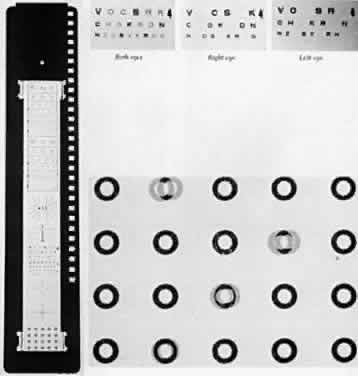
A-O VECTOGRAPHIC PROJECT-O-CHART SLIDE

The A-O Vectographic Project-O-Chart Slide* (Fig. 1) is another method for the study of a scotoma in patients with the monofixation syndrome. It is used in conjunction with a nondepolarizing aluminized screen. The polarized letters of the Polaroid Vectograph slide provide a rapid and dependable differentiation between patients with bifixation and those with monofixation. Each character on the slide has self-contained light polarizations; some are polarized at 90° to others. Viewed through analyzers, some images are made visible to one eye and invisible to the other, while some characters are visible to both eyes. This method provides a test environment closely approximating the normal binocular situation. The patient with bifixation reads the entire 20/50 (6/ 15) visual line without hesitation, although two letters are seen only by the right eye, two others only by the left eye, and the remaining two letters by both eyes. The patient with monofixation deletes the two letters that are imaged only in the nonfixating eye. Occasionally, the monofixating patient who rapidly alternates fixation from one eye to the other reads all six letters, but usually comments that as two letters disappear two others appear. This response misleads the examiner if the patient does not spontaneously comment about the everchanging letters appearing and disappearing.


* American Optical Company.
† Metric equivalent in parentheses after Snellen notation.

Fig. 1. The A-O Vectographic Project-O-Chart Slide.

STEREOACUITY TESTS

Stereoacuity is measured in seconds of arc of image disparity. The factor determining the stereoacuity in the patient with monofixation syndrome is the macular scotoma in the visual field of the nonfixating eye. Bifixation allows the high resolving powers of each macula to detect minute degrees of retinal image disparities, yielding a stereoacuity range between 14 and 40 seconds of arc. In monofixation, the retinal image disparity is detected by studying the images on retinal areas having low resolving power, giving stereoacuities between 60 and 3000 seconds of arc. It has been stressed that stereoacuity is a reliable indicator of either monofixation or bifixation.11

Polaroid vectographs offer a convenient, accurate, and simple method to determine stereoacuity. The vectographs produce the image disparity, and the patient sees them through polaroid analyzers in normal room illumination. If the patient wears glasses, the analyzers are fitted over them. The stereoacuity is measurable at either 6 m or 40 cm.

Distant stereoacuity is measured by the A-O Vectographic Project-O-Chart Slide (see Fig. 1) that presents a range of stereotargets between 240 and 60 seconds of arc.

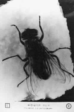
Near stereoacuity is measured with Polaroid vectographs by either the Titmus Stereotest* or the Randot Stereotest. The house fly Titmus Stereotest (Fig. 2) produces approximately 3000 seconds of arc of retinal image disparity at 40 cm. The circles of the Stereotest (Fig. 3) presents a range of stereotargets producing retinal image disparity between 800 and 40 seconds of arc at 40 cm.


* Titmus Stereotest, Stereo Optical Company, Inc, Chicago, IL
† Randot Stereotest, Stereo Optical Company, Inc, Chicago, IL

Fig. 2. The housefly Titmus Stereotest.

Fig. 3. The circles, Titmus Stereotest.

The circles in the Randot Stereotest present random dot stereotargets (Fig. 4) as opposed to the contoured targets of the Titmus Stereotest circles. The Randot targets provide a range of retinal image disparity between 400 and 20 seconds of arc at 40 cm. The Randot eliminates the lateralization clues of Titmus circles that can give false—positive results. Another random dot near stereoacuity test is the TNO Stereotest utilizing color targets and analyzers rather than Polaroid vectographs to produce the retinal image disparity, giving a range between 480 to 15 seconds of arc.

Fig. 4. The Randot Stereotest.


‡ TNO Stereotest, Lameris, Utrecht, The Netherlands

Back to Top
TREATMENT
The primary objective of treatment should be to induce the patient to become simultaneously attentive to the similar images on each macula, that is, to cease monofixating and begin bifixating.

However, all attempts to accomplish this objective have failed. The treatment can be divided into managing motor and sensory problems.

Improvement of the motor problem is not usually necessary since the maximal deviation in the normal binocular seeing situation never appears to exceed 8Δ; this is usually within the range of being easily reduced to zero by the patient's fusional vergence. Occasionally, the alternate cover deviation in a patient may be a horizontal deviation of 20Δ or more; this causes intermittent diplopia when there is a lapse of the fusional vergence that was maintaining an 8Δ or less cover-uncover deviation. This rare patient may benefit from surgery designed to eliminate the alternate cover deviation. Prismatic correction of the motor imbalance may be used in lieu of surgery, but indications for this procedure are equally rare. Except for the rare patient who demands considerable fusion effort to control the large deviation, no benefit is derived from correction of the usually small alternate cover deviation by either surgery or prisms because monofixation persists. The motor imbalance apparently is not the cause of the syndrome.

Inasmuch as these patients have adequate fusional vergence amplitudes, there is rarely a need to prescribe fusional vergence exercises. However, if the exercises are prescribed, the amplitudes increase with the same ease as in the bifixating patients.

Sensory treatment includes monocular and binocular therapy. Monocular therapy is essentially treatment of amblyopia. Unless the monofixating child alternates fixation from one macula to the other, the nonpreferred eye becomes amblyopic. Occlusion therapy adequately manages this sensory complication. If amblyopia tends to return when occlusion therapy is terminated, partial occlusion is maintained until the patient is 9 years old. Occlusion therapy for one hour each day is adequate to prevent recurrence of amblyopia.

Treatment of amblyopia, a monocular sensory defect, does not affect the scotoma, which is a binocular sensory defect in the patient with the monofixation syndrome. The established orthoptic therapeutic approach for overcoming the scotoma due to suppression is training the patient to recognize diplopia, but experience has shown that the patients with monofixation syndrome are refractory to learning to recognize diplopia, other than physiologic diplopia, or diplopia induced by displacing the image outside the scotoma with prisms. This is probably due to the fact that the scotoma is not caused by suppression (active cortical inhibition) but is the manifestation of the patient's capability to be attentive to only one macula at a time rather than simultaneously perceive the images on both maculas.

Anisometropic patients may be converted to alternating use of their monofixating maculas by spectacle or contact lens prescription. Supplying equally clear images simultaneously to each macula usually does not improve the chance for bifixation any more than compensating for the small deviation with prism spectacles, but some rare exceptions to this generalization have been documented.

Back to Top
PROGNOSIS
The most impressive prognostic feature of patients with the monofixation syndrome is their static alignment state. Over the years, their eyes continue to remain aligned as well as if they were bifixating, regardless of the associated factors of strabismus, anisometropia, a unilateral macular lesion, or absence of all three. Extramacular fusion alone seems to be just as effective as the combination of extramacular and central fusion in maintaining straight eyes.

Apparently, the monofixating patient has such a poor prognosis for ever becoming a bifixating patient that no therapy for the disorder appears justified, other than providing the ideal optic correction and occlusion therapy for amblyopia.

Back to Top
REFERENCES

1. Ogle KN: Fixation disparity. Am Orthopt J 4:35, 1954

2. Jampolsky A: Esotropia and convergent fixation disparity of small degree: Differential diagnosis and management. Am J Ophthalmol 41:825, 1956

3. Jampolsky A, Flom BC, Freid AN: Fixation disparity in relation to heterophoria. Am J Ophthalmol 43:97, 1957

4. Jampolsky A: Management of small-degree esodeviations. In Haik GM (ed): Strabismus Symposium of the New Orleans Academy of Ophthalmology, p 125. St. Louis, CV Mosby, 1962

5. Parks MM, Eustis AT: Monofixational phoria. Am Orthopt J 11:38, 1961

6. Burian HM: Normal and anomalous correspondence. In Allen JH (ed): Strabismus Ophthalmic Symposium, p 179. St Louis, CV Mosby, 1950

7. Lang J: Evaluation in small angle strabismus or microtropia. In Strabismus Symposium, pp 219–222. New York, S Karger, 1968

8. Helveston EM, von Noorden GK: Microtropia. Arch Ophthalmol 78:272, 1967

9. Parks MM: The monofixation syndrome. In Symposium on Strabismus, Transactions, New Orleans Academy of Ophthalmology, p 127. St Louis, Mosby, 1971

10. Irvine SR: Amblyopia exanopsia: Observations on retinal inhibition, scotoma, projections, light difference discrimination and visual acuity. Trans Am Ophthalmol Soc 46:527, 1948

11. Parks MM: Stereoacuity as an indicator of bifixation. In Strabismus Symposium, pp 258–260. New York, S Karger, 1968

Back to Top