Chapter 8
Binocular Vision Adaptations in Strabismus
MARSHALL M. PARKS
Main Menu   Table Of Contents

Search

BINOCULAR VISION
VISUAL SYMPTOMS IN STRABISMUS
ADAPTATIONS OF THE EXTRAMACULAR BINOCULAR FUSION REFLEX TO STRABISMUS
REFERENCES

Strabismus patients with established binocular vision before the onset of misaligned eyes may adapt their binocular vision to conform to the deviation of their eyes. The adaptations are in response to the annoying symptoms caused by the persistent functioning of normal binocular vision after acquiring the strabismus. Before discussing the adaptation, a brief review of normal binocular vision is in order.
Back to Top
BINOCULAR VISION
Binocular vision is the cortical integration of similar images on each retina into a unified perception. Binocular vision is composed of three independent components and is contained in two separate reflexes. The components of binocular vision are simultaneous perception, fusion, and stereopsis. The distinct reflexes of macular and extramacular binocular vision result in the duality of binocular vision. The many differences between the macular and extramacular binocular vision reflexes are discussed in Chapter 5.

Pertinent to this chapter on the adaptations of binocular vision in strabismus and the most important difference between macular and extramacular fusion reflexes is the absence of any adaptation by the macular fusion reflex in the strabismic patient. The reflex is impossible to adapt to the newly acquired strabismus because it ceases to function the moment the eyes deviate from being straight. For an adaptation of the binocular vision reflex to occur, the reflex must continue functioning during strabismus, which causes annoying symptoms that are eliminated by the adaptation. Only the extramacular fusion reflex continues functioning after the onset of the strabismus. Hence, the adaptations of binocular vision in strabismus are restricted to the extramacular fusion reflex.

Binocular vision is an acquired reflex that normally develops during the first 3 to 4 months of life. Its development demands certain requisites. The infant must be capable of seeing with each eye, and both eyes must be aligned with one another, permitting similar retinal images to project onto corresponding retinal areas during the critical period for binocular vision development, which extends to approximately 2 years of age. Patients with congenital or very early onset strabismus do not receive the essential stimulation from similar images projecting onto corresponding retinal areas; consequently, their binocular vision reflexes do not develop. Absence of binocular vision is elicited by sensory tests yielding responses that prove the absence of simultaneous perception of images on each retina (Chapter 9).

Patients1,2 without binocular vision experience none of the untoward visual symptoms that are associated with newly acquired strabismus in those who have developed binocular vision. Their only visual handicap is absence of stereopsis, which they are as unable to comprehend as the colorblind person is unable to comprehend a deficiency in color perception.

Despite the absence of binocular vision, if the attention directed to the object of regard results in fixating first with one eye for a moment and then fixating with the other eye for the next moment, such alternate fixation ensures normal monocular macular function in each eye. However, if the attention to the object of regard was fixated exclusively with one eye during the amblyogenic age, amblyopia will be apparent in the unused eye.

Back to Top
VISUAL SYMPTOMS IN STRABISMUS
Once the extramacular fusion reflex has developed, annoying visual symptoms are apparent when the eyes deviate from being straight or when placing a 15Δ prism before one eye. Viewing with the strabismic eyes, or through the prism before straight eyes, precludes the opportunity for similar retinal images to project onto corresponding retinal areas. The similar images are now seen doubled in space (diplopia), along with an overlay of dissimilar images appearing to be superimposed in the same place in space (visual confusion).

DIPLOPIA

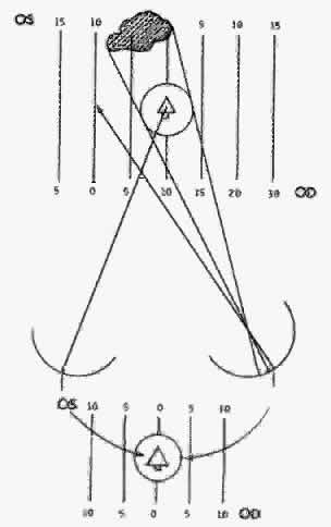
Diplopia results from the simultaneous perception of the images of the object of regard projecting onto noncorresponding retinal areas of the strabismic eyes. The simultaneous perception of the two similar images under these circumstances yields the impression that the object of regard is simultaneously located at two points in space (Fig. 1).

Fig. 1. Diplopia results from simultaneous perception of similar images of the object of regard, that is, a Christmas tree, projecting onto noncorresponding retinal areas. Esotropia causes homonymous diplopia; clear image on macula of right eye is illustrated by solid lines, blurred image on nasal retina of left eye is illustrated by broken lines. The illustrated diplopic images are displayed according to the patient's drawing of them. Inset. Heteronymous diplopia in exotropia. (From Symposium on Strabismus, Transactions of the New Orleans Academy of Ophthalmology. St Louis: CV Mosby, 1971.)

The size of the retinal area in one eye that corresponds to a retinal point in the other eye rapidly expands for retinal loci increasingly peripheral to the fovea. This determines the degree of retinal image disparity that either the macular or extramacular fusion reflex find tolerable. It accounts for Panum's visual space. Thereby, the depth of Panum's visual space is very limited for the macular fusion reflex to function within compared with the ever-expanding depth of fusional space within which the extramacular fusion reflex operates. This explains how an esotropic deviation may be sufficiently small to allow a spatial target peripheral to the object of regard (yet within Panum's fusional space) to project the peripheral target's images onto corresponding retinal areas while the object of regard projects its images onto noncorresponding macular areas (Fig. 2).

Fig. 2. The heterotropic deviation may be sufficiently small to cause the object of regard to be diplopic, but the peripheral objects displaced equidistant to the object of regard from the horopter may still be within Panum's area. Thus it is possible for a patient with a small heterotropic deviation to have extramacular fusion in the absence of macular fusion. (From Symposium on Strabismus, Transactions of the New Orleans Academy of Ophthalmology. St Louis: CV Mosby, 1971.)

According to Ogle,3 foveal horizontal image disparity greater than 20 minutes of arc (2/3 of a prism diopter) is not fusible. From my clinical investigation it appears that extramacular image disparity is fusible up to approximately 480 minutes of arc4,5 (8 prism diopters). The vast differences in tolerance of horizontal image disparity between macular and extramacular fusion suggest they are under the control of separate binocular vision reflexes. This reasoning is strengthened by the frequency of clinical cases that are devoid of a macular fusion reflex but have a normal extramacular fusion reflex.

The purpose of the macular fusion reflex is to produce and maintain bifixation. If the reflex is absent or intermittently shut down for any reason, the patient monofixates. Monofixation is manifest by a three-degree facultative macular scotoma present only in the nonfixating eye during binocular viewing while the intact normal extramacular fusion reflex remains within 0 to 8 prism diopters of horizontal eye alignment.

Such a large range of tolerance for horizontally disparate retinal images to be fused in the monofixator is easily demonstrated using stereopsis tests. Although the stereoacuity is poor in the monofixator compared with the bifixator,6 it does exist as long as the horizontal deviation of the eyes does not exceed 8 prism diopters. Along with the intact stereopsis, the fusional vergence amplitudes also remain normal in the monofixator as long as the horizontal deviation of the eyes does not exceed 8 prism diopters. In my experience, normal fusional vergence amplitudes and stereopsis perception occur only in normal retinal correspondence (NRC) but not in the adapted abnormal retinal correspondence (ARC).

Stereopsis can occur only if the horizontally disparate images in the monofixator's peripheral vision are from targets within Panum's fusional space. Although the center of the Panum's area is scotomatized, the peripheral Panum's area exists in small angles of deviation. To maintain this small horizontal deviation of 0 to 8 diopters (D) requires a good functioning fusional vergence reflex that can be generated by NRC but not by ARC.

VISUAL CONFUSION

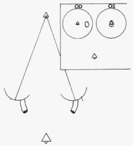
Visual confusion occurs in strabismus as a result of the projection onto corresponding retinal areas of simultaneously perceived dissimilar images of two different spatial objects that normally project onto noncorresponding retinal areas. Consequently, these different objects in space that create dissimilar images are perceived as located at the same place in space. Visual confusion does not exist for all portions of the visual field. The object of regard visually perceived by the fixating eye cannot simultaneously perceive a superimposed different image projecting on the fovea of the deviating eye. From the physiologic fact of binocular vision, as stated in Chapter 5, Fig. 13, only similar images projecting on the foveas are perceived simultaneously. No binocular function results from dissimilar images projecting onto these retinal areas. Consequently, from the moment the strabismic eyes deviate the patient is spared the visual confusion of a different spatial object's image conflicting with the object of regard. The three-degree macular scotoma in the esotropic left eye (Fig. 3) can be plotted using a binocular perimetric technique. This is a physiologic scotoma, present in all strabismic patients immediately at the moment the alignment of the eyes exceeds the threshold of 2/3 of a prism diopter. Therefore sustaining the bifixation reflex requires a very refined ocular alignment control along with an elegant level of similarity of the foveal images. The combination of these two inputs provide the essential stimuli for bifixation. The moment one or both are lacking, the bifixation reflex shuts down and a macular scotoma appears in the nonfixating eye. It is not a pathologic scotoma that evolves gradually to resolve the annoying visual symptom caused by strabismus. It is not an adaptation of the macular fusion reflex that gradually developed to eliminate visual confusion. In short, it is not a suppression scotoma.

Fig. 3. An absolute physiologic scotoma of the macula of the deviated eye develops immediately when the eye deviates, preventing perception simultaneously of a dissimilar image on the deviated eye's macula, compared with the macular image of the fixating eye. (From Symposium on Strabismus, Transactions of the New Orleans Academy of Ophthalmology. St Louis: CV Mosby, 1971.)

RETINAL IMAGE SIMILARITY VERSUS DISSIMILARITY

In addition to image disparity, another stimulus that pertains to the alignment of the eyes with certain thresholds required for fusion is image dissimilarity. The threshold for image dissimilarity acceptable for fusion is different for macular fusion compared with extramacular fusion. Dissimilar image parameters are size, shape, clarity of focus, illumination brilliance, and color. Each has its threshold of dissimilarity that eliminates fusion. Anisometropia, media disturbances within the ocular tissues, optical appliances, and so forth are sources of image dissimilarity that affect fusion.

Anisometropia is probably the maximal cause of image dissimilarity affecting the macular and extramacular binocular vision reflexes. Weakley7 has shown that the macular binocular vision reflex threshold for being shut down by anisometropia is approximately 1.5 D spherical equivalent (SE), but the extramacular binocular vision reflex continues to function until a much greater quantity of SE anisometropia is induced. The test for this is use of a vectographic method for measuring the stereoacuity. The macular level of stereoacuity disappears at approximately 1.5 to 2.0 D SE, but gross stereoacuity continues. The three-degree macular scotoma instantly appears in the more unfocused eye and bifixation is replaced by monofixation. Consequently, two different stimuli are essential for the macular fusion reflex to function.

Function of the macular binocular fusion reflex requires simultaneous input from the two essential stimuli. First, the alignment of the eyes must not exceed 2/3 of a prism diopter (retinal image disparity greater than 20 seconds of arc), and second, anisometropia must be no greater than approximately 2 D SE7 (retinal image dissimilarity). The moment either of the two essential stimuli are absent macular binocular vision (bifixation) ceases to function and a macular scotoma appears in the nonfixating eye. However, these levels of retinal image disparity and dissimilarity are subthreshold for affecting the continued function of the extramacular binocular vision reflex (within 8 prism diopters of the eye deviation and unknown image dissimilarity). In the meantime the extramacular fusion reflex continues to function because its thresholds for fusing disparate images and/or dissimilar images are greater than those of the macular fusion reflex. The fusional vergence amplitude and stereoperception of the extramacular fusion reflex continue as long as the ocular deviation is 8 prism diopters or less and the image dissimilarity is approximately 4 D SE or less of induced anisometropia.*


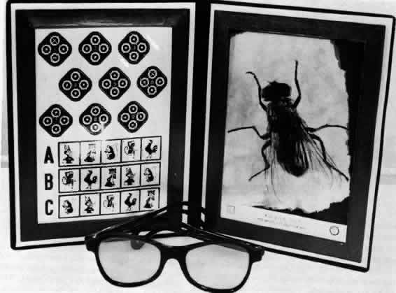
*Note: In self-experimentation with the stereo fly test held at 16 in., my 40-second stereoacuity continued when incremental-plus-spherical lens power was placed before one eye until + 2 D of induced anisometropia was reached. The stereoacuity precipitously diminished to the 70- to 100-second range, which restored to 40 seconds on removing the + 2 D add and diminished to the 70- to 100-second range the instant the + 2 D add was replaced. With incremental addition of more + D add the 70- to 100-second stereoacuity range persisted until reaching the + 4 D level of induced anisometropia, at which time precipitously all stereoperception disappeared. The 3000-second stereotest fly went flat and appeared as a picture of a fly; when the + 4 D add is reduced to a + 3 D add, the stereoperception of the fly instantly returned. My interpretation of the results are that + 2 D-induced anisometropia is just beyond threshold of image dissimilarity tolerated by the macular binocular vision reflex, but + 4 D of induced anisometropia is required to exceed the threshold of image dissimilarity tolerated by the extramacular binocular vision reflex.

Back to Top
ADAPTATIONS OF THE EXTRAMACULAR BINOCULAR FUSION REFLEX TO STRABISMUS

PHYSICAL ADAPTATIONS

The onset of strabismus in a person who has developed binocular vision produces three distinct annoying symptoms: central vision diplopia, peripheral vision diplopia, and peripheral vision confusion. Two factors offer partial relief from central vision diplopia. One is the perceptual difference in the sharpness and clarity of the two similar images of the object of regard because one object is projected onto the fovea of the fixating eye and the other is projected onto the extramacular area of the nonfixating eye. However, the visual acuity difference that results between the two eyes pertains only to contoured images and not to the images from a light with no contour. Also, it pertains to viewing the images during daylight. Under these visual circumstances, the better-seen image provides the important clue for the strabismic patient to locate correctly the object of regard in space. However, during scotopic vision or when viewing contourless objects, the patient is without the differential clues of sharpness and clarity to direct attention to the correct image.

The ability to be attentive to only one of the diplopic images of the object of regard is subconsciously enhanced by a rapid blink of the nondominant eye. The image that correctly localizes the object of regard is immediately identified, and as long as the patient's attention remains directed to this image, correct localization of the object continues. This tactic functions satisfactorily as long as the object of regard remains isolated and stationary during either photopia or scotopia, but if the patient is viewing a series of moving contourless objects, such as multiple oncoming headlights during nighttime driving, only sustained voluntary closure of the nonfixating eye may avert catastrophe.

CORTICAL ADAPTATIONS

Once the extramacular fusion reflex is established it never is completely surrendered. It is capable of being altered in various ways to alleviate the annoying symptoms a strabismic patient experiences because of its continued function while the eyes are misaligned. The symptoms are diplopia, both from the spatial area of conscious regard and visual space surrounding it, and visual confusion from only the visual space surrounding the area of conscious regard. Collectively, they all contribute to the patient's insecurity in orientation in visual space. If the patient could shut down the extramacular fusion reflex, as the macular fusion reflex was shut down the moment the eyes deviated, the symptoms would not exist. However, the continued function of the extramacular fusion reflex after the eye alignment no longer is controllable by the fusional vergences results in this constellation of symptoms. Fortunately, the extramacular fusion reflex possesses the virtue of being able to adapt its normal physiologic function to eliminate the annoying symptoms that a newly acquired strabismus causes. Two adaptations exist, and they develop gradually at approximately the same rate after the onset of the strabismus. Adapting for the symptom of the central visual field diplopia is a cortical phenomenon, called suppression, that affects a restricted area in the deviated eye's peripheral retina. The other cortical adaptation, called ARC, eliminates the peripheral visual field diplopia and visual confusion.

Time and exposure to the annoying symptoms are required for these adaptations to develop. The time required for their gradual development is minimal in young children compared with older children and adults. Children seem more tolerant to the symptoms than adults and relief for children arrives within days, whereas adults may take weeks to receive relief. Adults are far more impatient than children. The newly acquired strabismic adult is prone to react to the diplopia and visual confusion by occluding one eye and never giving the adaptations an opportunity to develop.

Suppression

Suppression is a positive inhibitory reflex that occurs within the extramacular fusion reflex and permits the cortex to ignore visual sensations dispatched from the retina of the nonfixating eye, on which are projected the images from the area of conscious regard. If either eye can maintain fixation, suppression is demonstrable in the nonfixating eye. In contrast to the physiologic macular scotoma that can be plotted in the deviated eye, the extramacular suppression scotoma is pathologic. It is absolute, but it is also facultative because it is nonexistent during monocular vision. Therefore, it may be plotted in the nonfixating eye only during binocular perimetry. The shape and the size of the suppression scotoma in esotropia (Fig. 4) differ significantly from the scotoma in exotropia. The nasal retina in esotropia usually has a scotoma of approximately 5 degrees, whereas the temporal retina in exotropia produces a scotoma extending peripherally from the hemiretinal line for the number of degrees required to include the image of the object of regard. This difference in size and shape of the scotomas in the nasal and temporal retinas is unexplainable.

Fig. 4. The suppression scotoma in an exotropic patient's deviated eye extensively involves the temporal retina up to the hemiretinal line; in the nasal retina of the esotropic patient (Inset) it is small and regional. (From Symposium on Strabismus, Transactions of the New Orleans Academy of Ophthalmology. St Louis: CV Mosby, 1971.)

Patients with vertical and/or cyclodeviations have the same suppression adaptation as the horizontal deviations. Also, the suppression scotoma is not rigidly located to a fixed retinal area. In patients with deviations that vary according to different gaze positions, the scotoma in the binocular field shifts across the retina according to the location where the object-of-regard image projects onto the extramacular area of the nonfixating eye. Also, it shifts from one eye to the other as rapidly as the patient alternates fixation, a feature shared also by the physiologic macular scotoma.

Unlike the pathologic suppression scotoma that developed in the extramacular binocular vision reflex, the physiologic macular scotoma remains rigidly in the same location, which is the macula of the deviated nonfixating eye. Recall that this scotoma is smaller (three degrees) than the suppression scotoma. The macular scotoma is not an adaptation of the extramacular fusion reflex to solve the annoying symptoms of diplopia or visual confusion. It does not gradually develop over time, as does the pathologic scotoma. Instead it appears the moment the macular fusion reflex shuts down (See Fig. 3). Finally, in the duality of binocular vision, the macular scotoma is a phenomenon within the macular fusion reflex, whereas the suppression scotoma is a phenomenon within the extramacular fusion reflex.

Unfortunately, the macular scotoma is often alluded to as a macular suppression scotoma, which is likely to confuse the student trying to comprehend the subject. The macular scotoma has nothing to do with the adaptation of the extramacular fusion reflex called suppression.

ABNORMAL RETINAL CORRESPONDENCE

Anomalous retinal correspondence is the cortical adjustment in a strabismic patient of the normal directional values supplied by the extramacular neuroepithelial retinal elements to permit fusion of the similar images projected onto noncorresponding areas. It does not occur within the macular fusion reflex. It is a phenomenon that occurs only within the extramacular fusion reflex. Abnormal retinal correspondence is the adaptation that eliminates the strabismic patient's peripheral visual field of both diplopia and visual confusion. Patients with vertical and cyclovertical strabismus, as well as those with horizontal strabismus, develop ARC to eliminate their peripheral vision symptoms.

No matter which strabismic eye fixates, whether both have foveal fixation capability or eccentricfixation exists in one eye, ARC can develop. It is manifest only during binocular viewing, because there is no abnormality in the directional values of the neuroepithelial elements of either eye during monocular viewing.

Once ARC has developed and extramacular single binocular vision is present in the strabismic patient, that person is always able to cortically adjust the directional values supplied by the neuroepithelial retinal elements to permit continuous fusion of similar images, despite different positions the eyes may assume. Subsequent to any change of eye alignment, such as after surgery, an ARC patient experiences diplopia and visual confusion until the cortical adjustment is adapted to match the directional values of the retinal neuroepithelial elements to the new alignment. This adjustment in the extramacular fusion reflex requires time (often several weeks for adults but usually only a few days for children), but the cortical adjustment is finally reconciled.

There is no apparent reason why ARC should not be a total cortical adjustment of the directional values of the retinal neuroepithelial elements to the degree that would allow fusion. Yet the results of certain testing techniques that do not simulate everyday seeing experience suggest that only a partial cortical adjustment occurs in some patients. From these results, the classification of harmonious ARC and unharmonious ARC has evolved to describe total and partial cortical adjustment. The results of recent nonlaboratory testing, offering everyday seeing experiences, indicate that the unharmonious ARC response is an artifact produced by the artificial seeing circumstance in the retinal correspondence test. The obsolete classic test for ARC with the haploscope evokes these artifactual responses because the patient views only a small portion of the visual field. Introduction of the Bagolini striated glasses test8 eliminated this artifact because they allow the full binocular field to be viewed by the patient (Chapter 9).

The ability of the extramacular binocular vision reflex (while in the NRC mode) to provide superb fusional vergence amplitudes has been discussed previously. In the ARC mode, however, the extramacular fusion reflex provides essentially zero fusional vergence amplitude. This may be explained by the fact that ARC patients have no need for fusional vergence amplitudes. It is more reasonable to expect the ARC patient to make a sensorial cortical adaptation rather than to expect a motor response to be made to alter the alignment on behalf of fusion. Therefore ARC fusion is a sensory extramacular fusion adaptation without a motor component, unlike NRC fusion, which possesses both a sensory and a motor component.

Another deficiency encountered in ARC patients involves stereopsis. I found no stereopsis appreciation in strabismic patients who had a simultaneous prism and cover horizontal deviation greater than 8Δ when tested with Polaroid vectographic stereo-targets that created a retinal image disparity of 6000 seconds of arc (using the Polaroid vectographic housefly of the Titmus Stereotest held 20 cm from the eyes) (Fig. 5).

Fig. 5. Titmus stereotest.

Abnormal retinal correspondence and suppression (Fig. 6) seem to develop simultaneously. With various testing techniques, they are easily demonstrated to coexist in all patients, despite the small suppression scotoma in the deviated eye of the esotrope that is confined to a nasal retinal area of 5 degrees, in contrast to the large suppression scotoma of the temporal retina in the deviated eye of the exotrope that extends up to the hemiretinal line (See Fig. 4).

Fig. 6. Abnormal retinal correspondence is the cortical adjustment in strabismus that permits fusion of similar images projecting onto noncorresponding retinal areas from object points peripheral to the area of conscious regard. Suppression eliminates the diplopia caused by object points within the area of conscious regard projecting onto noncorresponding retinal areas. (From Symposium on Strabismus, Transactions of the New Orleans Academy of Ophthalmology. St Louis: CV Mosby, 1971.)

Patients with intermittent strabismus may have ARC and suppression while the eyes are deviated but NRC and no suppression when the eyes are straight. Also, patients with deviation angles that vary in different positions of gaze may have ARC values and suppression scotoma localization that change according to the deviation angle as the eyes are moved into various positions. This is commonly observed in strabismic patients whose horizontal deviation has an associated A or V pattern or those with a vertical deviation that reverses in right and left gaze as a result of oblique muscle dysfunctions.

Esotropic patients with 30 to 40Δ of deviation may use the optic disc's physiologic blind spot of the deviated eye as the suppression scotoma (Fig. 7), removing the need for their visual cortex to adapt to eliminate the central visual field diplopia.9 However, their retinal area peripheral to the optic disc may develop the ARC adaptation to eliminate the peripheral visual field diplopia and visual confusion. Such a patient has no cortical adaptation equivalent to suppression but does have the cortical adaptation of ARC, an example of a situation in which both cortical adaptations do not concurrently exist. Consequently, one cannot state that suppression and ARC invariably are associated cortical adaptations of the extramacular fusion reflex.

Fig. 7. The physiologic blind spot offers an excellent ready-made immediate scotoma for the esotropic patient with 25 to 40Δ of deviation. (From Symposium on Strabismus, Transactions of the New Orleans Academy of Ophthalmology. St Louis: CV Mosby, 1971.)

ARC is able to reverse itself to NRC when the alignment is restored to within 8 or less prism diopters of horizontal deviation. Surgery, glasses, or prism therapy, either singly or in combination, can achieve this result. With the return of normal alignment, fusional vergence amplitudes and extramacular stereopsis appear, which suggests that NRC has resumed. However, bifixation does not return unless the constant strabismus persisted no longer than 3 months, a little-appreciated but significant difference between the macular and extramacular fusion reflexes.

Recall that once the extramacular fusion reflex has developed, it is never later surrendered by the patient who acquires strabismus. However, the NRC fusion reflex may be adapted to eliminate symptoms resulting from the simultaneously perceived diplopia and visually confusing images. This is accomplished as the NRC mode is replaced by the ARC and suppression mode. In the meantime, however, the macular binocular reflex, having no capacity to adapt its continued function in the newly acquired strabismus, immediately shuts down functioning the moment the eyes deviate more than 2/3 of a prism diopter. Remaining shut down constantly for 3 months destroys the reflex.

Currently it has become recognized that delaying surgery to align the intermittent exotropia can result in deterioration to constant exotropia. When bifixation has been absent for 3 months, the best obtainable result will be the monofixation syndrome.10,11,12 Also, these studies show that if the surgery overcorrects the intermittent exotropia to a constant esotropia, making bifixation impossible for 3 months or more, the best obtainable result will be the monofixation syndrome. Similarly, the 3-month critical period of absence of bifixation causing its permanent loss has been confirmed by using surgically induced anisometropia to produce monovision for managing presbyopia.13 Inducing surgical anisometropia of SE equal to or more than 1.5 D can be destructive to the bifixation reflex.14 An optically induced anisometropia for treating presbyopia with monovision presumably carries less risk for causing permanent destruction of the bifixation reflex than surgically induced anisometropia, because glasses or contact lenses seldom are worn constantly for 3 months, allowing some opportunity for bifixation and continued function of the reflex.

Back to Top
REFERENCES

1. Ing MR, Costenbader FD, Parks MM et al: Early surgery for congenital esotropia. Am J Ophthalmol 61:1419, 1966

2. Botet RV, Calhoun JH, Harley RD: Development of monofixation syndrome in congenital esotropia. J Pediatr Ophthalmol Strabismus 18:49, 1981

3. Ogle KN: Fixation disparity. Am Orthopic J 4:35, 1954

4. Parks MM: Monofixation syndrome. Tr Am Ophthalmol Soc 67:609, 1969

5. Parks MM: Monofixation syndrome. In Tasman W, Jaeger EA (eds): Duane's Clinical Ophthalmology. Vol 1, 4th ed. Philadelphia: Lippincott, 1994

6. Parks MM: Stereoacuity as an indicator of bifixation. International Strabismus Symposium, Arruga A. New York: S. Karger, 1968:258–260

7. Weakley DR: The association between anisometropia, amblyopia, and binocularity in the absence of strabismus. Trans Am Ophthalmol Soc 97:987, 1999

8. Bagolini B: Anomalous correspondence. Definitions and diagnostic methods. Doc Ophthalmol 23:346, 1967

9. Swan KC: The blind spot syndrome. Arch Ophthalmol 40:371, 1948

10. Richard JM, Parks MM: Intermittent exotropia: surgical results in different age groups. Ophthalmology 90:178, 1984

11. Ing MR, Nishimura J, Okino L: Outcome study of bilateral lateral rectus recession in intermittent exotropia in children. Trans Am Ophthalmol Soc 95:433, 1997

12. Abroms AD, Mohney BC, Parks MM et al: Timely surgery in intermittent and constant exotropia for superior sensory outcome. Am J Ophthalmol 131:111, 2001

13. Parks MM: Monovision: The case for two binocular vision systems. Binocular Vis Strabismus Q 15:13, 2000

14. Fawcett SL, Herman WK, Alfieri CD et al: Stereoacuity and foveal fusion in adults with long-standing surgical monovision. J Am Assoc Pediatr Ophthalmol Strabismus 5:342, 2001

Back to Top