Duction
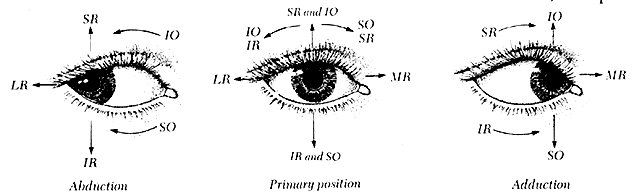
The term duction refers to the movement of one eye. A prefix is attached to this word to indicate the direction in which the eye is moved (Fig. 1). Duction is accomplished by simultaneous and equally graded contraction and relaxation of antagonistic muscles, in accord with Sherrington's law of reciprocal innervation.
|
Adduction
Adduction is a horizontal movement directed medially from the vertical axis. It is primarily accomplished by contraction of the medial rectus muscle and relaxation of the lateral rectus muscle.
Recessing the insertion does not reduce the adduction significantly until the recession exceeds 6 mm. In the primary position the muscle contacts the globe 6 mm as it arcs around the nasal sclera to insert. The medial rectus is a tight muscle; a maximum resection of 6 mm is possible without also producing resistance to abduction and slight retraction of the eye into the orbit (enophthalmos), with a reduction of the vertical dimension of the palpebral fissure.
A small amount of secondary adduction is accomplished by contraction of the vertical rectus muscles: in upgaze by the superior rectus muscle, in downgaze by the inferior rectus muscle, and in the primary position by their combined contraction.
Abduction
Abduction is a horizontal movement directed laterally from the vertical axis. It is accomplished by contraction of the lateral rectus muscle and relaxation of the medial rectus muscle.
Recessing the insertion does not reduce the abduction significantly until the recession exceeds 7 to 8 mm. In the primary position the muscle contacts the globe approximately 8 mm as it arcs around the temporal sclera to insert. The lateral rectus is not a tight muscle like the medial rectus, so excessive resection is less inclined to result in retraction of the eye into the orbit. However, resection of more than 8 mm does offer clinically detectable resistance to adduction.
A small amount of secondary abduction is accomplished by contraction of the oblique muscles: in upgaze by the inferior oblique muscle, in downgaze by the superior oblique muscle, and in the primary position by their combined contraction.
Supraduction
Supraduction (sursumduction) is a vertical movement (elevation) directed superiorly from the horizontal axis of the eye. It is the result of the combined contraction of the superior rectus and inferior oblique muscles and the combined relaxation of the inferior rectus and superior oblique muscles. However, as the eye moves into an abducted position, the superior rectus becomes the prime elevator muscle, and as the eye moves into adduction, the inferior oblique is the principal elevator muscle.
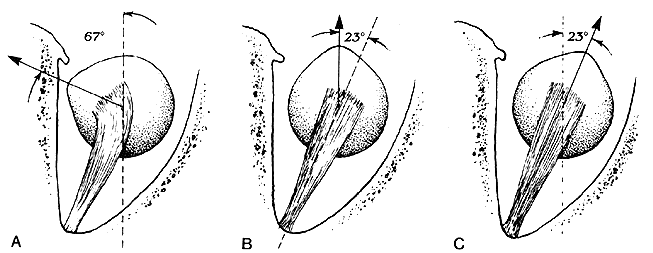
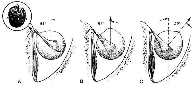
Since the superior rectus muscle courses forward from the orbital apex at an angle of 23° temporal to the medial wall of the orbit and inserts anterior to the center of rotation of the globe, the movement of the eye produced by the contraction of the muscle varies according to its horizontal starting position (Fig. 2). Starting from a position of 23° of abduction, the only movement is elevation. Starting from a position of 67° of adduction, the only movement is intorsion. Starting from the primary position, the movement is combined elevation and intorsion plus slight adduction; the adduction results from the midline of the muscle being medial to the center of rotation of the globe when the eye is in the primary position.
|
Recessing the superior rectus muscle decreases all its functions; resecting the muscle enhances all primary position functions plus offers resistance to the relaxation of its functions. Recessions and resections of the superior rectus muscle in excess of 4 mm reduce the normal full vertical excursion of the eye in abduction; those in excess of 6 mm produce a detectable change from the preoperative primary position relationship between the superior limbus and the position of the upper eyelid. Postoperatively, the vertical dimension of the palpebral fissure remains unchanged from the preoperative value but the level of the eye is vertically changed; this causes the sclera to present above the limbus following excessive recession and causes the superior pupillary border to be tangential with the upper eyelid following excessive resection.
Nasal transposition of the superior rectus muscle by one-half width enhances the adduction function, while temporal transposition of the same amount eliminates the secondary adduction, the effect being most conspicuous in midline upgaze.
Infraduction
Infraduction (deorsumduction) is a vertical movement (depression) directed inferiorly from the horizontal axis. It is the result of the combined contraction of the inferior rectus and superior oblique muscles. However, as the eye moves into an abducted position, the inferior rectus becomes the prime depressor muscle, and in adduction, the superior oblique is the principal depressor muscle.
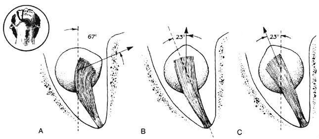
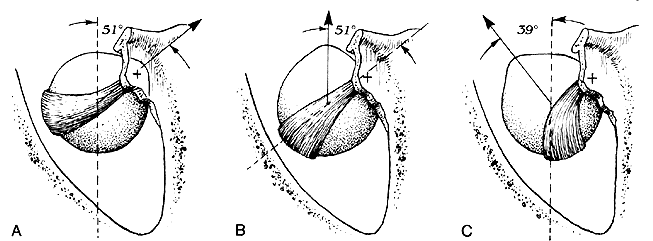
Since the inferior rectus muscle courses forward from the orbital apex at an angle of 23° temporal to the medial wall of the orbit and inserts anterior to the center of rotation of the globe, the movement of the eye produced by the contraction of the muscle varies according to its horizontal starting position (Fig. 3). Starting from a position of 23° of abduction, the only movement is depression. Starting from a position of 67° of adduction, the only movement is extorsion. Starting from the primary position, the movement is combined depression and extorsion plus slight adduction; the adduction results from the midline of the muscle being medial to the center of rotation of the globe when the eye is in the primary position.
|
Recessing the inferior rectus muscle decreases all its functions; resecting the muscle enhances all primary position functions plus offers resistance to the relaxation of its functions. Unless the inelastic cords extending between the external capsular surface of the inferior rectus muscle and lower eyelid tarsus skin and the inferior orbital septum are completely severed, any quantity of recession or resection will lower or raise the lower eyelid, causing either an increase or a decrease in the vertical dimension of the palpebral fissure. Recession of the inferior rectus muscle in excess of 4 mm reduces the total excursion of downgaze in abduction; however, as much as a 10-mm resection only minimally reduces the expected total excursion of upgaze.
Nasal transposition of the inferior rectus muscle by one-half tendon width enhances the adduction function, while temporal transposition of the same amount eliminates the secondary adduction, the effect being most conspicuous in midline downgaze.
Incycloduction
Incycloduction (intorsion) is a torsional movement of the eye about the anteroposterior axis that displaces the superior pole of the eye medially. Intorsion is the result of the combined contraction of the superior oblique and superior rectus muscles and the combined relaxation of the inferior oblique and inferior rectus muscles. However, as the eye moves into an abducted position, the superior oblique muscle becomes the prime intortor, and in adduction, the superior rectus muscle is the principal intortor.
Since the reflected tendon of the superior oblique muscle delivers the direction of the pulling power to the scleral surface when the muscle contracts, the movement of the eye varies according to its horizontal starting position (Fig. 4). Starting from a position of 39° of abduction, the only movement is intorsion. Starting from a position of 51° of adduction, the only movement is depression. Starting from the primary position, the movement is combined intorsion and depression plus slight abduction; the abduction results from the tendon being posterior to the rotation center of the globe when the eye is in the primary position.
|
Unlike the tendons of the rectus muscles, the superior oblique tendon does not lend itself well to recession or resection. Tenotomy and tucking of the tendon are the usual weakening and strengthening procedures performed on the superior oblique muscle. Tenotomy is performed medial to the superior rectus muscle on the cord portion of the tendon; the tuck may be performed on the tendon either medial or lateral to the superior rectus muscle. A tenotomy does not completely eliminate all function of the superior oblique, because some pull power persists when the muscle contracts through the connections of the tendon to the Tenon's capsule and the intermuscular septum. Tucking the tendon causes a relative decrease in the elevation excursion of the adducted globe; the severity is directly proportional to the quantity of the tuck.
The anterior half of the reflected tendon of the superior oblique muscle has a greater intorsion function, and the posterior half has a greater depressor function, according to Fink.1 Emanating from the thesis is an operative procedure described by Harada and Ito that either advances or anteroplaces the anterior half of the tendon to enhance the intorsion function.2
The secondary abduction function of the superior oblique muscle is apparent in midline downgaze by hyperfunctioning (overaction), causing excess abduction, and hypofunctioning (palsy), causing deficient abduction. The midline downgaze abduction abnormality associated with either overaction or palsy is eliminated by tenotomy or tucking of the tendon, respectively.
Excycloduction
Excycloduction (extorsion) is a torsional movement of the eye about the anteroposterior axis that displaces the superior pole of the eye laterally. Extorsion is the result of the combined contraction of the inferior oblique and inferior rectus muscles and the combined relaxation of the superior oblique and superior rectus muscles. However, as the eye moves into an abducted position, the inferior oblique muscle becomes the prime extortor, and in adduction, the inferior rectus muscle is the principal extortor.
When the inferior oblique muscle contracts, the resulting movement of the eye varies according to its horizontal starting position (Fig. 5). Thus, starting from a position of 39° of abduction, the only movement is extorsion. Starting from a position of 51° of adduction, the only movement is elevation. Starting from the primary position, the movement is combined extorsion and elevation plus slight abduction; the abduction results from the muscle being posterior to the center of rotation of the globe when the eye is in the primary position.
|
Weakening the inferior oblique muscle is accomplished by recession, myotomy, myectomy, or denervation plus extirpation. Recession can be graded according to the severity of the hyperfunction, ranging between 6 and 14 mm. Disinsertion without suturing the end of the muscle to the sclera is of little value, because the muscle reattaches near the original insertion site, nullifying an initially satisfactory result. The same fate is shared by denervation, since reinnervation occurs within a matter of months. Myotomy anywhere along the course of the muscle is followed by subsequent reunion of the cut ends, which also occurs in minimal myectomies. Myectomies of several millimeters are more effective but do not permit a graded quantity of surgery. The most complete elimination of the overaction is accomplished by extirpating all muscle tissue between the inferior oblique's penetration of Tenon's capsule and insertion. Denervation of the muscle is required in performing the extirpation, and this procedure is justified only in the case of the most severe overaction of the inferior oblique muscle. Strengthening procedures, such as resections and advancements, for an underacting inferior oblique are difficult and have proved rather useless.
The secondary abduction function of the inferior oblique muscle is apparent in midline upgaze by hyperfunctioning, causing excess abduction. The excessive upgaze abduction associated with the overaction is eliminated by weakening the muscle.
Jampel does not agree with the foregoing classic teaching regarding the vertical and torsional movements of the eyes.3, 4 He has challenged the concept of oblique muscle action. Based on mathematical considerations, studies of models of the eye, experimental studies on monkeys, and limited clinical observations, he proposes the following: (1) Elevation and depression, even in adduction, are mainly a function of the superior and inferior rectus muscles. (2) The torsional component of the oblique muscles is the same throughout the range of horizontal eye movement. (3) The abducting component of the oblique muscles does not decrease on adduction but actually increases.
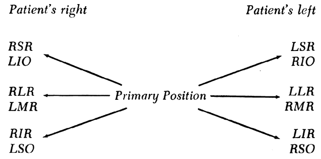
Version
Version refers to simultaneous movement of both eyes in the same direction; a prefix indicates the direction of the conjugate movement. The muscles in each eye that are the prime movers undergo equally graded contractions in accord with Hering's law of innervation, and for each contracting muscle there is an opposite and equally graded antagonist (Sherringtons law). The complete range of versions, indicating only the contracting muscles, is shown in Figure 6.
|
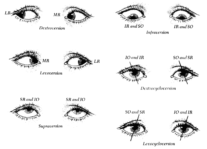
Horizontal versions are dextroversion and levoversion. Dextroversion is accomplished by contraction of the right lateral rectus and left medial rectus muscles and relaxation of the right medial rectus and left lateral rectus muscles. Levoversion is accomplished by contraction of the left lateral rectus and right medial rectus muscles and relaxation of the left medial rectus and right lateral rectus muscles.
Vertical versions are supraversion (sursumversion) and infraversion (deorsumversion). Supraversion is accomplished by bilateral contraction of the elevator muscles (the superior rectus and inferior oblique muscles) with simultaneous relaxation of the depressor muscles (the inferior recti and the superior obliques). As the eyes move into dextroversion, the right superior rectus and left inferior oblique muscles are the prime elevators; in levoversion, the left superior rectus and right inferior oblique muscles are the prime elevators. Infraversion is accomplished by an increase of innervation to the four depressor muscles with an equal decrease of innervation to the four elevator muscles. In dextroversion the prime depressors are the right inferior rectus and left superior oblique muscles; in levoversion, the left inferior rectus and right superior oblique muscles are the prime depressors.
Cycloversion is the simultaneous and equal tilting of the corneal superior poles either to the right or to the left (either dextrocycloversion or levocycloversion). Dextrocycloversion is accomplished by contraction of the extortors of the right eye (i.e., the inferior rectus and inferior oblique) and of the intortors of the left eye (i.e., the superior rectus and superior oblique) and by relaxation of the intortors of the right eye (i.e., the superior rectus and superior oblique muscles) and of the extortors of the left eye (i.e., the inferior rectus and inferior oblique muscles). In dextroversion the most effective dextrocycloverters are the right inferior oblique and left superior rectus muscles. In levoversion the most effective dextrocycloverters are the left superior oblique and right inferior rectus muscles. Levocy-cloversion is accomplished by an increase of innervation to the extortors of the left eye and the intortors of the right eye with a simultaneous decrease of innervation to the intortors of the left eye and the extortors of the right eye. In dextroversion the primary levocycloverters are the left inferior rectus and right superior oblique muscles; in levoversion the left inferior oblique and right superior rectus muscles are the prime levocycloverters.
Vergence
Vergence is the simultaneous and equal movement of the eyes in opposite directions; a prefix attached to “vergence” indicates the direction of the disjugate movement. The muscles that are the prime movers undergo equally graded contractions in accord with Hering's law of innervation, and for each contracting muscle there is an opposite and equally graded relaxing antagonist (Sherrington's law). All possible vergence movements are shown in Figure 7, which indicates only the contracting muscles.
|
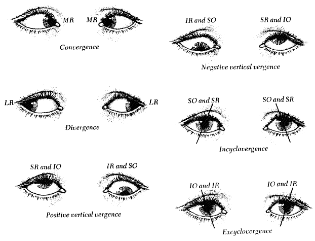
Horizontal vergences are convergence and divergence. Convergence is accomplished by contraction of the medial rectus muscles and relaxation of the lateral rectus muscles. Divergence is accomplished by contraction of the lateral rectus muscles and relaxation of the medial rectus muscles.
Vertical vergences are designated as positive and negative. Positive vertical vergence is elevation of the right eye with simultaneous depression of the left; negative vertical vergence is depression of the right eye with simultaneous elevation of the left. Each is accomplished by elevators contracting in one eye and depressors contracting in the other with equal and simultaneous relaxation of their antagonistic muscles.
Cyclovergence is equal and simultaneous tilting of the corneal superior poles either inward or outward (either incyclovergence or excyclovergence). Incyclovergence is accomplished by simultaneous contraction of all intortors (superior recti and superior obliques) and relaxation of all extortors (inferior recti and inferior obliques). Excyclovergence is accomplished by an increase of innervation to the extortors and an equal decrease of innervation to the intortors.