|
|
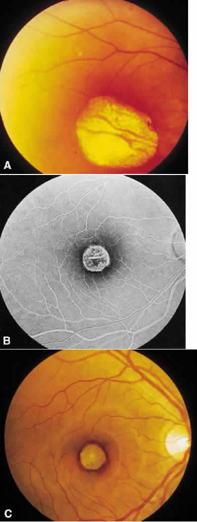
| Fig. 5. Central areolar choroidal dystrophy. A. There is a well-circumscribed macular sheen surrounded by a hyperpigmented border but no evidence of choroidal atrophy in this 23-year-old man. B. The angiogram indicates some degree of choriocapillaris atrophy since there is persistent visualization of the choroid vessel. C. As the lesion progresses, as is seen in this 62-year-old woman, the well-circumscribed area of chorioretinal atrophy overlying the bare sclera is easily seen. |