|
|
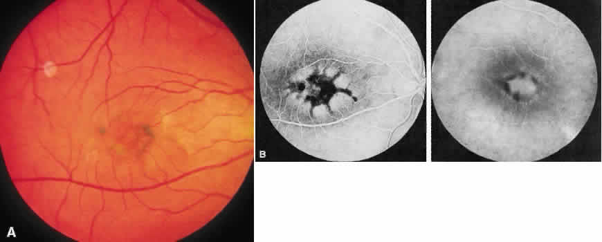
| Fig. 4. Pigment pattern dystrophies. Each eye of this 27-year-old man shows that he had irregularly shaped pigment accumulation (A), which is made more vivid on fluorescein angiography (B). The vision was 20/20 in the right eye and 20/50 in the left eye. The electroretinogram and electrooculogram were normal, and other family members were asymptomatic but unavailable for examination. |