|
|
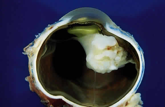
| Fig. 26. Teratoid medulloepithelioma. Histopathology (see Fig. 27) shows that the white foci in this ciliary body tumor are hyaline cartilage. A delicate cyclitic membrane is present, and the hyaloid artery persists. (From Shields JA, Eagle RC Jr, Shields CL et al: Fluorescein angiography and ultrasonography of malignant intraocular medulloepithelioma. J Pediatr Ophthalmol Strabismus 33:193, 1996) |