|
|
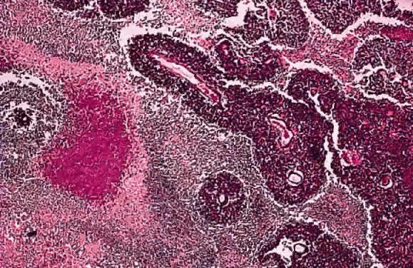
| Fig. 14. Retinoblastoma. Photomicrograph shows basophilic sleeves of viable tumor surrounding vessels. Extensive tumor necrosis is present. The necrotic area is eosinophilic and contains a reddish-purple focus of dystrophic calcification. (Hematoxylin and eosin, × 10) |