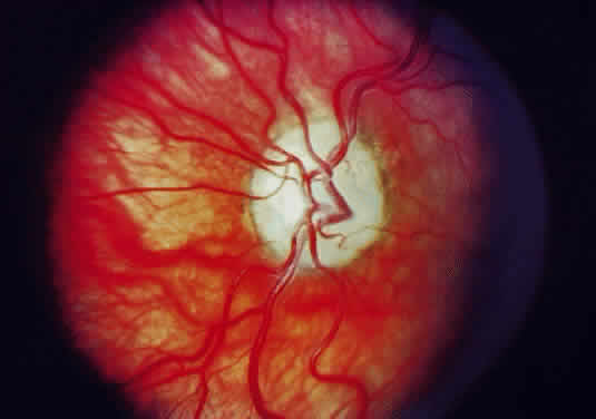
Fig. 42.
Fundus appearance of patient with primary meningioma of the optic nerve sheath. Note posterior ciliary shunt vessels.